A Myb Transcription Factor of
Phytophthora sojae
,
Regulated by MAP Kinase PsSAK1, Is Required for
Zoospore Development
Meng Zhang, Jing Lu, Kai Tao, Wenwu Ye, Aining Li, Xiaoyun Liu, Liang Kong, Suomeng Dong,
Xiaobo Zheng, Yuanchao Wang*
Key Laboratory of Integrated Management of Crop Diseases and Pests, College of Plant Protection, Nanjing Agricultural University, Ministry of Education, Nanjing, China
Abstract
PsSAK1, a mitogen-activated protein (MAP) kinase fromPhytophthora sojae, plays an important role in host infection and zoospore viability. However, the downstream mechanism of PsSAK1 remains unclear. In this study, the 3’-tag digital gene expression (DGE) profiling method was applied to sequence the global transcriptional sequence ofPsSAK1-silenced mutants during the cysts stage and 1.5 h after inoculation onto susceptible soybean leaf tissues. Compared with the gene expression levels of the recipientP. sojaestrain, several candidates of Myb family were differentially expressed (up or down) in response to the loss of PsSAK1, including of a R2R3-type Myb transcription factor,PsMYB1. qRT-PCR indicated that the transcriptional level ofPsMYB1decreased due toPsSAK1silencing. The transcriptional level ofPsMYB1increased during sporulating hyphae, in germinated cysts, and early infection. Silencing ofPsMYB1results in three phenotypes: a) no cleavage of the cytoplasm into uninucleate zoospores or release of normal zoospores, b) direct germination of sporangia, and c) afunction in zoospore-mediated plant infection. Our data indicate that thePsMYB1 transcription factor functions downstream of MAP kinase PsSAK1and is required for zoospore development ofP. sojae.
Citation:Zhang M, Lu J, Tao K, Ye W, Li A, et al. (2012) A Myb Transcription Factor ofPhytophthora sojae, Regulated by MAP Kinase PsSAK1, Is Required for Zoospore Development. PLoS ONE 7(6): e40246. doi:10.1371/journal.pone.0040246
Editor:Sung-Hwan Yun, Soonchunhyang University, Republic of Korea
ReceivedApril 10, 2012;AcceptedJune 3, 2012;PublishedJune 29, 2012
Copyright:ß2012 Zhang et al. This is an open-access article distributed under the terms of the Creative Commons Attribution License, which permits unrestricted use, distribution, and reproduction in any medium, provided the original author and source are credited.
Funding:This work was supported by the National Natural Science Foundation of China (30900932) to S.D., the Special Fund for Agro-scientific Research in the Public Interest (3–20) from China to Y.W., and Project Funded by the Priority Academic Program Development of Jiangsu Higher Education Institutions. The funders had no role in study design, data collection and analysis, decision to publish, or preparation of the manuscript.
Competing Interests:The authors have declared that no competing interests exist.
* E-mail: [email protected]
Introduction
Oomycetes are economically important because they are destructive to crops and nursery stocks. Examples include
Phytophthora sojae (causes soybean root rot), Phytophthora infestans
(causes potato late blight), andPhytophthora ramorum(causes sudden oak death). Approximately 116 pathogenicPhytophthoraspecies are currently known or described. New species, or new variants of known species, emerge continuously [1]. P. sojae is a soil-borne plant pathogen that causes soybean stem and root rot. Since its discovery in Indiana in 1948 and Ohio in 1951, Phytophthora rot has been frequently reported globally throughout most soybean-growing regions [2]. This disease may cause plant stand losses and complete yield reductions in susceptible soybean cultivars in poorly drained fields, costing millions of dollars each year. Therefore, slowing the assault ofP. sojaepathogens is important.
P. sojae produce asexual sporangia which breed and release zoospores. Although in some species such asP. infestans, sporangia are released freely from aerial hyphae and serve as agents of dispersal, the sporangia ofP. sojaeare not readily released from the hyphae. Zoospores are the most important route of infection of roots, especially when the soil is flooded. In P. sojae, zoospores swim chemotactically toward compounds including the isoflavones daidzen and genistein released by roots of their host plants [3]. Due to their energy needs and fragility, asexual sporangia are
ineffective resting structures. Instead the thick-walled and durable sexual spores, called oospores, can act as resting structures. As they remain viable between growing seasons, oospores are an important inoculum for disease. Oospores later germinate to produce either a hyphal tube, which can directly infect a plant, or a germ sporangium, which release zoospores [4]. Therefore, the normal development of sporangia plays key roles in the spread ofP. sojae.
The sporangium ofP. sojaeare multinucleated cells formed as the cytoplasm from the subtending hypha flows into the expanding hyphal apex [5]. After chilling sporangia to approximately 4uC for 30 min, the fusion of cleavage vesicles and release of zoospores occur spontaneously in distilled water. Nuclei within the sporan-gium organize into a regularly spaced distribution; some membranous elements differentiate in the cytoplasm to produce 10–30 zoospores. Although not as common, sporangia ofP. sojae
the germ tube. Having penetrated the epidermis, nutrients are acquired through the formation of haustoria [5]. Therefore, the normal sporulation of sporangia with release of normal and functional zoospores is important to complete theP. sojaelife cycle. Understanding the molecular mechanisms of sporangial devel-opment and release normal zoospores may facilitate develdevel-opment of new strategies for Phytophthora disease control. It is known that MAP kinase signal transduction cascades greatly influence gene expression, metabolism, cell division, cell morphology, and cell survival, and participate in regulating important pathogenic processes in fungi [6]. MAPK is activated by MAPK kinase (MEK or MAPKK), which is activated in turn by MEK kinase (MEKK or MAPKKK). The sequential activation of the MAPK cascade eventually activates transcription factors and the expres-sion of specific sets of genes in response to environmental stimuli [7]. There are direct links between Arabidopsis MAP kinase 4 (MPK4) and innate immunity based on releasing transcription factors in the nucleus upon activation [8]. InMagnaporthe grisea, the
MST11-MST7-PMK1MAP kinase (MAPK) cascade is essential for appressorium formation and plant infection [9]. One putative downstream transcription factor regulated by Pmk1 is Mst12 (Ste12 homolog), which is essential for pathogenesis.MST12may regulate genes involved in penetration and infectious growth, but another transcription factor(s) must function downstream ofPMK1
to regulate appressorium formation [7]. This MAPK pathway in
M. griseahas been characterized in detail. MAPK gene homologs have also been characterized in other phytopathogenic fungi. In
Colletotrichum lagenarium, the conidia of cmk1 mutants fail to germinate on plant and glass surfaces [10]. InBipolaris oryzae, the rice leaf spot pathogen,BMK1, is required for plant infection and conidiation [11]. In the necrotrophic pathogen, Fusarium grami-nearum(wheat scab fungus), gpmk1deletion mutants fail to infect roots [12]. InBotrytis cinerea, Delta bcsak1 mutants are significantly impaired in vegetative and pathogenic development: they are blocked in conidia formation, and are unable to penetrate unwounded plant tissue [13]. These studies indicate that the MAPK pathway may be conserved in many phytopathogenic fungi and regulate spore sporulation and other plant infection processes.
In contrast, little is known about the MAPK pathway elements or how they are regulated in oomycetes. Bioinformatic searches against theP. infestansgenome revealed that it contains 15 MAPK, 6 MAP2K, 5 MAP3K, and 4 MAP4K [14]. However, their biological functions are unclear. InP. sojae, a stress-activated MAP kinase was identified (namedPsSAK1) in a previous study, which represents a novel group of MAP kinases [15]. PsSAK1 is currently the only characterized MAP kinase inPhytophthora, which is responsible for plant infection processes. It was up-regulated in zoospores, cysts, and during the early infection stages. PsSAK1 -silenced mutants showed faster encystment, lower germination ratios, longer germ tubes, and colonization defects on both wounded and unwounded soybean leaves. PsSAK1 is an important regulator of zoospore development and pathogenicity inP. sojae. To better understand its roles, the transcriptomes of
PsSAK1-silenced mutant and the corresponding parental control P6497 in two stages were profiled and compared to identify components regulated by PsSAK1. It revealed that several family members of the Myb family were differentially expressed (up or down) in response to the loss of PsSAK1. In this study, a Myb transcription factor protein down-regulated in the transcriptomes of PsSAK1-silenced mutant is identified and characterized. To elucidate the mechanism of PsSAK1 inP. sojaedevelopment and pathogenicity, the expression of Phytophthora sojaeMYB protein 1 (PsMYB1) was silenced through stable transformation inP. sojae.
The functional analysis of silenced mutants demonstrates that PsMYB1, regulated by PsSAK1, plays key roles in several life-cycle stages that are related to zoospore-mediated plant infection.
Results
Identifying Downstream of the Stress-activated MAP Kinase PsSAK1
A stress-activated MAP kinase was indentified and is key regulator of zoospore development and pathogenicity in P. sojae
[15]. PsSAK1-silenced transformants show aberrant zoospore development and less osmotic adaptation. Firstly, zoospore behavior was severely affected by silencing PsSAK1. When zoospores were released from sporangia ofPsSAK1-silenced lines, they appeared as less-active, obese protoplasmic balls, and were not as bean-shaped as zoospores of P6497. Cyst germination was affected by silencing and germ tubes or hyphae of the mutants failed to penetrate host hypocotyls epidermal cells. Secondly, under stress mediated by H2O2or Nacl treatment, the expression
of PsSAK1 noticeably increases. Thirdly, PsSAK1-silenced strains were growth-impaired in the presence of 0.2 M Nacl compared with the wild-type and control strains. We demonstrate that PsSAK1-mediated pathway exists inP. sojaeto help regulate the zoosporogenesis, response to stress and infect host.
The 3’-tag digital gene expression (DGE) profiling method, which uses oligo-dT to generate libraries that are enriched in the 3’ untranslated regions of polyadenylated mRNAs and produces 21-bp cDNA tags, can measure gene expression level for whole transcriptome [16]. To understand the roles of PsSAK1, we applied DGE profiling for the PsSAK1-silenced line T31 using RNAs isolated from cysts and 1.5 h after inoculation onto susceptible soybean leaf tissues (mCY and mIF1.5 h), with the same stages of P6497 wild-type as the parental control (CY and IF1.5 h, [17]). Cysts stage was chosen because of the quicker encystment and a lower germination ratio of PsSAK1-silenced transformants. Meanwhile, 1.5 hours after inoculation was chosen as an early time of infection stage based on their phenomenon that could not penetrate the host surface [15]. From the four libraries, 7,493 to 9,846 genes were detected by the DGE profiling tags and a total of 11,081 genes were detected in at least one library (Table S1).
To identify the genes regulated byPsSAK1, the gene expression levels between CY-mCY and IF1.5 h-mIF1.5 h were compared respectively to analyze the differentially expressed genes. Two fold was the dividing line with P value#0.01, employing the False Discovery Rate (FDR) correction. In total, compared to the P6497 wild-type libraries, the numbers of down-regulated (up-regulated) genes were 2,641 (1,350) and 5,217 (825) in mCY and mIF1.5 h, respectively. Moreover, 1,624 and 241 genes were down- and up-regulated in both stages (Figure 1A, Table S1). This suggests that more genes are down-regulated than up-regulated, which also indicates that MAP kinase PsSAK1 activates rather than represses downstream genes whenP. sojaeattaches to the host surface, forms cysts, and penetrates the host surface [15].
Active MAPKs frequently translocate from the cytoplasm to the nucleus to phosphorylate nuclear targets, transcription factor is a important one [6]. Transcription factors containing two or three imperfect tandem repeats of the Myb DNA-binding domain (named R2R3 and R1R2R3, respectively) regulate important processes in growth and development [18]. Specific processes regulated by Myb proteins in plant include response to environ-mental stresses inArabidopsis[19], cell and petal morphogenesis in
Antirrhinum majus[20], drought resistance and salt tolerance in rice [21]. Some lower eukaryotes also express Myb proteins control stress pathway [22].Therefore, we speculate that, Myb proteins may be involved in the stress-associated MAPK PsSAK1pathway in P. sojae. To test this hypothesis, by using bioinformatics, annotation of theP. sojaegenome database, Fungal Transcription Factor Database (FTFD), SMART (http://smart.embl-heidelberg. de), and manual evaluation of gene models, 68 Myb TFs with
variable numbers of Myb DNA-binding domains were predicted (Table S2). Most (47) of the proteins have a single Myb domain, whereas the remaining 9, 10, 1, and 1 proteins have two, three, four, and five Myb domains, respectively. On the basis of differential DGE analysis of the PsSAK1-silenced line T31, approximately 44 Myb TFs were significantly down-regulated or up-regulated responsing to the loss of PsSAK1. These results demonstrate that Myb transcription factor family may play important roles in PsSAK1 pathway (Table S1).
Identification of the Myb transcription factor PsMYB1
DGE analysis revealed that seven Myb transcription factors were down-regulated significantly in both PsSAK1 silencing libraries, including Ps140839 or PsMYB1 (also named PsMYB-like 2–7in Table S2) (140839-incorrect in Table S1). The gene Figure 1. The expression changes and sequence ofPsMYB1.(A) Overview of expression changes during two stages ofPsSAK1-silenced strain T31. Illustrated are the stages examined (cysts and 1.5 h after inoculation onto susceptible soybean leaf tissues) and number of up- or down-regulated genes during each stage. (B) Alignment ofPsMYB1orthologs. The R2 and R3 Myb DNA-binding domains are underlined by red and blue lines, respectively. The orthologs are from P. infestans (PITG_06748.1),P. ramorum(Pr_86538), H. arabidopsidis (Ha_811065), Pythium ultimum (PYU1_T010492), andAlbugo laibachii(A.laibachiiNc 14). (C) Validation of DGE data by quantitative reverse-transcription polymerase chain reaction (qRT-PCR).PsMYB1transcripts in thePsSAK1-silenced line are down-regulated dramatically in cysts stage and IF1.5 h stage based on the qRT-PCR. Results of an independent-samplest-test for each species. *P,0.05; **P,0.001.
doi:10.1371/journal.pone.0040246.g001
was then cloned from P6497 mycelial cDNA and found previous gene model in the genome database was incorrect, and the corrected PsMYB1 encodes a 513 amino acid protein with three introns and contains two imperfect tandem repeats of the R2R3 Myb DNA-binding domains (GenBank accession number JX069980) (Figure 1B). Therefore, we updated the gene expression patterns in DGE profiling and found that the expressions of correct gene were down-regulated significantly in two stages (140839-correct in Table S1). To confirm this gene expression patterns derived from DGE profiling and the regulated relationship between PsMYB1 and PsSAK1, we checked the expression level of PsMYB1 in the cysts or 1.5 hours after inoculation of PsSAK1-silenced line T31 by qRT-PCR. The results showed that the transcription patterns of
PsMYB1 were consistent with that in the DGE profiling (Figure 1C). qRT results in combination with the data of DGE profiling indicate that the transcript of PsMYB1 depend on the presence of a functional PsSAK1 pathway. Each
PsMYB1 ortholog was identified in the sequenced oomycete species including P. infestans, Hyaloperonospora arabidopsidis, and
Pythium ultimum. Two were predicted in P. ramorum but one sequence possessed an unclear region. Meanwhile, only one gene (CCA17728.1) in Albugo laibachii, which is also an oomycete, was identified by BLASTp against the NCBI database with an E-value of 1E-50 as a cutoff (Table S2). Protein sequences alignment among PsMYB1 and its orthologs showed that the R2 and R3 domains had the highest amounts of conservation, whereas the remainder of the gene had relatively low conservation (Figure 1B).
Expression Profiling and Silencing ofPsMYB1inP. sojae
P6497
To address the roles of thePsMYB1 gene, we employed real-time quantitative RT-PCR using RNAs isolated from distinct developmental stages including mycelia, sporulating hyphae, swimming zoospores, cysts and germinating cysts, as well as soybean (Williams) tissues infected with P. sojae. PsMYB1
transcripts were detected in all of the tested libraries but were mostly absent from the zoospores. Expression of PsMYB1
increased during sporulation, in germinated cysts, and 3–6 h after infection (Figure 2A).
To evaluate the function of PsMYB1, the P. sojae (P6497)
PsMYB1-silenced strains were obtained through polyethylene glycol (PEG)-mediated protoplast stable transformation [23]. A total of 152 putative transformants that grew in the presence of Geneticin (50mg/ml) were selected for further evaluation. Of 152 putativePsMYB1 transformants, 66 PsMYB1-integrated transfor-mants were obtained using genome PCR. Real-time quantitative RT-PCR was used to evaluate the level of PsMYB1 mRNA accumulation. Finally, three transformants (T54, T77, and T101) failed to produce the given amplicon when the normal number of PCR cycles were applied with sporulating hyphae RNA as initial template (Figure 2B). Therefore, three independentP. sojaestrains deficient in PsMYB1 expression were successfully obtained. Furthermore, we have confirmed that lacking PsMYB1 did not affect the transcript of PsSAK1 by comparing the transcriptional level ofPsSAK1with and without functional PsMYB1 (Figure 2C).
PsMYB1 is Necessary for Normal Asexual Sporangial Development
Phenotypes of the silenced transformants in comparison to controls were evaluated throughout the P. sojae life cycle. The controls included the P6497 parental strain (WT), the PsSAK1
-silenced line (T31), and the transformed strain that failed to exhibit
PsMYB1silencing (T132). Comparison of the asexual and sexual growth amongPsMYB1-silenced and non-silenced strains indicat-ed that silencing of PsMYB1 was not associated with asexual hyphal growth or sexual development (Table 1).
However, the release of zoospores was affected byPsSAK1and
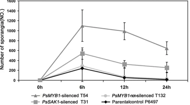
PsMYB1silencing. Instead of releasing swimming zoospores, the majority of sporangia of all three PsMYB1-silenced strains germinated directly. During the first 24 h after inducing sporangia production by chilling at 4uC for 30 min and removing to room temperature, we found approximately 59% of sporangia could not release zoospores compared with approximately 5.6% of that in parental control P6497. Over time, the top of the sporangium germinated to form hypha and the cytoplasm flowed from the sporangium to the hyphal tip. After one week, empty sporangia could be observed which consisted only of cell walls. Quantita-tively, the numbers of sporangia of strains were counted at 0, 6, 12, and 24 h after washing medium to induce sporangial development (Figure 3). We found that the sporangia of all three controls had releasing zoospores during first 6 h, but not in the threePsMYB1 -silenced lines.PsMYB1-silenced line T54 had the most sporangia (1099.336317.22) compared to the positive control PsSAK1 -silenced line T31 (539.336117.89), negative control PsMYB1
non-silenced transformant T132 (292.33614.57), and the parental control P6497 (239.00623.07). This result is consistent with those of the other two time points. We also found that the parental control wild-type had few sporangia could not release even 24 h after washing with sterile distilled water (13.33617.09), which is much lower than the number in T54 (647.006130.03). Thus, the
PsMYB1-silenced transformant T54 andPsSAK1-silenced line T31 produced significantly more directly germinated sporangia than wild-type after 6, 12, and 24 h (T31, p,0.05 and p,0.001 for T54, respectively) (Figure 3). This suggests that normal asexual sporangia development was affected differently by silencing of
PsMYB1orPsSAK1.
Silencing ofPsMYB1Impaired Zoosporogenesis and Zoospore Development
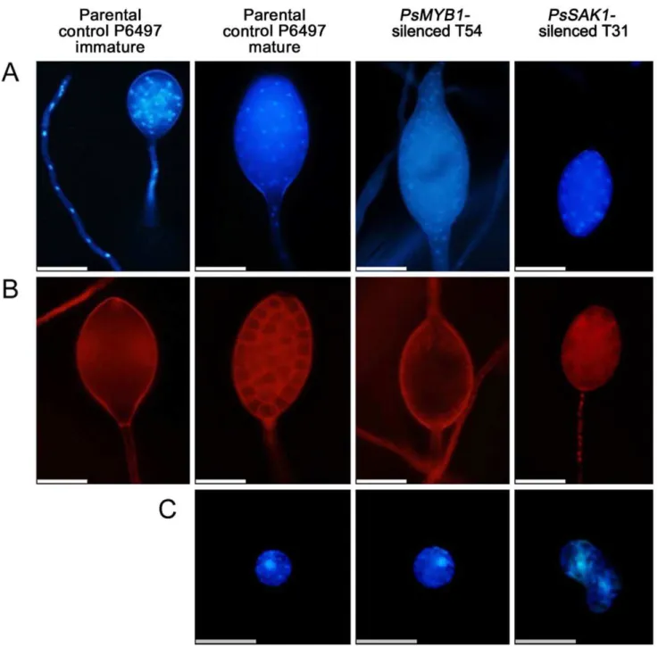
In Phytophthora cinnamomi, zoosporogenesis involves compart-mentalization of the mature multinucleate sporangium into a number of biflagellate and uninucleate zoospores [24]. During direct sporangial germination, the flagella and cleavage system degradation prior to germ tube formation [25]. We hypothe-sized that PsMYB1 is associated with zoosporogenesis. To explore this hypothesis, specific fluorescent dyes were used to study the effect of PsMYB1 on the organization of internal organelles during sporangial cleavage and zoospore formation. No significant differences in the cellular structure of the controls and PsMYB1-silenced sporangia were observed during the early stages. Comparing the silenced strains with controls, sporangia were multinucleated with undifferentiated cytoplasm. Further-more, the size, form, and structure were similar (immature parental control P6497of Figure 4A and 4B). However, after inducing sporangial cleavage, there were significant differences between sporangia of the controls and the PsMYB1-silenced strains. Nuclei within the sporangium of P6497 were regularly spaced; meanwhile, the cytoplasm was differentiated to form an average of 10–30 fully developed zoospores. By contrast, the sporangial cytoplasm of thePsMYB1-silenced line T54 remained undifferentiated and the nuclei remained disordered. For the
Figure 2. qRT-PCR assay of thePsMYB1gene.(A) Expression pattern ofPsMYB1based on qRT-PCR. The ten stages are: mycelia (MY), sporulating hyphae (SP), zoospores (ZO), cysts (CY), germinating cysts (GC), and IF1.5 h to IF24h (samples from 1.5, 3, 6, 12, and 24 h after infection of soybean leaves). (B) Expression ofPsMYB1in strains transformed with extra copies ofPsMYB1. qRT-PCR was performed using sporangiating hyphae RNAs from wild-type isolate (P6497) and transformants (PsMYB1silenced, T54, T77, and T101; PsMYB1 non-silenced, T132) as initial template. (C) PsSAK1 expression level in thePsMYB1-silenced line. qPCR was performed based on theDDCT method. Error bars are calculated from three replicate qRT-PCR assays.
doi:10.1371/journal.pone.0040246.g002
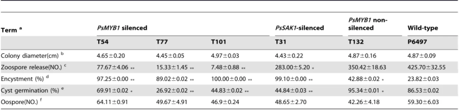
Table 1.Comparison of asexual and sexual growth amongPsMYB1-silenced and non-silenced strains.
Terma PsMYB1silenced PsSAK1-silenced
PsMYB1
non-silenced Wild-type
T54 T77 T101 T31 T132 P6497
Colony diameter(cm)b 4.6560.20 4.4560.05 4.9760.03 4.4360.22 4.8760.16 4.8760.09
Zoospore release(NO.)c 77.67
64.06** 15.3361.45** 7.4860.88** 283.0065.20* 350.42618.63 425.70632.55
Encystment (%)d 97.2560.00
** 89.0260.02** 100.0060.00** 99.1060.00** 42.8860.02* 23.8260.03 Cyst germination (%)e 69.9160.02* 26.9260.02** 44.8360.02** 44.8460.03** 95.3460.01* 86.5360.02
Oospore(NO.)f 64.11
60.91 49.6764.91 46.960.24 48.6562.70 42.2664.18 59.3066.03
aStrains are the P6497 wild-type (parental) or transformants derived from P6497 (by protoplast transformation). Independent-samples
t-test between wild-type P6497 and each mutant was performed and marked by "*" and "**", referring to p,0.05 and p,0.001, respectively. Values in the table are the mean
6standard error. bBased on 5 days of growth in 10% V8 juice agar medium.
cNumber of 5
65 mm colonies releasing zoospores after washing for 24 h at 25uC. The numbers of zoospores in 10 ul of sterile distilled water were counted. dPercent of encysted zoospores in 50 ul of zoospores suspension after 1 h incubation at 25
uC with 80% humidity.
ePercent of cysts forming germ tubes after 2 h incubation with vortexing to induce encystment, based on counting a minimum of 100 cysts from each strain. fNumber of oospores was counted in 10% V8 juice medium for 10 days.
doi:10.1371/journal.pone.0040246.t001
approximately 9% of the zoospores contained two nuclei instead of one, which were not able to swim but instead settled to the bottom of the dish immediately after release from the sporangia (Figure 4C). Our results demonstrate that loss of either PsSAK1
MAP kinase or PsMYB1 leads to abnormal P. sojae zoospor-ogenesis.
Phytophthora sporangia may emit germ tubes directly through the exit pore plug material and sporangia walls when incubated in a nutritive medium with high osmotic pressure [25]. Flagella and cleavage vesicles form during the initial stages of direct germination in the same manner as they do in indirect germination. Later, however, the flagella degenerate and cleavage of the cytoplasm is not complete [26]. The result above demonstrates that PsMYB1 plays key role in regulating zoosporogenesis. Well then, whether thePsMYB1 would express and execute function or not when the sporangia of P. sojae
germinate directly. To test the proposition and determine how specific connection between PsMYB1 and sporangia cleaving, we added exogenous mannitol to induce the sporangia of P6497 germinated directly and detected the expression level ofPsMYB1
in this situation, compared with that in sporangia germinated indirectly. 0.8M mannitol was added to sterile distilled water which was used to wash the 2-day-old wild-type hyphae to induce the sporangia germinated directly, using sterile distilled water alone to induce the sporangia of P6497 germinated indirectly. After 5 h we observed abundant formation of sporangia in both treatments. RNAs were isolated to examine the gene expression levels of PsMYB1, usingPsSAK1 expression as a positive control. As shown in Figure 5, the abundance of
PsMYB1 transcript in the sporangia exposed to 0.8 M mannitol was significantly lower than in the treatment by ddH2O2 only,
with down-regulation .2.5-fold. That suggests the transcription of PsMYB1 is suppressed in direct sporangial germination stage of P. sojae. The result clarified the connection between PsMYB1 and sporangia cleaving from the contrary aspect.
Although most sporangia directly germinated germ tubes, there were also few sporulate zoospores. This was quantitatively assayed
for each transformant by counting the zoospores released from a 565 mm colony. The amounts of zoospores released from the silenced strains (T54: 77.6764.06, T77: 15.3361.45, T101: 7.4860.88) were significantly less than those released from the controls (T132: 350.42618.63 and P6497: 425.70632.55) and T31 (283.0065.20) based on an independent-samples t-test (P,0.001) (Table 1).
The time span between zoospore release and encystment was significantly affected by silencing. Quantitatively, the silenced strains showed more rapid encystment than the control strains. The mean percent of zoospores that changed into cysts in the mutant strains during 1 h after release were 97.2560.00 (T54) and 89.0260.02 (T77) compared with 48.2260.02 in the negative control strain T132 and 23.8260.03 in the parental control P6497, which represents a significant difference based on an independent-samplest-test for each species (P,0.001). Especially for T101, all zoospores formed cysts within 0.5 h after releasing from the sporangia (Table 1). By analyzing these zoospores using microscopy, we found that zoospores released from the abnormal sporangia could not swim efficiently but instead remained near their release point, which was consistent with thePsSAK1-silenced line.
Furthermore, the cyst germination rates were lower in the mutants than in the negative control strains (Table 1). Although the average rate of cyst germination of each PsMYB1-silenced strain varied under the same conditions, there was a clear difference betweenPsMYB1-silenced lines and the negative control strains; e.g., mutants T77 (26.9260.02), T101 (44.8360.02), and T54 (69.9160.02) compared with T132 (95.3460.01) and P6497 (86.5360.02). This suggests that the number of germinated cysts were reduced significantly by silencing of PsMYB1 based on an independent-samplest-test for each species (P,0.001). Meanwhile, the positive control PsSAK1-silenced line T31 had a similar phenotype.
Overall, these results demonstrate that PsMYB1 is regulated by PsSAK1 and affects zoospore viability.
Figure 3. Effect ofPsMYB1silencing on sporangial development.The numbers of sporangia were counted at 0, 6, 12, and 24 h after inducing P. sojaeinto the sporangial forming stage.
PsMYB1 is Essential for Zoospore-mediated Plant Infection
Silencing also affected zoospore-mediated pathogenicity, as would be expected for strains defective in zoospore release and cyst germination. To test the pathogenicity, we independently applied mycelia and zoospore suspensions of three PsMYB1-silenced transformants (T77, T54, and T101), one PsSAK1-silenced line (T31), PsMYB1 non-silenced transformant (CK: T132), and the parental control P6497 on etiolated hypocotyls at 25uC to enhance disease progression. In the zoospore assay, all silenced
transfor-mants failed to infect based on the shorter lesion lengths compared to the wild-type strain P6497 (Figure 6A). We hypothesized that this was due to the reduced cyst germination ofPsMYB1-silenced transformants. That was confirmed by comparing the cyst germination ratio and virulence on soybean plants (Table 1 and Figure 6A). However, when inoculated with hyphal tip plugs, both negative control strains (T132 and P6497) andPsMYB1-silenced transformants caused spreading water-soaked lesions that are characteristic of virulent (Figure 6B).
Figure 4. Microscopic observation of the distribution of nuclei and the cleavage system within sporangia or zoospores inP. sojae.
(A) The nuclei distributions in sporangium of early stage of silenced-lines or controls, take parental control P6497 as an example; and the zoosporogenesis stages of P6497,PsMYB1-silenced transformant T54, andPsSAK1-silenced line T31 were observed with DAPI staining of the nuclei. (B) Cleavage-system degeneration during germ tube formation in test strains was seen with FM4-64 to selectively visualize the plasma membrane. The bars at the bottom of (A-B) represent 50mm. (C) Nuclear distribution in zoospores. The bar at the base of the panel represents 20mm.
doi:10.1371/journal.pone.0040246.g004
Discussion
Our results demonstrate that PsSAK1 elicits a robust transcrip-tional response, part of which is functranscrip-tionally important for zoospore development. Loss of PsSAK1 induced (repressed) the expression of 2,641(1,350) genes during the cysts stage and 5,217 (825) genes in the IF1.5 h library in P. sojae compared to the control. Several family members of the Myb transcription factor were also differentially expressed (up or down) in response to the loss of PsSAK1. DGE profiling revealed a R2R3-type Myb transcription factor named PsMYB1 was down-regulated signifi-cantly in two PsSAK1 silencing libraries. PsMYB1 transcripts increased during sporulating hyphae, germinated cysts and 3–6 h after infection, but were mostly absent during the zoospores stage. Loss ofPsMYB1 resulted in the sporangia of mutants remaining undifferentiated with disordered nuclei after inducing zoospore formation for 6–8 h when that of the parental control P6497 had released numerous zoospores. These results demonstrate for the first time that sporangial development and release of normal zoospores inP. sojaedepend on the PsMYB1 pathway, regulated by PsSAK1.
The Myb transcription factor plays an important role in zoospore development of oomycetes. This is supported by the function ofPsMYB1orthologs in other oomycetes. In P. infestans, semi-quantitative RT-PCR was performed against the PsMY-B1ortholog, PiMyb2R3(PITG_06748.1), during asexual sporula-tion or germinasporula-tion.PiMyb2R3transcripts persisted in sporangia, during zoosporogenesis (i.e., cleaving sporangia), and in swimming zoospores, but decreased during the germinated cysts stage. When sporangia were placed in broth media to induce the production of mycelia from sporangia,PiMybR2R3mRNA disappeared. This is consistent with the sporangial direct germination phenotype of the
PsMYB1-silenced line. For H. arabidopsidis, which lacks the zoospore stage, there is only 14 Myb transcription factors left in the genome, compared with 68 inP. sojae.PsMYB1expressed at a
higher level in sporangia stage, which is supported by a prior microarray study showing that most of the zoospore-specific genes inP. infestanswere induced during sporulation. Meanwhile, most sporulation-induced genes are absent from H. arabidopsidis com-pared to P. infestans, which is consistent with the inability of H. arabidopsidisto produce zoospores [18]. In this study, we focused on the biological function ofPsMYB1based on its silencing and the analysis of its gene expression patterns. From the findings that inhibition ofPsSAK1 resulted in defective zoospores and loss of
PsMYB1 resulted in malfunctions of zoospore development, we identified a role for thePsMYB1, regulated by the MAP kinase
PsSAK1, in zoospore development.
A challenging step in further understanding the MAPK pathway is the identification of downstream genes that are regulated by MAPK. Here, the analysis of 39 tag digital gene expression profiling using a PsSAK1 mutant obtained by PEG-mediated protoplast stable transformation led to the identification of a downstream gene of the MAPK PsSAK1 pathway,PsMYB1.
PsMYB1is transcriptionally suppressed after loss ofPsSAK1, and silencing of PsMYB1 does not affect the transcriptional level of
PsSAK1, which suggests partially that PsMYB1 is positively regulated by PsSAK1 (Figure 1C and Figure 2C). These data suggest that activation of the MAPK pathway leads directly or indirectly to increase production of PsMYB1 that in turn leads to zoosporogenesis and development of normal zoospores. Silencing ofPsMYB1results in undifferentiated sporangia that cannot form and release zoospores. Compared with PsSAK1-silenced lines which release less-active zoospores and contain obese protoplasmic balls, PsMYB1-silenced transformants display stronger malfunc-tions. However, loss ofPsMYB1has a weaker effect on virulence compared with thePsSAK1-silenced line. Thus, it is possible that the PsSAK1 MAPK pathway also regulates other genes which are important pathogenic factors inP. sojae. It is also possible that there is crosstalk between two pathways. Like inCryptococcus neoformans, inhibition of calcineurin induces the phosphorylation of Mpk1 and Figure 5. Analysis ofPsMYB1gene expression during sporangial direct germination.The RNAs of sporulating hyphae were prepared by repeatedly washing 2-day-old hyphae incubated in 10% V8 broth with sterile distilled water and incubating the washed hyphae in the light at 25uC for 4–8 h until sporangia developed on most of the hyphae. "M+", washing the P6497 hyphae with sterile distilled water containing 0.8 M mannitol
and induce the sporangia of P6497 to germinate directly. "CK", washing mycelia of the wild-type with sterile distilled water and induce the sporangia of P6497 to germinate indirectly. Evaluating expression ofPsMYB1in P6497 sporangia of germinated indirectly and that of germinated directly. Expression was measured by quantitative real time PCR and normalized to the level in the sporangia of germinated indirectly of wild-type P6497. Bars indicate standard errors from three independent replicates.
the induction ofFKS1, suggestive of an interplay between the CWI pathway and calcineurin [27]. Similar to these mechanisms, it is possible that PsMYB1 is activated by another pathway(s) when PsSAK1 is inhibited. The DGE profiling analysis supports this concept. Loss ofPsSAK1 up-regulated 1,350 and 825 genes in the cyst and IF1.5 h stages, respectively.
Our results demonstrate that PsMYB1, regulated by PsSAK1, is functionally important for zoospore development and release. We note that the phenotype of inhibition of zoospore development and release, and specifically that of direct germination can be recreated via manipulation of exogenous calcium. InP. infestans, inhibitors of calcium pathways strongly inhibited zoospore release [28]. It was found that inPhytophthora cinnamomi, a longer lasting rise in Ca2+
is required during cytoplasmic cleavage to regulate cytokinesis. Conversely, the calcium channel blockers were also found to inhibit zoosporogenesis and encystment [29]. In Phytophthora parasitica, Ca2+
treatments affected the production of zoospores from sporangia at two different stages: during cleavage of the sporangium protoplasm into uninucleate cells and at the stage when zoospores were released by dissolution of the sporangium papillum [30]. Here, we note that thePsSAK1-silenced lines and
PsMYB1-silenced strains display the similar phenotype. Thus it is possible that PsSAK1 mediates its affects by affecting proteins which mediate signaling of calcium, and vice versa. We intend to further explore this hypothesis in our future work.
We have found that PsMYB1, regulated by PsSAK1, plays key roles in zoospore development, but how PsSAK1 regulated PsMYB1 to affect zoosporogenesis is not clear. As is known, transferring sporangia to pure water initiates zoospore release after a short time, which demonstrates that very few conditions are need for triggering this process. The most important preconditions for zoospore release are a turgor pressure in the sporangium high enough to initiate the process and an osmotic gradient between sporangium and external medium high enough to allow a significant water uptake into the sporangium [25]. In the previous study, we found that PsSAK1 is required for P. sojaeresponse to extracellular osmotic stimuli because the expression of PsSAK1
noticeably increases under stress mediated by H2O2 or Nacl
treatment, and loss ofPsSAK1lead to sensitive to 0.2M Nacl [15]. Perhaps, loss ofPsSAK1,P. sojaecould not response the change of extracellular osmotic pressure. Thus, there may be not an osmotic gradient between sporangium and external medium, and the turgor pressure of sporangium is not high enough to initiate the PsMYB1 in regulating zoosporogenesis. In near-isotonic solutions, the cleavage of zoospores may be incomplete.
Our DGE profiling and silencing lines data indicate that PsMYB1, regulated by MAP kinase PsSAK1, is responsible for zoosporogenesis and the development of normal zoospores, and affects zoospore-mediated plant infection. These results demon-strate for the first time that PsSAK1 pathway plays an important role in P. sojae zoospores development by regulating PsMYB1.-Further studies on PsMYB1 inP. sojaewill yield important insights into the regulation mechanism of PsMYB1 and increase our understanding of its signaling pathway.
Materials and Methods
Preparation ofPhytophthora sojaeIsolates, Plant Materials, and Virulence Tests
Phytophthora sojae P6497 was from Brett Tyler (Oregon State University, USA). P. sojae isolates were maintained on 10% V8 agar media at 25uC in the dark [31]. To identify the asexual and sexual growth, each strain ofP. sojaewas grown on plates (9 cm in diameter) containing 10 ml V8 juice agar at 25uC. Individual agar disks (5 mm in diameter) were removed from the edge of an actively growing culture into the center of the plate. After 5 days, the colony diameter was measured. Zoospores were produced by placing three V8 agar disks (5 mm in diameter) from the edge of an actively growing culture of each strain into a plastic plate containing 150ml of 10% V8 juice for one disk, and then incubating these dishes in darkness at 25uC for 24 h. After using 150ml of sterile distilled water to wash the mycelium three times, another 150ml of sterile distilled water was added to induce the sporangia in light at 25uC for 24 h. Next, the agar disks were removed and the zoospores suspension was measured. We manually counted the number of zoospores in 10ml of zoospores suspension. We analyzed zoospore encystment and the germina-tion of cysts as described by Hua [23]. Five individual agar disks (5 mm in diameter) of test strains were placed into glass plates (9 cm in diameter) containing 10 ml of 10% V8 juice broth in darkness at 25uC for 10 days to induce oospore formation. Three replicates were made for each transformant, and every replicate contained three culture plates. We mixed the culture of three plates and added it to a homogenate cup for 5 min (3000 r/min), after which we counted the number of oospores in the 10ml of homogenate. All determinations of each strain were performed in triplicate which contained three duplications. We took the mean of the three duplications as the result of one replicate. For infection assays, the hypocotyls of soybean (Glycine max) cultivar Williams (rps) were used to evaluate the virulence of P. sojae cultures. Quantitative virulence of strains was measured using a lesion length assay, as described previously [32]. Etiolated seedlings (grown at 25uC in the dark at 80% humidity for 3–4 days) were inoculated 2 cm below the beginning of the root zone for the wild-type and the transformants. Each was spot incubated with hyphal tip plugs or 100 zoospores, sealed in plastic bags, and incubated at 25uC in the dark for 2 days. The disease lesion length from the inoculation point up and down the hypocotyls was then measured. Each treatment contained at least 5 plants. All treatments were replicated in triplicate independently. All statistical analyses were performed with PASW statistics 18 (SPSS Inc., USA).
Digital Gene Expression Profiling and Bioinformatics Analysis
The preparation of biological material, libraries, and RNA sequencing was conducted according to published methods [17]. For the wild-type stages (CY and IF1.5 h), we used the raw tags and the calculated gene expression levels, which were published previously [17]. The sequences of PsMYB1 orthologs from different organisms were obtained from the NCBI, Broad Institute databases, and DOE Joint Genome Institute, using the BLASTp Figure 6. Pathogenicity assay.(A) Disease lesion lengths in etiolated soybean hypocotyls infected with zoospores of wild-type and transformed strains ofP. sojae, 48 h after inoculation. (B) Disease lesion lengths in etiolated soybean hypocotyls infected with hyphal tip plugs of wild-type and transformed strains ofP. sojae, 48 h after inoculation. Results of an independent-samplest-test for each species. *P,0.05; **P,0.001. Results of statistical comparison between each mutant and P6497 are in the test. The large water-soaked lesions coating the surface of the etiolated seedling represent a successful infection. The photos were taken after incubation at 25uC for 48 h in the dark.
program. Multiple sequence alignments were generated and drawn by BioEdit 7.0.1.
Nucleic Acid Manipulation
Genomic DNA extraction was performed from hyphae grown in 10% V8 liquid medium, as described previously [33]. Total RNA was isolated using a PureLink RNA Mini Kit (Invitrogen, USA) following the manufacturer’s protocol. To confirm the relative abundance ofPsMYB1transcripts derived from the DGE libraries and thePsMYB1expression patterns during 10 develop-mental and infection stages ofP. sojae, the total RNA samples were prepared according to published methods [17].
P. sojaeTransformation, Quantitative RT-PCR, and Gene Expression Analysis
ForPsMYB1silencing,Phytophthora sojaetransformation plasmid pHamPsMYB1 (part sequence of PsMYB1,1084 bp of negative strand, driven by Ham34 promotor) was contructed as follows. The NptII gene of pHAMT35N was replaced with 1084 bp of
PsMYB1[34]. ThePsMYB1-silencing sequence was confirmed by EST, using the primers MYB1-2F,59 -GGCAACAGCAACAG-CAGCGG-39 and MYB1-2R,59 -TACGGGGAGTTCAT-CATGCCC-39. The transformation experiments were performed as described previously [35] except for the use of two different concentrations of G418 before transfer to the V8 agar medium with 50mg/ml G418. After the protoplast overnight regeneration, we suspended them in liquid Pea/0.5 M Mannitol (PM) media (40uC) containing 30mg/ml G418 and then plated the suspension. When visible colonies were observed after 2 to 3 days’ incubation at 25uC, we overlaid the plate with Agar Pea/0.5 M Mannitol (PM) media (40uC) containing 50mg/ml G418 and plated again at 25uC for 2–3 days. Next, the transformants were propagated on V8 agar with 50mg/ml G418 at 25uC. This modification decreases false positive rates. Of these, approximately 33% were found to contain the transgene. PutativeP. sojae PsMYB1-silenced lines were screened as follows. First, genomic PCR screening of all transformants was performed with oligonucleotides HMF (59 -TTCTCCTTTTCACTCTCACG-39) and HMR (59 -AGACA-CAAAATCTGCAACTTC-39), which are primers for the Ham34 promoter and terminator, respectively. Next, to investigate the silencing efficiency of thePsMYB1transgene in the transformants, total RNAs of positive inserted transformants in the sporulating hyphae stage were extracted and isolated. qRT-PCR was performed as described previously [36], except using oligonucle-otides MYB1qRTF(59-GAACCAGAGCAACCAGAATG-39) and MYB1qRTR (59- TACGGGGAGTTCATCATGCC-39).
Microscopy
For cell biology assays, sporangium and zoospores were stained with the blue-fluorescent DAPI nucleic acid stain 49, 6-diamidino-2-phenylindole, dilactate (Invitrogen), to visualize the distribution of nuclei within sporangium and the number of nuclei in zoospores
of the transformants or parental control P6497. We equilibrated the sporulating hyphae of different stages briefly with phosphate-buffered saline (PBS) and diluted the DAPI stock solution to 300 nM in PBS. Approximately 300ml of this dilute DAPI staining solution was added to the coverslip preparation. After incubating for 1–5 minutes, we rinsed the sample several times in PBS and then viewed the sample using an Olympus 1X71inverted microscope at 330–385 nm. Red-fluorescent FMH 4–64 dye, which selectively visualizes the plasma membrane, was used to monitor the cleavage system in sporangia. First, we prepared the samples as described above. At the same time, a working staining solution of 5mg/ml dye in ice-cold HBSS was prepared, which was kept on ice. Next, the coverslip with the sample was immersed in the staining solution on ice. After 1 minute, we removed the coverslip from the staining solution and imaged the sample without washing. The filter sets used were 510–550 nm.
To determine differences in sporangial development between P6497 and the silenced lines intuitively and quantitatively, 262 mm hyphal tip plugs of stains were cut to inoculate 150ml of sterile clarified 10% V8 juice. Stationary mycelia cultures were incubated at 25uC in the dark for 24 h. Next, we washed the hyphae with sterile distilled water three times and incubated them in the light at 25uC with 100ml of sterile distilled water. Samples were taken after 0, 6, 12, and 24 h incubations, and pictures were taken using a stereo microscopic Olympus SZX10. Subsequently, the number of sporangia that did not release was counted under the microscope. This assay was repeated at least three times, and the statistical analyses were performed with PASW statistics 18 (SPSS Inc., USA).
Supporting Information
Table S1 Gene expression profiling in two stages for the
PsSAK1-silenced transformant T31 vs. P. sojae P6497.
PsSAK1and PsMYB1 were labeled by red, and the other family members of Myb were labeled by blue color.
(XLS)
Table S2 The predicted Myb TF family genes.
(XLS)
Acknowledgments
We thank Professor Daolong Dou (College of Plant Protection, Nanjing Agricultural University) for revising this manuscript. We thank Xin Zhang, Yujuan Hao (Key Laboratory of Integrated Management of Crop Diseases and Pests, College of Plant Protection, Ministry of Education, Nanjing Agricultural University) for technical support.
Author Contributions
Conceived and designed the experiments: MZ SD YW XZ. Performed the experiments: MZ JL KT WY AL XL LK. Analyzed the data: MZ WY KT SD. Contributed reagents/materials/analysis tools: MZ SD YW KT WY. Wrote the paper: MZ WY YW SD.
References
1. Kroon LP, Brouwer H, de Cock AW, Govers F (2012) The GenusPhytophthora
Anno 2012. Phytopathology 102: 348–364.
2. Schmitthenner AF (1999)PhytophthoraRot of Soybean. Plant Health Progress Available: http://wwwplantmanagementnetworkorg/pub/php/management/ phytophthora/Posted 1 June 2000.
3. Tyler BM (2007)Phytophthora sojae: root rot pathogen of soybean and model oomycete. Mol Plant Pathol 8: 1–8.
4. Judelson HS, Blanco FA (2005) The spores ofPhytophthora: weapons of the plant destroyer. Nat Rev Microbiol 3: 47–58.
5. Hardham AR (2007) Cell biology of plant-oomycete interactions. Cell Microbiol 9: 31–39.
6. Qi M, Elion EA (2005) MAP kinase pathways. J Cell Sci 118: 3569–3572.
7. Zhao X, Mehrabi R, Xu JR (2007) Mitogen-activated protein kinase pathways and fungal pathogenesis. Eukaryot Cell 6: 1701–1714.
8. Qiu JL, Fiil BK, Petersen K, Nielsen HB, Botanga CJ, et al. (2008)Arabidopsis
MAP kinase 4 regulates gene expression through transcription factor release in the nucleus. EMBO J 27: 2214–2221.
9. Zhao X, Xu JR (2007) A highly conserved MAPK-docking site in Mst7 is essential for Pmk1 activation inMagnaporthe grisea. Mol Microbiol 63: 881–894. 10. Xu JR (2000) Map kinases in fungal pathogens. Fungal Genet Biol 31: 137–152. 11. Moriwaki A, Kihara J, Mori C, Arase S (2007) A MAP kinase gene, BMK1, is required for conidiation and pathogenicity in the rice leaf spot pathogenBipolaris oryzae. Microbiol Res 162: 108–114.
12. Urban M, Mott E, Farley T, Hammond-Kosack K (2003) The Fusarium graminearum MAP1 gene is essential for pathogenicity and development of perithecia. Mol Plant Pathol 4: 347–359.
13. Segmuller N, Ellendorf U, Tudzynski B, Tudzynski P (2007) BcSAK1, a stress-activated mitogen-stress-activated protein kinase, is involved in vegetative differenti-ation and pathogenicity inBotrytis cinerea. Eukaryotic Cell 6: 211–221. 14. Judelson HS, Ah-Fong AM (2010) The kinome ofPhytophthora infestansreveals
oomycete-specific innovations and links to other taxonomic groups. BMC Genomics 11: 700.
15. Li A, Wang Y, Tao K, Dong S, Huang Q, et al. (2010) PsSAK1, a stress-activated MAP kinase ofPhytophthora sojae, is required for zoospore viability and infection of soybean. Mol Plant Microbe Interact 23: 1022–1031.
16. Asmann YW, Klee EW, Thompson EA, Perez EA, Middha S, et al. (2009) 3 ’ tag digital gene expression profiling of human brain and universal reference RNA using Illumina Genome Analyzer. Bmc Genomics 10: 531.
17. Ye W, Wang X, Tao K, Lu Y, Dai T, et al. (2011) Digital Gene Expression Profiling of thePhytophthora sojaeTranscriptome. Mol Plant Microbe Interact 24: 1530–1539.
18. Xiang Q, Judelson HS (2010) Myb transcription factors in the oomycete
Phytophthorawith novel diversified DNA-binding domains and developmental stage-specific expression. Gene 453: 1–8.
19. Chen WQ, Provart NJ, Glazebrook J, Katagiri F, Chang HS, et al. (2002) Expression profile matrix ofArabidopsistranscription factor genes suggests their putative functions in response to environmental stresses. Plant Cell 14: 559–574. 20. Baumann K, Perez-Rodriguez M, Bradley D, Venail J, Bailey P, et al. (2007) Control of cell and petal morphogenesis by R2R3 MYB transcription factors. Development 134: 1691–1701.
21. Hu HH, Dai MQ, Yao JL, Xiao BZ, Li XH, et al. (2006) Overexpressing a NAM, ATAF, and CUC (NAC) transcription factor enhances drought resistance and salt tolerance in rice. Proceedings of the National Academy of Sciences of the United States of America 103: 12987–12992.
22. Yoshioka S, Taniguchi F, Miura K, Inoue T, Yamano T, et al. (2004) The novel Myb transcription factor LCR1 regulates the CO2-responsive gene Cah1, encoding a periplasmic carbonic anhydrase inChlamydomonas reinhardtii. Plant Cell 16: 1466–1477.
23. Hua C, Wang Y, Zheng X, Dou D, Zhang Z, et al. (2008) APhytophthora sojae G-protein alpha subunit is involved in chemotaxis to soybean isoflavones. Eukaryot Cell 7: 2133–2140.
24. Hyde GJ, Gubler F, Hardham AR (1991) Ultrastructure of Zoosporogenesis in
Phytophthora-Cinnamomi. Mycological Research 95: 577–591.
25. Erwin DC, S.Bartnicki-Garcia, Tsao PH (1985)Phytophthora. The American Phytopathological Society: 392 p.
26. Hemmes DE, Hohl HR (1969) Ultrastructural changes in directly germinating sporangia ofPhytophthora parasitica. Amer J Bot 56: 300–313.
27. Fuchs BB, Mylonakis E (2009) Our paths might cross: the role of the fungal cell wall integrity pathway in stress response and cross talk with other stress response pathways. Eukaryotic Cell 8: 1616.
28. Judelson HS, Roberts S (2002) Novel protein kinase induced during sporangial cleavage in the oomycetePhytophthora infestans. Eukaryot Cell 1: 687–695. 29. Walker CA, Van West P (2007) Zoospore development in the oomycetes. Fungal
biology reviews 21: 10–18.
30. von Broembsen SL, Deacon JW (1997) Calcium interference with zoospore biology and infectivity ofPhytophthora parasiticain nutrient irrigation solutions. Phytopathology 87: 522–528.
31. Erwin D, Ribeiro O (1996)Phytophthora diseases worldwide. SP, MN: American Phytopathological Society Press.
32. Vega-Sa´nchez ME, Redinbaugh MG, Costanzo S, Dorrance AE (2005) Spatial and temporal expression analysis of defense-related genes in soybean cultivars with different levels of partial resistance toPhytophthora sojae. Physiological and Molecular Plant Pathology 66: 175–182.
33. Brett M. Tyler HF, and Michael D Coffey (1995) Inheritance of Avirulence Factors and Restriction Fragment Length Polymorphism Markersin Outcrosses of the OomycetePhytophthora sojae. Mol Plant-Microbe Interact 8: 515–423. 34. Judelson HS, Tyler BM, Michelmore RW (1991) Transformation of the
oomycete pathogen,Phytophthora infestans. Mol Plant-Microbe Interact 4: 602– 607.
35. Dou D, Kale SD, Wang X, Chen Y, Wang Q, et al. (2008) Conserved C-terminal motifs required for avirulence and suppression of cell death by
Phytophthora sojaeeffector Avr1b. Plant Cell 20: 1118–1133.