Neelanjana Ghosh1,2, Thomas J. McKillop2¤, Thomas A. Jowitt2, Marjorie Howard2, Heather Davies2, David F. Holmes2, Ian S. Roberts2, Jordi Bella1,2*
1Manchester Interdisciplinary Biocentre, University of Manchester, Manchester, United Kingdom,2Faculty of Life Sciences, University of Manchester, Manchester, United Kingdom
Abstract
The genome sequences of enterohaemorrhagicE. coliO157:H7 strains show multiple open-reading frames with collagen-like sequences that are absent from the common laboratory strain K-12. These putative collagens are included in prophages embedded in O157:H7 genomes. These prophages carry numerous genes related to strain virulence and have been shown to be inducible and capable of disseminating virulence factors by horizontal gene transfer. We have cloned two collagen-like proteins fromE. coliO157:H7 into a laboratory strain and analysed the structure and conformation of the recombinant proteins and several of their constituting domains by a variety of spectroscopic, biophysical, and electron microscopy techniques. We show that these molecules exhibit many of the characteristics of vertebrate collagens, including trimer formation and the presence of a collagen triple helical domain. They also contain a C-terminal trimerization domain, and a trimerica-helical coiled-coil domain with an unusual amino acid sequence almost completely lacking leucine, valine or isoleucine residues. Intriguingly, these molecules show high thermal stability, with the collagen domain being more stable than those of vertebrate fibrillar collagens, which are much longer and post-translationally modified. Under the electron microscope, collagen-like proteins fromE. coliO157:H7 show a dumbbell shape, with two globular domains joined by a hinged stalk. This morphology is consistent with their likely role as trimeric phage side-tail proteins that participate in the attachment of phage particles toE. colitarget cells, either directly or through assembly with other phage tail proteins. Thus, collagen-like proteins in enterohaemorrhagicE. coligenomes may have a direct role in the dissemination of virulence-related genes through infection of harmless strains by induced bacteriophages.
Citation:Ghosh N, McKillop TJ, Jowitt TA, Howard M, Davies H, et al. (2012) Collagen-Like Proteins in PathogenicE. coliStrains. PLoS ONE 7(6): e37872. doi:10.1371/journal.pone.0037872
Editor:Anthony R. Poteete, University of Massachusetts Medical School, United States of America
ReceivedMarch 26, 2012;AcceptedApril 25, 2012;PublishedJune 6, 2012
Copyright:ß2012 Ghosh et al. This is an open-access article distributed under the terms of the Creative Commons Attribution License, which permits unrestricted use, distribution, and reproduction in any medium, provided the original author and source are credited.
Funding:Neelanjana Ghosh was supported by a Dorothy Hodgkin Postgraduate Award to the University of Manchester (ref. EP/P500966/1), funded by Research Councils UK (http://www.rcuk.ac.uk). Thomas J. McKillop was supported by a Masters in Biological Sciences Award to the University of Manchester (ref. BB/ E527355/1), funded by the Biotechnology and Biological Sciences Research Council (http://www.bbsrc.ac.uk). The funders had no role in study design, data collection and analysis, decision to publish, or preparation of the manuscript.
Competing Interests:The authors have read the journal’s policy and have the following conflict: data presented in this study provided the initial idea for a novel methodology that was developed in the laboratory of the corresponding author. JB is listed as an inventor on a patent application protecting the technology (which is not discussed here), filed by the University of Manchester (International Patent Application No PCT/GB2011/052217; Bacterial Collagen, date 14 November 2011). This competing interest does not alter the authors’ adherence to all PLoS ONE policies on sharing materials, methods, and data. The authors are free to share everything described in this publication.
* E-mail: [email protected]
¤ Current address: Defence Science and Technology Laboratory, Salisbury, United Kingdom
Introduction
EnterohaemorrhagicE. coli(EHEC) is responsible for gastroin-testinal disorders in humans that range from abdominal pain and diarrhoea to haemorrhagic colitis and haemolytic uremic syn-drome [1,2,3]. The EHEC serotype most often linked with outbreaks of severe disease isE. coliO157:H7. The genomes of the
E. coliO157:H7 strains EDL933 and Sakai are 0.9 Mb larger than that of the non-pathogenic laboratory E. coli strain K-12 [4,5]. That extra genetic material is the result of horizontal gene transfer (HGT) probably mediated by bacteriophages: the Sakai strain genome includes 18 prophages and 6 prophage-like elements integrated into different sites of the bacterial chromosome [5,6], while the EDL933 genome contains 18 prophages and prophage-like elements [4]. Up to 463 phage-associated genes are present in the O157:H7 strains for only 29 in the K-12 strain [3,7].
Several virulence genes of the O157:H7 strain are located into these prophages and prophage-like elements, notably the Shiga toxin (verocytotoxin) genesstx1andstx2[8], and various effector
proteins that are injected into the host cells by a type III secretion system [9,10]. Collectively, EHEC strains are considered new pathogens that have emerged from less virulent strains by progressive acquisition of virulence factors via HGT. There is significant evidence that variation of the prophage sequences is a main factor for the genomic and virulence diversity of EHEC [6,7,11,12,13]. The acquired specific virulent attributes allow EHEC strains to adapt to new niches and to broaden the spectrum of disease.
helical axis and are connected through a ladder of intermolecular hydrogen bonds roughly perpendicular to that axis [15,16,17,18]. The conformation of the collagen triple helix imposes a repetitive amino acid sequence pattern where glycine residues (Gly, G) occur at every third position. This (Gly-X-Y)npattern is recognized as the signature of collagen proteins and domains.
A surprising number of collagen-like sequences have been detected outside the metazoan realm, notably in bacterial and viral genomes [19,20,21]. These ‘‘prokaryotic collagens’’ exhibit in their (Gly-X-Y)n regions significant differences in residue content and distribution with respect to vertebrate collagens, and yet they seem to show the basic molecular characteristics of true collagen proteins [22,23]. The functions and potential contribution to virulence of these prokaryotic collagens are currently under study, but they seem to participate in pathogenesis in unexpected ways. Thus, collagen-like glycoproteins from Bacillus anthracis are components of the exosporium that are able to interact with integrin receptors on professional phagocytes [24,25], while collagen-like surface proteins fromStreptococcus pyogenesare able to promote bacterial adhesion and internalization to respiratory epithelial cells [26,27,28].
Open reading frames with collagen-like sequences in the
genomes of E. coli O157:H7 and other EHEC strains are
automatically annotated as ‘‘hypothetical tail fibre proteins’’. These collagen-like sequences seem a distinctive feature of EHEC strains and several bacteriophages, and have not been detected in K-12 or other non-pathogenic strains. They are normally included in the prophage or prophage-like elements of the EHEC genomes and would be expected to participate in phage morphogenesis during prophage induction. Indeed there is evidence of changes in levels of expression for some of these collagen-like protein transcripts under certain experimental conditions, normally in association with other prophage genes [29,30,31,32].
While most of the prophages in the EHEC genomes appear to be defective, often lacking genes apparently critical for phage induction and viability, phage induction from EHEC strains has been demonstrated and Shiga-toxin converting phages can be detected free in the extraintestinal environment [12,33]. Further-more, potentially defective phages have been shown to be inducible, to release virus particles of different morphologies and, after release, to infect otherE. colistrains, [34]. The same study also suggests that recombination and other inter-prophage interactions may make possible the biological activation of defective prophages [34].
Thus, prophages embedded in EHEC genomes have the potential of disseminating virulence factors through bacterial infection and HGT. Their morphogenetic proteins are largely uncharacterized and deserve investigation. Here, we present a first biochemical analysis of the collagen-like proteins of EHEC prophages, which we will refer collectively as EPclPs (EHEC Prophage collagen-like Proteins).
Results
Domain Architecture of Collagen-like Proteins in EHEC Genomes
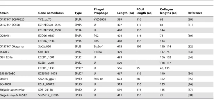
Several open reading frames potentially encoding collagen-like proteins have been identified by automatic sequence annotation in the genomes of EHEC strains. Those from the Sakai and EDL933 genomes will be discussed here, but many related sequences have been identified in other strains. Their primary structures show one or more collagen-like domains (Col) with the repeating collagen signature sequence (Gly-X-Y)n, flanked at both ends by a series of non-collagenous, conserved domains (Figure 1 and Table 1).
Domains PfN, Pf2 and PfC have been described on the basis of sequence conservation and are associated to fibre tail proteins from phages. They appear in automatic annotation of EPclPs. Figure 1 shows the different protein architectures and the nomenclature used here to refer to them, plus two representative sequences. The most common architecture (EPclA) appears in multiple copies in each genome, with more than 90% amino acid sequence identity across copies. Table 2 gives the complete list of EPclP sequences from the Sakai and EDL933 genomes, whereas representative examples of other architectures and strains are given in Table 3.
The EPclA architecture shows a single collagen triple helical sequence capped by PfN and PfC domains at the N- and C-termini, respectively. Between the PfN domain and the Col domain there is a region of low-complexity. Analysis of its amino acid sequence suggests a coiled-coil conformation (see below), and thus will be referred here as PCoil domain. The EPclB architecture shares the presence of PfN, PCoil and PfC non-collagenous domains, and contains two Col domains separated by a Pf2-type repeat. Protein sequences within each type of architecture show variable lengths of their Col and PCoil domains (Tables 2 and 3). Differences in length are typically multiples of three for Col domains and multiples of seven for PCoil domains, which is consistent with the lengths of the repetitive motifs in collagen and coiled-coil sequences, respectively. In sequences with two Col domains it is common that the first one contains a single interruption of the (Gly-X-Y)nrepeating pattern, with a conserved Gly-X-Pro-Gly-Gly-Pro-X-Gly sequence.
Only a few sequences conform to the EPclC and EPclD architectures (Table 3), which are characterized by different C-terminal regions with no sequence homology to the PfC domains of EPclA or EPclB architectures. Also, the PCoil domain is often missing in EPclC architectures. The EPclD sequence Stx2-86_gp21, from the Stx2-86 prophage in the Shiga toxin-producing
E. colistrain O86:H– (accession codes Q08J84, YP_794068), has a 322-amino acid Col domain (Table 3), by far the longest collagen-like sequence of all EPclPs. This long domain also shows a single Gly-X-Pro-Gly-Gly-Pro-X-Gly interruption. No examples of EPclC or EPclD sequences are found in the EDL933 or Sakai genomes.
The PfN and Pf2 domains are not exclusive to collagen-like proteins fromE. coliprophages and were first identified in the side tail fibre protein coded by the stf gene from l bacteriophages [35,36] (accession code P03764). Virions with a functionalstfgene show jointed tail fibres, expanded receptor specificity, and adsorb more rapidly to E. coli cells. Homologous proteins have been identified in embedded prophages of manyE. colistrains, including the laboratory reference strain K12 (protein stfR/ynaB, accession code P76072).
Other than their consistent presence in prophage tail fibre proteins, little is known about the structure and function of the PfN and Pf2 domains. Some sequence similarity between PfN and a regulatory domain of eukaryotic carboxypeptidases may be indicative of a proteolysis-related function, but to date there is no experimental evidence for this. Due to the presence of these domains, EPclP sequences are automatically annotated as putative tail fibre proteins.
Amino Acid Composition and Positional Preference in the Collagen Domains of EPclPs
(Table 4). By contrast, Pro is relatively infrequent in the Y position. Both X and Y positions also show a strong preference for charged amino acids, aspartate/glutamate (Asp/Glu, D/E) in the X position and lysine/arginine (Lys/Arg, K/R) in the Y position. Alanine (Ala, A) is also relatively frequent at both X and Y positions, and both glutamine (Gln, Q) and threonine (Thr, T) show a clear preference for the Y position. Interestingly, cysteine, phenylalanine, histidine, tryptophan and tyrosine are absent in collagen domains from EPclPs. At the triplet level, the most common are GPK (15%), GPQ (12%), GPA (12%), GER (8%)
and GET (8%). The triplet pattern GP(Q/P)–GPK–G(D/E) is repeatedly observed in the collagen domains of EPclPs.
The position-specific amino acid preferences in EPclPs are quite different from those seen in animal collagens, as shown for example by the human sequences (Table 4 and [37]). The most obvious difference is in the Pro distribution: human collagens have a clear preference for Pro residues in both the X and Y positions, close to 30% and 35% respectively. There is some variation between fibrillar and non-fibrillar collagens and with the collagen-like proteins, but Pro residues are invariable more common in the Y position of human collagens. The reason is well known: Pro
Figure 1. Collagen-like proteins from prophages embedded in the genomes ofE. coliO157:H7 and other EHEC strains, referred here as EPclA to EPclD (EHEC Prophage collagen-like A to D).(A) Domain architectures. The collagen triple helical domains are labelled ‘‘Col’’, and domains predicted to adopt ana-helical coiled-coil conformation (see text) are labelled ‘‘PCoil’’ (for phage coiled-coils). Key to other domain labels (Table 1): PfN, phage fibre N-terminal domain; PfC, phage fibre C-terminal domain; PfC2, phage fibre C-terminal domain, variant 2; Pf2, phage fibre repeat 2. (B) Sequence of a representative collagen-like protein with EPclA architecture (ECs2717), from the genome ofE. coliO157:H7 Sakai. (C) Sequence of a representative collagen-like protein with EPclB architecture (Z1483), from the genome of E. coli O157:H7 EDL933. Amino acid sequences corresponding to the different predicted domains are colour-coded as in (A).
residues in the Y position are often modified post-translationally to 4-hydroxyproline (Hyp, O), which contributes to the thermal stability of the collagen domains. Charged residues are also frequent in the X and Y positions of human collagens, with the same positional preferences as in collagen domains from EPclPs (Asp/Glu more often in X, Lys/Arg more often in Y). However, they are overall less frequent and their preferential position is less strict (Table 4). Other amino acids significantly contribute to the sequence variability at each position.
The expected average conformational parameters of the triple helical Col domains can be calculated from the distribution of imino acids along their sequences [38]. The expected values are – 106ufor the average twist and 2.88 A˚ for the average height, same as those predicted for human fibrillar collagens [38]. Thus, despite the differences in amino acid composition and positional preference, the overall conformation of the triple helical Col domains is expected to be very similar to that of human fibrillar collagens.
Collagen sequences found in other viral proteins are more similar to those from EPclPs, although the preference for Pro in the X position is not that strong. Viral collagens share with EPclPs the low proportion of Pro residues in the Y position, large number of charged amino acids, and relatively common occurrence of Gln and Thr in the Y position. Collagens from gram-positive bacteria, which include the well-studied examples of Bacillus anthracis or
Streptococcus pyogenes [22,24,39], show a lower presence of Pro residues in the X and Y positions, much lower proportion of charged amino acids, and a higher proportion of Ala residues in the X position and Gln and Thr in the Y position.
The main difference between human collagens and the three groups of non-animal collagens in Table 4 is the lack of preference for Pro in the Y position (as already noted in an earlier analysis of viral and bacterial collagen structural motifs [21]). Bacteria and viruses do not have the prolyl-hydroxylase enzymes required for hydroxylation of Pro in the Y position of a collagen triple helix, and therefore there is no contribution to the stability of their
Table 1.Domains observed in collagen-like proteins from the genomes of EHEC strains.
Domain Names InterPro Pfam
Col Collagen triple helix IPR008160 PF01391
PCoil Putative coiled-coil region (see text)
PfN Phage fibre N (Phage_tail_N; Prophage tail fiber N-terminal) IPR013609 PF08400 PfC Phage fibre C (Phage_fiber_C; Putative prophage tail fiber C-terminus) IPR009640 PF06820 PfC2 Phage fibre C, variant 2 (see text)
Pf2 Phage fibre 2 (Phage_fiber_2; Phage tail fiber repeat 2) IPR005068 PF03406
Currently available accession codes on the InterPro and Pfam databases are given. Domain architectures are shown in Figure 1. doi:10.1371/journal.pone.0037872.t001
Table 2.Collagen-like proteins from the genomes ofE. coliO157:H7 EDL933 and Sakai strains, and their corresponding prophage locations.
Strain Gene name/locus Type Prophage Length (aa) PCoil length (aa)
Collagen
lengths (aa) Reference
O157:H7 EDL933 Z0982 EPclA CP-933K 437 116 111 [4]
Z1382 EPclA* CP-933M 437 116 111
Z1483 EPclB BP-933W 645 109 157, 114
Z2147 EPclA CP-933O 437 116 111
Z2340 EPclA CP-933R 437 116 111
Z3074 EPclA CP-933U 437 116 111
Z3309, Z3307 EPclA* CP-933V 437 116 111
Z6027 EPclA CP-933P 437 116 111
O157:H7 Sakai ECs0844 EPclA Sp3 437 116 111 [5]
ECs1123 EPclA Sp4 437 116 111
ECs1228 EPclB Sp5 645 109 157, 114
ECs1808 EPclA Sp9 437 116 111
ECs1992 EPclA Sp10 437 116 111
ECs2159, ECs2158 EPclA* Sp11 407 116 81
ECs2231 EPclA Sp12 407 116 81
ECs2717 EPclA Sp14 437 116 111
ECs2941 EPclA Sp15 437 116 111
collagen domains by Hyp residues. Collagen domains from EPclPs appear to compensate this lack of prolyl hydroxylation with a larger proportion of Pro residues in the X position. The high ratio of charged amino acids and the relatively high occurrence of Ala, Gln and Thr in EPclPs and bacterial and viral collagens may be indicative of different mechanisms for stability of their collagen domains [40,41,42].
Interestingly, the metazoan collagen sequence closest to EPclPs comes from a sea anemone,Nematostella vectensis (NCBI accession code XP_001625905, incomplete sequence), with 56% identity to the collagen domain of the EPclA protein ECs2717 and containing a repetitive [GP(Q/E)-GPK-GDT-GIT]12sequence, reminiscent
of the commonly observed triplet pattern mentioned above.
A Low Complexity Region is Predicted asa-helical Coiled-coil Domain (PCoil)
The region between the predicted PfN and Col domains in the most common architectures, EPclA and EPclB, shows an unusual low-complexity sequence with predominance of Ala (32%), Ser (19%) and Glu (13%) amino acids that often appear in tandems or in stretches of up to four consecutive identical residues (Figure 1). Different coiled-coil predicting algorithms (PCoils, Marcoil, Multi-Coil) give high scores for the region between residues 101 and 245 in both EPclA and EPclB (Figure 2). This region includes the low-complexity sequence between the PfN and Col domains plus the last 34 residues of PfN, and shows a loose seven-residue
Ala-X-X-Table 3.Examples of collagen-like proteins from otherE. coliandShigellastrains, and their prophage locations.
Strain Gene name/locus Type
Phage/
Prophage Length (aa) PCoil length (aa)
Collagen
lengths (aa) Reference
O157:H7 EC970520 YYZ_gp70 EPclA YYZ-2008 389 116 63 [80]
O157:H7 EC508 ECH7EC508_5575 EPclA U 407 116 81 [81]
ECH7EC508_3568 EPclA U 470 116 144
O26:H11 ECO26_0887 EPclA P02 404 116 78 [10]
ECO26_1634 EPclA P06 440 116 114
O157:H7 Okayama Stx2Ip020 EPclB Stx2Q-1 678 109 190, 114 [82]
ECOR-9 ORF-401 EPclC P-Eiba 479 117, 75 [83]
O81 ED1a ECED1_1681 EPclC U 493 106, 102 [84]
ECED1_2081 EPclC U 520 118, 117
ECED1_1138 EPclC* U 566 95 48, 135
55989/EAEC EC55989_1078 EPclC* U 467 116 140 [84]
O86:H– Stx2-86_gp21 EPclD Stx2-86 673 88 322 [85]
EC4100B ECoL_05072 EPclD U 519 116 135 [86]
Shigella dysenteriae SDB_03138 EPclD U 519 116 135 [87]
Shigella boydiiBS512 SbBS512_E1096 EPclD U 411 116 27 [88]
U: unassigned prophage.
*EPclC variant, containing a PCoil domain and either one or two collagen domains. doi:10.1371/journal.pone.0037872.t003
Table 4.Position-specific amino acid preferences in collagen triple-helical domains of EPclPs, human collagens, and collagen-like proteins from different groups of organisms.
Collagen group X Y
Pro Asp, Glu
Arg,
Lys Ala Gln Thr Other Pro (Hyp)
Asp, Glu
Arg,
Lys Ala Gln Thr Other
EPclPs 48.1 35.6 0.3 10.3 0.1 0.1 5.5 7.4 0.1 31.0 21.2 14.5 20.2 5.7
Viruses* 22.1 35.3 6.0 7.1 1.5 3.1 24.9 9.3 7.8 38.1 5.5 13.0 8.9 17.4
Bacteria, gram positive 31.0 14.1 5.1 23.1 2.6 1.4 22.7 3.7 5.9 9.7 6.2 18.4 48.3 7.8
Human collagens
Fibrillar 31.1 18.5 6.6 9.8 3.4 1.6 28.9 33.5 6.9 21.3 9.1 7.3 4.1 17.8 Non-fibrillar 24.6 19.7 6.6 5.8 4.1 2.4 36.9 42.2 5.4 22.4 4.7 6.5 2.8 16.0
Human collagen-like proteins{
27.5 21.6 10.6 6.1 3.0 2.0 29.2 33.7 4.8 26.1 5.3 7.9 3.5 18.8
The X and Y letters refer to the consensus sequence pattern (Gly-X-Y)ncharacteristic of collagen triple-helical domains. The numbers indicate percentage occupation of
the X or Y position by a given amino acid type. *Excluding EPclPs from bacteriophages. {
Include molecules such as C1q, mannose binding proteins, collectins, macrophage scavenger receptors, or acetyl cholinesterase, which contain in their sequence a collagen domain but are not formally classified as collagen types.
Ala/Ser-X-X-Ser periodicity, where residues in the X positions are often charged. On account of the coiled-coil predictions we will refer to the low-complexity region between PfN and Col as the
PCoil domain. The MultiCoil and SCORER 2.0 prediction
algorithms favour a trimeric rather than dimeric coiled-coil structure for PCoil. Secondary structure prediction by Jpred3
suggests that the PfN domain has mainly ab-sheet structure for the first 80 residues and somea-helical conformation from residues 90 onwards, whereas the PCoil region is predicted to be mainlya -helical.Jpred3does not predict any secondary structure for the PfC domains (data not shown).
EPclA is a Trimeric Protein that Dissociates When Denatured
The quaternary structure of rEPclA was investigated by sedimentation equilibrium analytical ultracentrifugation (AUC) at increasing concentrations of guanidinium chloride (GuHCl) (Figure 3). The relative molar mass ofrEPclA at 0 M GuHCl was 13866 kDa, corresponding to the predicted molecular weight of a trimer ofrEPclA molecules (3647 kDa). As the concentration of GuHCl increased, a transition from trimer to monomer was observed and the relative molar mass ofrEPclA at 5 M GuHCl was 43 kDa, which is consistent with the predicted molecular weight of therEPclA monomer. Thus, rEPclA trimers dissociate into monomers as the GuHCl concentration increases; the trimer-to-monomer transition point was estimated at around 2.5 M GuHCl.
An independent measurement of the molecular weight of
rEPclA was carried out by size exclusion chromatography followed by multiangle laser light scattering (SEC/MALLS) (Figure S1). The molecular weight obtained from MALLS is consistent with a trimer ofrEPclA (Table 5). A proteolytic fragment from rEPclA that included only the Col and PfC domains (Col–PfC fragment, Figures S2 and S4) could be produced in enough amounts for biophysical characterization. Analysis by SEC/MALLS of frac-tions containing the Col–PfC fragment (Figure S1) showed it to be trimeric as well (Table 5), with a molecular weight of 64 kDa consistent with three times the molecular weight of monomer Col– PfC (21–22 kDa, predicted from the peptide fingerprinting data obtained from mass spectrometry, Figure S2).
The molecular weights obtained from AUC and MALLS experiments were consistent with the predicted values for non-glycosylatedrEPclA trimers and monomers. Lack of glycosylation ofrEPclA was confirmed by periodic acid-Schiff staining analysis (data not shown).
Domains PCoil and PfC from EPclA are Trimerization Modules
Molecular weights of several recombinant fragments containing different combinations of domains were determined by SEC/ MALLS (Figure S3 and Table 5). The data indicates that PfC is a trimerization domain, forming trimeric assemblies both when fused to a thioredoxin tag (Trx–PfC) or after removal of thioredoxin by thrombin digestion. The PfN–PCoil fragments were also trimeric, whereas the PfN domains were mainly in the monomer state (Figure S3). This data suggests that PCoil is also a trimerization domain.
EPclA Shows a CD Spectrum Consistent with Collagen anda-helical Conformations
The secondary structures ofrEPclA and the Col–PfC fragment were analysed by circular dichroism (CD). Interpretation of the results is easier if the Col–PfC CD data is considered first. A
sample of Col–PfC was purified from a preparation of full-length
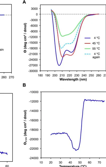
rEPclA by nickel-affinity and size exclusion chromatographies. Its CD spectrum was measured at different temperatures between 195 and 260 nm. The concentration of the Col–PfC sample was calculated as 0.2 mg/ml from its UV absorption at 280 nm and an estimated molar extinction coefficient e= 11000 M-1cm-1. The
CD spectrum of Col–PfC at 4uC (Figure 4A) shows the
characteristic features of triple helical collagen: a small maximum of positive ellipticity at 220 nm and a deep minimum of negative ellipticity at around 199 nm [43]. Both these features are associated with the polyproline II conformation [44] characteristic of the collagen triple helix. The Col–PfC fragment includes mainly the collagen domain (Col) of EPclA and the C-terminal PfC domain, and thus its CD spectrum suggests that the Col domain adopts indeed a collagen-like, triple helical structure. The triple helical features disappeared from the CD spectrum upon increase of temperature, as shown by the CD curve at 55uC (Figure 4A). Interestingly, immediate cooling of the same sample back to 4uC recovered completely the triple helical structure, with a CD spectrum practically indistinguishable from the initial one (Figure 4A).
To examine the thermal denaturation of the collagen triple helix in the Col–PfC domain, the CD of another sample of purified Col–PfC fragment (also 0.2 mg/ml) was monitored at 220 nm as a function of continuously increasing temperature, from 4uC to 60uC. The thermal curve showed a single sharp transition at 42uC, which typically corresponds to the decrease of ellipticity at 220 nm and loss of collagen triple helical structure (Figure 4B).
The CD spectrum ofrEPclA is different. A diluted sample of
rEPclA was purified by nickel-affinity and size exclusion chroma-tographies and its CD spectrum was measured between 195 and 260 nm at 4uC (Figure 5A). The concentration of the rEPclA sample was calculated as 0.04 mg/ml from its UV absorption at 280 nm and a molar extinction coefficient e= 17000 M-1cm-1, deduced from the amino acid sequence of rEPclA. The CD spectrum showed two minima of negative ellipticity at 205 nm and 224 nm, the first one being deeper, and a small local maximum between the two minima, at 216 nm. To investigate this region in more detail a second sample with higher concentration, 0.3 mg/ ml, was analyzed at different temperatures. When the sample was heated to 45uC, the height of the 216 nm maximum decreased significantly, the intensities of the two minima became more similar to each other, and their positions shifted to 210 nm and 222 nm, respectively (Figure 5A). This spectrum resembled more that of an a-helical coiled-coil conformation. Upon further increase of the temperature the overall ellipticity became less negative, and the two minima started to disappear and vanished completely when reaching 55uC (Figure 5A). The spectrum did not change upon further increase of temperature to 65uC. The slight decrease in ellipticity at 216 nm around 45uC and the more similar intensities of the two minima at that temperature suggest changes in the secondary structure that are consistent with the loss of the triple helical conformation in the Col domain while maintaining ana-helical conformation (Figures 4 and 5). Sucha -helical structure appears to be more stable and does not disappear completely until 55uC. Immediate cooling of the same sample from 65uC back to 4uC recovered approximately half of the initial CD spectrum (Figure 5A).
fragment at the same temperature; a second transition at 52uC showed a sharp and pronounced increase in ellipticity, corre-sponding to the loss of a-helical structure of the PCoil and PfN domains (Figure 5B). Thus, thea-helical structure of the PCoil and PfN domains is more stable than the collagen triple helix of the Col domain. The transition temperature of the Col domain is the same inrEPclA and its Col–PfC fragment, and seems unaffected by the presence of the PCoil and PfN domains. The melting transitions of thea-helical and collagen structures therefore appear to be largely independent.
The PfN–PCoil Region is Clearlya-helical and is Consistent with a Coiled-coil Structure
The secondary structures of the recombinant fragments PfN– PCoil and PfN were also studied by CD spectroscopy. Recombinant PfN–PCoil and PfN fragments were each purified with nickel-affinity and size-exclusion chromatographies. Concentrations of the PfN–PCoil and PfN samples were measured as 0.2 mg/ml and
0.3 mg/ml respectively, from their absorption at 280 nm and molar extinction coefficientse= 7000 M-1cm-1, initially calculated from the amino acid sequences of the PfN–PCoil and PfN recombinant fragments and adjusted using the observed UV absorption of samples in 8 M urea (see Materials and Methods). The CD spectrum of PfN–PCoil at 4uC in phosphate buffer (Figure 6A) shows the characteristic features of ana-helical protein, with two minima at 208 nm and 222 nm and a maximum at 195 nm. This spectrum is consistent with the prediction of ana-helical coiled-coil conforma-tion for the PCoil region. Thea-helical features were still present in a spectrum measured at 45uC, although the signal intensities at the two minima started to decrease (data not shown). These features disappeared when reaching 60uC (Figure 6B), and were mostly recovered upon cooling the sample back to 20uC (data not shown). The spectrum of the PfN domain (Figure 6A) was also consistent with ana-helical structure but the intensity of the two minima at 208 nm and 222 nm was much lower than in the PfN–PCoil spectrum, indicating a lowera-helical content in this domain. This spectrum also vanished at 60uC (Figure 6C), but it was not recovered upon cooling back to 20uC (data not shown). Thus, the main contribution to the CD signal comes from the PCoil domain, most likely through the formation of a trimeric a-helical coiled-coil structure that disappears at a temperature of 60uC but is regained when the temperature is lowered again.
To examine the thermal transitions of the PfN–PCoil domain, the CD of two more samples of purified recombinant PfN–PCoil (0.35 mg/ml concentration) and PfN (0.1 mg/ml) were monitored at 222 nm as a function of continuously increasing temperature, from 5uC to 95uC and then cooling back to 5uC at the same speed (1uC per minute). The heating thermal curve showed a single sharp transition at around 49uC (Figure 7), corresponding to the loss of the strong a-helical CD spectrum. The cooling thermal curve also showed a single sharp transition at around 45uC indicating partial re-gaining of thea-helical structure (the baseline in Figure 7 does not recover its initial value). This data suggests reversibility for the thermal transition of PfN–PCoil, mainly for the formation of the trimeric a-helical coiled-coil structure in the
Figure 3. Analysis by analytical ultracentrifugation of the average molar mass of a sample of purifiedrEPclA as a function of increasing concentration of guanidinium chloride (GuHCl).Weight-averaged molar mass was determined using a single ideal species model (see Methods). Mean value masses for the upper and lower plateaux were 13866 kDa and 4361 kDa respectively (averages of three measures). The molecular mass of nativerEPclA (0 M GuHCl) is consistent with three times that of denaturedrEPclA (see text). The transition midpoint concentration is 2.3860.09 M GuHCl.
doi:10.1371/journal.pone.0037872.g003
Table 5.Average molecular weights of different recombinant fragments, calculated from the SEC/MALLS data.
Molecule
AverageMw (kDa) SEC/MALLS
PredictedMw (kDa)
from sequence Oligomer state
rEPclA 145 47.3 Trimer
Col–PfC 62 ,21 Trimer
PfN–PCoil 88 28.0 Trimer
PfN 17 17.2 Monomer
Trx–PfC 64 21.0 Trimer
PfC 24 7.2 Trimer
Domain compositions of each molecule, including protein fusion tags, are shown in Supplementary Figure S5.
PCoil domain; the PfN domain does not recover completely after thermal denaturation and slow cooling. The actual value of the temperature of the transition is sensitive to the heating or cooling rate. A repeat of the heating experiment with a slower speed (0.33uC per minute) gave a transition temperature of 52uC for the PfN–PCoil fragment (data not shown).
Structural Organization of rEPclA and its Fragments Col– PfC and PfN–PCoil
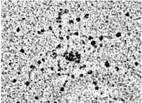
Full-lengthrEPclA was analyzed by rotary shadowing electron microscopy. Examination of the electron micrographs ofrEPclA showed a ‘‘dumbbell-shaped’’ structure with two globular particles joined by a semi-flexible stalk, in which it is possible to distinguish two regions of different thickness (Figures 8 and 9). Sequence analysis of the PCoil region and the CD spectrum of PfN–PCoil
Figure 4. Far-UV CD analysis of the Col–PfC fragment after purification fromrEPclA by SEC.(A) CD spectra at 4uC, 55uC and 4uC after immediately cooling back the sample (see text). The vertical axis measures mean residue ellipticityHin degrees cm2dmol-1. The CD data was collected between 195 and 260 nm, with a protein concentration of 0.2 mg/ml in 10 mM Tris, 150 mM NaCl, pH 7.4. Measurements were taken in a 0.5 mm path length cell. (B) Thermal denaturation of the Col–PfC fragment, monitored by CD at 220 nm as a function of increasing temperature between 4uC and 60uC, with a protein concentration of 0.2 mg/ml in 10 mM Tris, 150 mM NaCl, pH 7.4, and a heating rate of 0.33uC/min.
doi:10.1371/journal.pone.0037872.g004
Figure 5. Far-UV CD analysis of rEPclA after purification by SEC. (A) CD spectra at 4uC, 45uC, 55uC and 4uC after immediately cooling back the sample (see text). The vertical axis measures mean residue ellipticityHin degrees cm2dmol-1. The CD data was collected between 195 and 260 nm, with a protein concentration of 0.04 mg/ml (4uC) or 0.3 mg/ml (the rest) in 10 mM Tris, 150 mM NaCl, pH 7.4. Measurements were taken in a 0.5 mm path length cell. (B) Thermal denaturation of rEPclA monitored by CD at 216 nm (the maximum between the two minima at 208 and 224 nm). The CD was measured as a function of increasing temperature between 20uC and 75uC, with a protein concentration of 0.3 mg/ml in 10 mM Tris, 150 mM NaCl, pH 7.4, and a heating rate of 0.33uC/min.
both suggest that the PCoil domain is a trimerica-helical coiled-coil structure, and the observed thicker region of the stalk is consistent with such coiled-coil helical structure, which is known to have a larger cross-section than a collagen triple helix [45]. The remaining thinner region corresponds to the collagen triple-helical domain.
Inspection of rEPclA molecules from different micrographs suggests a hinge between the two regions of the stalk (collagen and coiled coil) that results in variable angles between the PCoil and Col domains and variable distances between the two globular domains (Figure 9). For most rEPclA molecules the distance between the centres of the PfN and PfC domains (N???C in Figure 9) has values around 50 nm, reaching 55 nm for the most extended, linear ones. The average N???C distance (4865 nm) is probably less significant than the values shown by the extended molecules. The N-terminal globular structure, made of three PfN domains, has an approximate diameter of 8 nm and is slightly bigger than the C-terminal globular structure made of three PfC domains (approximate diameter 7 nm) (Figure 9). Taking into account the radii of the globular domains,rEPclA molecules can reach a length of,62 nm when totally extended. However, most rEPclA molecules appear slightly bent at the hinge between the PCoil and Col domains and are, overall, a bit shorter (55–60 nm, including the globular domains).
Rotary shadow electron micrographs of Col–PfC fragments showed molecules very reminiscent of those of rEPclA but with only one globular domain (PfC) connected to a stalk (Col) (Figure 10A,B). The stalk region appears slightly unravelled where thea-helical coiled coil and the PfN domain have been removed by endogenous proteolysis of full-length rEPclA. The observed morphology confirms the previous assignment of N- and C-terminal domains forrEPclA images and that the thin region of the stalk corresponds to the collagen triple helix.
Rotary shadowing electron micrographs of PfN–PCoil frag-ments identified molecular shapes consistent with the N-terminal half ofrEPclA molecules (Figure 10C,D). PfN domains (approx-imately the first 134 residues of EPclA) form a trimeric globular structure attached to an elongated stalk containing the PCoil domain. The micrographs show several instances of PfN–PCoil fragments apparently joined at the tails of their PCoil domains (Figure 10C). This association may result from some interaction between partially unravelled or unfolded chains at the terminal end of the PCoil domains.
From measures on the electron micrographs of the Col–PfC and PfN–PCoil fragments it is possible to estimate the lengths of the PCoil and Col regions as approximately 16 nm and 30 nm respectively (Figure 9). The length of the collagen domain is consistent with the predicted length of a collagen triple helix of 111 residues (11162.9 A˚ = 32 nm, where 2.9 A˚ is an approximate measure of the height of an individual residue in a collagen triple helix [38]. Similar estimates can be obtained from measures on the thick and thin regions of the stalk in the rEPclA micrographs (14 nm for the PCoil region and 28 nm for the Col domain). The slightly shorter values suggest some overlap between domains in
Figure 6. Far-UV CD spectra of the PfN–PCoil and PfN fragments after purification by SEC:(A)PfN and PfN–PCoil at 206C;(B)PfN–PCoil at 206C and 606C;(C)PfN at 206C and 606C.
In all panels the vertical axis measures mean residue ellipticityHin degrees cm2 dmol-1. The CD data was collected between 195 and 260 nm, with protein concentrations of 0.2 mg/ml (PfN–PCoil) or 0.3 mg/ml (PfN), in 20 mM phosphate buffer (Na2HPO4/NaH2PO4), 100 mM NaCl, pH 7.4. Measurements were taken in a 0.5 mm path length cell.
therEPclA stalk that cannot be resolved in the rotary shadowing micrographs. The apparent thickness of the PCoil and Col domains in the electron micrographs over-estimates the true cross-section dimensions of these domains, as it includes the thickness of the shadowing replica. Nevertheless, a slight difference is observed between these estimates for the PCoil (3.360.5 nm) and Col (2.760.3 nm) domains. Cross-section values seen in high-resolution structures of these domains are closer to 1.3–1.7 nm for a collagen triple helix and 2.2–2.7 nm for a trimeric a-helical coiled coil.
Structural Organization of rEPclB High-molecular Weight Aggregates
Expression ofrEPclB both by IPTG induction or auto-induction produced a good yield of soluble protein. However, purification of
rEPclB from the soluble fraction by nickel-affinity and size exclusion chromatography showed that all the protein went to form soluble, high-molecular weight aggregates that eluted in the void volume of the size exclusion columns (data not shown). The protein aggregated to such an extent that it was not possible to identify any additional peak or shoulder suitable for molecular mass determination by MALLS. Aggregation was worse in samples produced by auto-induction (presumably due to the increased protein production). Attempts to reduce the degree of aggregation by lowering the protein concentration, adding EDTA (to rule out His6-mediated metal chelation aggregation), adding
glucose in high concentration, or changing the ionic strength of the buffers, were all unsuccessful: it was not possible to obtain enough non-aggregatedrEPclB for molecular weight determination or for CD studies. Interestingly, the high molecular weight aggregates were soluble and the protein did not precipitate out of solution, even after high-speed centrifugation. The bands observed in
SDS-PAGE experiments suggest that SDS treatment extracts mono-mericrEPclB from the high molecular weight aggregates.
To investigate a possible structural organization of these aggregates, a sample of IPTG-inducedrEPclB was used for rotary shadowing electron microscopy. The sample contained exclusively high-molecular weight aggregates that appeared in the electron micrographs as large masses of protein that, nevertheless, appeared to have a relatively narrow size distribution (300– 500 nm in diameter, data not shown). Close inspection of the smallest aggregates (probably at an early stage of formation) revealed an internal structure that could be reconciled with entangled, multiple flexible linear beaded molecules (Figure 11). In the vicinity of these aggregates it was possible to discover individual features reminiscent of therEPclA molecular morphol-ogy, but with three globular ‘‘domains’’ instead of two, connected by two flexible stalks (Figure 11). The terminal globular structures would correspond to the PfN and PfC domains, and the internal one would include the Pf2 domains. The flexible stalks would correspond to the two Col domains and the PCoil domain predicted in therEPclB sequence. All these structures (andrEPclB) are presumed to be trimeric due to the presence of the PfC, Col and PCoil domains, all shown to trimerize in rEPclA. The structural organization forrEPclB would therefore be similar to that seen forrEPclA. However, an effective protocol to increase the amount of non-aggregated protein will be necessary to demonstrate these assumptions and to properly characterize the molecular architecture ofrEPclB (work in progress).
Discussion
Multiple open reading frames with collagen-like amino acid sequences have been identified automatically in the genomes of several EHEC strains. These open reading frames are incorpo-rated in the sequence regions of prophage and prophage-like
Figure 7. Thermal denaturation and renaturation of recombinant PfN–PCoil (orange) and PfN (red) monitored by CD at 222 nm (corresponding to a minimum in both CD spectra).The CD was measured in a 1 mm path length cell as a function of increasing temperature between 5uC and 95uC (left) and then decreasing temperature between 95uC and 5uC (right). The temperature was changed at a rate of 1uC per minute. Both PfN–PCoil (0.35 mg/ml) and PfN (0.1 mg/ml) were in 10 mM Tris, 150 mM NaCl, pH 7.4. PfN–PCoil showed a sharp transition at around 49uC corresponding to the loss ofa-helical coiled-coil structure. The CD signal was almost completely recovered upon cooling, with a sharp transition about 45uC. This behaviour is indicative of a reversible structural transition for thea-helical coiled-coil. The PfN fragment gradually lost its CD signal with a transition midpoint of about,52uC. The gradual nature of this transition suggests denaturation rather than a cooperative unfolding. The PfN CD signal was not regained upon cooling.
elements embedded in these EHEC genomes (Tables 2 and 3) and presumably code for proteins involved in phage morphogenesis. Two recombinant proteinsrEPclA andrEPclB, representative of the most common domain architectures EPclA and EPclB (Figure 1), were amplified from a sample of genomic DNA from the O157:H7 Sakai strain, cloned into appropriate protein expression vectors, and the resulting recombinant proteins analyzed biochemically. The aims were to demonstrate their biochemical viability and structural integrity, to confirm the presence of molecular characteristics typical of collagen-like proteins, to investigate their quaternary structure, conformation, morphology and thermal stability, and to analyze some of their individual domains.
BothrEPclA andrEPclB are produced as soluble proteins inE. coli, althoughrEPclB has a strong tendency to form large, soluble aggregates. Thus, most of the biochemical analysis has been done on rEPclA. Our data demonstrate that EPclA shows the main characteristics of collagen-like proteins: it forms stable trimers in solution that dissociate upon denaturation (Figure 3), and its collagen-like sequence adopts a collagen triple helical conforma-tion, as demonstrated by CD spectroscopy (Figure 4). These data confirm that the collagen-like sequence (Col) of EPclA is a true collagen domain, and strongly suggests that collagen-like sequenc-es in other EPclPs will adopt the same conformation.
The molecular morphology of rEPclA has been visualized by rotary shadowing electron microscopy. rEPclA is a trimeric dumbbell-shaped molecule, with two globular domains joined by a semi-flexible ‘‘stalk’’, or rod-shaped domain (Figures 8–9). This connecting stalk is made in fact of two triple-helical domains: the collagen triple-helical domain (Col) and a trimerica-helical coiled coil (PCoil) encompassing the region between the Col domain and the N-terminal PfN domains. The CD spectrum of rEPclA (Figure 5) is largely dominated by the combination of CD spectra from ana-helical coiled coil and a collagen triple helix. Coiled-coil prediction algorithms give high scores to the last 30 amino acids of the PfN domain and to the region between the PfN and Col domains, both for EPclA and EPclB (Figure 2). Our data indicates that, at least in EPclA, the region between PfN and Col forms indeed a trimerica-helical coiled coil, as demonstrated by SEC/ MALLS (Figure S3) and CD spectroscopy (Figure 6). We predict that similar PCoil domains in EPclB and other EPclPs will also form trimerica-helical coiled coils. The combination of collagen triple helices adjacent to trimeric coiled coils is not unusual and there are many proteins for which such structural arrangement is predicted [45,46], where thea-helical coiled coil functions mainly as an oligomerization domain.
The thermal stability of the Col domain of EPclA is higher than expected when compared to eukaryotic collagens, especially after considering the lack of prolyl hydroxylation (discussed below). The thermal denaturation data ofrEPclA (Figures 4, 5, 6, 7) shows two sharp transitions, a first one at 42uC and a second one at 52uC (Figure 5). These two transitions correspond respectively to the loss
Figure 8. Rotary shadowing electron microscopy ofrEPclA.(A, B) Different micrographs showing dumbbell-shaped structures corre-sponding torEPclA trimers. The globular shapes correspond to the PfN and PfC terminal domains, presumably forming trimeric structures themselves. Flexible stalks connecting these globular structures contain the trimeric collagen triple-helical region (Col) and the trimerica-helical coiled-coil region (PCoil) of rEPclA. The concentration of rEPclA was 5mg/ml. Scale bar = 100 nm. (C) Detailed view of anrEPclA trimer. The arrows indicate the globular terminal domains and the thin (Col) and thick (PCoil) regions of the stalk, respectively. The globular domains can be identified as N- or C-terminal by their position with respect to the two stalk zones.
of the collagen triple helix and the loss of thea-helical coiled coil. The sharpness of both melting curves indicates highly cooperative transitions for each case (including local chain dissociation as a result of conformational change). Nevertheless, EPclA will remain trimeric at temperatures between the melting of its Col (collagen
triple helical) and PCoil (a-helical) domains. Identical transition temperatures are observed separately in two fragments: Col–PfC shows a single transition at 42uC, and PfN–PCoil shows a single transition at around 50uC, both consistent with the loss of conformation of their helical domains. The coincidence in
Figure 9. Molecular dimensions ofrEPclA and its domains.(A) Magnified view of a representativerEPclA molecule from a rotary shadowing electron micrograph. (B) The same molecule with the background masked out showing the different molecular dimensions analyzed below. (C) Average dimensions obtained from multiple measures on electron micrographs: N???C and,Coil???Col are averages of 224 measures on eightrEPclA micrographs;DC,LColandTColare averages of 76, 35 and 74 measures, respectively, on three Col–PfC micrographs (Figure 10A);DN,LCoilandTCoilare averages of 200 measures on one PfN–PCoil micrograph (Figure 10B). (D) Histograms showing the distribution of N???C and,Coil???Col values on the sample of 224rEPclA molecules.
transition temperatures with those of full-lengthrEPclA suggests that the two transitions are essentially independent of each other, and that the presence or absence of either the Col or PCoil domains does not change the thermal stability of the other one.
The thermal stability of the Col domain of EPclA appears remarkable. To remain functional at body temperatures, mam-malian collagens have a high proportion of imino acids in their collagen domains (.20% in humans) and require a complicated mechanism of post-translational hydroxylation of proline residues [47,48,49]. The enzymes required for prolyl hydroxylation are not present inE. coliand yet, the melting temperature of EPclA Col domain (42uC) is higher than that of the much longer mammalian collagens (,37uC, around body temperature [50]). Glycosylation
of threonine residues, another mechanism of collagen stabilisation [40], is not observed in rEPclA either. Thus, the higher than expected thermal stability of the Col domain of EPclA could be the consequence of an unusually high proportion of Pro residues in the X position (Table 4), a high proportion of charged residues Arg, Asp, Glu and Lys (Table 4), and possibly a stabilizing effect by the PfC domains (see below). A stabilization mechanism based on a high proportion of charged residues has been proposed for the collagen triple helical domain ofS. pyogenes collagen-like protein Scl2 [42], but the overall proportion of charged amino acids in the collagen domain of Scl2 (30%) is higher than the overall proportion of charged amino acids in the collagen domains of EPclPs (22%).
The thermal stability of the PCoil domain in EPclA is even higher. It is currently unknown if these high transition tempera-tures for the Col and PCoil domains have any functional significance. As EPclPs are likely to participate in phage morphogenesis, such high thermal stability may be required to ensure appropriate assembly of the phage particles during prophage induction from EHEC, while inside the host intestine, or to facilitate survival of free phages in the extraintestinal environment.
Electron micrographs of rEPclA show a degree of variable bending in the connecting stalk (Figure 9) that suggests the presence of a ‘‘molecular hinge’’ between the PCoil and Col domains. This hinge could be simply an area of structural discontinuity and increased flexibility between the collagen triple helix and the three-stranded a-helical coiled coil. Some discon-tinuity is expected: the transition between the two helical domains needs to account for a change in axial chain staggering, from three chains in register in the a-helical coiled coil to the one-residue stagger characteristic of the collagen triple helix [45]. It is also likely that the PCoil to Col transition changes its superhelical handedness, from a right-handed collagen superhelix to the most common left-handed coiled-coil superhelix. Although right-hand-ed coilright-hand-ed coils have been describright-hand-ed, they show undecad (11) or pentadecad (15) residue periodicity instead of the canonical heptad (7) periodicity of left-handed coiled coils [51,52]. The sequence of the PCoil domain is unusual for an a-helical coiled coil, almost devoid of hydrophobic residues like leucine or isoleucine and with
Figure 10. Rotary shadowing electron microscopy of Col–PfC and PfN–PCoil fragments.(A) Micrograph showing the morphology of Col–PfC fragments (concentration 1mg/ml). (B) Detailed view of one Col–PfC fragment. The globular shape corresponds to a trimer of PfC domains and the stalk corresponds to the trimeric collagen triple-helical domain (Col). The N-terminal end of the stalk shows a short, unravelled tail, where the PfN and PCoil domains have been removed. (C) Micrograph showing the morphology of PfN–PCoil fragments (concen-tration 5mg/ml). (D) Detailed view of three PfN–PCoil fragments. The globular shapes correspond to trimers of PfN domains and the short tails correspond to the PCoil domains.
Figure 11. Rotary shadowing electron microscopy ofrEPclB.(A) Internal structure of a small aggregate ofrEPclB molecules. The micrograph suggests multiple flexible molecules, reminiscent of those observed forrEPclA, with dark globular structures (presumably globular domains of PfN, PfC and Pf2), and poorly defined linear structures (presumably stalks containing the PCoil and Col domains). TherEPclB molecules seem to aggregate heavily through one of the globular domains. (B) Possible examples of individualrEPclB molecules observed, isolated from the large aggregates, in some electron micrographs. (C) Interpretation of the observed in terms of three globular domains (PfN, Pf2 and PfC) connected by two stalk regions. Approximate molecular dimensions are shown for comparison purposes withrEPclA.
a high proportion of Ala and Ser residues (Figure 1). Thus, visualization of a clear repeating periodicity is difficult. Neverthe-less, most of the PCoil domain and the last 30 residues of the PfN domain conform loosely to a seven-residue Ala-X-X-Ala/Ser-X-X-Ser periodicity that is given a high score by coiled-coil prediction algorithms. Additionally, differences in length between PCoil domains are multiples of seven (Tables 2 and 3), also consistent with a heptad repeat. Such periodicity would favour a left-handed superhelix for the PCoil domain. Whatever its structural details, the existence of a hinge or flexible discontinuity between the PCoil and Col domains is consistent with the independent thermal transitions observed in these two domains.
The globular domains of the rEPclA dumbbell are made of trimers of PfN and PfC domains. Analysis of the recombinant fragments PfN, PfN–PCoil, Trx–PfC and PfC show that PfC is a trimerization domain whereas PfN on its own is largely monomeric (Table 5). CD analysis of the PfN and PfN–PCoil fragments (Figure 8) shows that the PCoil domain is largely responsible for thea-helical features of the PfN–PCoil spectrum. The PfN spectrum also suggests some a-helical content in PfN domains (mostly predicted on the last 30 residues) that does not seem sufficient to form a three-stranded coiled-coil structure when the PCoil domain is not present. The CD spectrum of Col–PfC is entirely consistent with that of a collagen triple helix and indicates that the PfC domain has little or noa-helical structure.
The rapid reversibility of Col–PfC thermal denaturation (Figure 4) suggests that either the PfC domain remains stable and folded over the range of temperature used (4–55uC), or it unfolds at the same time as the Col domain but is able to refold rapidly when the temperature decreases. In either case the PfC domain is able to nucleate back the refolding of the triple helix in the Col domain. A similar behaviour has been observed with the N-terminal globular domain of theS. pyogenescollagen-like protein Scl2 [42]. The thermal denaturation of the PfN–PCoil fragment is
partially reversible, and it appears that only thea-helical coiled-coil structure of the PCoil domain is quickly regained upon cooling (Figure 9). Thus, both the PfC and PCoil domains can be considered trimerization modules in EPclPs, and in the case of PfC, it may contribute in part to the high melting temperature of the Col domain.
Recent studies on collagen-like proteins of some pathogenic bacteria have revealed a variety of functions for these proteins, including binding extracellular matrix proteins or adherence to mammalian cells [25,27,28]. It is not currently known if EPclPs have any functional role forE. coliitself, or they are strictly phage morphogenetic proteins. Currently available evidence of EPclP expression in EHEC seems to be linked to phage induction [29,30,34], and the presence of common domains in sequences of EPclPs and side tail fibre proteins of lambda phages is a strong indicator of EPclPs as structural proteins in tail fibres from phages. The high thermal stability of EPclPs is also consistent with such a role.
All prophages and phages containing EPclP sequences are described asCaudovirales, or tailed bacteriophages. Bacteriophage 933W, isolated from the O157:H7 strain EDL933, was amongst the first EPclP-containing phages to be studied [53,54,55,56]. Particles of 933W observed under the electron microscope show regular hexagonal heads (probably icosahedral), about 70 nm wide, and short tails 22–28 nm long and 13–17 nm wide [55,56]. Often, 933W virions clump together through some form of tail-tail interaction [56]. Phage induction from EHEC strains results in virion particles with different morphologies [12,34,57,58], that are usually classified as members of the Podoviridae or Siphoviridae
families, with short or long non-contractile tails, respectively. Phages of these families often show tail fibres extending laterally from the sides of the tails (Figure 12), although none of the published electron micrographs of phages from EHEC strains has sufficient detail for their visualization. Tail fibres are often used for the phage particles to attach to the target cells, and this attachment triggers further events leading to injection of the viral DNA in the host cell periplasm.
The presence of PfN and Pf2 domains both in EPclP sequences and in side tail fibre proteins oflbacteriophages [35,36] strongly suggests (although do not absolutely prove) that EPclPs are major components of prophage side tail fibres and probably participate in adhesion to the E. colicell surface, either directly or through assembly with other prophage tail fibre proteins. At least one instance of direct interaction has been proposed, between EPclB proteins of short-tailed Shiga toxin-carrying phages and the conservedE. coliprotein YaeT. The study, which refers to EPclB as tail-spike protein, concludes that YaeT is the surface molecule recognized by the majority of these phages [59].
The use of the collagen triple helix by bacteriophages to build trimeric fibrillar proteins is remarkable. Trimerization is a highly prevalent characteristic of bacteriophage tail proteins and adhesins [60,61] and novel trimeric folds have been discovered in bacteriophage fibre proteins [62,63,64,65]. The a-helical coiled coil is present in bacteriophage fibre proteins such as fibritin [66] and our data confirms that phages have added collagen helices to their armoury of trimeric folds, in combination with a-helical coiled coils and other capping domains like PfN and PfC, the latest being a trimerization module itself. Furthermore, similarity with other viral fibrous tail proteins would suggest that the PfN domain contains the viral attachment site and the PfC domain is involved in binding toE. colicell surface proteins [63]. The modular nature of EPclPs and other side tail fibre proteins, with different combinations of the same domains present in both closely and distantly related genomes, is consistent with a degree of
recombi-Figure 12. Typical morphologies ofpodoviridaeandsiphoviridae
particles (reproduced with permission from ViralZone, Swiss Institute of Bioinformatics: www.expasy.org/viralzone, [79]). The representative 933W phage and most field isolates show apodoviridae morphology, with isometric capsids of about 60–70 nm in diameter and short tails of 10–30 nm in length [12,56]. EPclPs would be the main components on the side tail fibres.
nation between multiple prophages on the same bacterial genome that often results in novel phages with an expanded host range and in new bacterial strains [34]. Prophage recombination and the efficiency of bacteriophages as HGT vehicles are responsible in part of the heterogeneity amongst EHEC strains and their rapid evolution. Thus, understanding the mechanisms of phage morpho-genesis and phage interaction with theE. colior EHEC cell surfaces may be more important than previously thought and deserve further investigation.
Materials and Methods
Sequence Retrieval and Analysis
Sequences of prophage collagen-like proteins from EHEC strains (EPclPs) were retrieved from the UniProt database [67]. These sequences were classified into different domain architectures (EPclA, EPclB, EPclC and EPclD, Figure 1) that were defined according to the occurrence and relative location of several conserved non-collagenous domains described in the InterPro [68] and Pfam databases [69] (Table 1). The probability of coiled-coil conformation and its oligomerization state in EPclP sequences were calculated with different prediction algorithms:PCoils [70],
Marcoil [71], MultiCoil [72] and SCORER 2.0 [73]. Secondary structure predictions for EPclP sequences were obtained from the
Jpred3 prediction server [74]. Default settings were used for all prediction algorithms.
Cloning of EPclA and EPclB Sequences from E. coli O157:H7
Recombinant EPclA and EPclB proteins were produced using laboratoryE. colistrains. Two DNA fragments coding for EPclA and EPclB were amplified from a sample of genomic DNA of
Escherichia coliO157:H7 strain RIMD 0509952 (Sakai), which was a gift from Charles W. Penn (School of Biosciences, University of Birmingham, UK). Forward and reverse primers were designed from the nucleotide sequences of the open reading frames ECs2717 (EPclA) and ECs1228 (EPclB) of theE. coliSakai strain genome [5], with accession numbers Q7ACX5 and Q8XAX7 (Uniprot), or NP_310744 and NP_309255 (NCBI), respectively. Appropriate Nde I and Xho I restriction sites were incorporated into the primer designs, with the resulting oligonucleotide
sequences 59-CAT ATG ATG GCA GTA AAG ATT TCA
GGT GTA CTG-39(EpclA forward), 59-CTC GAG TTC TCC
TGT TCT GCC TGT ATC ACT GCC-39(EpclA reverse), 59 -CAT ATG ATG ACG ATG GAT CCG GGG GAG TAT
GCG-39 (EPclB forward), and 59-CTC GAG TCA TTC TCC
TGT TCT GCC TGT ATC ACT -39 (EPclB reverse). The
ECs2717 sequence was chosen amongst the six EPclA open reading frames from the Sakai genome as it had a putative promoter sequence (TGTTATGAC) 38 nucleotides upstream of the predicted start of the coding sequence. The products of PCR amplification were ligated into the pET-28a(+) expression vector (Novagen), and the correct frame and ligation of the EPclA and EPclB clones were confirmed by sequencing. The recombinant EPclA construct (rEPclA) was designed with hexahistidine tags both at the N- and C-terminus, whereas the recombinant EPclB construct (rEPclB) was designed with only an N-terminal hexahistidine tag (Figure S4). The entire nucleotide sequence for the rEPclA fragment could be obtained from the sequencing data and it was shown to contain twelve changes with respect to the closest deposited sequence (ECs2717). These changes resulted in four changes in the amino acid sequence (Figure S5). Each of these amino acid changes is a common substitution in other EPclA sequences from the Sakai and other O157:H7 strains.
Thus, these changes correspond to normal sequence variability amongst EPclA proteins and are not artefacts introduced during PCR amplification.
Cloning of PfN, PfN–PCoil, and PfC Fragments from EPclA
Separate recombinant constructs were prepared for three fragments of EPclA containing different predicted domains: PfN (residues 1-140), PfN–PCoil (residues 1-250) and PfC (residues 363-426) (Figure S4). All three fragments were amplified by PCR from therEPclA clone using designed forward and reverse primers containing appropriate restriction sites:NheI andXhoI (PfN and PfN–PCoil) orBamHI andEcoRI (PfC). Sequences used for the
different primers were 59-CTC GTC GCT AGC ATG GCA
GTA AAG ATT TCA-39(PfN and PfN–PCoil forward), 59-CTC
GTC CTC GAG TCA CTG ACT GGC TGA-39(PfN reverse),
59-CTC GTC CTC GAG TCA CAC CAC GGT GGG-39(PfN–
PCoil reverse), 59-CTC GTC GGA TCC ATC CGT TTT CGT
CTG GGGC-39 (PfC forward), and 59-CTC GTC GAA TTC
CTA ATC CAG CCC CTT AAC ATC-39 (PfC reverse). The
products of PCR amplification were ligated into the pET-28a(+) expression vector (Novagen) and the correct frame and ligation of the clones were confirmed by sequencing. The PfC construct failed to produce any detectable expression and the PfC sequence was subsequently cloned into the fusion protein expression vector pHisTrx, an in house derivative of pET-32a (Novagen) encoding
E. colithioredoxin (trx) with an N-terminal hexahistidine tag and a thrombin cleavage site, followed by a unique multiple cloning site [75] (Figure S4).
Expression and Purification of Recombinant Proteins
Recombinant proteins and fragments were produced inE. coli
BL21(DE3) cells (rEPclA andrEPclB) or JM109(DE3) cells (PfN– PCoil, PfN and Trx–PfC), using both IPTG induction (small-scale test expression and large-(small-scale expression) and auto-induction (only large-scale expression) [76]. Best expression conditions for IPTG induction were achieved by inoculating 5 ml cultures with single bacterial colonies expressing the recombinant proteins, followed by incubation at 37uC overnight, and then inoculating 500 ml cultures in 2-litre flasks with 1% overnight pre-culture, followed by incubation at 37uC until log phase, and induction with 1 mM IPTG followed by further incubation for 4 hours at 30uC. Best expression using auto-induction was achieved by inoculation with 1% overnight pre-culture of 500 ml pre-cultures supplemented with auto-induction solutions (1 M each of Na2HPO4, KH2PO4, NH4Cl, Na2SO4
and MgSO4, 20 mM CaCl2, 50% glycerol, 1 M glucose and
20% lactose) followed by incubation overnight at 37uC. All recombinant products mainly localised to the soluble fraction, auto-induction expression being more effective in producing larger amounts of soluble protein. Recombinant proteins were purified by nickel-affinity chromatography (QIAGEN) using a 5 ml column and following the manufacturer protocols, followed by size-exclusion chromatography using a 120-ml HiLoad 16/60 Superdex 200 column (GE Healthcare), with 10 mM Tris, 150 mM NaCl, pH 7.4 as elution buffer. SDS-PAGE analysis showed that the bands corresponding to rEPclA and rEPclB migrated with apparent molecular weights of ,66 kDa and ,100 kDa, respectively (Figure S2 and data not shown), which
by auto-induction produced significant amounts of two endog-enous proteolytic fragments whose partial sequences were identified by peptide mass spectrometry (see below and Figure S2). A fragment containing only the Col and PfC domains from
rEPclA (Col–PfC fragment, Figures S2 and S4) could be isolated in enough quantities from full-length rEPclA for subsequent biophysical characterisation. Conditions for consistent production of the two proteolytic fragments could not be established, and most samples of rEPclA produced by auto-induction and IPTG induction did show one major band in SDS-PAGE analyses of purified samples, corresponding to full length rEPclA (Figure S2A). In these cases, levels of the proteolytic fragments were too low for characterization and isolation.
Gel electrophoresis, Immunoblot Analysis and Protein Identification
Protein samples were denatured by heating at 90uC for 10 minutes and electrophoretically separated in 0.1% SDS, 4–12% gradient NuPAGE Bis-Tris polyacrylamide gels (Invitrogen). Proteins were visualised via Coomassie Brilliant Blue staining, and their electrophoretic migrations compared to those of prestained molecular weight markers (Precision Plus All Blue standards, BioRad). All proteins and fragments containing the Col or PCoil domains showed a slower than normal electropho-retic migration and higher than expected apparent molecular weights in the gels. Thus, identities of individual protein bands had to be confirmed by in-gel trypsin digestion followed by reverse phase chromatography of the tryptic peptides and sequence identification with mass spectrometry in the Biomolec-ular Analysis Facility of the Faculty of Life Sciences, University of Manchester (BAF-FLS). Bands of interest were excised from the gels, reduced with 10 mM dithiothreitol and alkylated with 55 mm iodoacetamide. Samples were digested overnight with trypsin at 37uC and then analysed using a CapLC (Waters) nanoLC system coupled to a Q-TOF Micro Mass Spectrometer (Waters). Peptides were separated by reverse phase chromatog-raphy using a 0.0756150 mm PepMap column (Dionex) and an acetonitrile gradient in 0.1% formic acid. Peptides eluting from the column were selected automatically for fragmentation. Data were searched against the UniProt database using the Mascot
engine (Matrix Science). Further confirmation by immunoblot-ting was performed via transfer of SDS-PAGE separated proteins onto nitrocellulose membranes (Whatman) followed by incuba-tion with a commercial anti-His6 tag antibody. Horseradish
peroxidase-coupled Fab-specific anti-mouse IgG (Sigma) was used as secondary antibody, and detection was achieved with SuperSignal West Pico Chemiluminescent Substrate (Thermo Scientific).
Protein Concentration
Protein concentration was determined by measuring UV absorption at 280 nm with a NanoDrop ND-1000 spectropho-tometer (Labtech International), using molar extinction coeffi-cients calculated from the amino acid sequences of the different recombinant proteins and fragments [77]. For recombinant fragments without tryptophan residues in their sequences, a corrected value of the molar extinction coefficient was derived from the UV absorption at 280 nm of equal concentration samples in aqueous buffers and in 8 M urea. In these cases the extinction coefficient calculated from sequence was considered accurate enough for determination of the protein concentration in 8 M urea [77], and an adjusted extinction coefficient was derived for the protein in water-based buffers.
Analytical Ultracentrifugation
Sedimentation equilibrium analysis of rEPclA was performed using an Optima XL-A ultracentrifuge (Beckman Instruments) from the BAF-FLS. The protein was in a 10 mM Tris, 150 mM NaCl, pH 7.4 buffer that was supplemented with increasing concentrations of guanidinium chloride (0–6 M). Sample volumes of 110ml were used in six-sector Epon-filled centrepiece cells equipped with quartz windows. All experiments were conducted at 20uC and sedimentation equilibrium data were collected at 10,000, 18,000 and 28,000 rpm. Weight-averaged molar mass (Mw) was determined usingHetero analysis (developed by J. Cole and J. Lary at the University of Connecticut), using a single ideal species model. A value of n= 0.7139 was used for the partial
specific volume ofrEPclA, calculated from its amino acid sequence usingSednterp, version 1.09.
Size Exclusion Chromatography and Multiangle Laser-Light Scattering
Molecular weights of the recombinant proteins were determined by size exclusion chromatography coupled to multiangle laser-light scattering (SEC/MALLS) in the BAF-FLS. Depending on their size, proteins were chromatographed in Superdex 200 10/300 GL (10 to 600 kDa), Superose 6 10/300 GL (5 to 5000 kDa), or Superdex 75 5/150 GL (3 to 70 kDa) columns, all from GE Healthcare, using 10 mM Tris pH 7.4, 150 mM NaCl, 1 mM EDTA as elution buffer. Elution from the column was continu-ously analysed in-line with a light scattering (LS) detector Dawn Heleos II (Wyatt Technology) and an Optilab rEX refractometer (Wyatt Technology). The LS intensity and eluant RI were analyzed using ASTRA software (version 5.21, Wyatt) to give a weight-averaged molar mass (Mw). Fractions of 0.5 ml were collected for further analysis.
Circular Dichroism (CD) Spectroscopy
Secondary structures and thermal denaturation of the recom-binant proteins were analyzed by CD spectroscopy. Samples were equilibrated in 10 mM Tris, 150 mM NaCl, pH 7.4 and purified via size-exclusion chromatography before analysis. For the PfN– PCoil and PfN recombinant proteins a phosphate buffer with 20 mM Na2HPO4/NaH2PO4, 100 mM NaCl pH 7.4, was also
used for CD analysis. CD spectra were recorded with a Jasco J-810 spectrometer equipped with a peltier temperature controller. Wavelength scans were performed using a 0.5 mm-path-length Starna quartz cell of acceptable birefringence for CD, and data were collected every 0.5 nm with a 1 nm bandwidth. Each spectrum was obtained from the accumulation of data from 10 scans; baseline was corrected using the spectrum of a Tris/NaCl buffer blank. Spectra were obtained at different temperatures, and thermal transition profiles were recorded between 4uC and 90uC at 220, 216 or 222 nm (depending on the protein being examined) with a data pitch of 0.5 nm, bandwidth of 1 nm, detector response time of 32 sec and temperature slope of 20uC/hr. Samples were cooled back to 4uC after the different transitions and final spectra were recorded at that temperature. Ellipticities in millidegrees were converted to mean residue molar ellipticities (degree cm2 dmol-1) by normalizing for the number of residues on each protein or fragment.
Rotary Shadowing Electron Microscopy
![Figure 2. Coiled-coiled predictions for the amino acid sequences of (A) EPclA (ECs2717) and (B) EPclB (ECs1228), using the PCoils [70] and Marcoil [71] algorithms](https://thumb-eu.123doks.com/thumbv2/123dok_br/16325741.187821/7.918.96.720.83.1019/figure-coiled-coiled-predictions-sequences-pcoils-marcoil-algorithms.webp)