Age-Dependent Defects of Regulatory B Cells
in Wiskott-Aldrich Syndrome Gene Knockout
Mice
Tadafumi Yokoyama1,2, Ayumi Yoshizaki3, Karen L. Simon1,2, Martha R. Kirby1, Stacie M. Anderson1, Fabio Candotti1,4*
1Genetics and Molecular Biology Branch, National Human Genome Research Institute, National Institutes of Health, Bethesda, Maryland, United States of America,2Medical Genetics Branch, National Human Genome Research Institute, National Institutes of Health, Bethesda, Maryland, United States of America,
3Department of Dermatology, Faculty of Medicine, University of Tokyo, Tokyo, Japan,4Division of Immunology and Allergy, University Hospital of Lausanne, Lausanne, Switzerland
Abstract
The Wiskott-Aldrich syndrome (WAS) is a rare X-linked primary immunodeficiency charac-terized by recurrent infections, thrombocytopenia, eczema, and high incidence of malig-nancy and autoimmunity. The cellular mechanisms underlying autoimmune complications in WAS have been extensively studied; however, they remain incompletely defined. We investi-gated the characteristics of IL-10-producing CD19+CD1dhighCD5+B cells (CD1dhighCD5+
Breg) obtained fromWasgene knockout (WKO) mice and found that their numbers were significantly lower in these mice compared to wild type (WT) controls. Moreover, we found a significant age-dependent reduction of the percentage of IL-10-expressing cells in WKO CD1dhighCD5+Breg cells as compared to age-matched WT control mice. CD1dhighCD5+
Breg cells from older WKO mice did not suppress thein vitroproduction of inflammatory cyto-kines from activated CD4+T cells. Interestingly, CD1dhighCD5+Breg cells from older WKO
mice displayed a basal activated phenotype which may prevent normal cellular responses, among which is the expression of IL-10. These defects may contribute to the susceptibility to autoimmunity with age in patients with WAS.
Introduction
The Wiskott-Aldrich syndrome (WAS) is a rare X-linked primary immunodeficiency that causes recurrent infections, thrombocytopenia, eczema, and high incidence of malignancy in affected patients. Autoimmune complications are also common in WAS and have been described in 40% to 70% of patients in retrospective cohort studies [1,2].
WAS is caused by mutations of theWASgene that encodes for the WAS protein (WASp), a
multidomain-containing protein that regulates the actin cytoskeleton in hematopoietic cells [3]. The role of WASp deficiency in the development of autoimmunity in WAS has been explored extensively. Others and we have demonstrated that WASp-deficient natural
OPEN ACCESS
Citation:Yokoyama T, Yoshizaki A, Simon KL, Kirby MR, Anderson SM, Candotti F (2015) Age-Dependent Defects of Regulatory B Cells in Wiskott-Aldrich Syndrome Gene Knockout Mice. PLoS ONE 10(10): e0139729. doi:10.1371/journal.pone.0139729
Editor:Pierre Bobé, INSERM-Université Paris-Sud, FRANCE
Received:July 24, 2015
Accepted:September 15, 2015
Published:October 8, 2015
Copyright:This is an open access article, free of all copyright, and may be freely reproduced, distributed, transmitted, modified, built upon, or otherwise used by anyone for any lawful purpose. The work is made available under theCreative Commons CC0public domain dedication.
Data Availability Statement:All relevant data are within the paper and its Supporting Information files.
Funding:This work was supported by the Intramural Funds of the National Human Genome Research Institute, NIH (Project Z01-HG000121), and the Swiss National Science Foundation (Grant no.
310030_156795).
regulatory T cells (nTreg) have defective suppression function of effector T cells and of B lym-phocyte proliferation [4,5,6,7,8]. Recent observations in WASp-deficient (WKO) mice have shown decreased numbers of interleukin (IL)-10-producing regulatory B cells (Breg), associ-ated with increased Th1 and Th17 cells, as well as exacerbassoci-ated pathology in an experimental mouse arthritis model [9,10]. Altogether, these findings indicate the importance of WASp at multiple levels of the regulatory cellular network of the immune system. We and others have also shown that both humans affected with WAS and mouse models of the disease present with intrinsic defects of B cell differentiation that are associated with increased production of autoantibodies [11,12,13]. In addition, we have observed that WASp deficiency results in age-dependent attrition of the immune system in humans [13,14]. These clinical observations are reflected in the WKO mouse model where we observed that animals older than 6 months of age show significantly higher titers of antinuclear (ANA) and anti dsDNA antibodies and increased severity of proliferative glomerulonephritis compared to age-matched wild type (WT) controls [15,16].
Based on these findings we set out to assess the presence, function, and effects of age on splenic CD1dhighCD5+B cells (CD1dhighCD5+Breg), a subset of regulatory B cells that have potent immune inhibitory capabilities through the secretion of IL-10 [17,18,19]. CD1dhighCD5+ Breg cells affect CD4+T cell production of interferon- (IFN-) and tumor necrosis factor- (TNF-) and are thought to thereby regulate the differentiation of CD4+T cells into Th1/Th2/Th17 cells. CD1dhighCD5+Breg cells are also known to play a critical role in the pathogenesis of various autoimmune mouse models, such as experimental autoimmune encephalomyelitis (EAE) and murine systemic lupus erythematosus [18,19].
However, while it has been reported that WKO mice have lower numbers of IL-10-produc-ing, B220+regulatory B cells [9,10], detailed information about the specific CD1dhighCD5+ subset of Breg cell in WKO mice is lacking. In this study, we demonstrate that CD1dhighCD5+ Breg cells are reduced in numbers in WKO mice and become functionally impaired in older animals, which provides additional insights into the mechanisms leading to immune dysregu-lation in WAS.
Materials and Methods
Mice
Was-/y,Was-/-(129S6/SvEvTac-Wastm1Sbs/J) (WKO), and 129S6/SvEvTac (WT) mice were
used in experiments approved by the National Human Genome Research Institute Animal Care and Use Committee. All mice were bred in a specific pathogen-free barrier facility and used from 6 weeks to under 1.5 years of age.
Cell preparations
Single-cell suspensions from spleens were generated by gentle dissection and treatment in ACK lysis buffer (Lonza, Walkersville, MD, USA) followed by passage through 70μm cell strainers (BD Biosciences, San Jose, CA, USA) and suspension in complete medium (RPMI 1640 media containing 10% FCS, 200μg/ml penicillin, 200 U/ml streptomycin, 4 mM L-gluta-mine and 5×10⁻⁵M 2-mercaptoethanol (all from Gibco, Gaithersburg, MD, USA).
To purify CD19+CD1dhighCD5+Breg cells, CD4-depleted cells were further subjected to cell sorting using a BD FACSAria III sorter (BD Biosciences) based on their expression of CD19, CD1d, and CD5 with obtained purities of>95%.
Flow cytometry analysis of cell surface markers and intracellular IL-10
synthesis
FITC-, PE-, APC-, PerCP-, or APC-Cy7-conjugated anti-CD19 (1D3), CD5 (53–7.3), CD1d (1B1), CD4 (RM4-5), CD45R/B220 (RA3-6B2), CD69 (H1.2F3), IL-10 monoclonal antibodies and isotype controls were obtained from BD Biosciences and Biolegend, San Diego, CA, USA. Staining of cell surface markers was performed using standard procedures. The gating strategy used to assess the frequency of CD1dhighCD5+Breg cells in mouse spleens are described in Figure inS1 Fig. For intracellular staining of IL-10 production, isolated splenocytes were resus-pended (2×10⁶cells/ml) in medium containing LPI (10μg/ml lipopolysaccharides (LPS; Sigma Aldrich, St. Louis, MO, USA), 50 ng/ml phorbol 12-myristate 13-acetate (PMA; Sigma Aldrich), 500 ng/ml ionomycin (Sigma Aldrich)), and 2μl of GolgiStop (BD Biosciences) and incubated with 5% CO2at 37°C for 5 hrs (LPI stimulation). Fc receptors were then blocked with purified rat anti-mouse Fc-receptor-specific mAb (CD16/CD32 (2.4G2) antibody; BD Biosciences) before cell-surface staining and then fixed and permeabilized with the Cytofix/ Cytoperm kit (BD Biosciences) according to the manufacturer’s instructions. Background staining was assessed using non-reactive, isotype-matched control mAbs. We defined IL-10-positive cells by gating to exclude>99.9% of non-reactive cells. All data were collected on
FACS Calibur and LSRII instruments (BD Biosciences) and analyzed with FlowJo software (TreeStar, Ashland, OR, USA).
ELISA
Purified CD1dhighCD5+Breg cells (1×10⁵cells/ml) were cultured in 1 ml of complete medium with or without LPI for 5 hrs. IL-10 concentration in culture supernatant was then quantified using the IL-10 OptEIA ELISA kit (BD Pharmingen, San Diego, CA, USA) following the manu-facturer’s protocol. All assays were conducted in triplicate.
Cell co-culturing assay
Isolated WT CD4+T cells and WT/WKO CD1dhighCD5+Breg cells were cultured in complete medium in 96-well flat-bottom plates in the presence of plate-bound anti-CD3 (0.1μg) and sol-uble anti-CD28 (1μg/ml) antibodies (eBioscience, San Diego, CA, USA). For co-culture experi-ments, WT CD4+T cells (1×10⁵cells in 100μl/well) and either WT or WKO CD1dhighCD5+ Breg cells were cultured at a ratio of 1:1 for 72 hrs.
After stimulation, concentration of cytokines in culture supernatants was measured using the IFN- ELISA MAXTMkit (Biolegend), and the mouse-TNF- kit (OptEIA ELISA; BD Phar-mingen) following the manufacturer’s protocols.
Statistical analysis
Data are shown as mean and standard deviation (SD). Significant differences (p<0.05) between
sample means were determined using Student’st-test. Percentage values were statistically
ana-lyzed after angular transformation and using the Mann-Whitney test.
Results and Discussion
The number of splenocytes obtained from WKO mice (8.5±3.68×10⁷cells/mouse; N = 45) was not significantly different from that of WT animals (9.4±3.64×10⁷cells/mouse; N = 76). How-ever, and as expected (3, 9, 10), the number of total splenic CD19+B cells observed in WKO mice (2.6±1.23×10⁷cells/mouse; N = 44) was significantly lower than in WT mice (3.7 ±1.83×10⁷cells/mouse; N = 75) (p<0.05).
The percentage of CD1dhighCD5+Breg cells within WKO splenic CD19+B cells was statisti-cally lower than in WT splenic B cells (1.3±0.75% vs 3.8±2.89%, p<0.05) (Fig 1a).
Conse-quently, the absolute number of CD1dhighCD5+Breg cells in WKO mice (0.3±0.22×10⁶cells/ mouse) was approximately 5 times lower than in WT animals (1.5±1.75×10⁶cells/mouse, p<0.05). Age did not appear to be a factor in this observation, as both younger (<6 months
of age) and older (>6 months of age) WKO mice presented significantly lower number of
CD1dhighCD5+Breg cells compared to WT controls (Fig 1b, p<0.05).
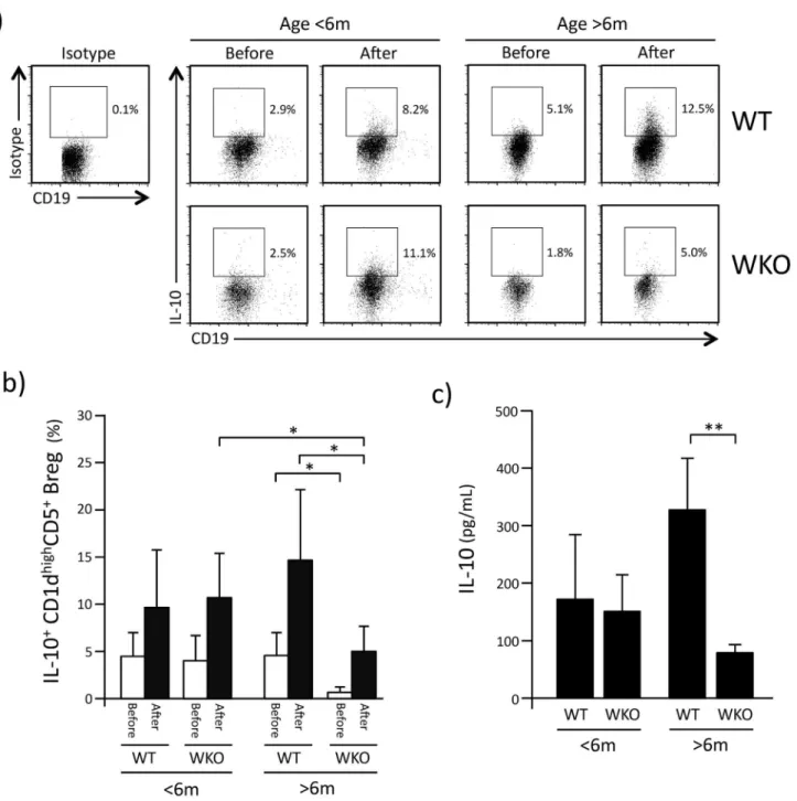
We then assessed the proportion of IL-10-producing CD1dhighCD5+Breg cells within WT and WKO splenocytes in resting conditions (Before) and 5hrs after stimulation with LPI (After,
Fig 2a). For both resting and stimulated splenocytes from younger animals, the proportion of
IL-10-producing CD1dhighCD5+Breg cells did not differ between WT and WKO mice (Fig 2b). However, in WKO mice older than 6 months, the percentage of IL-10+CD1dhighCD5+Breg cells both in resting and stimulated splenocytes was statistically lower than that detected in age-matched WT control mice. Moreover, the percentage of IL-10+CD1dhighCD5+Breg cells in older WKO mice cells was also statistically lower compared to younger WKO mice (p<0.05)
(Fig 2b).
Accordingly, while the concentration of IL-10 detected in the culture medium of stimulated CD1dhighCD5+Breg cells from younger WKO mice did not differ from WT control mice, IL-10 production by CD1dhighCD5+Breg cells from older WKO mice was statistically lower than that observed in age-matched WT CD1dhighCD5+Breg cells (Fig 2c, p<0.002).
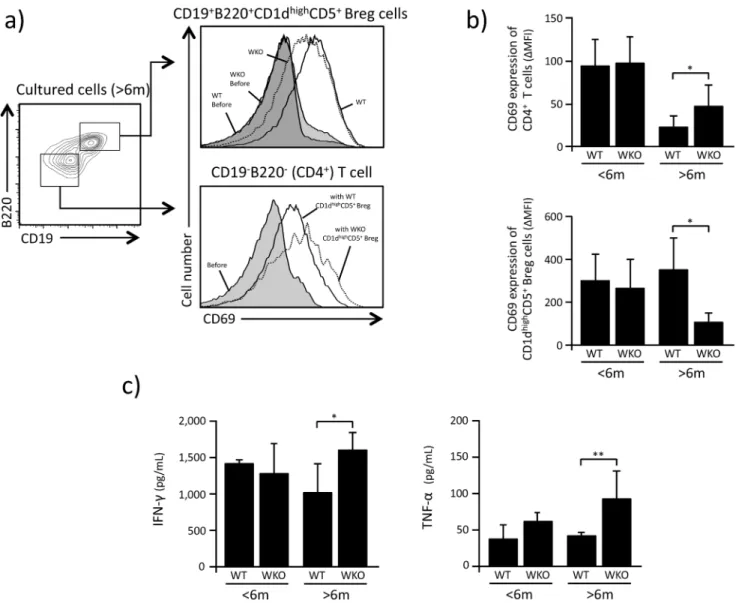
CD1dhighCD5+Breg cells have been reported to suppress the activation of CD4+T cells and affect their production of IFN- and TNF- (19). Based on the observations described above, we hypothesized that CD1dhighCD5+Breg cells from older WKO mice would show defective IL-10-mediated suppressing capabilities compared to CD1dhighCD5+Breg cells from older WT animals, as well as younger WKO mice. Indeed, in our co-culture experiments of CD4+T cells and CD1dhighCD5+Breg cells, WKO CD1dhighCD5+Breg cells from older WKO mice showed lower suppression of CD69 expression on WT CD4+T cells compared to CD1dhighCD5+Breg cells from age-matched WT (Fig 3a and 3b, p<0.05). We also noted that the CD69 expression
levels on cultured CD1dhighCD5+Breg cells from older WKO mice was statistically lower than that of CD1dhighCD5+Breg cells from age-matched WT controls (p<0.05). Of note, no
differ-ences were observed between the effects of CD1dhighCD5+Breg cells from younger WKO and WT mice on the expression of CD69 on the surface of co-cultured CD4+ T cells (Fig 3a
and 3b).
We also found that the concentration of IFN- and TNF- in the medium of co-cultures of WT CD4+T and WKO CD1dhighCD5+Breg cells from older mice was statistically higher than that from co-cultures of control WT CD4+T and WT CD1dhighCD5+Breg cells (IFN-: p<0.05,
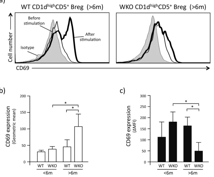
To investigate whether the lower production of IL-10 by CD1dhighCD5+Breg cells from older WKO mice was due to reduced effects of LPI stimulation, we assessed the expression levels of CD69 on the surface of CD1dhighCD5+Breg cells before and after LPI stimulation (Fig 4a).
Interestingly, we found that the CD69 expression levels on CD1dhighCD5+Breg cells from older WKO mice before LPI stimulation was statistically higher than that of CD1dhighCD5+ Breg cells from age-matched WT controls and younger WKO mice (Fig 4b, p<0.001). In
con-trast, after LPI stimulation, the CD69 expression levels of CD1dhighCD5+Breg cells from older WKO mice were statistically lower than that of CD1dhighCD5+Breg cells from age-matched WT and younger WKO controls (Fig 4c, p<0.05 and 0.001, respectively).
Our observations show that age plays a significant role in shaping the CD19+CD1dhighCD5+ B cell compartment of WKO mice. When analyzed at>6 months of age, WKO mice present
significantly lower percentages of IL-10 producing CD1dhighCD5+Breg cells compared to both WT control and young WKO mice. This condition results in reduced suppression of CD4+ T-cell activation and decreased inhibition of IFN- and TNF- production. Altogether, older WKO mice show significantly impaired regulatory B cell function that may contribute to the onset of the known autoimmune complications observed in this animal model and possibly to the age-dependent exacerbation of the autoimmune phenotype that we have previously described [15,20].
Further studies are needed to uncover the mechanisms leading to these findings. As shown by Bouma et al. [9], WKO mice of less than 6 months of age show a more severe experimental arthritis compared to control mice that can be ameliorated by adoptive transfer of Breg cells. By comparing equal numbers of WT and WKO CD1dhighCD5+Breg cells, we demonstrate here that WKO CD1dhighCD5+Breg cells from young WKO mice maintain the ability of secreting normal amounts of IL-10. The increased severity of the autoimmune arthritis Fig 1. Characteristics of CD1dhighCD5+Breg cells in WKO mice.a: Representative results of the percentages of CD1dhighCD5+Breg cells detected within total splenic CD19+B cells of WT and WKO mice. b: Total number of splenic CD1dhighCD5+Breg cells in younger (<6 months old) and older (>6 months old) mice. Average numbers (±SD) of CD1dhighCD5+Breg cells from 23
–49 mice are shown.*p<0.002 (Student’st-test).
observed in these mice, therefore, can simply be due to the reduction of Breg cell frequency observed by Bouma et al. and the lower absolute numbers of CD1dhighCD5+Breg cells identi-fied by us in WKO mice. Interestingly, we demonstrated that IL-10 secretion of WKO
CD1dhighCD5+Breg cells from older mice becomes significantly defective. Similar to the obser-vations we previously made in WKO nTreg cells that have significantly reduced ability of Fig 2. IL-10 production by WKO CD1dhighCD5+Breg.a: Representative flow-cytometry histograms showing IL-10-producing cells identified within CD1dhighCD5+Breg cells of younger (<6 months old) and older (>6 months old) mice before and 5 hrs after stimulation with LPS, PMA, and ionomycin. b: Percentage of IL-10-positive cells within CD1dhighCD5+Breg. White bars indicate the percentage (±SD) of IL-10-positive cells before stimulation (Before). Black bars indicate the percentage (±SD) IL-10 positive cells 5 hrs after stimulation with LPS, PMA and ionomycin (After). Each histogram represents data observed in 10 to 27 mice. c: IL-10 production after stimulation of CD1dhighCD5+Breg cells with LPS, PMA and ionomycin. Bar graphs indicate mean (±SD) of 3–5 experiments in which CD1dhighCD5+Breg cells were pooled from 6–10 mice were analyzed.*p<0.05,**p<0.002 (Student’st-test).
granzyme B degranulation [8], the defective secretion of IL-10 by WKO CD1dhighCD5+Breg cells may be attributable to defective cytokine trafficking and release due to aberrant cytoskeletal reorganization in the absence of WASp expression. However, the absence of similar findings in CD1dhighCD5+Breg cells from younger WKO mice makes this explanation somewhat less com-pelling. On the other hand, our findings that WKO CD1dhighCD5+Breg cells from older mice present with significantly higher levels of CD69 expression in basal conditions suggest that the functional impairment of CD1dhighCD5+Breg cells from older WKO mice may be at least in part due to abnormal activation, possibly translating in defective responses to further stimula-tion. The reasons underlying the increased levels of cellular activation in CD1dhighCD5+Breg cells from older WKO mice are at present unclear.
Fig 3. CD1dhighCD5+Breg suppression activity.a: Representative results showing the fluorescent intensity of CD69 expression on WT CD4+T cells and CD1dhighCD5+Breg cells 72 hrs after co-culture with anti-CD3/CD28. b: Fluorescence intensity of CD69 expression on the surface of CD4+T cells and CD1dhighCD5+Breg cells. Fluorescence intensity was expressed as delta mean fluorescence intensity (
ΔMFI), which was calculated by subtracting the intensity of expression in unstimulated cells from that of cultured cell populations. Bar graphs indicate mean±SD.*p<0.05 (Studentt-test). c: Concentrations of cytokine (IFN- and TNF-) measured in the medium of CD1dhighCD5+Breg cells and CD4+T cells co-cultured with LPS, PMA and ionomycin. Bar graphs indicate mean±SD of 3–5 experiments in which CD1dhighCD5+Breg cells pooled from 6
–10 mice were analyzed.*p<0.05,**p<0.02 (Student’st-test).
Conclusions
Our observations uncovered additional characteristics of the defective compartment of regula-tory B cells that develops in the absence of WASp expression and provides possible insights into the development of autoimmunity with increasing age in patients affected with WAS.
Supporting Information
S1 Fig. Gating strategy for evaluation of CD1highCD5+Breg cells.
(PDF)
Fig 4. Activation status of CD1dhighCD5+Breg cells before/after LPI stimulation.a: Representative flow-cytometry histograms showing the expression of CD69 on CD1dhighCD5+Breg cells. Gray curves indicate isotype staining of the CD1dhighCD5+Breg cells. Thin and thick lines indicate CD69 expression before and after LPI stimulation, respectively. b: CD69 expression intensity on CD1dhighCD5+Breg cells before LPI stimulation expressed as geometric mean. c: Change in intensity of CD69 expression on CD1dhighCD5+Breg cells after LPI stimulation. Data are expressed as
ΔMFI, calculated as inFig 3b. Data are mean and SD of samples obtained from 6–10 mice in each assays and repeated 3–5 times.*p<0.001 (Student’st-test).
Acknowledgments
The authors wish to thank the staff of the NHGRI Office of Laboratory Animal Medicine and the Animal Technicians of the NIH Building 49 Central Animal Facility.
Author Contributions
Conceived and designed the experiments: TY FC. Performed the experiments: TY MRK SMA. Analyzed the data: TY FC. Contributed reagents/materials/analysis tools: AY KLS. Wrote the paper: TY AY KLS FC.
References
1. Schurman SH, Candotti F. Autoimmunity in Wiskott-Aldrich syndrome. Curr Opin Rheumatol. 2002 Jul; 15(4):446–53. PMID:12819473
2. Dupuis-Girod S, Medioni J, Haddad E, Quartier P, Cavazzana-Calvo M, Le Deist F, et al. Autoimmunity in Wiskott-Aldrich syndrome: risk factors, clinical features, and outcome in a single-center cohort of 55 patients. Pediatrics. 2003 May; 111(5 Pt 1):e622–7. PMID:12728121
3. Ramesh N, Geha R. Recent advances in the biology of WASP and WIP. Immunol Res. 2009; 44(1– 3):99–111. doi:10.1007/s12026-008-8086-1PMID:19018480
4. Adriani M, Aoki J, Horai R, Thornton AM, Konno A, Kirby M, et al. Impaired in vitro regulatory T cell func-tion associated with Wiskott-Aldrich syndrome. Clin Immunol. 2007 Jul; 124(1):41–8. PMID:17512803
5. Maillard MH, Cotta-de-Almeida V, Takeshima F, Nguyen DD, Michetti P, Nagler C, et al. The Wiskott-Aldrich syndrome protein is required for the function of CD4+CD25+Foxp3+regulatory T cells. J Exp Med. 2007 Feb 19; 204(2):381–91. PMID:17296786
6. Marangoni F, Trifari S, Scaramuzza S, Panaroni C, Martino S, Notadangelo LD, et al. WASP regulates suppressor activity of human and murine CD4(+)CD25(+)FOXP3(+) natural regulatory T cells. J Exp Med. 2007 Feb 19; 204(2):369–80. PMID:17296785
7. Humblet-Baron S, Sather B, Anover S, Becker-Herman S, Kasprowicz DJ, Khim S, et al. Wiskott-Aldrich syndrome protein is required for regulatory T cell homeostasis. J Clin Invest. 2007 Feb; 117 (2):407–18. PMID:17218989
8. Adriani M, Jones KA, Uchiyama T, Kirby MR, Silvin C, Anderson SM, et al. Defective inhibition of B-cell proliferation by Wiskott-Aldrich syndrome protein-deficient regulatory T cells. Blood. 2011 Jun 16; 117 (24):6608–11. doi:10.1182/blood-2010-12-322834PMID:21515824
9. Bouma G, Carter NA, Recher M, Malinova D, Adriani M Notarangelo LD, et al. Exacerbated experimen-tal arthritis in Wiskott-Aldrich syndrome protein deficiency: Modulatory role of regulatory B cells. Eur J Immunol. 2014 Sep; 44(9):2692–702. doi:10.1002/eji.201344245PMID:24945741
10. Du HQ, Zhang X, An YF, Ding Y, Zhao XD. Effect of Wiskott-Aldrich syndrome protein deficiency on IL-10-producing regulatory B cells in humans and mice. Scand J Immunol. 2015 Jun; 81(6):483–93. doi: 10.1111/sji.12282PMID:25728049
11. Westerberg LS, de la Fuente MA, Wermeling F, Ochs HD, Karlsson MC, Snapper SB, et al. WASP con-fers selective advantage for specific hematopoietic cell populations and serves a unique role in mar-ginal zone B-cell homeostasis and function. Blood. 2008 Nov 15; 112(10):4139–47. doi:10.1182/ blood-2008-02-140715PMID:18772454
12. Recher M, Burns SO, de la Fuente MA, Volpi S, Dahlberg C, Walter JE, et al. B cell-intrinsic deficiency of the Wiskott-Aldrich syndrome proteins (WASp) causes severe abnormalities of the peripheral B-cell compartment in mice. Blood. 2012 Mar 22; 119(12):2819–28. doi:10.1182/blood-2011-09-379412 PMID:22302739
13. Simon KL, Anderson SM, Garabedian EK, Moratto D, Sokolic RA, Candotti F. Molecular and pheno-typic abnormalities of B lymphocytes in patients with Wiskott-Aldrich syndrome. J Allergy Clin Immunol. 2014 Mar; 133(3):896–9.e4. doi:10.1016/j.jaci.2013.08.050PMID:24210885
14. Wada T, Schurman SH, Garabedian EK, Yachie A, Candotti F. Analysis of T-cell repertoire diversity in Wiskott-Aldrich syndrome. Blood. 2005 Dec 1; 106(12):3895–7. PMID:16091449
15. Nikolov NP, Shimizu M, Cleland S, Bailey D, Aoki J, Strom T, et al. Systemic autoimmunity and defec-tive Fas ligand secretion in the absence of the Wiskott-Aldrich syndrome protein. Blood. 2010 Aug 5; 116(5):740–7. doi:10.1182/blood-2009-08-237560PMID:20457871
17. Yoshizaki A, Miyagaki T, DiLillo DJ, Matsushita T, Horikawa M, Kountikov EI, et al. Regulatory B cells control T-cell autoimmunity through IL-21-dependent cognate interactions. Nature. 2012 Nov 8; 491 (7423):264–8. doi:10.1038/nature11501PMID:23064231
18. Yanaba K, Bouaziz JD, Matsushita T, Tsubata T, Tedder TF. The development and function of regula-tory B cells expressing IL-10 (B10 Cells) requires antigen receptor diversity and TLR signals. J Immu-nol. 2009 Jun 15; 182(12):7489–72. doi:10.4049/jimmunol.0900270PMID:19494269
19. Matsushita T, Horikawa M, Iwata Y, Tedder TF. Regulatory B cells (B10 cells) and regulatory T cells have independent roles in controlling experimental autoimmune encephalomyelitis initiation and late-phase immunopathogenesis. J Immunol. 2010 Aug 15; 185(4):2240–52. doi:10.4049/jimmunol. 1001307PMID:20624940