Comparative transcriptome analysis
of papilla and skin in the sea cucumber,
Apostichopus japonicus
Xiaoxu Zhou1,2,*, Jun Cui2,*, Shikai Liu3,*, Derong Kong2, He Sun2, Chenlei Gu2, Hongdi Wang2, Xuemei Qiu1,2, Yaqing Chang1,2, Zhanjiang Liu3and Xiuli Wang1,2
1Key Laboratory of Mariculture & Stock Enhancement in North China’s Sea, Ministry of Agriculture, Dalian Ocean University, Dalian, China
2College of Fisheries and Life Science, Dalian Ocean University, Dalian, China
3The Fish Molecular Genetics and Biotechnology Laboratory, Aquatic Genomics Unit, School of Fisheries, Aquaculture and Aquatic Sciences and Program of Cell and Molecular Biosciences, Auburn University, Auburn, Alabama, United States
*These authors contributed equally to this work.
ABSTRACT
Papilla and skin are two important organs of the sea cucumber. Both tissues have ectodermic origin, but they are morphologically and functionally very different. In the present study, we performed comparative transcriptome analysis of the papilla and skin from the sea cucumber (Apostichopus japonicus) in order to identify and characterize gene expression profiles by using RNA-Seq technology. We generated 30.6 and 36.4 million clean reads from the papilla and skin and de novo assembled in 156,501 transcripts. The Gene Ontology (GO) analysis indicated that cell part, metabolic process and catalytic activity were the most abundant GO category in cell component, biological process and molecular funcation, respectively. Comparative transcriptome analysis between the papilla and skin allowed the identification of 1,059 differentially expressed genes, of which 739 genes were expressed at higher levels in papilla, while 320 were expressed at higher levels in skin. In addition, 236 differentially expressed unigenes were not annotated with any database, 160 of which were apparently expressed at higher levels in papilla, 76 were expressed at higher levels in skin. We identified a total of 288 papilla-specific genes, 171 skin-specific genes and 600 co-expressed genes. Also, 40 genes in papilla-skin-specific were not annotated with any database, 2 in skin-specific. Development-related genes were also enriched, such asfibroblast growth factor,transforming growth factor-b,
collagen-a2andIntegrin-a2, which may be related to the formation of the papilla and skin in sea cucumber. Further pathway analysis identified ten KEGG pathways that were differently enriched between the papilla and skin. The findings on expression profiles between two key organs of the sea cucumber should be valuable to reveal molecular mechanisms involved in the development of organs that are related but with morphological differences in the sea cucumber.
Subjects Aquaculture, Fisheries and Fish Science, Biochemistry, Genomics
Keywords Comparative transcriptome, High-throughput sequencing, Gene expression, Papilla, Skin, Sea cucumber (Apostichopus japonicus)
Submitted12 November 2015
Accepted17 February 2016
Published7 March 2016
Corresponding author
Xiuli Wang, [email protected]
Academic editor
Tzi Bun Ng
Additional Information and Declarations can be found on page 15
DOI10.7717/peerj.1779
Copyright
2016 Zhou et al.
Distributed under
INTRODUCTION
The sea cucumbers group (Echinodermata, Holothuroidea) comprises approximately 1,250 species (Du et al., 2012). Sea cucumbers are mostly processed into a dry product called trepang, beˆche-de-mer or hai-san, which is widely recognized as a delicate food with a medicinal effect for human consumption. Sea cucumbers have been harvested for commercial use for a thousand years, and they are now widely cultured in more
than 70 countries (Steven et al., 2012). The sea cucumber Apostichopus japonicus
(Holothuroidea, Aspidochirotida) is intensively cultured in many East Asian countries and is naturally found along the coasts of China, Japan, Korea and Russia of Northeast Asia (Sloan, 1984;Chang et al., 2009). It is intensively cultured as an important
aquaculture species in many countries of East Asia.
The pentamerous radial symmetry is considered as one of the characteristics of echinodermata. In sea cucumber, pentamerous symmetry is usually determined based on the presence of five meridional ambulacra bearing podia (Steven et al., 2012). Papillae represent the podia on the dorsal surface, and generally have no locomotive function. WithA. japonicus, fleshy and conical papillae, with a sensory spina at its apex, are present in two loose rows on the dorsal surface and two rows at the lateral margins of the ventral surface (Steven et al., 2012). Previous studies have investigated the morphological characteristics of papilla in theA. japonicus(Vanden-Spiegel et al., 1995;Chang et al., 2011; Steven et al., 2012). In the papillae, the ciliated cells and histamine-like immunoreactivity neurons are in contact with the nerve plexus (Hyman, 1955;Luke et al., 2012).
Therefore, the dorsal papillae have long been associated to a sensory role, which may involve chemoreception and mechanoreception (Vanden-Spiegel et al., 1995).
The thicker body wall ofA. japonicusconsists of a thin cuticle over the epidermis and a thick dermis underneath. The cuticle and epidermis as the outer tissues of the
A. japonicus are represented by skin (Steven et al., 2012). The skin forms a protective barrier, forming the first line of defence against the environment. Previous studies have been conducted on skin, with the main focus on the intrinsic mechanisms underlying immune response to skin ulceration and peristome tumescence (Liu et al., 2010; Zhang et al., 2013).
The papillae are closely associated with the skin in sea cucumber. Both organs are mainly composed of collagen (up to 70%), and are the major component of the body wall. Moreover, the papillae and skin are formed by similar elements and homologous cell types, such as keratinocytes, epidermis and dermis, all derived from the ectoderm (Chang et al., 2004;Lowdon et al., 2014). Despite the common embryonic origin of the two organs, they exhibit clear morphological differences and play distinct functions. The molecular mechanisms underlying differentiation between the papilla and skin remain largely unknown. The lack of reference genome and the limited genetic resources ofA. japonicus
2008;Wang, Gerstein & Snyder, 2009). Recently, several RNA-Seq based transcriptome analyses have been conducted in theA. japonicus, including studies on histology
(Sun et al., 2011;Sun et al., 2013), immunology (Li et al., 2012), physiology (Zhao et al., 2014a;Zhao et al., 2014b), embryonic development and gene marker discovery
(Du et al., 2012;Zhou et al., 2014). The first transcriptome sequencing of theA. japonicus
intestine and body wall was performed bySun et al. (2011). Thereafter, the global dynamic changes during all stages of intestine regeneration were further investigated (Sun et al., 2013). To identify candidate transcripts potentially involved in aestivation and generate a wide coverage of transcripts involved in a broad range of biological processes, eight cDNA libraries were constructed and sequenced byDu et al. (2012). Immune-related genes and pathways in response to pathogen infection were identified (Zhou et al., 2014;Gao et al., 2015). Moreover, many physiological networks were identified and characterized in theA. japonicus on the basis of transcriptomic resources (Wang et al., 2015;Yang et al., 2015).
Here, in this work, we report comparative transcriptome analysis of the papilla and skin. A relatively large number of genes that displayed distinct expression profiles between the papillae and skin were identified. Further enrichment analysis identified pathways such as tight junction and p53 signaling pathway could be involved in the development of the papilla and skin. This work provided the essential genomic resources for further investigations into the molecular interactions and multiple biological process of appendages such as the papilla and skin in the A. japonicus.
MATERIALS AND METHODS
Sample collectionA total of 45 sea cucumbers (average weight of 25 g) provided by the Key Laboratory of Mariculture in North China (Dalian, Liaoning) were used in the present study. In order to have a good reference transcriptome, the skin around the papillae, papilla and tube foot tissues were collected for RNA-Seq. We randomly group these 45 sea cucumbers into three groups as replicates. Within each group,∼1 g tissue was dissected
from each individual, respectively. Tissues collected from each group were of every individual were pooled (one pool per tissue) and placed in 2 ml of RNAlaterÒ
Solution (Ambion, Carlsbad, CA, USA) for overnight at 4C followed by transferring to
−80C
until RNA extraction.
RNA-Seq
Transcriptome assembly and annotation
Low quality reads and adaptors were trimmed before assembly. Trimed reads were de novo assembled by Trinity software using default parameters (Grabherr et al., 2011) and used as a reference for gene expression analysis. Transcirptome was annotated using Basic Local Alignment Search Tool (BLAST) searches against the NCBI Non-Redundant (NR) database, Swiss-Prot, KEGG (the Kyoto Encyclopedia of Genes and Genomes) and GO (Gene Ontology), Clusters of Orthologous Groups (COG) and Eukaryotic Orthologous Groups (KOG) with e-value cutoff of 1e-5.
Differentially Expressed Gene (DEG) analysis
Gene expression was determined by the FPKM (Fragments Per kb of transcript per Million mapped fragments) method. The gene expression differences between the papilla and skin tissues were identified following the formula:
Fold change ¼ Log2
FPKMPapilla FPKMSkin
DEGs were determined with the absolute fold change values greater than 2.0, and FDR (false discovery rate) lesser than 0.01 (Cui et al., 2014;Cui et al., 2015).
To further investigate DEGs identified between papilla and skin, genes were
compared to those identified from theA. japonicusintestine RNA-Seq dataset (accession NO.GSE44995) from a previous study bySun et al. (2013). The intestine is responsible for the metabolic rate depression under deep aestivating conditions (Chen et al., 2013) and plays a role in organ regeneration (Sun et al., 2013). In the present study, we used the intestine as a major site in the internal environment of A. japonicusto further investigate DEGs identified between papilla and skin. All assembled sequences of
A. japonicus published in Sun et al. (2013) were downloaded as a database to blast the DEGs; the differential expression of DEGs among the papilla, skin, and intestine was estimated using the formula:
Score¼Log2
FPKMTissues RPKMIntestines
Where FPKMTissuesindicates the FPKM of papilla or skin; RPKMIntestinesindicates the RPKM of intestine. Significant candidates were determined as the absolute score greater than 4.0.
qRT-PCR validation
And three technical replications were performed for each qRT-PCR validation. PCR was conducted as follows: 94C for 30 s, 45 cycles of 94C for 5 s, annealing temperature (showed in Table S1) for 15 s, and 72 C for 15 s.
Gene enrichment analysis
The gene enrichment analysis was conducted using KEGG database. The over-presentation of the DEGs was determined in the specific pathways. The level of enrichment was indicated by enrichment factor, and p-value was used to calculate the significance of enrichment. The top 10 KEGG enrichments were selected to carry out further analysis.
RESULTS
Sample sequencing
RNA-Seq of the papilla and skin samples yielded over 70 million pair-end reads with average length of 125 bp (Table 1). Similar number of reads was obtained from both tissues, with over 33 million reads from papilla and over 37 million from skin. After trimming, 30.6 and 36.4 million high-quality reads were retained from papilla and skin, respectively. Totally, 7.7 billion bases generated from the papilla, 9.2 billion bases generated for the skin and 8.6 billion bases generated for the tube foot were used for down-stream analysis of de novo assembly and mapping. Data obtained from papilla, skin and tube foot were deposited to the sequence read archive (SRA) with the accession numbers SRX1097860andSRX1081978.
Transcriptome assembly and annotation
The de novo assembly resulted in a total of 156,501 transcripts, with the average length of 910.77 bp and N50 length of 1,694 bp (Table 2). The length distribution of transcripts and unigenes are shown inFig. 1. Over 84% of reads from both tissues were successfully mapped back to the de novo transcriptome assembly.
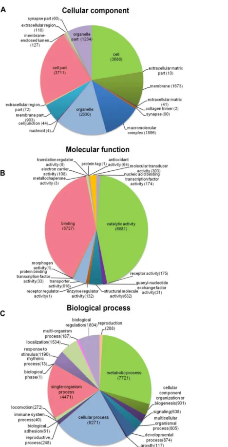
The transcriptome assembly was annotated by BLASTX against NCBI NR, Pfam, Swiss-Prot, KEGG, COG and KOG databases with E-value threshold of 1e-5. Annotation resulted in the identification of 92,343 unigenes (unique transcripts matched with known proteins). From all the 92,343 unigenes, 30,706 were found to have homologs in NR database, 22,261 found to posses functional domains in Pfam database; 18,944 unigenes showed significant matches to Swiss-Prot database, 22,361 to KOG, 11,190 to KEGG, 10,876 to COG and 12,410 unigenes were associated with GO terms (Table 2). Taken together, a total of 33,584 unigenes had at least one significant matches to these databases (Table 2). The unigenes annotated with NR database accounted for the largest proportion (91.4%), followed by Pfam and Swiss-Prot (Fig. 2).
study,Du et al. (2012)found that membrane-bounded organelle was the most represented GO term in cell component; the major category in biological process was the primary metabolic process; and genes involved in hydrolase activity accounted for major proportion in molecular function. To be noted, because the samples used inDu et al. (2012)study were collected from different developmental stages and adult tissues (intestines, respiratory trees and coelomic fluid), there may be some biases.
Identification of DEGs
A total of 1,059 DEGs were identified between the papilla and skin. The MA plot showed significant DGE (blue) against all non-significant DEG (red) (Fig. 4A). Among identified DEGs, 739 were expressed at significantly higher in papilla, while 320 genes were expressed at significantly higher levels in skin (Table S2). The number of genes with higher expression levels in papilla was over twice than the number of that in skin. Papilla, as the projections of body wall, included more unique contents than skin, such as the calcareous ossicles, which are hidden in the dermis of body wall, papillae and tentacles (Steven et al., 2012). We also analysed the expression profiles of 1,059 DEGs in each tissue. Papilla-specific genes represent the DEGs that there is no expression in the skin, and that goes for skin-specific. A total of 288 papilla-specific DEGs were expressed only in papilla,
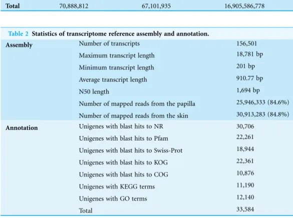
Table 2 Statistics of transcriptome reference assembly and annotation.
Assembly Number of transcripts 156,501
Maximum transcript length 18,781 bp
Minimum transcript length 201 bp
Average transcript length 910.77 bp
N50 length 1,694 bp
Number of mapped reads from the papilla 25,946,333 (84.6%)
Number of mapped reads from the skin 30,913,283 (84.8%)
Annotation Unigenes with blast hits to NR 30,706 Unigenes with blast hits to Pfam 22,261
Unigenes with blast hits to Swiss-Prot 18,944
Unigenes with blast hits to KOG 22,361
Unigenes with blast hits to COG 10,876
Unigenes with KEGG terms 11,190
Unigenes with GO terms 12,140
Total 33,584
Table 1 Summary of the RNA-Seq data.
Number of reads Number of reads after trimming
Number of nucleotides after trimming (bp)
Papilla 33,504,127 30,657,027 7,723,425,469
Skin 37,384,685 36,444,908 9,182,161,309
while 171 DEGs were found to be only expressed in skin (skin-specific). A total of 600 DEGs were expressed in both papilla and skin (Fig. 4B). Apparently, the number of skin-specific (53.44%) genes is higher than papilla-specific genes (38.97%).
Of the 1,059 DEGs, 61 DEGs were annotated to homologous genes instrongylocentrotus purpuratus, a model species that is closely related to A. japonicus. Hsp gp96,Hsp26,
ALDOA(aldolase class-1 protein) andtenasxinwere annotated withA. japonicus. Our results revealed thatHsp gp96andALDOAwere 3.93- and 4.45-Fold up-regulated in papillae, respectively. In constrast,Hsp26andtenascinwere−2.39- and−3.62-Fold
down-regulated in skin, respectively. In addition, 236 differentially expressed genes were not annotated with any database, 160 of which were apparently higher in papilla. Further analysis revealed that 40 of which were papilla-specific and two were skin-specific.
Putative genes related to development that may be associated with the formation of the papilla were identified (Table 3). Detailed information of develop-related genes was provided inTable S3. Our results revealed thatcuticle collagen 2andalpha-2 collagenwere highly expressed in papilla with 5.76 and 2.55, respectively. Several genes that know to be related to the collagen development (Hinz et al., 2003;Hinz, 2009;Leask & Abraham, 2004), such asFibroblast Growth Factor(FGF),Transforming Growth Factor-b(TGF-b) andIntegrin-a2(ITGA2) were found to be significantly expressed. SeveralRas-related genes such asRan,Rab1a,Arf3,Ran1, Ras, RhoA, Rho Guanine nucleotide exchange factors
(RhoGEF), Rho GTPase, Rho GTPase activation protein (RhoGAP) and Ran-binding protein 1 (RanBP1), which play key roles in the development by regulating growth and morphogenesis, were also identified in our study (Table S4). All Ras-related DEGs were expressed at lower levels in skin except forRhoGEFsthat were reported to be associated with cancer, pathogen infection or neural system related diseases and development (Reichman et al., 2015). Understanding of the function ofRas-related genes will
facilitate to unravel the mechanisms of some physiological and pathological process in the skin of A. japonicus.
To further verify DEGs data, we compared our results with those DEGs identified in the intestine ofA. japonicusfrom a previous study Sun et al. (2013)(results are shown in
Table 4). Seven DEGs showed the same score trend as that of the fold change in papilla. The reason for this observation could be due to the lack of a complete RPKM data (Sun et al., 2013).
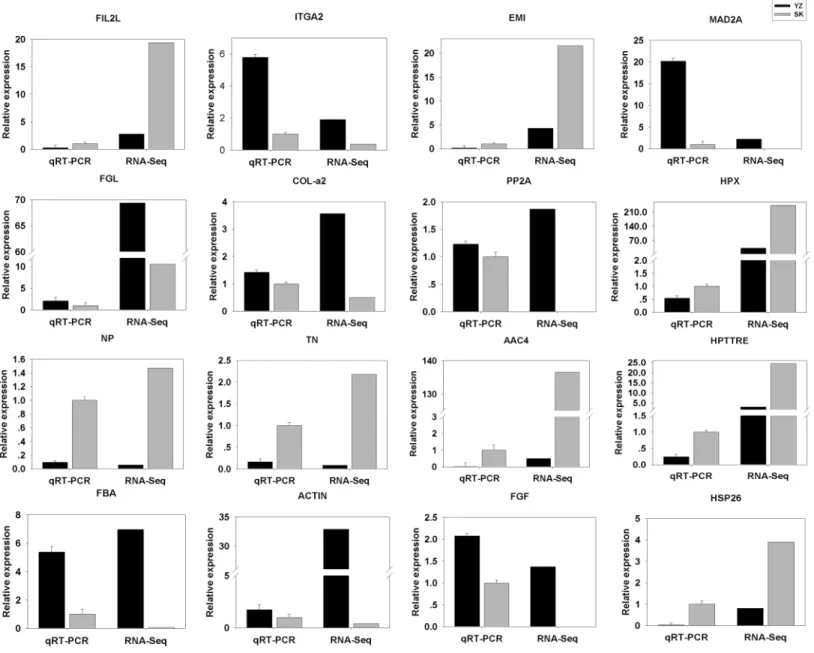
qRT-PCR validation
To validate the DEGs results obtained, we randomly selected 16 DEGs for validation using qRT-PCR. As shown inFig. 5, the DGEs identified from qRT-PCR analysis were correlated well with those obtained from qRT-PCR, indicating the reliability and accuracy of the RNA-Seq method used in the present study.
Enrichment analysis of DEGs
A total of 296 DEGs identified were mapped to 133 pathways. KEGG enrichment pathway analysis was also carried out to investigate their potential functional roles. The top 10 enrichment pathways were selected by a hypergeometric test (p < 0.05) (Table 5). One of
which is the ribosome pathway, which was related to the protein biogenesis and was observed to be involved in intestine regeneration (Sun et al., 2013) and aestivation (Chen et al., 2013;Zhao et al., 2014a;Zhao et al., 2014b) in theA. japonicus. In addition, tight junction and p53 signaling pathways were also detected in enrichment pathways analysis (detailed information is provided inTable S5).
Table 3 Differentially expressed genes between the papilla and skin that are involved in development.
Unigene ID Gene symbol Foldchange
c14695.graph_c0 cdk 4.96
c40875.graph_c0 cyc-B 6.26
c75877.graph_c0 cyc-A 6.346
c76406.graph_c0 cytC 2.76
c76859.graph_c0 gadd45a 4.82
c12901.graph_c0 ck2bl 4.40
c14611.graph_c0 MAGUKs 4.34
c18023.graph_c0 PP2A 4.54
c19039.graph_c0 claudin 6.31
c37832.graph_c0 actin 6.05
c16255.graph_c0 FGF 5.26
c58770.graph_c0 ITGA2 2.12
c15897.graph_c0 TGF-b 6.21
c54738.graph_c0 col-a2 2.55
c54237.graph_c0 tub-a −4.53
c77661.graph_c0 emmhc 4.49
c54933.graph_c0 eef2 −5.43
c42633.graph_c0 gtf 8 4.96
c57892.graph_c0 ubeE2 −4.18
c76626.graph_c0 ctATPase 4.62
c38162.graph_c0 cul-a2 5.76
Table 4 The result of DEGs with significantly different expression by comparison with the intestine.
Unigene ID Isotig ID Annotate Foldchange Score
c45050.graph_c0 isotig25664 Cell death abnormality protein 1 8.62 14.40
c64723.graph_c0 isotig15743 Sushi domain (SCR repeat) 7.15 4.09
c67657.graph_c0 isotig19241 Fibrinogen-like protein A 3.02 4.96
c66534.graph_c0 isotig15670 Hypothetical protein CAPTEDRAFT_211426 2.84 11.35
c73725.graph_c2 isotig27287 Sulfotransferase family 2.56 12.76
c60095.graph_c0 isotig09563 Hypothetical protein BRAFLDRAFT_231341 2.54 4.31
c60588.graph_c0 isotig18328 Histone-lysine N-methyltransferase 2.16 13.36
Note:
DISCUSSION
In this study, we conducted comparative transcriptome analysis between papilla and skin, two important organs of sea cucumber. A total of 1,059 differentially expressed genes were identified between the two organs. This result lay the foundation to identify genes that were potentially involved in the development of the papilla and skin. The generated genomic resources should be valuable for other genetic and genomic studies in the A. japonicus.
Figure 5 Comparison between RNA-Seq results and qRT-PCR validation results.X-axis shows genes in two tissues validated in this study; Y-axis shows Log2Ratio of expression of SK (skin) versus YZ (papilla). AAC4PL, AAC-rich mRNA clone AAC4 protein-like; Hsp26, heat shock protein 26;
NP, novel protein; TN, Tenascin; EMI, EMI domain; Hp TTRE, hypothetical protein TTRE_0000953901; FGL, Fibrinogen-like protein A; HpX975-24482, hypothetical protein X975_HpX975-24482, partial; PP2A, Serine/threonine-protein phosphatase; FIL2L, Fibrinogen-like protein A; Col-aFiCollagen
As previously reported, excessive deposition resulting from abnormal balance of growth factors and cell proliferation can improve local hyperplastic collagen production in skin in response to injury in mammalians (Tuan & Nichter, 1998). Keloids (Seifert & Mrowietz, 2009; Shih & Bayat, 2010) and Hypertrophic Scar (HS) (O’Leary, Wood & Guillou, 2002), are characterized by fibroblastic proliferation and accumulation of Extracellular Matrix (ECM), especially excessive deposition of collagen. However, such prominences are regarded as benign tumors (Diao et al., 2011). It has been suggested that factors such asFGF,ITGA2,TGFandS-adenosylmethionine(a-SMA) can cause those lesions (Hinz et al., 2003;Hinz, 2009; Leask & Abraham, 2004). The FGF activity was first identified from bovine pituitary in 1974 (Gospodarowicz & Moran, 1974).FGFsignaling is required for different developmental stages during embryogenesis (Sun et al., 1999;Naiche, Holder & Lewandoski, 2011;Niwa et al., 2011;Vega-Herna´ndez et al., 2011). Compared with normal dermal fibroblast, TGF-bis believed to induce collagen production and increase the contractile activity in keloid fibroblasts (Bran et al., 2010; Sandulache, Parekh & Li-Korotky, 2007). In additon, TGF-b associated with
connective tissue growth factor (CCN2) has been revealed to stimulate a-SMA, collagen
expression (Jiang et al., 2008).ITGA2is the main cell adhesion molecule that takes part in the modulation of collagen contraction and the activity of myofibroblast in HS (Cooke, Sakai & Mosher, 2000). The expression levels ofITGA2 were also found up-regulated in hypertrophic scar fibroblasts, compared with normal skin tissues in human. In our study, we found thatcol-a2was expressed at a higher level in the papilla, and we also observed differential expression patterns of genes involved in collagen synthesis as the major differences between the papilla and skin. The expression of FGF,TGF-b
andITGA2, associated with collagen development, were all expressed at higher levels in the papilla. Compare to previous studies of local hyperplastic collagen in mammals, we speculate that these collagen-related genes may play critical roles in stimulating the production of collagen in papilla and might be involve in the morphological
differentiation between the two organs.
Table 5 Enrichment analysis of genes with significantly differential expression between the papilla and skin.
Pathway KO Enrichment factor p-value
Ribosome ko03010 0.39 1.47E-13
Oocyte meiosis ko04114 0.27 2.07E-06
Cell cycle ko04110 0.40 0.000557178
Glycolysis/Gluconeogenesis ko00010 0.43 0.000610528
p53 signaling pathway ko04115 0.28 0.003222732
Tight junction ko04530 0.38 0.004679565
NOD-like receptor signaling pathway ko04621 0.27 0.010624351
Regulation of actin cytoskeleton ko04810 0.44 0.017102208
RNA transport ko03013 0.68 0.029611824
Besides the development-related genes as discussed above, some immune-related genes are also identified as being differentially expressed between papilla and skin in this study, such as Hspgp96, Hsp26, ALDOAandtenascin. Many lines of evidences support that Hsps act as natural immunoregulatory agents, increasing the awareness of innate immune cells to pathogens (Ciancio & Chang, 2008;Prohaszka & Fust, 2004; Zugel & Kaufmann, 1999). ALDOA plays a role in glycolysis pathway (Oparina et al., 2013). Further investigation is required to explore the pathological researches for papilla and skin. Through analysis of the DEGs against intestine transcriptome data from a previous study (Sun et al., 2013), we found Fibrinogen-like protein A(fglA) showed the same score trend as that of fold change in papilla. FglA is a member of the fibrinogen-related protein superfamily, plays crucial roles including innate immune response, regeneration and blood clotting (Yamamoto et al., 1993). Previous studies have demonstrated that fglA is widely distributed in A. japonicusbody wall, intestines, longitudinal muscles and respiratory tree of A. japonicus(Wu et al., 2014). Our results also show that fglAwas 3.02 fold change and 4.96 score up-regulated in papilla, respectively. The role of fglAin the development of papilla remains unclear and further investigation is required to understand its functional roles.
Enrichment KEGG analysis revealed that tight junction and p53 signaling pathway were highlighted in enrichment pathways. In humans, the content of G2-M arrested cells in keloid skin was higher than in normal skin (Varmeh et al., 2011). Keloid fibroblasts showed a higher rate of senescence and lower proliferative capacity in comparison to normal fibroblasts (Varmeh et al., 2011). In our study, a set of genes, including growth arrest and DNA-damage-inducible protein (gadd45), cyclin dependent kinase 1 (cdk), cyclin-B (cyc-B), cyclin-A (cyc-A) and cytochrome C (cyt-C), were all expressed at higher levels in the papilla (Table 1). These genes are involved in the p53 signaling pathway. Once p53 signaling pathway is activated, it can induce either cell cycle arrest or apoptosis in the damaged cell. In humans, cyc-B and cdk2 kinase influence a cell’s progress through the cell cycle, which is especially important in several skin cancers (Ely et al., 2005;
Casimiro et al., 2014). Cyc-B forms the regulatory subunits and cdk2 form the catalytic subunits of an activated heterodimer. The cyc-B has no catalytic activity, and cdk2 is inactive in the absence of a partner cyc-B. Once activated the cdk2/cyc-B complex control cell cycle (Abreu Velez & Howard, 2015). Gadd45 is a ubiquitously expressed 21 protein with a key role in response to genotoxic agents, and it is involved in many biological processes related to maintenance of genomic stability and apoptosis. It has been shown that gadd45’s inhibits cdk2 kinase activity through alteration of cyc-B subcellular localization, inducing the arrest of the cell cycle in G2-M state (Jin et al., 2000;Smith et al., 1994). These results indicated that the level of cell cycle arrest at the G2-M in the papilla might be higher than in the skin. It’s speculated that papilla fibroblasts commit to a higher rate of senescence, which may cause fibroblast-related genes eventually stop expressing and maintain external morphology of the papilla.
found that the expression of Serine/threonine-protein phosphatase (PP2A) in papilla is higher than that in the skin. Many reports showed that PP2A regulatesAtaxia
Telangiectasia Mutated (ATM),Ataxia Telangiectasia Rad3 related(ATR),Check Point Kinase-1 (CHK1), andCheckpoint Kinase-2(CHK2) after DNA damage, and activate the checkpoint of G2-Massociated with the p53 signaling pathway. The process activated by PP2A may also regulate the external morphological of papilla and skin of
A. japonicus.
CONCLUSION
In this study, we performed comparative transcriptome analysis of the skin and papailla
A. japonicusby using RNA-Seq. In total, 156,501 transcripts and 92,343 unigenes were assembled. A total of 1,059 differentially expressed genes were indentified between the two important organs ofA. japonicus. We identified 236 novel genes (not annotated with any database), 160 of which were expressed at higher levels in papilla. Further tissue-expression analysis identified 288 papilla-specific genes and 171 skin-specific genes. Gene pathway enrichment analysis revealed several gene pathways that were involved in development. In addition, many DEGs involved in the process of p53 signaling pathway and tight junction were also identified, which were ported to be relative to keloid skin in humans. This result provided insight into genes and pathways that may be
associated with the formation of the papilla and skin in sea cucumber, laying the foundation for further investigation to understand the development of the papilla in
A. japonicus. Moreover, the generation of larger-scale transcriptomic data presented in this work enriched genetic resources of the echinodermata species, which should be valuable to comparative and evolutionary studies in echinoderms.
ACKNOWLEDGEMENTS
We thank Dr. Daniel Garcia de la serrana (Fish Muscle Research Group, Scottish Oceans Institute, University of St Andrews, UK) for revising and polishing the manuscript. We would like to acknowledge Prof. Tzi Bun Ng and two anonymous reviewers for helpful comments on the manuscript.
ADDITIONAL INFORMATION AND DECLARATIONS
Funding
This project was supported by the State 863 High-Technology R & D Project of China (No. 2012AA10A412) and the Program for Liaoning Excellent Talents in University, China (No. LR2014022). The funders had no role in study design, data collection and analysis, decision to publish, or preparation of the manuscript.
Grant Disclosures
Competing Interests
The authors declare that they have no competing interests.
Author Contributions
Xiaoxu Zhou conceived and designed the experiments, performed the experiments, analyzed the data, contributed reagents/materials/analysis tools, wrote the paper, prepared figures and/or tables, reviewed drafts of the paper.
Jun Cui analyzed the data, wrote the paper, prepared figures and/or tables, reviewed drafts of the paper.
Shikai Liu analyzed the data, contributed reagents/materials/analysis tools, wrote the paper, prepared figures and/or tables, reviewed drafts of the paper.
Derong Kong performed the experiments, reviewed drafts of the paper. He Sun performed the experiments, reviewed drafts of the paper. Chenlei Gu performed the experiments, reviewed drafts of the paper.
Hongdi Wang contributed reagents/materials/analysis tools, reviewed drafts of the paper.
Xuemei Qiu contributed reagents/materials/analysis tools, reviewed drafts of the paper. Yaqing Chang contributed reagents/materials/analysis tools, reviewed drafts of the
paper.
Zhanjiang Liu wrote the paper, reviewed drafts of the paper.
Xiuli Wang conceived and designed the experiments, analyzed the data, contributed reagents/materials/analysis tools, wrote the paper, reviewed drafts of the paper.
DNA Deposition
The following information was supplied regarding the deposition of DNA sequences: 1. Raw reads of papilla deposited in sequence read archive database (SRX1097860). 2. Raw reads of skin deposited in sequence read archive database (SRX1097860).
Supplemental Information
Supplemental information for this article can be found online athttp://dx.doi.org/ 10.7717/peerj.1779#supplemental-information.
REFERENCES
Abreu Velez AM, Howard MS. 2015.Tumor-suppressor genes, cell cycle regulatory checkpoints, and the skin.North American Journal of Medical Sciences7(5):176–188
DOI 10.4103/1947-2714.157476.
Anisimov SV. 2008.Serial Analysis of Gene Expression (SAGE): 13 years of application in research.
Current Pharmaceutical Biotechnology9(5):338–350DOI 10.2174/138920108785915148. Bran GM, Sommer UJ, Goessler UR, Ho¨rmann K, Riedel F, Sadick H. 2010.TGF-b1 antisense
impacts the SMAD signalling system in fibroblasts from keloid scars.Anticancer Research
30:3459–3463.
Chang Y, Ding J, Song J, Yang W. 2004.Biology and Aquaculture of Sea Cucumbers and Sea Urchins. Beijing: Ocean Press, 35–38 (in Chinese).
Chang YQ, Zhu F, Yu J, Ding J. 2009.Genetic variability analysis in five populations of the sea cucumber Stichopus (Apostichopus japonicus) from China, Russia, South Korea and Japan as revealed by microsatellite markers.Marine Ecology30(4):455–461DOI 10.1111/j.1439-0485.2009.00292.x. Chang YQ, Shi SB, Zhao C, Han ZH. 2011.Characteristics of papillae in wild, cultivated and
hybridsea cucumbers (Apostichopus japonicus).African Journal of Biotechnology10:13780–13788. Chen M, Zhang X, Liu J, Storey KB. 2013.High-throughput sequencing reveals differential
expression of miRNAs in intestine from sea cucumber during aestivation.PLoS ONE
8(10):e76120DOI 10.1371/journal.pone.0076120.
Ciancio MJ, Chang EB. 2008.Do heat shock proteins play any role in gut inflammation?
Inflammatory Bowel Diseases14(Suppl 2):S102–S103DOI 10.1002/ibd.20697.
Cooke ME, Sakai T, Mosher DF. 2000.Contraction of collagen matrices mediated bya2 beta1 A anda(v) beta3 Ins.Journal of Cell Science113:375–383.
Cui J, Liu S, Zhang B, Wang H, Sun H, Song S, Qiu X, Liu Y, Wang X, Jiang Z, Liu Z. 2014. Transciptome analysis of the gill and swimbladder of takifugurubripes by RNA-seq.PLoS ONE
9(1):e85505DOI 10.1371/journal.pone.0085505.
Cui J, Xu J, Zhang S, Wang K, Jiang Y, Mahboob S, Al-Ghanim KA, Xu P. 2015.Transcriptional profiling reveals differential gene expression of amur ide (Leuciscus waleckii) during
spawning migration.International Journal of Molecular Sciences16(6):13959–13972
DOI 10.3390/ijms160613959.
Diao JS, Xia WS, Yi CG, Wang YM, Li B, Xia W, Liu B, Guo SZ, Sun XD. 2011.Trichostatin a inhibits collagen synthesis and induces apoptosis in keloid fibroblasts.Archives of
Dermatological Research303(8):573–580DOI 10.1007/s00403-011-1140-1.
Du H, Bao Z, Hou R, Wang S, Su H, Yan J, Tian M, Li M, Wei W, Lu W, Hu X, Wang S, Hu J. 2012.Transcriptome sequencing and characterization for the sea cucumberApostichopus japonicus.PLoS ONE7(3):e33311DOI 10.1371/journal.pone.0033311.
Ely S, Di Liberto M, Niesvizky R, Baughn LB, Cho HJ, Hatada EN, Knowles DM, Lane J, Chen-Kiang S. 2005.Mutually exclusive dependent kinase 4/cyclin D1 and cyclin-dependent kinase 6/cyclin D2 pairing inactivates retinoblastoma protein and promotes cell cycle dysregulation in multiple myeloma.Cancer Research65:11345–11353
DOI 10.1158/0008-5472.CAN-05-2159.
Gao Q, Liao M, Wang Y, Li B, Zhang Z, Rong X, Chen G, Wang L. 2015.Transcriptome analysis and discovery of genes involved in immune pathways from coelomocytes of sea cucumber (Apostichopus japonicus) after vibrio splendidus challenge.International Journal of Molecular Sciences16(7):16347–16377DOI 10.3390/ijms160716347.
Gospodarowicz D, Moran J. 1974.Effect of a fibroblast growth factor, insulin, dexamethasone, and serum on the morphology of BALB/c 3T3 cells.Proceedings of the National Academy of Sciences71(12):4648–4652.
Grabherr MG, Haas BJ, Yassour M, Levin JZ, Thompson DA, Amit I, Adiconis X, Fan L, Raychowdhury R, Zeng Q, Chen Z, Mauceli E, Hacohen N, Gnirke A, Rhind N, di Palma F, Birren BW, Nusbaum C, Lindblad-Toh L, Friedman N, Regev A. 2011.Full-length
transcriptome assembly from RNA-Seq data without a reference genome.Nature Biotechnology
29:644–652DOI 10.1038/nbt.1883.
Hinz B, Dugina V, Ballestrem C, Wehrle-Haller B, Chaponnier C. 2003.Alpha-smooth muscle actin is crucial for focal adhesion maturation in myofibroblasts.Molecular Biology of the Cell
Hinz B. 2009.Tissue stiffness, latent TGF-b1 activation, and mechanical signal transduction: implications for the pathogenesis and treatment of fibrosis.Current Rheumatology Reports
11:120–126DOI 10.1007/s11926-009-0017-1.
Hyman LH. 1955.Invertebrates, Vol 4 Echinodermata. New York: McGraw-Hill, 763.
Jiang D, Jiang Z, Han F, Zhang Y, Li Z. 2008.HGF suppresses the production of collagen type III and alpha-SMA induced by TGF-beta1 in healing fibroblasts.European Journal of Applied Physiology103(5):489–493DOI 10.1007/s00421-008-0733-7.
Jin S, Antinore MJ, Lung FDT, Dong X, Zhao H, Fan F, Colchagie AB, Blanck P, Roller PP, Fornace AJ Jr, Zhan Q. 2000.The GADD45 inhibition of Cdc2 kinase correlates with GADD45-mediated growth suppression.Journal of Biological Chemistry275:16602–16608
DOI 10.1074/jbc.M000284200.
Leask A, Abraham DJ. 2004.TGF-bsignaling and the fibrotic response.The FASEB Journal
18(7):816–827DOI 10.1096/fj.03-1273rev.
Li C, Feng W, Qiu L, Xia C, Su X, Jin C, Zhou T, Li T. 2012.Characterization of skin ulceration syndrome associated microRNAs in sea cucumberApostichopus japonicus
by deep sequencing.Fish and Shellfish Immunology33(2):436–441
DOI 10.1016/j.fsi.2012.04.013.
Liu H, Zheng F, Sun X, Hong X, Dong S, Wang B, Tang X, Wang Y. 2010.Identification of the pathogens associated with skin ulceration and peristome tumescence in cultured sea cucumbersApostichopus japonicus(Selenka).Journal of Invertebrate Pathology105(3):236–242
DOI 10.1016/j.jip.2010.05.016.
Lowdon RF, Zhang B, Bilenky M, Mauro T, Li D, Gascard P, Sigaroudinia M, Farnham PJ, Bastian BC, Tlsty TD, Marra MA, Hirst M, Costello JF, Wang T, Cheng JB. 2014.Regulatory network decoded from epigenomes of surface ectoderm-derived cell types.Nature
Communications25:5442DOI 10.1038/ncomms6442.
Luke A, Hoekstra LA, Moroz LL, Heyland A. 2012.Novel insights into the echinoderm nervous system from histaminergic and FMRFaminergic-like cells in the sea cucumber
Leptosynapta clarki.PLoS ONE7(9):e44220DOI 10.1371/journal.pone.0044220.
Naiche LA, Holder N, Lewandoski M. 2011.FGF4 and FGF8 comprise the wavefront activity that controls somitogenesis.Proceedings of the National Academy of Sciences108(10):4018–4023
DOI 10.1073/pnas.1007417108.
Niwa Y, Shimojo H, Isomura A, Gonza´lez A, Miyachi H, Kageyama R. 2011.Different types of oscillations in notch and Fgf signaling regulate the spatiotemporal periodicity of somitogenesis.Genes25:1115–1120DOI 10.1101/gad.2035311.
Oparina NY, Snezhkina AV, Sadritdinova AF, Veselovskii VA, Dmitriev AA, Senchenko VN, Mel’nikova NV, Speranskaya AS, Darii MV, Stepanov OA, Barkhatov IM, Kudryavtseva AV. 2013.Differential expression of genes that encode glycolysis enzymes in kidney and lung cancer in humans.Genetika49(7):814–823
DOI 10.7868/s0016675813050111.
O’Leary R, Wood EJ, Guillou PJ. 2002.Pathological scarring: strategic interventions.
European Journal of Surgery168(10):523–534DOI 10.1002/ejs.6161681002.
Prohaszka Z, Fust G. 2004.Immunological aspects of heat-shock proteins-theoptimum stress of life.Molecular Immunology41:29e44DOI 10.1016/J.MOLIMM.2004.02.00. Reichman M, Schabdach A, Kumar M, Zielinski T, Donover PS, Laury-Kleintop LD,
Lowery RG. 2015.A High-Throughput Assay for Rho Guanine Nucleotide Exchange Factors Based on the Transcreener GDP Assay. Journal of Biomolecular Screening 20(10):1294–1299
Sandulache VC, Parekh A, Li-Korotky H. 2007.Prostaglandin E2 inhibition of keloid fibroblast migration, contraction, and transforming growth factor (TGF)-beta1-induced collagen synthesis.
Wound Repair and Regeneration15(1):122–133DOI 10.1111/j.1524-475X.2006.00193.x. Seifert O, Mrowietz U. 2009.Keloid scarring: bench and bedside.Archives of Dermatological
Research301(4):259–272DOI 10.1007/s00403-009-0952-8.
Shih B, Bayat A. 2010.Genetics of keloid scarring.Archives of Dermatological Research
302(5):319–339DOI 10.1007/s00403-009-1014-y.
Sloan NA. 1984.Echinoderm fisheries of the world: a review in echinodermata. In: Keegan BF, O’Connor BDS, eds.Echinodermata: Proceedings of the 5th International Echinoderm Conference,
Balkema, Rotterdam, The Netherlands.Rotterdam: A A Balkema Publishers, 109–124.
Smith ML, Chen IT, Zhan Q, Bae I, Chen CY, Gilmer TM, Kastan MB, O’Conor PM, Fornace AJ. 1994.Interaction of the p53-regulatedprotein Gadd45 with proliferating cell nuclear antigen.
Science266(5189):1376–1380DOI 10.1126/science.7973727.
Steven W, Purcell SW, Yuves S, Chantal C. 2012.Commercially Important Sea Cucumber of the World. Rome: FAO Species Catalogue for Fishery Purposes, 2–4, 6, 11, 86.
Sun L, Chen M, Yang H, Wang T, Liu B, Shu C, Gardiner DM. 2011.Large scale gene expression profiling during intestine and body wall regeneration in the sea cucumberApostichopus japonicas.Comparative Biochemistry and Physiology Part D Genomics Proteomics6(2):195–205
DOI 10.1016/j.cbd.2011.03.002.
Sun L, Yang H, Chen M, Ma D, Lin C. 2013.RNA-Seq reveals dynamic changes of gene expression in key stages of intestine regeneration in the sea cucumberApostichopus japonicas.PLoS ONE
8(8):e69441DOI 10.1371/journal.pone.0069441.
Sun X, Meyers EN, Lewandoski M, Martin GR. 1999.Targeted disruption of Fgf8 causes failure of cell migration in the gastrulating mouse embryo.Genes13:1834–1846DOI 10.1101/gad.13.14.1834. Tuan TL, Nichter LS. 1998.The molecular basis of keloid and hypertrophic scar formation.
Molecular Medicine Today4(1):19–24DOI 10.1016/S1357-4310(97)80541-2.
Vanden-Spiegel D, Flammang P, Fourmeau D, Jangoux M. 1995.Fine structure of the dorsal papillae in the holothurioidHolothuriaforskali(Echinodermata).Tissue and Cell27(4):457–465
DOI 10.1016/S0040-8166(95)80066-2.
Varmeh S, Egia A, McGrouther D, Tahan SR, Bayat A, Pandolfi PP. 2011.Cellular senescence as a possible mechanism for halting progression of keloid lesions.Genes Cancer
2(11):1061–1066DOI 10.1177/1947601912440877.
Vega-Herna´ndez M, Kovacs A, De Langhe S, Ornitz DM. 2011.FGF10/FGFR2b signaling is essential for cardiac fibroblast development and growth of the myocardium.Development
138:3331–3340DOI 10.1242/dev.064410.
Wang Z, Gerstein M, Snyder M. 2009.RNA-Seq: a revolutionary tool for transcriptomics.Nature Reviews Genetics10:57–63DOI 10.1038/nrg2484.
Wang H, Zhang W, Li C, Lv Z, Jin C. 2015.Identification and characterization of a novel Foxo transcription factors inApostichopus japonicas.Fish and Shellfish Immunology44(1):164–171
DOI 10.1016/j.fsi.2015.02.006.
Wu Y, Yao F, Mei Y, Chu B, Cheng C, Liu Y, Li X, Zou X, Hou L. 2014.Cloning and
expression analysis of the gene encoding fibrinogen-like protein A, a novel regeneration-related protein fromApostichopus japonicas.Molecular Biology Reports41(4):2617–2627
DOI 10.1007/s11033-014-3120-y.
Yang L, Li C, Chang Y, Gao Y, Wang Y, Wei J, Song J, Sun P. 2015. Identification and characterization a novel transcription factor activator protein-1 in the sea cucumber
Apostichopus japonicas.Fish and Shellfish Immunology 45(2):927–932
DOI 10.1016/j.fsi.2015.06.015.
Zhang P, Li C, Zhu L, Su X, Li Y, Jin C, Li T. 2013.De novo assembly of the sea cucumber apostichopus japonicas hemocytes transcriptome to identify miRNA targets associated with skin ulceration syndrome.PLoS ONE8(9):e73506DOI 10.1371/journal.pone.0073506. Zhao Y, Yang H, Storey KB, Chen M. 2014a.RNA-seq dependent transcriptional analysis
unveils gene expression profile in the intestine of sea cucumberApostichopus japonicas.
Comparative Biochemistry and Physiology, Part D10:30–43DOI 10.1016/j.cbd.2014.02.002. Zhao Y, Yang H, Storey KB, Chen M. 2014b.Differential gene expression in the respiratory
tree of the sea cucumberApostichopus japonicasduring aestivation.Marine Genomics
18(Part B):173–183DOI 10.1016/j.margen.2014.07.001.
Zheng W, Wang Z, Collins JE, Andrews RM, Stemple D, Gong Z. 2011.Comparative
transcriptome analyses indicate molecular homology of zebrafish swimbladder and mammalian lung.PLoS ONE6(8):e24019DOI 10.1371/journal.pone.0024019.
Zhou ZC, Dong Y, Sun HJ, Yang AF, Chen Z, Gao S, Jiang JW, Guan XY, Jiang B, Wang B. 2014. Transcriptome sequencing of sea cucumber (Apostichopus japonicus) and the identification of gene-associated markers.Molecular Ecology Resources14(1):127–138
DOI 10.1111/1755-0998.12147.