249
EARLY AND LATE TAMOXIFEN RESISTANCE IN BREAST CANCER
Z. ABU RABI, M. MARKIĆEVIĆ, TIJANA VUJASINOVIĆ, SILVANA LUKIĆ, LJILJANA STAMATOVIĆ and DRAGICA NIKOLIĆ-VUKOSAVLJEVIĆ
Institute of Oncology and Radiology of Serbia, 11000 Belgrade, Serbia
Abstract - In our study we investigated the role of the estrogen receptor (ER), progesterone receptor (PR) and clinico-histological parameters in breast cancer patients treated with tamoxifen during the early (2.5 years) vs. late (2.5-5 years) follow-up. The negative status of both ER and PR and tumors equal to or bigger than 2 cm defined the phenotypes and consequently the groups of patients with the worst clinical course of the disease: ER-negative PR-negative, ER-negative pT2 and PR-negative pT2. These high-risk subgroups were related to early follow-up indicating de novo resistance. It is relevant to point out that examined predictive indicators did not show significant importance in the late follow-up study.
Key words: Breast cancer, estrogen receptor, progesterone receptor, prediction, resistance
UDC 616.18-006-08
INTRODUCTION
Nearly 70% of breast tumors express estrogen (ER) and progesterone (PR) receptors (Massarweh, 2006). For this reason endocrine therapies have been used for more than a 100 years and they are the most effective treatment for breast cancers (Osborne, 2006). For almost 20 years the standard therapy for patients expressing ER/PR is the one with SERMs (selective estrogen-receptor modu-lators), which are designed to block the ER function (EBCTCG, 2005). The most potent SERM is tamoxifen, which was approved for use in advanced breast cancer (Jordan, 1994). In the years after, tamoxifen was established as the best endocrine therapy for the adjuvant treatment of primary breast cancers (Osborne, 1998). Although it signi-ficantly reduces distant metastasis and the death of breast cancer patients who received tamoxifen for 5 years, the problem of early (de novo) as well as late (acquired) resistance is still important. With the first presentation to tamoxifen therapy only 50% of ER-positive tumors are responsive and after some time the responsive tumors can become resistant, leading to breast cancer progression and death (Osborne, 1998). In light of these problems with
classical SERM drugs, today we have a new generation of anticancer agents. Aromatase inhibi-tors can reduce the level of estrogen and inhibit the ligand-induced activation of ER. A SERD (selective ER downregulator) like fulvestrant can bind to the ER, completely blocking its function and inducing its degradation (Campos, 2003; Osborne, 2000; Baum, 2003; Buzdar, 2003).
Table 1. Histological characteristics of breast carcinomas
therapy, but the role of PR is more controversial (Rakha, 2007). It is much easier to decide between treatment strategies in the case of double positive or negative phenotypes than in the case of ER-posi-tive/PR-negative or ER-negative/PR-positive tu-mors.
Today, the presence of multiple endocrine therapies provides us with the opportunity to choose among different treatment strategies. The timing of the occurrence of distant metastasis has great clinical relevance and it can help clinicians to decide which kind of therapy each patient should receive (Kennecke, 2008). In our study, we analyzed the molecular biomarkers ER and PR and clinical-histological characteristics of breast cancers in order to subgroup patients with an increased risk of early vs. late occurrence of distant metastasis in the group of postmenopausal patients bearing breast carcinomas with detectable ER.
PATIENTS AND METHODS
Patients
Our study included 113 patients with histologically verified primary operable breast carcinomas. All of the patients were postmenopausal women who received adjuvant tamoxifen therapy. The course of the disease was followed for 5 years, for each individual patient. The patient’s follow-up was conducted every 3 months during the first 2 years, and every 6 months during the next 3 years. After the curative surgery, tumor samples were stored in liquid nitrogen, until assayed.
Methods
Clinical-histological predictive parameters: The age and the menopausal status were obtained from each patient’s medical record. A patient was considered to be postmenopausal if menstruation had been absent for at least six months. The patients’ age ranged from 43 to 81 years, with a median age of 62. Histological specimens were reviewed and then classified according to the criteria of the International Union Against Cancer for TN stages (UICC, 1987) and the histological type (Scarf, 1968). The histological characteristics of breast carcinomas are shown in Table 1.
Molecular biomarkers: ER and PR
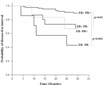
Figure 1. Disease-free interval probability as a function of the ER and PR status in patients with breast carcinoma in the first 2.5 years of follow up study
Table 2.Steroid hormone receptor values (range, median) in breast carcinomas
Statistical evaluations: The probabilities of a disease-free interval were estimated using the Kaplan-Meier method and were compared by the log-rank test. The correlation between the quantitative values of ER and PR was examined by the Spearman rank correlation test. A P value less than 0.05 was considered as statistically significant.
RESULTS
One hundred and thirteen patients were included in this investigation. All of these patients had detectable levels of ER and PR in breast carcinomas and had undergone the adjuvant tamoxifen therapy after surgical treatment. The course of the disease for all patients was followed until the occurrence of distant metastasis (distant metastasis as an end point), with a follow-up period of 60 months. Our focus in the study was directed to two subgroups of patients. The first one included patients who were analyzed within the first 2.5 years of the follow-up period. The second subgroup consisted of patients who were disease-free after the 2.5 years of follow-up, and who were monitored until the end of the follow-up period. The ranges and median values of the ER and PR levels are
given in Table 2. A positive correlation was found between the ER and PR levels in the whole group of patients (p<0.001, data not shown).
The course of disease for the first 2.5- year period of the follow-up study
The DFI probabilities for the subgroups of patients according to the cut-off value reached a statistically significant difference in the case of ER (p=0.002, data not shown). The subgroup of patients with po-sitive ER status (n=100) had higher DFI pro-babilities. Similar results were obtained for PR (p<0001) where the PR-positive subgroup of patients (n=91) had statistically significantly higher probabilities of DFI (data not shown).
Of the classical clinical-histological parameters, lymph node status, histological type and the histological grade of the tumors did not cause differences in the analyses of DFI probabilities (data not shown). The tumor size defined two subgroups of patients with pT1 (n=58) showing a statistically significantly better prediction than pT2 patients (n=53, p=0.03, data not shown).
Figure 2. Disease-free interval probability as a function of the pT and ER status in patients with breast carcinoma in the first 2.5 years of follow up study
Figure 3. Disease-free interval probability as a function of the pT and PR status in patients with breast carcinoma in the first 2.5 years of follow up study
negative (p<0.001), and the ER-positive PR-negative (n=15, p=0.01) subgroups of patients.
We also investigated the course of disease of four phenotypes defined by tumor size and ER status (Figure 2). Out of these four phenotypes, the patients with large tumors (pT2) with ER-negative status (n=10) had the worst course of the disease. There was a statistically significant difference in the DFI probabilities between the ER-positive pT1 (n=55) and ER-negative pT2 subgroups of patients (p<0.001). Comparison of the patients with large tumors (pT2) and either positive (n=42) or negative ER status revealed a statistically significant diffe-rence in the DFI probabilities (p=0.003). The ER-negative pT1 subgroup had only 3 patients so we did not include it in our analysis.
By comparing the subgroups of patients defined by tumor size and PR status, we found statistically significant differences in the DFI probabilities bet-ween the following subgroups of patients: PR-posi-tive pT1 (n=47) vs. PR-negaPR-posi-tive pT1(n=11, p=0.03); PR-positive pT1- vs. PR-negative pT2 (n=11, p<0.001), and positive pT2 (n=42) vs. PR-negative pT2 (p=0.01) (Fig. 3).
The course of disease for the period of 2.5-5-years of the follow up study
There were no significant differences in the DFI probabilities between the subgroups of patients defined by tumor size, histological type and grade, or lymph node status. Statistically significant dif-ferences in the DFI probabilities determined within the first 2.5 years of the follow-up study, for the subgroups of patients defined by ER and PR status, are lost (data not shown).
The comparison of the DFI probabilities for patients with tumors of different size and with dif-ferent PR status revealed no statistically significant differences (data not shown). The subgroups of patients (PR-positive pT1 or pT2 and PR-negative pT1 or pT2) caused no statistically significant differences, as seen from the analysis within the first 2.5 years of the follow-up study (data not shown).
DISCUSSION
There are limited data in identifying subgroups of postmenopausal breast cancer patients who are at the highest risk of early occurrence of distant metastasis and who could benefit from the different additive endocrine treatment approach (Mansell, 2008).
The main purpose of our study was to deter-mine the importance of the quantitative values of the ER and PR expression and clinico-histological characteristics of breast carcinomas in the emer-gence of distant metastasis in postmenopausal breast cancer patients. A secondary objective was to identify the different breast cancer phenotypes that could point to the early (de novo) or late (acquired) resistance to tamoxifen.
Evaluating the relevance of the ER and PR status within the first 2.5 years of the follow-up period, we found that these biomarkers were in-dependent predictors of early resistance. The nega-tive ER and PR status points to an early occurrence of distant metastasis, making the ER and PR strong predictors in relation to tamoxifen treatment, as shown in some studies (Mauriac, 2007; Kennecke, 2008). The tumor size indicates a poor prediction for larger tumors, pT2, consistent with the study of Mansell et al., (Mansell, 2008). The power of ER, PR and tumor size as predictors is lost after the initial 2.5 years of tamoxifen therapy. Lymph node status, histological grade and type of tumor did not addi-tionally subgroup the patients. Considering treat-ment strategies, it is much easier to decide in the case of the double positive/negative phenotypes than in case of single-positive phenotypes. It has been reported that 75-85% of tumors with the double-positive phenotype respond to hormonal manipulation, whereas only 40% of the single-posi-tive phenotypes respond in the same way (Dowsett, 2006). In the subgroup of ER-positive tumors, among postmenopausal patients there is conside-rable evidence of the significant predictive power of PR (Ponzone, 2006; Cui, 2005). It is shown that PR negativity is a marker of early (de novo) tamoxifen resistance during the first 3 years of treatment and that there is a decline of risk after the first 3 years (Tovey, 2005). Similar results are reported in the ATAC trial where it was confirmed that the PR
status defines a group of tumors with distinctive pathological features that can benefit from a more aggressive approach or a different kind of therapy (Dowsett, 2005).
When the four phenotypes defined by ER and PR status were analyzed, the ER-positive PR-negative phenotype had lower probabilities of DFI than ER-positive PR-positive tumors, as seen in the ATAC trial (Dowsett, 2005). The expression of PR gives an additional predictive value to ER. There are several explanations for the ER-positive PR-ne-gative phenotype. Since the expression of PR is ER-dependent, the simplest explanation is that the ER is non-functional and unable to stimulate PR production, so the tumor is no longer dependent on estrogen for growth and survival (Cui, 2005). But, as proven in the recent trial, ER-positive PR-ne-gative tumors respond much better to estrogen withdrawal than to tamoxifen (Dowsett, 2003). This indicates that these tumors are still estrogen dependent and that the loss of ER function is not the whole explanation for resistance to tamoxifen (Cui, 2005).
acti-vity of tamoxifen, which can lead to proliferation and resistance to therapy (Campbell, 2001). This growth factor signaling can be amplified even more by switching from nuclear to membrane-initiated steroid signaling of ER (Cui, 2005). The same effect of growth factor signaling on the PR expression was confirmed in some in vitro studies (Konecny, 2003; McClelland, 2001).
Our results also show a sharp decrease of significance of the PR status after 2.5 years of the follow-up period. This indicates that PR is a time-dependent predictor of the risk of the appearance of distant metastasis. This assumption is confirmed in the study of Coombes et al., (Coombes, 2004). Analysis of the DFI probabilities, within the pheno-types defined by ER status and tumor size, revealed a high-risk group of patients bearing larger tumors with a negative status of ER. The estrogen receptor status can additionally subgroup patients within the pT2 group. The statistically significant differences in the DFI probabilities between the ER-negative pT2 and other phenotypes were also diminished after 2.5 years of the follow-up study. On the other hand, the PR status enables the subgrouping of patients within both the pT1 and pT2 subgroups of patients during the first 2.5 years of the follow up.
To summarize, our research confirmed a sig-nificant role of the ER and tumor size as predictive parameters. By combining these parameters, we can obtain additional information regarding the patients’ prognosis. The estrogen receptor is still the most powerful predictive biochemical marker in terms of tamoxifen therapy. But our study confirms that the PR is also a significant predictive marker that can possibly indicate the aberrant growth factor signaling in the group of ER-positive patients. Our data support the conclusion that ER-positive PR-negative and pT2 either ER- or PR- negative patients constitute subgroups with a high risk of de novo tamoxifen resistance. This ER-, PR- and pT- related risk is indubitable time-dependent and confined to the first 2.5 year of the therapy. Other prospective clinical trials are required in order to define the short- and long-term hormone
therapeutic strategies tailored for each ER-positive breast cancer patient.
Acknowledgments: This study was supported by a grant from the Ministry of Science and Technological Development, project number 145018: “Molecular biomarkers of the growth, invasiveness and metastatic potential in breast carcinoma: biological and clinical aspects”.
REFERENCES
Baum, M., and A. Buzdar, (2003). The current status of
aromatase inhibitors in the management of breast cancer. Surg. Clin. North. Am. 83, 973-994.
Buzdar, A.U. (2003). Advances in endocrine treatments for
postmenopausal women with metastatic and early breast cancer. Oncologist.8, 335-341.
Campbell, R.A., Bhat-Nakshatri, P., Patel, N.M.,
Constantinidou, D., Ali, S., and H. Nakshatri, (2001).
Phosphatidylinositol 3-kinase/AKT-mediated activation of estrogen receptor a: a new model for anti-estrogen resistance. J. Biol. Chem.276, 9817-24.
Campos, S.M., and E.P. Winer, (2003). Hormonal therapy in
postmenopausal women with breast cancer. Oncology.
64, 289-299.
Coombes, R.C., Hall, E., and L.J. Gibson, (2004). A randomized
trial of exemestane after two to three years of tamoxifen therapy in postmenopausal women with primary breast cancer. N. Engl. J. Med.350, 1081-92.
Cui, X., Schiff, R., Arpino, G., Osborne, C.K., and A.V. Lee,
(2005). Biology of progesterone receptor loss in breast cancer and its implications for endocrine therapy. J.
Clin. Oncol.23, 7721-35.
Dowsett, M., Cuzick, J., Wale, C., Howell, T., Houghton, J., and
M. Baum, (2005). Retrospective analysis of time to
recurrence in the ATAC trial according to hormone receptor status: a hypothesis- generating study. J. Clin.
Oncol. 23, 7512–7517.
Dowsett, M., Houghton, J., Iden, C., Salter, J., Farndon, J.,
A’Hern, J., Sainsbury, R., and M. Baum, (2006). Benefit
from adjuvant tamoxifen therapy in primary breast cancer patients according oestrogen receptor, progesterone receptor, EGF receptor and HER2 status.
Ann. Oncol.17, 818-826.
Dowsett, M. (2003). On behalf of the ATAC trialists group:
Analysis of time to recurrence in the ATAC (Arimidex, Tamoxifen, Alone or in Combination) trial according to estrogen receptor and progesterone receptor status.
Early Breast Cancer Trialist’ Collaborative Group (EBCTCG)
(2005). Effects of chemotherapy and hormonal therapy for early breast cancer on recurrence and 15-year survival: an overview of the randomized trials. Lancet 365, 1687-1717.
EORTC Breast Cancer Cooperative Group. (1980). Revision of
the standard for the assessment of hormone receptors in human breast cancer. Eur. J. Cancer. 9, 1513-1515.
Houghton, J., ATAC Trialists’ Group. (2006). Initial adjuvant
therapy with anastrozole (S) reduced rates of early breast cancer recurrent and adverse events compared with tamoxifen (T)—data reported on behalf of the ATAC (‘Arimidex’, Tamoxifen, Alone or in Combination) Trialists’ group. Ann. Oncol.17, (Suppl. 9) (abstract 243PD), 94.
Jordan V.C. (1994). The development of tamoxifen for breast
cancer therapy, In: Long-term tamoxifen treatment for breast cancer (Eds. V.C. Jordan), 3-26. University of Wisconsin Press, Wisconsin.
Kennecke, H., McArthur, H., Olivotto, I.A., Speers, C., Bajdik, C., Chia, S.K., Ellard, S., Norris, B., Hayes, M., Barnett,
J., and K.A. Gelmon, (2008). Risk of early recurrence
among postmenopausal women with estrogen receptor-positive early breast cancer treated with adjuvant tamoxifen. Cancer.112, 1437-44.
Konecny, G., Pauletti, G., Pegram, M., Untch, M., Dandekar, S., Aguilar, Z., Wilson, C., Rong, H.M., Bauerfeind, I., Felber, M., Wang, H.J., Beryt, M., Seshadri, R., Hepp, H.,
and D.J. Slamon, (2003). Quantitative association
between HER-2/neu and steroid hormone receptors in hormone receptor-positive primary breast cancer. J.
Natl. Cancer. Inst. 95, 142-153.
Lapidus, R.G., Nass, S.J., and N.E. Davidson, (1998). The loss of
estrogen and progesterone receptor gene expression in human breast cancer. J. Mammary. Gland. Biol. Neoplasia. 3, 85-94.
Mansell, J., Monypenny, I.J., Skene, A.I., Abram, P., Carpenter,
R., Gattuso, J.M., Wilson, C.R., Angerson, W.J., and J.C.
Doughty, (2008). Patterns and predictors of early
recurrence in postmenopausal women with estrogen receptor-positive early breast cancer. Cancer. 117, 91-8. Massarweh, S., Schiff, R. (2006). Resistance to endocrine
therapy in breast cancer: exploiting estrogen receptor/growth factor signaling crosstalk. Endocr. Relat. Cancer. 13, 15-24.
Mauriac, L., Keshaviah, A., Debled, M., Mouridsen, H., Forbes, J.F., Thürlimann, B., Paridaens, R., Monnier, A., Láng, I., Wardley, A., Nogaret, J.M., Gelber, R.D., Castiglione-Gertsch, M., Price, K.N., Coates, A.S., Smith, I., Viale, G.,
Rabaglio, M., Zabaznyi, N., and A. Goldhirsch, BIG 1-98
Collaborative Group; International Breast Cancer Study
Group. (2007). Predictors of early relapse in
post-menopausal women with hormone receptor-positive breast cancer in the BIG 1–98 trial. Ann. Oncol. 18, 859–867.
McClelland, R.A., Barrow, D., Madden, T.A., Dutkowski, C.M.,
Pamment, J., Knowlden, J.M., Gee, J.M., and R.I.
Nicholson, (2001). Enhanced epidermal growth factor
receptor signaling in MCF7 breast cancer cells after long-term culture in the presence of the pure antiestrogen ICI 182,780 (Faslodex). Endocrinology.142,
2776-2788.
Osborne, C.K., R. Schiff, (2005). Estrogen-receptor biology:
Continuing progress and therapeutic implications. J.
Clin. Oncol. 23, 1616-1622.
Osborne, C.K. (1998). Tamoxifen in the treatment of breast
cancer. N. Engl. J. Med. 339, 1609-18.
Osborne, C.K., Zhao, H., and S.A. Fuqua, (2000). Selective
estrogen receptor modulators: Structure, function, and clinical use. J. Clin. Oncol. 18, 3172-3186.
Petz, L.N., Ziegler, Y.S., Schultz, J.R., Kim, H., Kemper, J.K., and
A.M. Nardulli (2004). Differential regulation of the
human progesterone receptor gene through an estrogen response element half site and Sp1 sites. J. Steroid.
Biochem. Mol. Biol. 88, 113–122.
Petz, L.N., Ziegler, Y.S., Schultz, J.R., and A.M. Nardulli, (2004).
Fos and Jun inhibit estrogen-induced transcription of the human progesterone receptor gene through an activator protein-1 site. Mol. Endocrinol.18, 521-532.
Ponzone, R., Montemurro, F., Maggiorotto, F., Robba, C., Gregori, D., Jacomuzzi, M.E., Kubatzki, F., Marenco, D.,
Dominguez, A., Biglia, N., and P. Sismondi, (2006).
Clinical outcome of adjuvant endocrine treatment according to PR and HER-2 status in early breast cancer.
Ann. Oncol. 17, 1631-1636.
Rakha, E.A., El-Sayed, M.E., Green, A.R., Paish, E.C., Powe, D.G., Gee, J., Nicholson, R.I., Lee, A.H., Robertson, J.F.,
and I.O. Ellis, (2007). Biological and clinical
characteristics of breast cancer with single hormone receptor positive phenotype. J. Clin. Oncol. 25, 4772-8.
Saphner, T., Tormey, D.C., and R. Gray, (1996). Annual hazard
rates of recurrence for breast cancer after primary therapy. J. Clin. Oncol. 14, 2738–2746.
Scarf, R.F., and H. Torloni, (1968). Histological typing of breast
tumors. In: International Histological Classification of Tumors, 2nd edn. World Health Organization., WHO, Geneva.
Tomlinson, I.P., Nicolai, H., Solomon, E. and W.F. Bodmer
(1996). The frequency and mechanism of loss of heterozygosity on chromosome 11q in breast cancer. J. Pathol. 180, 38-43.
Tovey, S., Dunne, B., Witton, C.J., Forsyth, A., Cooke, T.G., and
when to implement treatment with aromatase inhibitors in invasive breast cancer? Clin. Cancer. Res. 11, 4835– 4842.
International Union Against Cancer (UICC) (1987). TNM
Classification of Malignant Tumors, 4 (Eds. P. Hermanek, L.H. Sobin). Springer-Verlag, New York.
Winqvist, R., Hampton, G.M., Mannermaa, A., Blanco, G., Alavaikko, M., Kiviniemi, H., Taskinen, P.J., Evans, G.A.,
Wright, F.A., Newsham, I., and W.K. Cavenee, (1995).
Loss of heterozygosity for chromosome 11 in primary human breast tumors is associated with poor survival after metastasis. Cancer. Res. 55, 2660-2664.
РАНА И КАСНА РЕЗИСТЕНЦИЈА НА ТАМОКСИФЕН КОД КАНЦЕРА ДОЈКЕ
З. АБУ РАБИ, М. МАРКИЋЕВИЋ, ТИЈАНА ВУЈАСИНОВИЋ, СИЛВАНА ЛУКИЋ, ЉИЉАНА СТАМАТОВИЋ и ДРАГИЦА НИКОЛИЋ-ВУКОСАВЉЕВИЋ
Институт за онкологију и радиологију Србије, 11000 Београд, Србија
У студији је испитивана улога ER, PR и клиничко-хистолошких параметара у групи болесница оболелих од карцинома дојке, лечених тамоксифеном, током раног (2,5 године) и касног (2,5-5 година) периода праћења тока болести. Негативни статуси ER и PR и величина тумора једнака или већа од 2 цм су дефинисали фенотипове, односно подгрупе болесница, са најлошијим клиничким током