VALUE OF SONOGRAPHIC ENDOMETRIAL FINDINGS IN PATIENTS
UNDER TAMOXIFEN THERAPY*
Arildo Corrêa Teixeira1, Linei A.B.D. Urban2, Ricardo Salfer Schwarz3, Caroline Pereira4, Thaís Cristina Cleto Millani4, Ana Paula Passos5
OBJECTIVE: To evaluate endometrial alterations by means of transvaginal ultrasound and to correlate them with hysteroscopic and histological findings in patients under tamoxifen therapy. MATERIALS AND METH-ODS: The present study was developed in the period between January 2003 and December 2005, including patients under tamoxifen therapy for breast cancer, and presenting with endometrial thickening > 5 mm. The sonographic findings were correlated with hysteroscopic and anatomopathological results. RESULTS: Twenty-five patients, mean age 62.6 years, were selected. The mean time elapsed from the diagnosis of cancer was 4.3 years, and use of tamoxifen, three years. Twenty patients (80%) were asymptomatic, and five (20%) presented with bleeding. Ultrasound showed that 16% of patients had endometrial thickening ranging between 5 mm and 8 mm, 40% between 9 mm and 15 mm, and 44% above 15 mm. Hysteroscopy showed 40% of patients with atrophy, 16% with cystic atrophy, 28% with polyps and 16% with hyperplas-tic lesion. Anatomopathological study showed 35.2% of patients with normal results, 5.8% with atrophy, 29.4% with polyps and 11.7% with hyperplasia. One case of adenocarcinoma (5.8%) was observed. CON-CLUSION: Combined ultrasound and hysteroscopy have proven to be important allies in the evaluation of patients under tamoxifen therapy. Ultrasonography presents low specificity for detecting endometrial thick-ening, while hysteroscopy is more accurate in the detection of polyps, hyperplasia and neoplastic alterations. Keywords: Ultrasound; Hysteroscopy; Tamoxifen.
Valor da ultra-sonografia na avaliação das alterações endometriais em pacientes tratadas com tamoxifeno. OBJETIVO: Avaliar as alterações endometriais por meio da ultra-sonografia transvaginal e correlacioná-las com os achados da histeroscopia e histologia, em pacientes submetidas a tratamento com tamoxifeno. MA-TERIAIS E MÉTODOS: No período de janeiro de 2003 a dezembro de 2005, foram incluídas pacientes com câncer de mama usuárias de tamoxifeno que apresentaram espessamento endometrial acima de 5 mm. Os achados foram correlacionados com os dados de histeroscopia e anatomopatologia. RESULTADOS: Foram selecionadas 25 pacientes com idade média de 62,6 anos. O tempo médio do diagnóstico do câncer foi de 4,3 anos e do uso de tamoxifeno, três anos. Vinte pacientes eram assintomáticas (80%) e as demais apre-sentaram sangramento (20%). À ultra-sonografia, 16% apreapre-sentaram espessamento endometrial entre 5 mm e 8 mm, 40% entre 9 mm e 15 mm, e 44% acima de 15 mm. Ao estudo com a histeroscopia, 40% apresen-taram atrofia, 16% atrofia cística, 28% pólipos, e 16% lesão hiperplásica. O estudo anatomopatológico apre-sentou-se normal em 35,2% dos casos e mostrou atrofia em 5,8%, pólipo em 29,4% e hiperplasia em 11,7%. Foi observado um caso de adenocarcinoma (5,8%). CONCLUSÃO: A ultra-sonografia associada à histeros-copia apresentam-se como importantes aliados na avaliação de pacientes usuárias de tamoxifeno. A detec-ção de espessamento endometrial à ultra-sonografia apresenta baixa especificidade, enquanto a histerosco-pia é mais acurada na detecção de pólipos, hiperplasia e alterações neoplásicas.
Unitermos: Ultra-sonografia; Histeroscopia; Tamoxifeno. Abstract
Resumo
* Study developed in the Department of Echography, Mater-nidade do Hospital de Clínicas da Universidade Federal do Pa-raná (UFPR), and Clínica DAPI – Diagnóstico Avançado por Ima-gem, Curitiba, PR, Brazil.
1. Professor of Gynecology and Obstetrics at Hospital de Clí-nicas da Universidade Federal do Paraná (UFPR), MD, Ultra-sonographist at Clínica DAPI – Diagnóstico Avançado por Ima-gem, Curitiba, PR, Brazil.
2. MD, volunteer for the Department of Radiology at Hospital de Clínicas da Universidade Federal do Paraná (UFPR), MD, Radiologist at Clínica DAPI – Diagnóstico Avançado por Imagem, Curitiba, PR, Brazil.
3. MD, Volunteer for the Service of Gynecological Endoscopy at Hospital de Clínicas da Universidade Federal do Paraná (UFPR), Curitiba, PR, Brazil.
4. MDs, Residents, Department of Gynecology and Obstetrics, Hospital de Clínicas da Universidade Federal do Paraná (UFPR), Curitiba, PR, Brazil.
5. MD, Trainee in Ultrasonography, Department of Gynecol-ogy and Obstetrics, Hospital de Clínicas da Universidade Federal do Paraná (UFPR), Curitiba, PR, Brazil.
INTRODUCTION
Tamoxifen has been utilized for almost three decades as an adjuvant treatment for breast cancer, and also is effective in the chemoprophylaxis of this disease. How-ever, this drug presents endometrial prolif-eration as a side effect, with a low, but real, risk for the development of endometrial
cancer(1,2). This risk is of almost two cases
of cancer for every 1000 women under tamoxifen therapy during a year, similar to the postmenopausal women submitted to single-dose hormone replacement ther-apy(2,3). Studies have demonstrated that the
majority of cases of endometrial cancers in women under tamoxifen therapy were found at their early stages, and the treat-ment has been successfully instituted; how-ever, in women who have developed en-dometrial cancer whose diagnosis was made at later stages, the prognosis has been poorer.
Mailing address: Dra. Linei A.B.D. Urban. Rua Padre Agosti-nho, 913, ap. 51, Mercês. Curitiba, PR, Brazil, 80430-050. E-mail: [email protected]
Considering these findings, the moni-toring of the endometrium of the patients under tamoxifen therapy is recommended aiming at the early diagnosis of these le-sions, either by means of ultrasound (US), or hysteroscopy, or uterine curettage, or endometrial biopsy. So, the main objective of the present study was to analyse the main echotextural changes in the endometrium by means of US, in a group of patients under tamoxifen therapy for determining the abnormalities that could occur in the uterine mucosa, besides the correspondent hysteroscopy, and eventually, anatomo-pathological findings.
MATERIALS AND METHODS
The present study retrospectively in-cluded 25 patients with breast cancer un-der tamoxifen therapy referred to the De-partment of Ultrasound at Hospital das Clínicas da Universidade Federal do Pa-raná (HC-UFPR) in the period between January 2003 and December 2005, and presenting with endometrial thickening > 5 mm. No patient had undergone hormone replacement therapy or presented gyneco-logic disease previously to the breast can-cer diagnosis. Clinical data evaluated were: age, time of diagnosis, duration of tamoxi-fen therapy, and presence of symptoms.
Transvaginal US was performed with a Sonoline Sienna Ultrasound Imaging Sys-tem (Siemens, Germany), with a multifre-quency 6.5 MHz transducer and color Dop-pler. The following items were evaluated: endometrial echotexture and endometrial thickness measurement on a longitudinal plane, larger anteroposterior diameter from the echogenic interface at the endome-trium-myometrium junction from one side to another. The patients were divided into three groups, according to the thickening degree: thickening between 5–8 mm, 9–15 mm, and above 15 mm. Echotexture was described as homogeneous, heterogeneous with cystic areas, and with an image of a polyp inside.
Hysteroscopy was performed with a Storz hysteroscope, with 4.1 mm-diameter, 5.0 mm diagnostic shirt, and a Xenon light source. The uterine cavity distension was accomplished with carbon dioxide (CO2) gas with intrauterine pressure being
main-tained at a level accepted as safe (between 80 mmHg and 100 mmHg). During the hys-teroscopic examination, areas of interest or under suspicion were biopsied. Hystero-scopic findings were described as normal (with no endometrial atypia) or abnormal (endometrial atrophy, cystic atrophy and/or hyperplasia).
The analysis of these tissues as per-formed by pathologists in the Department of Pathological Anatomy at UFPR. The results were considered as normal, pro-vided a regular endometrial maturation was found, or abnormal, in case of atrophy, polyps, simple hyperplasia with or without atypias, fibroids, or endometrial carci-noma.
RESULTS
The mean age of the 25 patients in-cluded in the present study was 62.6 years (age range = 37–77 years). The mean time for the diagnosis of cancer was 4.3 years (ranging between one and 13 years) and mean time under tamoxifen therapy was three years (ranging between one and seven years). All of the patients received a 20 mg/ day-dose. Twenty patients were asymptom-atic and five presented with bleeding at the moment of examination.
Transvaginal ultrasound demonstrated a mean endometrial thickness of 16.4 mm
(ranging between 5 mm and 58 mm). Among the patients, 16% (4/25) presented endometrial thickening between 5 mm and 8 mm, 40% (10/25) between 9 mm and 15 mm, and 44% (11/25) > 15 mm. Echotex-ture was homogeneous in 80% (20/25) of cases, with cystic areas in 16% (4/25) and polyp image in 4% (1/25).
Hysteroscopic study performed at last 56 days after the transvaginal US demon-strated 40% (10/25) of cases with atrophy, 16% (4/25) with cystic atrophy, 28% (7/25) with polyp, and 16% (4/25) with hyperplas-tic lesion.
Anatomopathological study was per-formed in 17 cases. The samples were in-sufficient in 11.7% (2/17) of cases. In the other cases, with appropriate samples, a normal endometrium was observed in 40% (6/15), atrophy in 6.6% (1/15), polyp in 33.3% (5/15) and hyperplasia in 13.3% (2/ 15). One case (6.6%; 1/15) of endometrioid adenocarcinoma.
The correlation between mean time under tamoxifen therapy and sonographic, hysteroscopic and histological findings is demonstrated on Table 1.
Table 2 correlates clinical data with imaging (US and hysteroscopy) and anatomopathological findings.
Table 3 demonstrates the correlation between sonographic, hysteroscopic and anatomopathological findings.
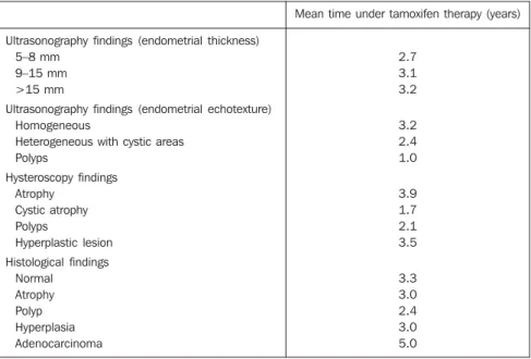
Table 1 Correlation between ultrasonography, hysteroscopy and anatomopathological findings, and average time of tamoxifen therapy in breast cancer patients.
Ultrasonography findings (endometrial thickness) 5–8 mm
9–15 mm >15 mm
Ultrasonography findings (endometrial echotexture) Homogeneous
Heterogeneous with cystic areas Polyps
Hysteroscopy findings Atrophy
Cystic atrophy Polyps
Hyperplastic lesion
Histological findings Normal
Atrophy Polyp Hyperplasia Adenocarcinoma
Mean time under tamoxifen therapy (years)
2.7 3.1 3.2
3.2 2.4 1.0
3.9 1.7 2.1 3.5
Table 2 Correlation among ultrasonography, hysteroscopy, anatomopathological findings, and the pre-sence of symptoms in breast cancer patients under tamoxifen therapy.
Ultrasonography findings (endometrial thickness) 5–8 mm
9–15 mm >15 mm
Ultrasonography findings (endometrial echotexture) Homogeneous
Heterogeneous with cystic areas Polyps
Hysteroscopy findings Atrophy
Cystic atrophy Polyps
Hyperplastic lesion
Histological findings Normal
Atrophy Polyp Hyperplasia Adenocarcinoma
Symptomatic patients*
40% (2/5) 20% (1/5) 40% (2/5)
60% (3/5) 20% (1/5) 20% (1/5)
0% (0/5) 0% (0/5) 60% (3/5) 40% (2/5)
0% (0/3) 0% (0/3) 66.6% (2/3)
0% (0/3) 33.3% (1/3)
Asymptomatic patients
10% (2/20) 45% (9/20) 45% (9/20)
85% (17/20) 15% (3/20)
0% (0/20)
50% (10/20) 20% (4/20) 20% (4/20) 10% (2/20)
50% (6/12) 8.3% (1/12) 25% (3/12) 16.6% (2/12)
0% (0/12)
* Patient reporting vaginal bleeding.
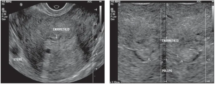
Figure 1. Sonographic study – Longitudinal plane demonstrating echogenic and heterogeneous en-dometrium, with small cystic areas inside, with well defined margins, with the atrophic pattern usu-ally observed in climac-teric women under tamo-xifen therapy.
DISCUSSION
Tamoxifen is successfully utilized in the adjuvant therapy of breast cancer, and its effect as a powerful selective estrogen re-ceptor modulator is well established in the literature. On the other hand, tamoxifen effect on other tissues such as the uterus, for example, is more complex, acting si-multaneously as a powerful estrogen ago-nist and antagoago-nist(2,4). The mechanism of
action, like the one of other antiestrogenic agents, would be the antagonism to estro-gen-specific receptors. It acts competitively binding to estrogen receptors in the tumor-like tissue or other tissues to form a nuclear complex that reduces the DNA synthesis, inhibits the estrogen effects, resulting in an interruption of the cellular growth. How-ever, several authors consider that the tamoxifen mechanism of action is complex and is still to be elucidated(4–6). The
pres-ence of hyperestrogenism-related endome-trial disease (polyps, hyperplasia and en-dometrial carcinoma) in 53.2% of the pa-tients included in the present study con-firms, in agreement with the literature, that tamoxifen therapy increases the incidence of endometrial lesion related to the agonist effect of this drug, although most of times there is no progression to cancer(2).
A consensus is still to be reached in the literature as regards the association be-tween tamoxifen therapy duration and de-gree of endometrial lesion(1,3,6). Gerber et
al.(3), in a prospective study with 274
women have not found a relation between tamoxifen therapy duration and endome-trial thickness. Donne et al.(1) have not
observed any relation between therapy du-ration and degree of endometrial lesion, but the incidence of endometrial cancer was higher in patients exposed to tamoxifen for more than four years. In the present study no relation was found between the presence of endometrial lesion and tamoxifen therapy duration. However, the sole case of carcinoma occurred in a patient who had been using tamoxifen for more than five years, suggesting that the prolonged use of tamoxifen may be related to an increase in the incidence of endometrial cancer.
The endometrial involvement second-ary to the tamoxifen therapy should be prompt and adequately diagnosed. Ultra-Table 3 Correlation between ultrasonography findings, hysteroscopy and histological results in breast
cancer patients under tamoxifen therapy.
Ultrasonography findings
Hysteroscopy findings Atrophy
Cystic atrophy Polyps
Hyperplastic lesion
Histological findings Normal
Atrophy Polyp Hyperplasia Adenocarcinoma
40% (10/25) 16% (4/25) 16% (4/25) 8% (2/25)
26.6% (4/15) 6.6% (1/15) 26.6% (4/15) 13.3% (2/15) 6.6% (1/15)
– – 8% (2/25) 8% (2/25)
13.3% (2/15) – – – –
– – 4% (1/25)
–
– – 6.6% (1/15)
sonography constitutes a non-invasive, low-cost imaging method that is well tol-erated by the patients, and, when trans-vaginally performed, can indirectly visual-ize the uterine cavity. Several authors have described endometrial alterations in breast cancer patients treated with tamoxifen. Zarbo et al.(7), in a prospective study, have
reported the follow-up of 219 women with breast cancer and treated with tamoxifen during six years, and described 26.9% prevalence of endometrial alterations rep-resented mainly by polyps and cystic atro-phy. In Brazil, Feitosa et al.(6) have
ob-served a 36.6% prevalence of endometrial alterations, 26.6% represented by polyps,
and 46% by cystic atrophy. The present study demonstrated 33.3% prevalence of polyps, 13.3% of hyperplasia, and 6.6% of carcinoma.
Among the evaluated sonographic find-ings, the main abnormalities observed in the present study were endometrial thick-ness and echotextural alterations. As re-gards thickness, it is known that the post-menopausal endometrium is shown as a subtle, white and homogeneous line with no more than 5 mm of thickness. Above 5 mm, the higher the thickness, the lower the US accuracy for ruling out the endometrial involvement. That is to say, an endometrial thickness of more than 5 mm does not
in-dicate the presence of endometrial lesion, but rather that an echographic study cannot rule out its presence. Several studies have demonstrated that 40%–54% of patients under tamoxifen therapy present endome-trial thickness between 6mm and 10 mm(8,9). Gerber et al.(3) have described a
mean thickness of 9.2 mm in a group of tamoxifen users, as compared with 3.5 mm in the control group. However, it should be taken into consideration that many of these cases, actually, represent only an atrophy confirmed by hysteroscopy and anatomo-pathological study. Aiming at avoiding unnecessary procedures, Gerber et al.(3)
have suggested a 10 mm cut-off to deter-Figure 2. Sonographic study demonstrating endometrium presenting heterogeneous texture with small cysts (A), besides an image of polyp surrounded by anechoic halo (B), a pattern usually found in patients under hormone replacement therapy.
A B
A B
mine the necessity of diagnostic deepening in tamoxifen users. On the other hand, Franchi et al.(10) have suggested a 9 mm
cut-off, after studying 163 patients both symp-tomatic and asympsymp-tomatic. The authors have divided them into two groups and observed histological alterations in 60% of patients in the group with > 9 mm, while only 6.1% of patients with ≤ 9 mm pre-sented alterations. However, it is important to note that the case of endometrial carci-noma observed in the present study was fund in a symptomatic patient with en-dometrial thickness of 7 mm, suggesting that symptomatic patients must be investi-gated, independently from their endome-trial thickness.
Echotextural findings have an increas-ing significance in the differentiation be-tween benign and malignant lesions. In postmenopausal patients with no history of hormone replacement therapy, the pattern of atrophy presented at US may mimic a thickened aspect, leading to a great num-ber of false-positive diagnoses, considering that when these women are submitted to hysteroscopy or uterine curettage, they present an endometrial atrophy pattern. The same echotextural endometrial pattern found in association with cystic areas in postmenopausal women, frequently simu-lating a neoplasm, is observed in tamoxifen users. The apparent increase in the endome-trial thickness associated with cystic areas is due to the powerful antiestrogenic effect of tamoxifen on the uterus, causing edema and cystification of endometrial glands, while the overlying epithelium remains atrophic(1,3,11). Cechini et al.(12) have found
41% of cases with echographically abnor-mal endometrium, 46% of which were false-positive. In our study, 56.6% of tamo-xifen users presented endometrial thicken-ing at US, correspondthicken-ing to atrophy and cystic atrophy at hysteroscopy, also demon-strating a high rate of false-positive results. Additionally, a low accuracy was observed in the identification of hyperplasia and polyps, because the majority of these find-ings could not be individualized at US.
Several studies have emphasized the significance of hysteroscopy in the evalu-ation of the endometrial cavity with guided-biopsy aiming at a definite
diagno-sis(1,5). However, this is an invasive and
expensive procedure, requiring specialized professionals and equipment. In the present study, hysteroscopy demonstrated high ac-curacy in the diagnosis of polyps, hyperpla-sia and neoplastic alterations.
The presence of symptoms, such as vaginal bleeding, increases the probability of finding endometrial lesions at imaging studies in tamoxifen users. Deligdisch et al.(2) have found more pathological
alter-ation in the group of symptomatic patients (52% of patients with polyps and 62% with carcinoma presented vaginal bleeding) than in the asymptomatic group. Gerber et al.(3)
have described atrophy in 73% of atomatic patients, while only 25% of symp-tomatic patients presented this finding. The present study observed that the presence of symptoms is associated with the presence of lesion at hysteroscopy (all the symptom-atic patients presented hyperestrogenism-related lesions characterized by polyps, hyperplasia or adenocarcinoma). However, symptoms absence does not mean absence of lesion, since 41.6% of asymptomatic patients presented polyps or hyperplasia at anatomopathological studies.
CONCLUSIONS
Patients who are being treated with tamoxifen require a rigorous endometrial management, because of the increased risk for development of endometrial cancer. Transvaginal ultrasound should be recom-mended as an initial endometrial screening of breast cancer patients treated with tamoxifen, considering that the disease prognosis is highly dependent on an early diagnosis.
In case endometrial alteration suggest-ing neoplasm is found, hysteroscopy is re-quired for direct visualization of the en-dometrial cavity as well as a guided-biopsy aimed at a definite diagnosis. The presence of vaginal bleeding increases the risk for endometrial lesion, therefore, anatomopa-thological study should be performed in all of these cases, independently from the im-aging findings.
The present study demonstrated that the sole utilization of US in the assessment of these patients and based only on the
in-crease in the endometrial thickness pre-sents a high rate of false-positive results (56%) leading to unnecessary surgical in-terventions. However there is a consensus in the literature that cut-offs above 9–10 mm require a more detailed diagnosis.
It is important to emphasize the need for an association between clinical symptoms and imaging findings as criteria for estab-lishing the strategy for management of this group of patients.
REFERENCES
1. Le Donne M, Lentini M, De Meo L, Benedetto V, Misiti M. Uterine pathologies in patients un-dergoing tamoxifen therapy for breast cancer: ultrasonographic, hysteroscopic and histological findings. Eur J Gynaecol Oncol 2005;26:623– 626.
2. Deligdisch L, Kalir T, Cohen CJ, de Latour M, Le Bouedec G, Penault-Llorca F. Endometrial histo-pathology in 700 patients treated with tamoxifen for breast cancer. Gynecol Oncol 2000;78:181– 186.
3. Gerber B, Krause A, Müller H, et al. Effects of adjuvant tamoxifen on the endometrium in post-menopausal women with breast cancer: a pro-spective long-term study using transvaginal ultra-sound. J Clin Oncol 2000;18:3464–3470. 4. Fung MFK, Reid A, Faught W, et al. Prospective
longitudinal study of ultrasound screening for endometrial abnormalities in women with breast cancer receiving tamoxifen. Gynecol Oncol 2003; 91:154–159.
5. Fong K, Kung R, Lytwyn A, et al. Endometrial evaluation with transvaginal US and hystero-sonography in asymptomatic postmenopausal women with breast cancer receiving tamoxifen. Radiology 2001;220:765–773.
6. Feitosa FEL, Juaçaba SF, Medeiros FC. Altera-ções endometriais em pacientes com câncer de mama tratadas com tamoxifeno. RBGO 2002;24: 233–239.
7. Zarbo G, Caruso G, Zammitti M, Caruso S, Zarbo R. The effects of tamoxifen therapy on the en-dometrium. Eur J Gynaecol Oncol 2000;21:86– 88.
8. Suh-Burgmann EJ, Goodman A. Surveillance for endometrial cancer in women receiving tamo-xifen. Ann Intern Med 1999;131:127–135. 9. Marconi D, Exacoustos C, Cangi B, et al.
Trans-vaginal sonographic and hysteroscopic findings in postmenopausal women receiving tamoxifen. J Am Assoc Gynecol Laparosc 1997;4:331–339. 10. Franchi M, Ghezzi F, Donadello N, Zanaboni F, Beretta P, Bolis P. Endometrial thickness in tamoxifen-treated patients: an independent pre-dictor of endometrial disease. Obstet Gynecol 1999;93:1004–1008.
11. Seoud M, Shamseddine A, Khalil A, et al. Tamo-xifen and endometrial pathologies: a prospective study. Gynecol Oncol 1999;75:15–19. 12. Cecchini S, Ciatto S, Bonardi R, et al. Risk of