SISTEM ´ATICA DE LISTROSCELIDINAE (ORTHOPTERA:
TETTIGONIIDAE) DA MATA ATL ˆANTICA
Disserta¸c˜ao apresentada `a Universidade Fe-deral de Vi¸cosa, como parte das exigˆencias do Programa de P´os-Gradua¸c˜ao em Entomo-logia, para obten¸c˜ao do t´ıtulo de Magister Scientiae.
VI ¸COSA
Ficha catalográfica preparada pela Seção de Catalogação e Classificação da Biblioteca Central da UFV
T
Fialho, Verônica Saraiva, 1984-
F438s Sistemática de Listroscelidinae (Orthoptera: Tettigoniidae) 2012 da Mata Atlântica / Verônica Saraiva Fialho. – Viçosa, MG, 2012.
vii, 112f. : il. (algumas col.) ; 29cm.
Inclui apêndices.
Texto em inglês e português.
Orientador: Karla Suemy Clemente Yotoko
Dissertação (mestrado) - Universidade Federal de Viçosa. Inclui bibliografia.
1. Ortóptero. 2. Marcadores genéticos. 3. Filogenia. 4. Inseto - Classificação. I. Universidade Federal de Viçosa.
II. Título.
SISTEM ´ATICA DE LISTROSCELIDINAE (ORTHOPTERA:
TETTIGONIIDAE) DA MATA ATL ˆANTICA
Disserta¸c˜ao apresentada `a Universidade Fe-deral de Vi¸cosa, como parte das exigˆencias do Programa de P´os-Gradua¸c˜ao em Entomo-logia, para obten¸c˜ao do t´ıtulo de Magister Scientiae.
APROVADA: 25 de julho de 2012.
Maria K´atia Matiotti da Costa
Cristiano Lopes Andrade
AGRADECIMENTOS
`
As institui¸c˜oes financiadoras que possibilitaram a execu¸c˜ao desse estudo CAPES (pela bolsa de mestrado), CNPq e FAPEMIG (pelo suporte financeiro aos projetos). Ao Programa de P´os-Gradua¸c˜ao em Entomologia da Universidade Federal de Vi¸cosa e ao Departamento de Entomologia, em especial `a Silvania pelas orienta¸c˜oes e aux´ılio com as quest˜oes burocr´aticas.
Ao amor primordial e supremo que sinto pelos meus pais e pelo meu lindo irm˜ao. Eu nunca me perdoarei pelos momentos perdidos ao lado de vocˆes, embora consci-ente de que alguns sacrif´ıcios s˜ao inevit´aveis. A dor da distˆancia machuca, mas a esperan¸ca de poder dar um m´ınimo de conforto a vocˆes um dia ´e que me fortalece. Obrigada pelo imenso amor e carinho, pela educa¸c˜ao, rigidez e castigos merecidos e, principalmente, por me ensinarem a lutar pelos meus sonhos.
Aos meus ador´aveis afilhados, Fellipe e Leonardo, pelo amor, abra¸cos e garga-lhadas ofertadas constantemente. Aos demais familiares que sempre me apoiaram e incentivaram, em especial ao maninho Claudimar, padim Vander, tio Messias, ma-drinhas Efigˆenia e Sandra, tia Quiquinha e Ione, Dayse, Aline, Janisse, Ruth e Jo˜ao Bosco.
`
A minha orientadora Karla Yotoko, que me recebeu de bra¸cos abertos e cotidia-namente me deu est´ımulo, carinho e pux˜ao de orelha. Certamente tudo serviu para “fortalecer o car´ater”. Agrade¸co pelos ensinamentos em evolu¸c˜ao, gen´etica, culin´aria japonesa e relacionamentos afetivos. Espero que tenha no¸c˜ao do quanto a admiro.
`
Ao querido Vin´ıcius e sua fam´ılia, pelo amor, carinho e apoio, principalmente nos momentos mais delicados dessa fase.
`
A Juliana Chamorro Rengifo, cuja amizade e parceria profissional foram cruciais para a realiza¸c˜ao desse trabalho. Agrade¸co pelo ensinamentos, for¸ca e companhei-rismo.
Ao professor Cristiano Lopes Andrade e aos integrantes do Laborat´orio de Siste-m´atica e Biologia de Coleoptera da UFV, pelos ensinamentos, acolhimento e suporte t´ecnico.
Ao meu doce lar em Vi¸cosa, Rep´ublica Bico Fino, e `as minhas irm˜as mais novas: Babi, Marcellinha, Lari, Lu, Lilian e Aline Cinquini, pela amizade, conselhos e todos os momentos de alegria que compartilhamos. E aos agregados Camis Fialho, Luquinhas, Lincoln, Caio e Ruan, Filiiispe, Teteu, Malu, Paty e Leiliane.
`
A fam´ılia Buzelin, pelas oportunidades de crescimento, mas em especial ao Min-duin, pelo carinho, amor e ternura.
`
As Panteras Flay e Eric˜ao, amigas cuja ausˆencia ainda n˜ao consegui superar. Vocˆes sempre ser˜ao ´unicas e insubstitu´ıveis. Nada apaga tudo o que vivemos! Vocˆes s˜ao meu porto-seguro.
Aos amigos Jˆo, Lena, Well, Izabell Miller, Rafael C´esar, Anayansi, Elisa, Maria Fernanda, Silvinha, Fabr´ıcio, Alan Voiski, Cristiano Agra, Gaia, Mirlene, Joel, Ro-g´erio Meneghin, William Rox, Celmilo, Camila St´efany e todos mais que certamente sabem que eu os amo.
Aos amigos que fiz na Entomologia Pedro, In´acio, Renata, Bob, Nat´alia Vi-cente, Ma´ıra, Andr´e, Dany, Lucimar e Farley, Katherine, Anderson Fernandes, Pu-ker, Pollyana, Cassiano, Alessandra Marins, Ester e V´ıvian.
Aos que j´a foram mas fizeram parte da minha forma¸c˜ao e crescimento, deixando saudade pra sempre no cora¸c˜ao: v´o Carolina, v´o Patroc´ınia, Sr. Oliveira, Maria Auxiliadora Magalh˜aes e Ricardo.
Aos marujos e Capit˜ao J. Dergam, amigos do Laborat´orio de Sistem´atica Mo-lecular ”Beagle”, integrantes e ex-integrantes, que foram igualmente importantes no processo.
culpados devo acrecentar: L´ucio Campos, Eraldo Lima, Daniel Avelar, Ana Fl´avia Bicalho, Maria L´ucia Wakisaka, Eder Assis, Junia Franco, Raquel Bernardo, F´a-bio Garcia, Luiz Silva, Janete Regina, Rosinei Rocha, Bernardo Castro, Humberto Mello, Cristiana Buzelin e An´ısio Nunes.
`
A toda equipe envolvida no Projeto Biota de Orthoptera do Brasil, em especial ao Prof. Carlos Sperber, Prof. Francisco Assis e Profa. K´atia Matiotti.
SUMÁRIO
Resumo...vi
Abstract...viii
Introdução Geral...8
CAPÍTULO 1 - Taxonomy of spiny predatory katydids (Tettigoniidae:
Listroscelidinae) from the Atlantic Forest………10
CAPÍTULO 2 - Sistemática de Listroscelidinae (Orthoptera: Tettigoniidae): um caso de congruência de caracteres morfológicos e moleculares...77
Considerações Finais...101
RESUMO
FIALHO, Verˆonica Saraiva, M. Sc., Universidade Federal de Vi¸cosa, julho, 2012. Sistem´atica de Listroscelidinae (Orthoptera: Tettigoniidae) da Mata Atlˆantica. Orientadora: Karla Suemy Clemente Yotoko. Coorientadores: Carlos Frankl Sperber e Jorge Abdala Dergam dos Santos.
ABSTRACT
FIALHO, Verˆonica Saraiva, M. Sc., Universidade Federal de Vi¸cosa, July, 2012. Systematic of Listroscelidinae (Orthoptera: Tettigoniidae) of the Brazil-ian Atlantic Forest. Adviser: Karla Suemy Clemente Yotoko. Co-advisers: Carlos Frankl Sperber and Jorge Abdala Dergam dos Santos.
INTRODUÇÃO GERAL
Em 2010 foi submetido ao Sistema Nacional de Pesquisa em Biodiversidade
(SISBIOTA) do CNPq o projeto multi-institucional intitulado Biota de Orthoptera
do Brasil (Edital MCT/CNPq/MMA/MEC/CAPES/FNDCT – Ação
Transversal/FAPs Nº 47/2010), composto por três sub-projetos: Diversidade
Biológica, Sistemática e Biogeografia de Ortópteros; Filogenia e Identificação
Molecular em Orthoptera (DNA Barcodes) e Determinantes da Biodiversidade de
Orthoptera do Brasil.
O projeto inclui a participação de quase todos os ortopterologistas do Brasil, sob
coordenação do professor Carlos Frankl Sperber, da Universidade Federal de
Viçosa (UFV). Esperam-se como resultados deste projeto a formação de
profissionais especializados e a ampliação do conhecimento da diversidade de
ortópteros brasileiros, a fim de contribuir e direcionar projetos de manejo e
conservação. Dessa forma, o projeto envolve coletas e processamento dos
espécimes, sistemática morfológica e molecular, além de estimativa de variáveis
ecológicas para teste de hipóteses explicativas em diferentes escalas espaciais.
Com a aprovação do projeto, fez-se necessário padronizar os procedimentos de
obtenção de sequências nucleotídicas para estudos de diversidade e evolução de
Orthoptera, conforme previsão do sub-projeto Filogenia e Identificação
Molecular em Orthoptera (DNA Barcodes).
O projeto requer coletas em todos os biomas brasileiros, exigindo um plano de
trabalho estruturado e personalizado, de modo a possibilitar a execução das
coletas sem infringir leis e respeitando restrições logísticas das coletas. Assim, a
partir de janeiro de 2011, iniciei este processo de padronização (Apêndice A) no
Laboratório de Bioinformática e Evolução (LBE/UFV), que culminou na
publicação do artigo Ethanol fuel improves arthropod capture in pitfall traps and
preserves DNA. Este trabalho retrata os principais achados dos testes utilizando
parceria com o Laboratório de Ortopterologia (UFV), e encontra-se no Apêndice
B desta dissertação.
Após a obtenção dos protocolos padronizados, participei de uma expedição de
campo para coleta de esperanças no bioma Mata Atlântica (MA), junto à estudante
de doutorado Juliana Chamorro Rengifo, do Laboratório de Sistemática e Biologia
de Coleoptera (LabCol/UFV). Durante as coletas, interessei-me pelos tetigonídeos
Listroscelidinae e decidi trabalhar com a taxonomia desses insetos, unindo a
abordagem morfológica e a molecular.
Assim, o Capítulo 1 traz o inventário com a descrição de novos taxa e uma
revisão taxonômica do grupo, focada nas espécies brasileiras. No Capítulo 2
concentra-se a análise molecular das espécies coletadas na MA, com inferências
filogenéticas apoiadas nos dados discutidos no Capítulo 1 e a proposição de uma
região para os DNA Barcodes. Por último, fiz as considerações finais a respeito de
CAPÍTULO 1
Systematics of spiny predatory katydids (Listroscelidinae: Tettigoniidae)
from Brazilian Atlantic forest
Veronica Saraiva Fialho1,3, Juliana Chamorro-Rengifo1,2,4, Cristiano Lopes-Andrade2, Karla Clemente Suemy Yotoko3
1 Programa de Pós-Graduação em Entomologia, Departamento de Entomologia.
Universidade Federal de Viçosa, 36570-000 Viçosa MG, Brazil. E-mail: veronicasaraiva@gmail.com; julianachamorro@yahoo.es
2 Laboratório de Sistemática e Biologia de Coleoptera, Departamento de
Biologia Animal, Universidade Federal de Viçosa, 36570-000 Viçosa MG, Brazil. E-mail: ciidae@gmail.com
3 Laboratório de Bioinformática e evolução, Universidade Federal de Viçosa,
36570-000 Viçosa MG, Brazil. E-mail: karla@ufv.br
4 Corresponding author, E-mail: julianachamorro@yahoo.es
Abstract
We describe eight new katydids species of Listroscelidinae Redtenbacher, 1891 collected at the Brazilian Atlantic forest: Cerberodon sp. nov. 1, Listroscelis sp. nov. 1, Listroscelis sp. nov. 2, Listroscelis sp. nov. 3, Listroscelis sp. nov. 4, Listroscelis sp. nov. 5, Listroscelis sp. nov. 6. A monospecific genus is described: Gen. nov. sp. 1. Redescriptions of species and new records of distribution are also provided. We propose Cerberodon cuiabensis Piza as a syn. nov. of Carliella
mandibularis Karny, and Cerberodon angustifrons Piza as a syn. nov. of Listroscelis atrata Redtenbacher. New distributional records of Megatympanon speculatum Piza are provided. We provide images of living individuals collected
in field and an identification key for genera and species treated in this work.
Keywords
Cerberodon, Listroscelis, Gen. nov., Megatympanon, Monocerophora, National
Introduction
Listroscelidinae Redtenbacher, 1891 are carnivorous katydids easily recognized
by their large mandibles and long spines on the legs. It is presumed they are
primarily insectivorous (Bruner 1915). The group was originally proposed as a
tribe inside Conocephalinae Burmeister, 1838 (“Conocephaliden”), due to the
foretibiae with enlarged spines and the small fastigium, compressed and narrow.
Initially the tribe was composed for genera that actually belong to
Listroscelidinae, Meconematinae Burmeister, 1838 and Hexacentrinae Karny,
1925. The name Listroscelini was erected based on Listroscelis Serville, 1831. In
1898, Saussure & Pictet grouped Listroscelis inside Listroscelites, separating this
genus from the others. However it was checked L. arachnoides syn.
(=Arachnoscelis arachnoides). Later on, the group gained the status of subfamily,
Listrocelinae (Kirby 1906), but adding an erroneous term.
Bruner (1915) elaborated a taxonomic key including only genera that
occur on American tropics and considered the group a family. After that, Karny
(1924) redefined Meconeminae (=Meconematinae) and transferred some genera
previously assigned to Listroscelinae.
Zeuner (1936a, b) re-evaluated the subfamilies of Tettigoniidae by
examining traditional characteristics of the head, venation of the wings and
structures of the prothoracic tracheal apparatus. Listroscelinae was included inside
the group Conocephaloid (= Xiphidiinae), also with Salomoninae (enclosing
Agraeciinae Redtenbacher, 1891), and Copiphorinae Karny, 1912. This
classification agreed with that of Karny (1924). Conocephaloids was considered
close related to Tettigonioids (Tettigoniinae, Decticinae, Saginae, Mecopodinae
and Phyllophorinae). In the other hand, a close relationship between
Listroscelinae and Tympanophorinae was proposed, based on the pattern of
venation of forewings (Zeuner 1936a). Zeuner (1940) reorganized the subfamily
and moves Xiphidiopsis Redtenbacher, 1891, Phlugis Stål, 1861 and Phlugiola
Meconeminae with Listroscelinae, considering that the differences between them
would be gradual.
The name Listroscelidinae begun to be used since the publication of the
key by Rentz (1979) for subfamilies of Tettigoniidae. Following the later, the
main characters supporting the group are the fore tibiae usually bearing 5-7 long
spines and the fastigium of vertex narrow and usually sulcated, middle and fore
femora of many species bearing heavy teeth. Nickle (1992) disagreed from Rentz
about the number of spines, suggesting 4-8 on the fore tibiae as a diagnostic
number. However, in the latter case Phlugis Stal, 1861 (Meconematinae) as
included in group.
Listroscelidinae is exclusively Pantropical, with most species distributed in
the Oriental and Austral regions (Eades et. al. 2012). On the southern North
America, only the genus Neobarrettia Rehn 1901 is known (Conh 1957). The
genus Arachnoscelis Karny, 1911 is known only from Central American (Costa
Rica and Panamá) and Western South America (Colombia and Peru). Species of
eight genera occurs in Brazil, which shelters the mighty species richness of
Listroscelidinae in America. Five genus were proposed each based on a single
specie: Carliella Karny, 1911, Isocarliella Mello-Leitão, 1940, Liostethomimus
Karny, 1914, Macrometopon Bruner, 1915 and Megatympanon Piza, 1958. Three
genus were originally based on at least two species: Cerberodon Perty, 1832
(three species), Listroscelis Serville, 1831 (four species) and Monocerophora
Walker, 1869 (two species). In most cases, the species were collected in the
Atlantic Forest biome.
Materials and Methods
Specimens and examination
Field work and collections were carried out at conservation units or reserves in the
were surveyed in the states of Bahia (BA), Espírito Santo (ES), Minas Gerais
(MG) and Rio de Janeiro (RJ), but specimens of Listroscelidinae were collected
only in ten Conservation Units (CU), as follows: 1, 2, 3, 4, in BA; 7, 10, 11, in
MG; 9, in ES; and 13, 15, in RJ.
All Listroscelidinae were captured manually. Images of live katydids were
taken in field. Then, specimens were killed and dried for preserving. Before
drying, one middle- or hind leg of each specimen was preserved in ethylic alcohol
(100%) under –20C°.
Listroscelidinae deposited in a few Brazilian entomological collections
were also borrowed and examined (see acronyms below). Specimens were
examined under a Zeiss Stemi DV4 stereomicroscope. Photos were taken under a
Zeiss Discovery V8 or a Zeiss Stemi 2000–C stereomicroscope, equipped with a
Canon EOS 1000D or a Zeiss MRc Axiocam, respectively. Images were stacked
with CombineZM freeware (Hadley 2010) or Zeiss AxioVision 4.8, respectively,
then edited in CorelDraw X5. Color images are available at the Orthoptera
Species File (Eades et al., 2012). Images of type material deposited at the
Museum of Vienna were provided by the curator. The identification key is for
Listroscelidinae of the Atlantic Forest.
Abbreviations, labels and depositories
We use the following abbreviations for measurements (in mm) and counting of
parts: TL, total body length; FF, length of fore femur; FT, length of fore tibia; HF,
length of hind femur; HT, length of hind tibia; PL, pronotal length at midline;
PW, maximum pronotal width; EyeW, minimum eye width; TegL, maximum
tegmina length; TegW, maximum tegmina width; SL, length of the stridulatory
file of male tegmen; NT, number of teeth in the stridulatory file; minT, minimum
tooth length; maxT, maximum tooth length; OL, length of the ovipositor. We took
the measurements of the largest parts of the body with graph paper, the
stridulatory line, teeth and EyeW were measured with the metric ruler of the
margins of the left and right femora (F) and tibiae (T). For forelegs, sFF; for
middle legs, sMF; sMT; sMTld (dorsolateral); and for hind legs, sHF; sHTd
(dorsal); sHTv (ventral). We provide the range of the variation found for either the
holotype and allotype, and the range of variation for males and females of the
whole type series or examined specimens. Some measures were obtained from the literature.At the descriptions we use “fastigium” as an abbreviation for fastigium verticis and “frons” for frontal fastigium.
Figure 1: Map showing the sampled areas. A: The studied area on the map of Brazil. B:
Each specimen of the type series received an additional label containing
the name of the species, authors and its status in the type series (holotype, allotype
or others). We used red labels for holotypes and blue labels for paratypes, if any.
We based each species description on the morphology of male holotype and
female allotype, if any. We provided the observed variation, if any, in a separate
section after each description. Specimens have been deposited in or belong to the
following institutional collections (with acronyms used in this paper):
- MNRJ Museu Nacional Rio de Janeiro (Rio de Janeiro, RJ, Brazil)
- UFES Coleção de Insetos (Vitoria, ES, Brazil)
Descriptions and Redescriptions
Listroscelidini Redtenbacher, 1891
Cerberodon Perty, 1832
Type species: Cerberodon viridis Perty, 1832
Redescription. General coloration of the specimen body could include the
following colors and its variations of dark and light: greenish, blackish, reddish
and orangish. Head. Fastigium laterally compressed, narrower and shorter than
the first antennomere. Eyes globose and frontally prominent, inserted separately
of the basal margin of antennal sockets. Apex of antennal sockets at the same
distance of half the length of the eyes. Sclerites of antennal sockets not in contact
at the midline. Head in frontal view apparently globose and broad, due to the
protruding vertex; width of the head eight-ninths its length. Frons subtriangular,
with a rounded tiny projection at the middle of its length, where the ocellus is
located, trough not easily visible. Face, genae and partial or total portion of
the apex elongated; in male, the apical portion of the left mandible is strongly
angulated, projected and curved upwards. Mandibles with a basal process at the
incise area. Maxillary and labial palpi greenish. Thorax. Pronotum with anterior
margin slightly curved inwards, posterior margin almost straight. Prozone with a
transverse furrow extending through the lateral lobe and sometimes reaching or
the anterior margin. Mesozone with a straight transverse furrow strongly curved to
the posterior margin of the pronotum; in lateral view, this furrow leading to a
dorsal strong depression. Metazone with a transverse furrow extending laterally at
the lateral lobe, reaching the lower margin. Lateral lobes with lower margin
straight, posterior margin slightly oblique; corners rounded and without sinus
humeralis. Wings fully developed or shortened; tegmina devoid of bright spot.
The coxae could bear an acute or rounded spine at ventral margin, on the basal
and distal portions. Legs robust and short; hind femora shorter than the length of
the body. Mid and fore femora with a broad longitudinal furrow along the ventral
surface. Both ventral margins of femora armed with long spines interspersed with
thin spines. Fore tibiae slightly curved inwards and bearing a small hole or spur
dorsally, below and close to each opening of the tympanum. Mid tibiae bearing
four to seven spines. Hind tibiae with spines at dorsal and ventral margins.
Genicular lobes of all legs ending in an acute spine. Tympanum at the outer
margin of tibiae; area around tympanum slightly inflated; narrow opening, width
one-eleventh the length of the opening. Each sternite with two stout spines, but
the ones at the metasternum being comparatively flattened. Abdomen. Cerci, in
male, bent in almost an acute angle, ending in an acuminate hook-like tip. Apical
portion with almost the same length of the basal one, the former with tubercles
and long bristles. Supranal (epiprocte) plate triangular or rounded. Paraprocts, in
male, triangular; lateral outer vertex with a spine curved downwards. Subgenital
plate of male wide; apical portion with a V-shape emargination. Styli short,
almost or shorter than one-quarter the length of the plate. Subgenital plate of
female triangular or subtriangular, with an emargination leading a V-shape cut
almost one-quarter the length of plate at the longitudinal midline. Male genitalia
with a membranous epiphallus and a sclerotized and long phallus, visible at dorsal
and lateral view, projected outwards, ending in a rough lobe. This genus includes
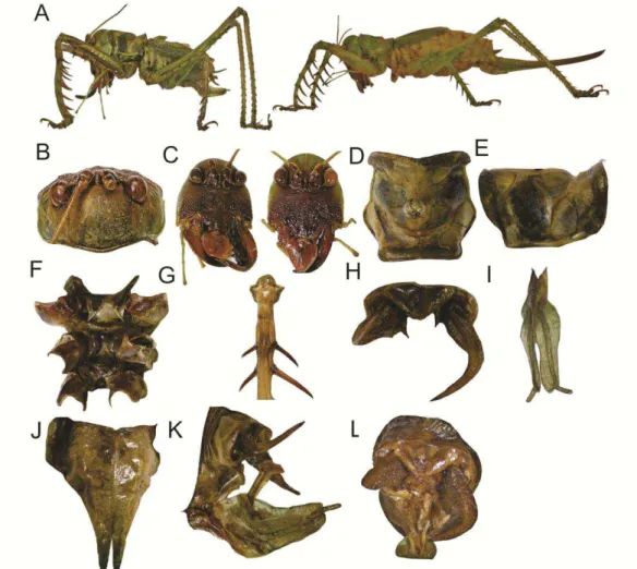
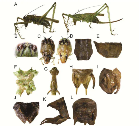
Cerberodon sp. nov. 1 (Figs 1 & 3)
Diagnosis. This species can be distinguished from C. viridis by the following
combination of characters: (i) fore tibiae with ventral surface dark brownish when
dead, or dark orange when live; (ii) male tegmina shorter than the length of the
abdomen not covering the last two abdominal segments, and in female not
covering the five last abdominal segments; (iii) supranal plate in male
semitriangular.
Description (holotype ♂ & allotype ♀). Holotype: TL 27; PL 9; PW 7; EyeW 4; TegL 14; SL 2.39; NT 66; minT 0.04; maxT 0.08; HF 22; HT 26; sFF, inner
margin 6, outer 6, small 2–0; sMF, inner margin, large 5, small 19, outer large 5,
small 6; sHF, inner margin, large 8–10, small 2–8, outer large 10–12, small 0–1;
sMT, inner margin 7, outer 4; sMTld 4; sHTd, inner margin 10–15, outer 12–13,
sHTv, inner margin 9–12, outer 12. Allotype ♀ TL 35; PL 8; PW 8; EyeW 4;
TegL 13; HF 22; HT 24; sFF, inner margin, large 5–6, small 2–3, outer large 5–6,
small 1–2; sMF, inner margin, large 19, small 18, outer large 4, small 4; sHF,
inner margin, large 11–13, small 0, outer large 11–12, small 0; sMTld; sHTd,
inner margin, outer, sHTv, inner margin, outer; OL 28. Coloration pattern
consisting of light greenish and dark brownish areas (Fig. 1A). Head. Dorsal area
light greenish (Fig.1B). Fastigium dark brownish. Antennal scape dark brownish.
Pedicele with frontal surface dark brownish and dorsal surface greenish.
Antennomeres greenish. Eyes dark brownish, the surface around them blackish.
Frons dark brownish, semitriangular, with an oval protruding tubercle at the
middle, and with the ocelus pole (Fig. 1C). Face, genae and clypeus dark
brownish. Mandibles light brownish with cutting edge darkish; outer margin
wrinkled. Clypeus dark brownish with apical portion darker. Labrum pinkish with
basal portion dark brownish. Mouthpart in ventral view dark brownish. Thorax.
Pronotum greenish (Fig. 1D–E). Fore coxae with basal portion greenish and apical
portion reddish; trochanters dark brownish. Fore femora greenish; lower margin
reddish; spines whitish with inner base blackish. Tibiae greenish with base darker;
spines light greenish. Mid femora greenish, lower margin and spines whitish,
ventral area greenish. Hind femora greenish, lower margin lighter, spines dark
third one darker. Tarsus claws greenish with tip darker. Sternum mostly greenish,
prosternum lighter (Fig. 1E); meso- and metasternum with posterior margin
reddish (Fig. 1F). Tegmina with primary and secondary veins light greenish,
spaces between veins dark brownish; stridulatory area darker. Abdomen. Tergites
dark brownish; sternites slightly reddish. Supranal plate wide, broader than
longer, subtriangular (Fig. 1H). Cerci of male with basal portion wider than the
apical, the latter flattened dorsoventrally, strongly curved inwards and ending in
an acute tip (Fig. 1H). Paraprocts with a protruding spine curved downwards.
Subgenital plate of male with a deep V-shape emargination (Fig. 1I), being half
the length of the plate; in female, with a V-shape emargination (Fig. 1J), being
one-quarter the length of the plate. Male genitalia as shown in Fig. 1K–L.
Ovipositor longer than the length of abdomen. Alive, the areas described as dark
brownish are reddish, almost dark orangish (face, mandibles, posterior margin of
the pronotum, ventral surface of fore femora, spines of tibiae, tarsi, part of the
sternum, abdominal tergites, abdominal spiracles, and cerci in male, dorsal and
ventral margin of the ovipositor). Eyes pinkish frontally, and whitish at dorsally;
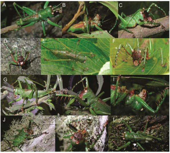
labrum lighter than after dead (Fig. 3A–F).
Type series. Holotype male labeled \Brasil, MG, Alto Caparaó, P.N Caparaó. 23–
26, XI, 2011. J. Chamorro leg. [handwritten on with paper] \ 1/Listro/Caparao
[typewritten on white paper] \ Cerberodon sp. nov. 1 Fialho, Chamorro-Rengifo
& Lopes-Andrade [handwritten on red paper]\. Allotype female labeled \Brasil,
MG, Alto Caparaó, P.N Caparaó. 4–8, II, 2012. V. Fialho. [handwritten on with
paper] \ Cerberodon sp. nov. 1 Fialho, Chamorro-Rengifo & Lopes-Andrade
[handwritten on blue paper].
Comments. Deimatic behavior was performed by male holotype, and was
documented in Fig. 3D. Similar behaviors have been observed in Neobarrettia
Rehn, 1901 (Cohn 1965), other Listroscelidinae, and species of different
subfamilies as in Mygalopsis ferruginea Redtenbacher, 1891 (Sandow and Bailey
1978), Acanthodis curvidens (Stål, 1875) (Robison 1969), and Panacanthus
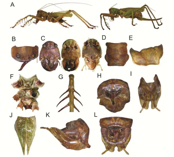
Cerberodon viridis Perty, 1832, Modified from description of Redtenbacher,
1891 (Figs 2 & 3)
Diagnosis. This species can be distinguished from Cerberodon sp. nov. 1 by the
following combination of characters: (i) fore tibiae with ventral area blackish; (ii)
tegmina longer than the abdomen; (iii) supranal plate of male rounded.
Redescription. (Base on images of material and additional specimens). Type and
General coloration consisting of light greenish and maroonish or dark brownish
areas (Fig. 2A). Head. Dorsal area greenish (Fig.2B). Fastigium dark brownish.
Scape and pedicele with frontal portion maroonish, and dorsal one greenish;
antennomeres light brownish. Eyes light brownish, with cuticle around the eyes
blackish. Frons dark brownish, semitriangular and protruding (Fig. 2C). Face,
genae and clypeus dark brownish (Fig. 2C). Mandibles light brownish with
cutting edge darkish; outer margin wrinkled. Labrum brownish. Mouthparts in
ventral view dark brownish. Thorax. Pronotum greenish (Fig. 2D, 2E). Legs, in
general, greenish with spines dark brownish; coxae and trochanters with ventral
area maroonish, the remaining greenish. Forelegs with ventral area of femora and
areas close to spines of tibiae blackish. Tarsi and tarsal claws dark brownish.
Sternum greenish, with some spots maroonish (Fig. 2F). Tegmina with primary
and secondary veins greenish, spaces between veins dark brownish, anal and
stridulatory area dark brownish. Abdomen. Tergites greenish; sternites in male
light reddish, and in female whitish. Supranal plate wide, broader than longer.
Cerci of male with basal portion wider than the apical one, strongly curved
inwards and ending in an acute tip. Subgenital plate of male with a deep V-shape
emargination, being shorter than half of the length of the plate; in female with a
V-shape emargination (Fig. 2J), being one-quarter the length of the plate. Male
genitalia as shown in figure 2L. Ovipositor longer than the length of the body.
Alive, the areas described above as dark brownish, reddish or maroonish when
dead, are dark reddish when alive (face, anterior and posterior margin of the
pronotum, spines at the legs, mouthparts in ventral view, areas at the sternum,
abdominal spiracles, abdominal sternites, dorsal and ventral margin of the
ovipositor). Eyes pinkish at frontal, and whitish at dorsal; labrum pinkish, lighter
Specimens examined. Specimens were collected in Brazil, Rio de Janeiro, Nova
Friburgo, Reserva Particular do Patrimônio Natural Bacchus, 16–19, XI, 2011. All
are labeled as follows: \Brasil, RG, R.P.P.N. Bacchus, 16–19, XI, 2011, J.
Chamorro leg. [handwritten on with paper] \ Cerberodon viridis [handwritten on
with paper]\ 1 Adult ♀: \3/Listro/Bacchus [typewritten on white paper] \ 1 immature male ♂ \ 4/Listro/Bacchus [typewritten on white paper] \ 2 immature female ♀ \ 1 and 2/Listro/Bacchus [typewritten on white paper]\. 2 male ♂ and 2 females ♀ (MNRJ): male 1 \ No. R. Anlé. Petrópolis. 1936 [handwritten on yellowish paper] \ Cerberodon viridis [handwritten on white paper] \ male 2 \
Petropolis 1.52. Frey Thomaz [handwritten on blue paper] no verso Inst. Osvaldo
Cruz [handwritten on yellowish paper] \ female 1 \ Vista chinesa 79. col. OTERO
[handwritten on yellowish paper] \ Cerberodon viridis [handwritten on white
paper] \ female 2 \ Petropolis. Est. do Rio. BRASIL. [typewritten] janeiro 1958.
Herta [handwritten] \ COLECÃO CAMPOS SEABRA [typewritten on white
paper].
Variation. Measurements of females (n=2, including the allotype, in this case,
only are shown the same measurements in Redtenbacher, 1891): TL 38–42
(including tegmina); PL 7–7.5; TegL 20–27; FF 15–16; FT 16–18; HF 22–24; OL
25.5–26. Other specimens, Measurements of males (n=2): TL 33; PL 8; PW 7;
EyeW 5; TegL 26–27; SL 2.71–2.78; NT 65–67; minT 0.02; maxT 0.18–0.19; HF
22–23; HT 24–26; sFF, inner margin, large 7-7, small 9–13, outer large 6–7, small
2-19; sMF, inner margin, large 6–7, small 11–12, outer large 6–7, small 17–24;
sHF, inner margin, large 10–14, small 0-2, outer large 9–14, small 0–3; sMTld 1–
4; sHTd, inner margin 12–14, outer 16-16, sHTv, inner margin 13–17, outer 11–
13. Measurements of females (n=3): TL 36–39; PL 7–9; PW 7; EyeW 4–5; TegL
22–27; HF 23–24; HT 25–28; sFF, inner margin, large 6-6, small 0-3, outer large
6-7, small 0-4; sMF, inner margin, large 6-6, small 14-20, outer large 6-7, small
18-23; sHF, inner margin, large 11-13, small 0-0, outer large 9-12, small 0-0;
sMTld 3-4; sHTd, inner margin 13- 14, outer 15- 17, sHTv, inner margin 14-17,
outer 11-12; OL 26-28.
Comments. Material type was collected from an unknown locality from Brazil.
Listroscelis Serville, 1831
Type species: Listroscelis armata Serville, 1834
Redescription. General coloration of the specimen body could include the
following colors and its variations of dark and light: greenish, blackish,
maroonish, yellowish and brownish. Head. Fastigium laterally compressed,
narrower and shorter than the first antennomere. Eyes globose and protruding,
located separately of the antennal sockets. Apex of the antennal sockets shorter
half than the length of the eyes. Sclerites of antennal sockets not in contact at the
midline. Head at frontal view lengthy, width five-ninths of the length; vertex
slightly visible at frontal, not protruding. Frons subtriangular, with a rounded
projection at the middle of the length, where the ocellus is located, not totally
visible, in some cases the ocellus is at the base of the frons. Face smooth, or
slightly wrinkled. Mandibles with apex elongated and curved inwards; in males of
some species mandibles symmetric, in other species asymmetric, the left mandible
with lateral lower portion lengthened and curved outwards, different than in
Cerberodon where the apex is lengthened. Mandibles with a basal process at the
cutting area. Thorax. Pronotum with anterior and posterior margins straight or
slightly curved inwards. Prozone with a transverse furrow slightly extending at the
lateral lobe, not reaching the lower margin. Mesozone with a transverse furrow,
extending at the lateral lobe, reaching or not the lower margin. Metazone with a
straight transverse furrow extending laterally at the lateral lobe, almost reaching
the posterior margin. Lateral lobes with lower margin almost straight; posterior
margin slightly oblique, with rounded corners without sinus humeral. Wings fully
developed; tegmina with a yellowish bright spot at the base. Coxae could bear a
spine that could be acute or rounded at ventral, on the basal and distal portion.
Legs slender, hind femora have the same length of the body or slightly longer.
Mid and fore femora with a ventral broad distinct longitudinal furrow at all the
length. All femora armed with long spines interspersed or not with thin spines.
Fore tibiae with area close to the spines blackish; a small hole or spur at dorsal,
below and close to each opening of the tympanum. Mid tibiae at ventral have four
to six spines. Hind tibiae have several spines at dorsal and ventral margin.
one-sixth the length of the opening; localized frontally at the tibiae; area around
the tympanum conspicuously inflated. Each sternite with two spines with tip
rounded; spines at the prosternum slender, and ones of the metasternum could be
flattened or slender. Abdomen. Cerci in males, with apex bent or slightly curved
inwards or downwards; tubercles and bristles covering the entire surface. Supranal
plate could be triangular, elongated or modified with lobes, emarginated or not.
Subgenital plate of male variable, emarginated in different ways; styli with length
variable. Paraprocts in males triangular and concealing the genitalia; a spine
curved downward at the lateral outer vertex of the plate. Subgenital plate of
female could be or not emarginated. Male genitalia with an epiphallus
membranous and a phallus sclerotized, long and projected outwards, with rounded
tips.
This genus includes L. armata Serville, 1831, L. atrata Redtenbacher, 1891, L.
carinata Karny, 1907, L. ferruginea Redtenbacher, 1891, Listroscelis sp. nov. 1,
Listroscelis sp. nov. 2, Listroscelis sp. nov. 3, Listroscelis sp. nov. 4 and
Listroscelis sp. nov. 5, Listroscelis sp. nov. 6.
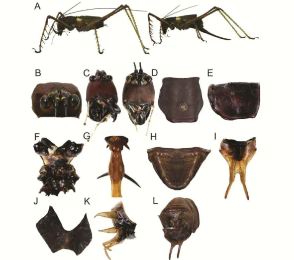
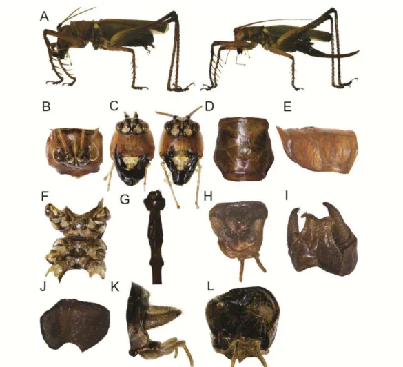
Listroscelis sp. nov. 1 (Figs 4 & 12)
Diagnosis. This species can be distinguished from the others species of
Listroscelis by the following combination of characters: (i) General coloration
dark maroonish, dark brownish and light and dark greenish; (ii) face, genae and
clypeus with transverse wrinkles arranged horizontally, parallel to the vertex; (iii)
left mandible, in male, with the pre-apical lateral portion elongated and bent,
apical portion projected upwards; (iv) yellowish spot being almost one-quarter the
length of the tegmen; (v) supranal plate in male triangular.
Description (holotype ♂ & allotype ♀). Holotype ♂: TL 25; PL 7; PW 5; EyeW 3; TegL 14; SL 2.11; NT 73; minT 0.02; maxT 0.09; HF 23; HT 26; sFF, inner
margin, large 4–5, small 24–25, outer large 5, small 30–32; sMF, inner margin,
large 5, small 23–26, outer large 4, small 28; sHF, inner margin, large 11–12,
small 21, outer large 12–13, small 12–19; sMTld 4; sHTd, inner margin 16–17,
5; EyeW 3; TegL 14; HF 23; HT 26; sFF, inner margin, large 4, small 11–14,
outer large 5–6, small 25–26; sMF, inner margin, large 5–6, small 29–30, outer
large 4–5, small 22–23; sHF, inner margin, large 7–15, small 5–12, outer large 11,
small 4–10; sMTld 4; sHTd, inner margin 18, outer 15–16, sHTv, inner margin 9–
10, outer 10–11; OL 21. General coloration consisting of dark maroonish and
greenish areas (Fig. 4A). Head. Dorsal area dark maroonish (Fig. 4B); vertex with
dorsal area dark yellowish, and sides blackish. Antennal scape and pedicele
blackish with indistinct dark brownish spots. Antennomeres blackish; sclerites of
antennal sockets blackish. Eyes dark yellowish with undefined dark brownish
spots; a dark yellowish oval area at the dorsal inner surface. Frons oval and
protruding, dark brownish, with a defined ocellus. Face and basal portion of
clypeus dark maroonish, with transversal wrinkles arranged parallel to the vertex
(Fig. 4C); middle and apical portion of clypeus yellowish, with undefined dark
brownish spots. Labrum almost blackish with basal portion dark brownish.
Mandibles blackish. Maxillary and labial palpi light greenish, with blackish spots
at the apical portion of each palpus. Mouthparts in ventral view, excepting the
appendages, dark brownish. Thorax. Pronotum dark maroonish (Fig. 4D–E).
Epimeron with the area close to the prothoracic spiracle fluorescent yellowish.
Tegmina with the portion between the costal region to the R vein, and between the
Sc vein to the lower margin, greenish fluorescents and blackish; region between
the R and Sc veins brownish. Primary veins brownish, secondary veins greenish,
space between veins blackish. Hind wings dark brownish. Sternum blackish;
spines and furrows light yellowish (Fig. 4F). Legs with ventral portion of coxae
blackish; mid and hind coxae with undefined dark yellowish spots, lateral and
ventral portions light brownish. Trochanters have similar color to that of the
coxae. Femora and tibiae of all legs with similar coloration pattern as follows:
dorsal portion light brownish, ventral one blackish and apical portion of ventral
edge light brownish. Large spine of each femur with a whitish oval spot at the
inner base. Tibiae light greenish with areas close to the spines light brownish;
spines dark brownish. Hind femora at lateral with a slightly brownish stripe
parallel to the ventral margin. Areas close to the tympanum light brownish (Fig.
4G). Tarsi have first, second and fourth tarsomeres light brownish, apical portion
spots. Tarsus claws dark brownish. Abdomen. Tergites with central portion light
brownish, and lateral portions dark brownish. Sternites blackish. Supranal plate of
male triangular (Fig. 4H). Cerci of male stout and elongated, dismissing in size
from the basal to the apical portion; tip projected and curved inwards (Fig. 4K).
Subgenital plate of male with basal portion dark brownish and the remaining light
brownish (Fig. 4I); an emargination leading a U-shape cut almost one-quarter the
length of the plate at the longitudinal midline. Styli almost one-third the length of
the plate. Ovipositor one-sixth times longer than the abdomen. Male genitalia as
shown in Fig. 4L.
Variation. Measurements of males (n=8, including the holotype): TL 23–26; PL
7; PW 5–6; EyeW 2–3; TegL 13–14; HF 22–24; HT 24–26; sFF, inner margin,
large 4–5, small 27–35, outer large 5, small 28–37; sMF, inner margin, large 4,
small 29–33, outer large 3–4, small 24–35; sHF, inner margin, large 4–15, small
21–25, outer large 10–13, small 9–22; sMTld 4; sHTd, inner margin 16–18, outer
15–17, sHTv, inner margin 9-11, outer 15-17. Stridulatory file (n=2) SL: 2.11–
2.23; NT: 73–78; minT 0.02–0.03. Measurements of females (n=2, including the
allotype): TL 27–28; PL 6–7; PW 5; EyeW 3; TegL 12–14; HF 22–23; HT 25–26;
sFF, inner margin, large 4–5, small 10–14, outer large 5–6, small 25–26; sMF,
inner margin, large 5–6, small 14–30, outer large 4–6, small 22–24; sHF, inner
margin, large 7–15, small 1–12, outer large 9–11, small 0–10; sMTld 4–4; sHTd,
inner margin 16–18, outer 15–16, sHTv, inner margin 9–12 , outer 9–11; OL 20–
21.
Type series. Specimens were collected in Brazil, Bahia, Camacan, Reserva
Particular do Patrimônio Natural Serra Bonita, 10–12, I, 2012. All are labeled as
follow: \Brasil, Camacan, BA, R.P.P.N Serra Bonita, 10–12, I, 2012, J. Chamorro
leg. [handwritten on with paper]\. Holotype male also labeled \17–Listro–Bonita
[typewritten on white paper] \ Listroscelis sp. nov. 1 Fialho, Chamorro-Rengifo &
Lopes-Andrade 2012 [handwritten on red paper]\. Allotype female labeled
\7/Listro/Bonita \ Listroscelis sp. nov. 1 Fialho, Chamorro-Rengifo &
Lopes-Andrade 2012 [handwritten on blue paper]\. Paratypes all labeled: \ Listroscelis
blue paper]\. 7 ♂ adults, codes: 1,2,3,4,5,13,14; 1♂ immature, code: 26; 1 ♀ adults, codes: 15; 1 ♀ immature, codes: 7,15.
Comments. Individuals of this species were abundant in field, unlike other
species of the genus. Alive as shown in Figure 12A–C.
Listroscelis sp. nov. 2 (Figs 5 & 12)
Diagnosis. This species can be distinguished from the others species of
Listroscelis by the following combination of characters: (i) general coloration
light greenish with few areas light brownish; (ii) face, genae and clypeus smooth;
(iii) mandibles symmetric; (iv) yellowish spot being almost one-twelfth the length
of the tegmen; (v) supranal plate in male elongated, the width of the plate
decreases rapidly from the middle to the tip; a deep emargination with an
oval-shape at the longitudinal midline.
Description (holotype ♂ & allotype ♀). Holotype ♂: TL 26; PL 7; PW 5; EyeW
3; TegL 22; SL 2.22; NT 136; minT 0.01; maxT 0.06; HF 23; HT 25; sFF, inner
margin, large 4–5, small 23–27, outer large 5, small 36; sMF, inner margin, large
5, small 32, outer large 4, small 27; sHF, inner margin, large 13–15, small 18–21,
outer large 12–13, small 7–10; sMTl d 5; sHTd, inner margin 16–18, outer 15,
sHTv, inner margin 10, outer 12-13. Allotype ♀: TL 27; PL 6; PW 5; EyeW 3;
TegL 22; HF 12; HT 28 sFF, inner margin, large 4-5, small 19–21, outer large 4–
5, small 26-28; sMF, inner margin, large 5, small 31, outer large 4, small 30; sHF,
inner margin, large 13–15, small 12–13, outer large 12, small 7–10; sMTld 6;
sHTd, inner margin 18, outer 16–17, sHTv, inner margin 10–11, outer 13; OL 26.
General coloration consisting of light greenish and light brownish areas (Fig. 5A).
Head. Dorsal area light greenish (Fig. 5B). Fastigium at dorsal portion whitish
and at laterals brownish. Antennal scape and pedicele blackish; antennomeres
dark brownish; sclerites of antennal sockets blackish. Eyes dark brownish with
irregular blackish spots. Frons semi-oval, light brownish, with a thin tubercle (Fig.
5C). Face almost smooth, slightly wrinkled, light brownish; sides light greenish.
Clypeus and labrum light brownish. Mandibles with basal portion and sides light
palpi greenish. Mouthparts in ventral view light brownish. Thorax. Pronotum
light greenish (Fig. 5D–E). Sternites and spines light greenish (Fig. 5F). Legs
including coxae and trochanters light greenish; spines at the femora and tibiae
light brownish. Areas close to the tympanum light brownish (Fig. 5G). Tarsi light
greenish. Tarsus claws light greenish with the tip dark brownish. Tegmina
including primary and secondary veins light greenish; spaces between veins dark
brownish. Abdomen. Tergites and sternites light greenish. Supranal plate of male
as shown in Fig. 5H. Subgenital plate of male wide with a V-shape emargination
at the longitudinal midline (Fig. 5I). Styli being one-fifth the length of the plate.
Cerci with apical portion slightly curved downwards (Fig. 5H, 5J). Ovipositor
one-thirteenth longer than the length of the abdomen. Male genitalia as shown in
Fig. 5K.
Type series. Holotype male labeled \Brasil, ES, Linhares, ReBio de Sooretama.
29–XI – 2–XII, 2011. J. Chamorro leg. [handwritten on with paper] \ 6–Listro–
Sooretama [typewritten on white paper] \ Listroscelis sp. nov. 2 Fialho,
Chamorro-Rengifo & Lopes-Andrade 2012 [handwritten on red paper]\. Allotype
female, same locality and data as the holotype, also labeled \1–Listro–Sooretama
[typewritten on white paper] \ Listroscelis sp. nov. 2 Fialho, Chamorro-Rengifo &
Lopes-Andrade 2012 [handwritten on blue paper]\.
Comments. Alive, both individuals had more vivid greenish coloration than after
dead (Fig. 12D–E).
Listroscelis sp. nov. 3 (Figs 6)
Diagnosis. This species can be distinguished from the others species of
Listroscelis by the following combination of characters: (i) general coloration
dark and light brownish and light greenish; (ii) face, genae and clypeus almost
smooth, slightly wrinkled; (iii) mandibles symmetric; (iv) yellowish spot being
almost one-sixteenth the length of the tegmen; (v) supranal plate in male
Description (holotype ♂): TL 26; PL 6; PW 5; EyeW 3; TegL 22; SL 2.68; NT 178; minT 0.02; maxT 0.08; HF 22; HT 24; sFF, inner margin, large 5-6, small
27, outer large 5, small 35–38; sMF, inner margin, large 5, small 29, outer large 5,
small 28; sHF, inner margin, large 10–12, small 22–25, outer large 16–17, small
12–22; sMTld 4; sHTd, inner margin 18, outer 16–18, sHTv, inner margin 9–12,
outer 11. General coloration consisting of dark and light brownish and light
greenish areas (Fig. 6A). Head. Dorsal area dark brownish (Fig. 6B). Fastigium
dorsally light yellowish and laterals dark brownish. Antennal scape and pedicele
light brownish; antennomeres dark brownish. Frons semitriangular, dark brownish
(Fig. 6C). Eyes reddish. Face dark brownish. Mouthparts in ventral view whitish.
Thorax. Pronotum dark brownish (Fig. 6D–E). Forelegs with coxae and
trochanters light brownish; femora light brownish, lower margin lighter. Spines at
the femora and tibiae dark brownish with tip darker. Tibiae with apical portion
dark brownish and the remaining light greenish; areas close to the spines blackish.
Tympanum with cuticle around the openings blackish. Mid and hind coxae and
trochanters yellowish. Midlegs light greenish. Hind femora and tibiae light
greenish, apical and basal portion dark brownish. Mid and hind spines light
brownish. Tarsi light brownish, the third one darker. Tarsus claws light brownish
with tip dark brownish. Tegmina with primary and secondary veins light greenish;
spaces between veins dark brownish; stridulatory area darker. Hind wings light
greenish with primary veins dark brownish. Prothoracic spiracle with the upper
and lateral portions dark brownish. Sternum yellowish with spines light greenish
(Fig. 6F). Abdomen. Tergites light brownish; sternites dark brownish. Subgenital
plate of male with a deep V-shape emargination (Fig. 6I); styli being one-third the
length of the plate. Cerci laterally flattened, with apical portion bended inward
(Fig. 6H, 6J). Male genitalia as shown in Fig. 6K.
Type series. Holotype male labeled \Brasil, BA, P.N. do Descobrimento. J.
Chamorro leg. 13–15, I, 2012 [handwritten on white paper] \
4/Listro/Descobrimento [typewritten on white paper] \ Listroscelis sp. nov. 3
Listroscelis sp. nov. 4 (Figs 7 & 12)
Diagnosis. This species can be distinguished from the others species of
Listroscelis by the following combination of characters: (i) general coloration
dark and light brownish; (ii) face, genae and clypeus smooth in both sexes, but in
male slightly wrinkled; (iii) left mandible, in male, with pre-apical lateral portion
elongated and bent, apical portion projected upwards; (iv) yellowish spot being
one-eleventh the length of the tegmina; (vi) Supranal plate in male triangular.
Description (holotype ♂ & allotype ♀). Holotype ♂: TL 28; ; PL 7; PW 5; EyeW 3; TegL 20; SL 2.20; NT 92; minT 0.04; maxT 0.11, HF 20; HT 21; sFF,
inner margin, large 4–5, small 15–18, outer large 5, small 17; sMF, inner margin,
large 5, small 22, outer large 5, small 21; sHF, inner margin, large 13, small 0–3,
outer large 12, small 0; sMTld 5; sHTd, inner margin 16, outer 14–15, sHTv,
inner margin 10, outer 11–13. Allotype ♀: TL 24; PL 6; PW 4; EyeW 3; TegL
20HF 19; HT 21; sFF, inner margin, large 4–5, small 14–16, outer large 4–5,
small 20–27; sMF, inner margin, large 4, small 20, outer large 4–5, small 17; sHF,
inner margin, large 0–13, small 0, outer large 0, small 0; sMTld 4;sHTd, inner
margin 16, outer 14, sHTv, inner margin 7–9, outer 10; OL 16. General coloration
consisting of dark and light brownish areas (Fig. 7A). Head. Dorsal area dark
brownish, with a whitish stripe extending from the tip of the fastigium to the
posterior margin of the head (Fig. 7B). Fastigium with laterals portions dark
brownish. Eyes dark brownish with undefined darker spots. Antennal scape and
pedicele in male almost blackish, pedicele with the inner area yellowish, in female
dark brownish with undefined blackish spots. Antennomeres light brownish.
Sclerites of antennal sockets blackish. Frons almost semitriangular, dark brownish
(Fig. 7C). Face dark brownish, slightly wrinkled in male, in female smooth.
Clypeus in male almost yellowish, with central portion whitish, in female with
basal portion at the angles blackish; labrum almost blackish with basal portion
yellowish. Mandibles blackish. Maxillary and labial palpi light greenish.
Mouthparts in ventral view yellowish. Thorax. Pronotum dark brownish (Fig.
7D–E); lateral lobes with a blackish stripe at the lower and posterior margin.
Epimeron with the area close to the prothoracic spiracle light brownish;
7F). Legs with coxae and trochanters yellowish with undefined dark brownish
spots, mid and hind coxae and trochanters darker. Femora and tibiae dark
brownish. Tibiae with spines light brownish. Femora with spines dark brownish.
Apical portion of femora darker. Hind femora with lower margin blackish. Tarsus
dark brownish; tarsal claws dark brownish with tip blackish. Tegmina light
brownish with primary veins light brownish, secondary veins light greenish,
spaces between veins light brownish. Abdomen. Tergites light brownish and
sternites dark brownish. Cerci stout, with tip strongly curved inwards (Fig. 7K–L).
Subgenital plate of male with a slight emargination V-shape at the longitudinal
midline (Fig. 7I); styli almost half the length of the plate. Ovipositor one-seventh
longer than the abdominal length. Male genitalia as shown in Fig. 7L.
Type series. Holotype male labeled \Brasil, MG, Araponga, P.E. Brigadeiro. 12–
15, XII, 2011. J. Chamorro leg. [handwritten on with paper] \ 2–Listro–Brigadeiro
[typewritten on white paper] \ Listroscelis sp. nov. 4 Fialho, Chamorro-Rengifo &
Lopes-Andrade 2012 [handwritten on red paper]\. Allotype female, same locality
and data as the holotype, and additionally labeled \1–Listro–Brigadeiro
[typewritten on white paper] \ Listroscelis sp. nov. 4 Fialho, Chamorro-Rengifo &
Lopes-Andrade 2012 [handwritten on blue paper]\.
Comments. Alive, abdominal tergites and sternites are light greenish; the whitish
stripe at the dorsal of the head is more vivid (Fig. 12F–G).
Listroscelis sp. nov. 5 (Figs 8 & 12)
Diagnosis. This species can be distinguished from the others species of
Listroscelis by the following combination of characters: (i) general coloration
dark and light brownish; (ii) face, genae and clypeus almost smooth, slightly
wrinkled; (iii) mandibles symmetric; (iv) yellowish spot being one-tenth the
length of the tegmen; (v) supranal plate broad, with an emargination leading an
oval-shape cut of almost one-quarter the length of the plate at the longitudinal
Description (holotype ♂ & allotype ♀). Holotype ♂: TL 32; PL 8; PW 6; EyeW 4; TegL 29; SL 3.86; NT 153; minT 0.04; maxT 0.16; HF 25; HT 26; sFF, inner
margin, large 4–5, small 24–27, outer large 6, small 30–39; sMF, inner margin,
large 5, small 37, outer large 5, small 27; sHF, inner margin, large 14, small 1–4,
outer large 14, small 2–4; sMTld 0; sHTd, inner margin 16–19, outer 15–17,
sHTv, inner margin 11–14, outer 11–13. Allotype ♀: TL 36; PL 8; PW 6; EyeW
3; TegL 29; HF 27; HT 28; sFF, inner margin, large 4–5, small 21–22, outer large
5, small 31–32; sMF, inner margin, large 4, small 32, outer large 4, small 27; sHF,
inner margin, large 14, small 10–14, outer large 13–14, small 4–13; sMTld 3;
sHTd, inner margin 16–17, outer 15–16, sHTv, inner margin 10–11, outer 13–14;
OL 25. General coloration consisting of light and dark brownish areas (Fig. 8A).
Head. Dorsal area dark brownish (Fig. 8B). Fastigium at dorsal portion yellowish
and laterals light brownish. Eyes dark brownish with undefined darker spots.
Antennal scape and pedicele light brownish with indistinct dark brownish spots;
antennomeres black brownish. Frons semitriangular with a defined ocelus (Fig.
8C). Face almost smooth with few wrinkles. Clypeus yellowish, labrum almost
light brownish with apical portion dark brownish, almost blackish. Mandibles
blackish. Maxillary and labial palpi yellowish. Mouthparts in ventral view light
brownish. Thorax. Pronotum dark brownish (Fig. 8D–E). Sternum and spines
light brownish (Fig. 8F). Epimeron with area close to the prothoracic spiracle light
brownish; prothoracic spiracle with the margin blackish. Legs including coxae
and trochanters light brownish; femora darker than the tibiae; spines of femora
dark brownish; spines of tibiae dark brownish with tip lighter. Area close to the
tympanum dark brownish (Fig. 8G). Tarsi light brownish, the third one darker.
Tarsal claws light brownish with tip darker. Tegmina light brownish; primary
veins dark brownish, secondary veins light brownish, spaces between veins dark
brownish. Hind wings light brownish. Abdomen. Tergites and sternites light
brownish. Cerci stout, apical portion narrower and curved downwards (Fig. 8H–I,
8K). Subgenital plate broad with a V-shape emargination (Fig. 8I). Stily almost
one-fifth the length of the plate. Ovipositor one-eleventh longer than the
Variation. Measurements of females (n=2, including the allotype): TL 36–39; PL
8–9; PW 6; EyeW 3; TegL 29–31; HF 27–30; HT 20–33; sFF, inner margin, large
4–5, small 21–27, outer large 5, small 31–39; sMF, inner margin, large 5–4, small
32–33, outer large 4, small 27–34; sHF, inner margin, large 10–14, small 10–17,
outer large 10–14, small 4–15; sMTld 6; sHTd, inner margin 16–18, outer 15–16,
sHTv, inner margin 10–11, outer 13–16; OL 25–27.
Type series. Specimens were collected in Brazil, Bahia, Parque Nacional do
Descobrimento, 13–15, I, 2012. All specimens are labeled as follow: \Brasil, BA,
P.N. do Descobrimento. J. Chamorro leg. 13–15, I, 2012 [handwritten on white
paper]\. Additionally labeled: Holotype male \23–Listro–Descobrimento
[typewritten on white paper] \ Listroscelis sp. nov. 5 Fialho, Chamorro-Rengifo &
Lopes-Andrade 2012 [handwritten on red paper]\. Allotype female \1–Listro–
Descobrimento [typewritten on white paper] \ Listroscelis sp. nov. 5 Fialho,
Chamorro-Rengifo & Lopes-Andrade 2012 [handwritten on blue paper]\.
Paratype female \2–Listro–Descobrimento [typewritten on white paper] \
Listroscelis sp. nov. 5 Fialho, Chamorro-Rengifo & Lopes-Andrade 2012
[handwritten on blue paper]\.
Comments. Alive, individuals have almost the same colors than after dead (Fig.
12H).
Listroscelis sp. nov. 6 (Figs 9–12)
Diagnosis. This species can be distinguished from the others species of
Listroscelis by the following combination of characters: (i) general coloration
dark and light brownish and light greenish; (ii) face, genae and clypeus smooth;
(iii) yellowish spot being one-quarter the length of the tegmen.
Description (holotype ♀): TL 25; PL 7; PW 4; EyeW 3; TegL 19; HF 22; HT 23; sFF, inner margin, large 4, small 6-8, outer large 4-5, small 8-9; sMF, inner
margin, large 4, small 20, outer large 4, small 15; sHF, inner margin, large 14,
small 5-7, outer large 11-12, small 0; sMTld 3; sHTd, inner margin 16, outer 16,
dark and light brownish and light greenish areas (Fig. 9A). Head. Dorsal area
dark brownish (Fig. 9B). Fastigium with the dorsal portion whitish, laterals dark
brownish. Eyes dark brownish, slightly reddish, with undefined blackish spots.
Antennal scape, pedicele and antennomeres dark brownish. Sclerites of antennal
sockets blackish. Frons oval and dark brownish (Fig. 9C). Face dark brownish,
sides lighter; area below to the eyes yellowish. Clypeus dark brownish with
undefined blackish spots. Labrum almost yellowish with basal portion dark
brownish. Mandibles blackish. Maxillary and labial palpi whitish. Mouthparts in
ventral view yellowish. Thorax. Pronotum dark brownish (Fig. 9D–E); lateral
lobes with a darker stripe at the lateral-posterior margin. Legs with coxae and
trochanters light greenish. Fore femora light greenish, with lower margin with a
black stripe; spines light greenish with the tip blackish; tibiae dark brownish. Mid
femora greenish with lower margin almost whitish; spines dark brownish with
inner base whitish; tibiae dark brownish. Hind femora dark brownish, with lower
margin light greenish; spines light greenish with tip dark brownish; tibiae light
brownish. Tarsi light brownish, the third one darker. Tarsal claws light brownish
with tip darker. Tegmina greenish; primary veins light brownish, secondary veins
light greenish, spaces between veins blackish. Sternites and spines light greenish
(Fig. 9F); spines of meta-sternum flattened. Abdomen. Tergites dark brownish
with undefined areas reddish; sternites light brownish. Ovipositor one-eleventh
longer than the abdomen.
Variation. Measurements of females (n=3, including the holotype): TL 22–29; PL
6–7; PW 4–5; EyeW 3; TegL 19–22; HF 22; HT 23–24; sFF, inner margin, large
4, small 6–20, outer large 4–5, small 8–24; sMF, inner margin, large 4–5, small
20–24, outer large 4, small 15–18; sHF, inner margin, large 12–14, small 1–7,
outer large 11–13, small 0; sMTld 4; sHTd, inner margin 13–17, outer 14–16,
sHTv, inner margin 10–13, outer 10–14; OL 16–19.
Type series. Specimens were collected in Brazil, RJ, Parque Nacional de Itatiaia.
7–13, XI, 2011. Holotype female labeled \Brasil, RJ, P.N. Itatiaia. 7–13, XI, 2011
J. Chamorro leg. [handwritten on with paper] \ 6/Listro/Itatiaia [typewritten on
2012 [handwritten on red paper]\. Paratypes females labeled as the holotype, with
codes \ 3,8/Listro/Itatiaia [typewritten on white paper]\.
Comments. Alive, as show in Fig. 12I.
Listroscelis carinata Karny, 1907 (Figs 10 –12)
Diagnosis. This species can be distinguished from the others species of
Listroscelis by the following combination of characters: (i) General coloration
dark brownish and greenish; (ii) face, genae and clypeus slightly wrinkled; (iii)
left mandible with the apical portion elongated outwards; (iv) yellowish spot
being almost one-eighth the length of the tegmen; (v) supranal plate of male
elongated, the width of the plate decreases rapidly from the middle to the tip; a
deep emargination with an linear-shape at the longitudinal midline; tip of the plate
acute.
Redescription (Based on images of material type and observation of other
specimens). It is unknown how many specimens were measured by Kirby 1907,
males (n=?, material type): TL 22–24; PL 6; TegL 20; HF 20–21. females (n=?,
material type): TL 23–28; PL 5.9–6.2; TegL 17.5–21; HF 21–22; OL 16–23.
General coloration consisting of dark brownish and dark greenish areas (Fig.
10A). Head. Dorsal portion dark brownish. Fastigium with dorsal portion
yellowish, and sides brownish (Fig. 10B). Antennal scape and pedicele brownish.
Antennomeres dark brownish; sclerites of antennal sockets blackish. Eyes dark
brownish; a dark yellowish oval area at the dorsal inner surface. Frons oval and
protruding, dark brownish. Head in frontal view apparently elongated, due to the
outstretched labrum (Fig. 10C). Face and clypeus dark brownish. Labrum with
basal portion light yellowish, the remaining dark brownish. Mandibles dark
brownish; apical portion of both mandibles elongated, and acuminated, but the left
one more elongated than the other. Maxillary and labial palpi dark brownish.
Thorax. Pronotum dark brownish (Fig. 10D–E). Epimeron with the area close to
the prothoracic spiracle light brownish. Tegmina dark brownish, with primary,
secondary veins and spaces between them brownish. Hindwings light brownish.
brownish, femora dark greenish, tibiae dark brownish. Additionally, fore femora
with a dark brownish stripe at the lower margin of the lateral portion, in female
this stripe is weaker. Spines of each femora and tibiae dark brownish. Areas close
to the tympanum dark brownish. Tarsus of all legs dark brownish, the third-one
darker. Tarsus claws dark brownish with the tip darker. Abdomen. Supranal plate
as shown in Fig. 10H. Cerci of male elongated, forceps-shape, with tip curved
downwards (Fig. 10K). Subgenital plate of male with an emargination leading a
V-shape cut less than one-half the length of the plate at the longitudinal midline
(Fig. 10I). Styli almost one-third the length of the plate. Ovipositor almost the
same length as the body. Male genitalia as shown in Fig. 10L.
Variation. Not including data of material type. Measurements of male (n=1): TL
23; PL 8; PW 5; EyeW 2.5; TegL 21; SL 2.82; NT 158; minT 0.03; maxT 0.08;
HF 21; HT 25; sFF, inner margin, large 3–3, small 0–6, outer large 3–3, small 14–
0; sMF, inner margin, large 4–4, small 20–17, outer large 5–5, small 24–18; sHF,
inner margin, large 11–12, small 5–5, outer large 10–10, small 7–9; sMTld 5–5;
sHTd, inner margin 16–16, outer 16–18, sHTv, inner margin 13–16, outer 13–15.
Measurements of females (n=6): TL 24–31; PL 6–7; PW 4–5; EyeW 2–3; TegL
18–20; HF 22–33; HT 23–25; sFF, inner margin, large 3–4, small 10–14, outer
large 4–4, small 15–17; sMF, inner margin, large 4–4, small 13–21, outer large 4–
5, small 19–21; sHF, inner margin, large 10-12, small 0–7, outer large 9–12, small
3–5; sMTld 5–5; sHTd, inner margin 16–17, outer 16–17, sHTv, inner margin 13–
15, outer 10–14; OL 18–22.
It is unknown the type locality of L. carinata, the only information available is: “Minas Gerais, Espirito Santo”. From now on, it is known at least two localities where this species could be found.
Specimens examined. 1 adult male ♂ and 2 Adults ♀ (MNRJ). Specimens
labeled are follow: male, \Collatina. E do. E. Santo. M. Rosa, Out. 36 [typewritten
on yellowish paper] \ No. Proc. 58/512 [type– and handwritten on yellowish
paper] \ Listroscelis carinata Karny, 1907 [handwritten on white paper]\. Female
1 \Collatina. E. do. Santo. M. Rosa, Out. 36 [typewritten on yellowish paper] \ No.
Proc. 58/510 [type– and hindwritten on yellowish paper] \ Listroscelis carinata