Ramaswamy Sharma1, Masahiro Tsuchiya2, Ziedonis Skobe1, Bakhos A. Tannous3, John D. Bartlett1*
1Department of Cytokine Biology, Forsyth Institute, and Department of Developmental Biology, Harvard School of Dental Medicine, Boston, Massachusetts, United States of America,2Division of Aging and Geriatric Dentistry, Tohoku University, Sendai, Japan,3Departments of Neurology and Radiology, Massachusetts General Hospital, and Program in Neuroscience, Harvard Medical School, Boston, Massachusetts, United States of America
Abstract
Background:It is not known why the ameloblasts responsible for dental enamel formation are uniquely sensitive to fluoride (F2). Herein, we present a novel theory with supporting data to show that the low pH environment of maturating stage ameloblasts enhances their sensitivity to a given dose of F2. Enamel formation is initiated in a neutral pH environment (secretory stage); however, the pH can fall to below 6.0 as most of the mineral precipitates (maturation stage). Low pH can facilitate entry of F2into cells. Here, we asked if F2was more toxic at low pH, as measured by increased cell stress and decreased cell function.
Methodology/Principal Findings:Treatment of ameloblast-derived LS8 cells with F2at low pH reduced the threshold dose of F2required to phosphorylate stress-related proteins, PERK, eIF2a, JNK and c-jun. To assess protein secretion, LS8 cells were stably transduced with a secreted reporter,Gaussia luciferase, and secretion was quantified as a function of F2dose and pH. Luciferase secretion significantly decreased within 2 hr of F2treatment at low pH versus neutral pH, indicating increased functional toxicity. Rats given 100 ppm F2in their drinking water exhibited increased stress-mediated phosphorylation of eIF2ain maturation stage ameloblasts (pH,6.0) as compared to secretory stage ameloblasts (pH,7.2). Intriguingly, F2-treated rats demonstrated a striking decrease in transcripts expressed during the maturation stage of enamel development (Klk4and
Amtn). In contrast, the expression of secretory stage genes,AmelX,Ambn,EnamandMmp20, was unaffected.
Conclusions:The low pH environment of maturation stage ameloblasts facilitates the uptake of F2, causing increased cell stress that compromises ameloblast function, resulting in dental fluorosis.
Citation:Sharma R, Tsuchiya M, Skobe Z, Tannous BA, Bartlett JD (2010) The Acid Test of Fluoride: How pH Modulates Toxicity. PLoS ONE 5(5): e10895. doi:10.1371/journal.pone.0010895
Editor:Xiaoping Pan, East Carolina University, United States of America ReceivedFebruary 2, 2010;AcceptedMay 9, 2010;PublishedMay 28, 2010
Copyright:ß2010 Sharma et al. This is an open-access article distributed under the terms of the Creative Commons Attribution License, which permits unrestricted use, distribution, and reproduction in any medium, provided the original author and source are credited.
Funding:This work was supported by grant DE018106 from the National Institute of Dental and Craniofacial Research (www.nidcr.nih.gov) to JDB. The funders had no role in study design, data collection and analysis, decision to publish, or preparation of the manuscript.
Competing Interests:The authors have declared that no competing interests exist. * E-mail: [email protected]
Introduction
Fluoride (F2) at concentrations of 0.7 to 1.2 ppm in drinking water is beneficial as an anti-cariogenic [1]. However, higher levels of F2can occur naturally in groundwater or on land, as is found in several areas in the world [2]. Chronic exposure to high dose F2 can result in dental fluorosis [3], skeletal fluorosis [4] as well as renal and thyroid toxicity [5]. However, the initial and most apparent effect of excess F2 is in dental enamel. Approximately 32% of children in the United States suffer from mild to severe forms of dental fluorosis [6], manifested as white spots of hypomineralized enamel to darkly stained and porous enamel that chips easily [7]. It is not known why tooth enamel is uniquely sensitive to F2.
Enamel formation occurs in stages. During the secretory stage, the enamel forming epithelial cells (ameloblasts) secrete large quantities of protein, including amelogenin, ameloblastin, enam-elin and matrix metalloproteinase-20 (MMP-20). Together, these proteins form an organic matrix within which thin enamel ribbons of hydroxyapatite crystallize. The pH during the secretory stage of enamel formation is approximately 7.23 [8]. Once the enamel ribbons attain their full length, ameloblasts transition to the maturation stage. During this stage, ameloblasts secrete
kallikrein-4 (KLKkallikrein-4) to degrade the matrix proteins and facilitate their resorption [9]. This allows enamel ribbons to grow in width and thickness and interlock to form mature hardened enamel [10]. Massive precipitation of hydroxyapatite mineral occurs during the maturation stage. Depending on the phosphate precursor, the creation of one mole of apatite releases 8–14 moles of H+
ions [11,12]. Therefore, during the maturation stage of enamel development, ameloblasts are exposed to an acid environment that can dip below pH 6.0 [8].
Therefore, if the pH of the extracellular matrix is lower than that of the cell cytoplasm, an intracellular-extracellular pH gradient is maintained that continuously drives HF into the cell. Over the course of months to years, the F2 concentration within an ameloblast could rise to many times that present in the extracellular matrix, leading to ameloblast cell stress.
Exposure to excess F2can trigger endoplasmic reticulum (ER) stress within ameloblasts and compromise protein secretion [13,14]. Secreted proteins pass through the ER. The ER functions as a quality control organelle and prevents misfolded proteins from traversing the secretory pathway [15]. Factors that adversely affect ER homeostasis cause ER stress and initiate an ER-to-nucleus signaling pathway, termed the unfolded protein response (UPR). Activation of the UPR results in transient attenuation of protein translation, enabling cells to cope with the existing protein load. The UPR also upregulates chaperones, augmenting the folding capacity of the ER. Accumulated proteins may also be removed via the ER-associated degradative pathway. UPR-mediated alleviation of ER stress may allow the cell to survive; prolonged ER stress can result in apoptosis [16,17].
Here, we ask if low pH reduces the threshold dose required to induce F2 -mediated stress and if this stress results in decreased protein secretion. We also ask if rat incisor maturation stage ameloblasts that are naturally exposed to a low pH are more sensitive to F2-induced stress than secretory stage ameloblasts.
Results
Low pH enhances F2-mediated stress
F2can induce ER stress and activate the UPR in ameloblastsin vivo
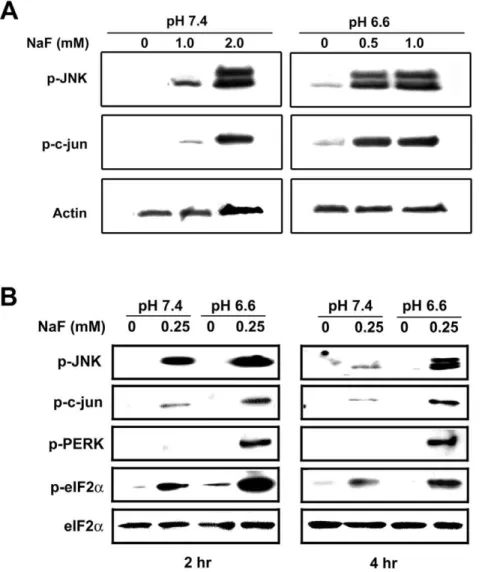
as well as in ameloblast-like LS8 cellsin vitro[13,14]. Activation of the UPR can result in the phosphorylation of JNK and c-jun [18,19,20]. To determine if low pH enhances F2-mediated stress, we treated LS8 cells with F2 at pH 6.6 or pH 7.4 and monitored phosphor-ylation of JNK and c-jun. Both proteins were phosphorylated at higher levels at low pH as compared to treatment at neutral pH. The phosphorylation observed at 2 hr with 2.0 mM F2at pH 7.4 were similar to that observed with 0.5 mM F2at pH 6.6 (Figure 1A). In addition, F2 treatment at low pH consistently resulted in more phosphorylation of these proteins at all doses assayed (Figure 1B).
Figure 1. Low pH enhances F2-mediated stress.(A) Immunoblots of LS8 cells treated with indicated doses of NaF for 2 hr at pH 7.4 or pH 6.6
were probed for phosphorylated JNK and c-jun. Actin bands are controls for protein loading. (B) Immunoblots of LS8 cells treated with 0.25 mM NaF for 2 hr or 4 hr were probed for phosphorylated forms of JNK, c-jun, PERK and eIF2a. Total eIF2abands are controls for protein loading. In all cases,
The serine/threonine kinase, PERK, is a primary sensor of the UPR that is activated by phosphorylation. Activated PERK phosphorylates the translation initiation factor, eIF2a, resulting in
a transient attenuation of protein translation. This allows cells to cope with existing accumulated proteins within the ER. As shown in Figure 1B, exposure to F2 for 2 hr or for 4 hr at pH 6.6, relative to pH 7.4, enhanced PERK and eIF2aphosphorylation.
Total levels of eIF2areflect protein loading. Taken together, these
results indicate that at low pH, lower doses of F2are required to activate stress-related proteins.
Low pH further decreases the F2-mediated reduction in
protein secretion
During the secretory stage, ameloblasts secrete large amounts of proteins such as amelogenin, enamelin and the enzyme, MMP-20, that help form the organic matrix. During the maturation stage, ameloblasts secrete KLK4, a proteinase that helps in the degradation and resorption of the organic matrix. Therefore, protein secretion is a key function of ameloblasts that is essential for enamel formation. We have previously shown that F2 decreases protein secretion in a dose-dependent manner at neutral pH [13].
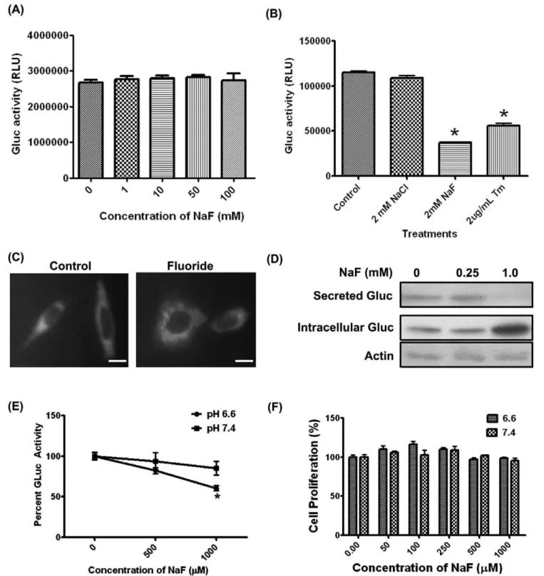
To determine if the F2-mediated decrease in protein secretion was further reduced by low pH, we stably transduced LS8 cells with either of two different secreted Gluc reporter constructs (LS8-Gluc-CFP or LS8-Gluc-YFP). Medium supernatant was assayed for Gluc activity. When recombinant Gluc was harvested and directly incubated with F2, no change in activity was observed (Figure 2A), demonstrating that F2does not affect Gluc enzymatic activity. Exposure of Gluc-transduced LS8 cells to NaF decreased Gluc secretion, as assessed by Gluc activity in the culture medium. However, treatment with NaCl did not affect Gluc secretion, indicating that F2but not Cl2was toxic to the cells (Figure 2B). Tunicamycin, an agent that induces ER stress by inhibiting N-linked glycosylation, was used as a positive control.
In untreated controls, Gluc-YFP was present throughout the cell, presumably within the secretory pathway. In contrast, treatment with 0.25 mM NaF for 6 hr caused peri-nuclear accumulation of Gluc (Figure 2C). Immunoblots for Gluc showed that F2caused a decrease in secretion and conversely, enhanced intracellular accumulation (Figure 2D). Together, these data indicate that F2 interfered with the secretion of Gluc and presumably, other endogenous secreted proteins, resulting in their intracellular accumulation.
To determine if low pH affected the F2-mediated decrease in protein secretion, we treated LS8-Gluc-CFP cells with F2 at pH 6.6 or 7.4 with 0.5 mM or 1 mM F2(Figure 2E). A significant decrease in Gluc activity (p,0.05) at pH 6.6 was observed within 2 hr as compared to Gluc activity at pH 7.4. This decrease in Gluc activity could not be attributed to changes in cell proliferation (Figure 2F). Therefore, Gluc secretion in the presence of F2was pH-dependent.
Maturation stage ameloblasts experience higher levels of F2-induced stress
To confirm our cell culture resultsin vivo, we compared stress-induced phosphorylation levels of eIF2a in ameloblasts of rats
drinking 0 or 100 ppm F2-treated water for 6 weeks. Rodent incisors grow continuously and therefore, are good models for studying fluorosis. However, a 10-fold higher F2dose is required in rodents to induce plasma F2levels equivalent to those found in humans [21]. This may be due to more efficient renal F2 clearance [22] and a shorter time-period of exposure to F2during
enamel formation. Rodent ameloblasts progress from the secretory stage to the final maturation stage in a matter of weeks whereas enamel development in humans may take years. F2 dose in rodents of 100 ppm is, therefore, representative of approximately 10 ppm in humans.
Sagittal sections of the continuously growing rodent incisors reflect all ameloblast developmental stages. Therefore, effects of F2on both secretory (pH,7.2) and maturation stage ameloblasts
(pH,6.0) can be visually compared in the same rodent incisor.
Staining for phosphorylated eIF2a was weak in secretory stage
ameloblasts whereas staining was much more intense in matura-tion stage ameloblasts and in the surrounding papillary layer (Figure 3). No significant eIF2aphosphorylation was observed in
ameloblasts from untreated control rats. These data suggest that the low pH environment of maturation stage ameloblasts sensitize them to the toxic effects of F2exposure.
Maturation stage ameloblasts exhibit decreased gene expression
F2toxicity can result in a decrease in mRNA expressionin vitro. For example, a decrease in insulin mRNA was reported when beta-cells of the pancreas were exposed to 1.35 mM NaF [23]. Therefore, we asked if F2 decreased the expression of genes involved in enamel development and importantly, if the decrease occurred in a pH-dependent manner. Enamel matrix proteins, amelogenin (AMELX), ameloblastin (AMBN), enamelin (ENAM) and matrix metalloproteinase-20 (MMP20), are pre-dominantly secreted during the secretory stage at neutral pH. Conversely, the cell-adhesion protein, amelotin (AMTN), and the matrix-degrad-ing enzyme, kallikrein-4 (KLK4), are secreted durmatrix-degrad-ing the acidic maturation stage. Gene expression was quantified by qPCR in secretory and maturation stage enamel organs of incisors from rats treated with 0, 50, 100 and 150 ppm F2ad libitum for 6 weeks. Expression levels of the secretory stage genes (Amelx,Ambn,Enam
and Mmp20) were not reduced by F2 treatment (Figure 4). However, F2 treatment significantly reduced the expression of both maturation stage genes. Expression of Klk4 decreased significantly at the lowest dose tested (50 ppm, p,0.05) and the expression ofAmtndecreased significantly at 100 ppm F2. These data are consistent with reports indicating UPR-mediated degradation of mRNAs encoding proteins destined for secretion or for proteins that localize to the plasma membrane [24,25]. Therefore, these results demonstrate F2decreases enamel matrix gene expression and that this decrease occurs in the maturation stage, when the pH is acidic.
Discussion
Figure 2. Low pH further decreases the F2-mediated reduction in protein secretion.(A) Recombinant Gluc was harvested from medium
supernatant and directly treated with the indicated doses of NaF at 37uC for 6 hr. No significant decrease in Gluc activity was observed, demonstrating that F2,by itself does not inhibit Gluc activity (B) LS8-Gluc-CFP cells were treated with NaCl, NaF or the ER stress-inducing agent,
tunicamycin for 6 hr; medium supernatant was then analyzed for Gluc activity (secretion). NaF and tunicamycin, but not NaCl, decreased Gluc secretion. (C) LS8-Gluc-YFP cells were treated with 0.25 mM NaF for 6 hr and imaged for YFP. NaF treatment localized the fusion protein within the peri-nuclear region. (D) LS8-Gluc-CFP cells were treated with the indicated doses of NaF for 24 hr and medium supernatants and cell lysates were immunoblotted and probed for Gluc. Actin served as the loading control. Note that F2treatment resulted in intracellular accumulation of Gluc. (E)
LS8-Gluc-CFP cells were treated with NaF at pH 6.6 or 7.4 for 2 hr. Gluc activity (secretion) in medium supernatant significantly decreased at pH 6.6 (p,0.05) (F) Cell proliferation, as measured by WST1 assay after 6 hr treatment, did not change significantly, indicating that the observed differences were not due to a proliferative advantage of one treatment group over another, in the short time period examined. All experiments were performed in triplicate and repeated three times. Scale bar for (C) represents 10mm.
Ameloblasts are unique because, during the maturation stage of enamel formation, they are in direct contact with the acidic mineralizing enamel matrix (pH,6.0) [8]. They are not as well-protected as other cells exposed to low pH, such as the cells lining the stomach. The latter are sheltered by a bicarbonate-rich mucus barrier that neutralizes the acid produced during digestion [37], and are continually replaced every 3–5 days. Ameloblasts, on the other hand, do not have any protective barriers and are not regenerated. Therefore, maturation stage ameloblasts may be directly exposed to F2under low pH conditions.
Several reports point toward a relation between F2and pH. For example, a decrease in pH facilitated the entry of F2into L929 fibroblasts [38]. In addition, F2-mediated cytotoxicity in osteosar-coma cells was enhanced by low pH [39]. F2 uptake in micro-organisms also occurs as a function of the culture medium pH gradient [40,41]; F2-resistant mutants become more sensitive to effects of F2at low pH [42].In vivo, F2absorption rate from the stomach increased as the gastric pH decreased [43]. Similarly, a decrease in serum pH increased F2absorption in the hamster cheek pouch and in the renal tubules of rat [44,45], rabbit [46], dog [47] and human [48,49,50]. Conversely, less fluoride was excreted as the urinary pH decreased [45,51]. Significantly, rats rendered acidotic by treatment with NH4Cl retain increased quantities of F2in their dental enamel [52]. Therefore, the more acidic the extracellular fluid, the greater the tissue fluoride concentration [53,54].
Here, we propose a novel, integrated mechanism based on pH and cell stress to explain the development of dental fluorosis. We hypothesize that F2 is converted to HF during the acidic maturation stage of enamel development and that HF flows down a steep pH concentration gradient from the enamel matrix into the ameloblast cytosol. The neutral pH inside the cell reverts HF to F2. Excess F2 within the cell interferes with ER homoestasis, inducing ER stress and activation of the UPR (Figure 5), resulting in compromised ameloblast function.
We validate our hypothesis by demonstrating that low pH enhanced F2-mediated stressin vitroandin vivo. Phosphorylation of eIF2a was observed in the papillary layer as well as in the
maturation stage ameloblasts. The complete absence of staining in the control (untreated) maturation stage ameloblasts as well as the papillary layer suggests that the staining is specific. However, the results are not surprising. Maturation stage ameloblasts are in contact with the papillary layer near the basal terminal bars [55]. Ameloblasts and papillary layer cells are extensively interconnect-ed by several large gap junctions [56]. The presence of numerous coated vesicles and also microvilli in the papillary cells suggest that they function similar to ameloblasts in the transport of ions, water and small nutrients during maturation [57]. Therefore, it is possible that fluoride ions within the ameloblast could reach the papillary cells through the gap junctions. This would result in papillary cell stress and consequently, lead to the phosphorylation
Figure 3. Maturation stage, but not secretory stage, ameloblasts from F2-treated rats exhibit stress.Rats were suppliedad libitumwith 0 or
100 ppm F2in their drinking water. Immunohistochemistry was performed on incisor sections with antiserum specific for phosphorylated eIF2 a. Note
significant staining in maturation stage ameloblasts and in the papillary layer but not in secretory stage ameloblasts of F2-treated rats. No staining was
observed in the untreated rats. Curly brackets indicate ameloblasts and square brackets indicate papillary layer. Scale bar represents 50mm.
of eIF2a. Moreover, carbon-dioxide produced within the
ameloblasts during metabolism can lead to the formation of bicarbonate ions and hydrogen ions, catalyzed by carbonic anhydrases (as shown below):
CO2zH2O<H2CO3<HzzHCO3
{
Ameloblasts contain at least 2 different carbonic anhydrases, CA2 and CA6 [58,59]. Because the blood capillary-rich papillary layer is in close proximity with the ameloblasts, it is likely that the H+ions are pumped to the capillaries and that this will cause a local decrease in the extracellular pH of the papillary layer as well.
We also showed that F2inhibited cell function (Gluc secretion) in a pH-dependent manner. Indeed, F2-mediated decrease in protein synthesis and/or secretion has been well-documented [27,28,32,60,61,62,63,64,65,66,67,68,69,70,71,72,73]. Important-ly, we demonstrated a decrease in enamel matrix transcripts during the maturation stage.
Taken together, our data show that F2 can regulate KLK4 activity by at least 3 different mechanisms. First, F2 can decrease KLK4 synthesis through stress-mediated phosphory-lation of the transphosphory-lation initiation factor, eIF2a. This results in
transient attenuation of global translation. Second, F2can also decrease KLK4 secretion from ameloblasts. Third, F2 can decrease the steady state levels of mRNAs expressed during the
Figure 4. Decreased expression of maturation but not secretory stage-specific genes.Rats were treated with 0, 50, 100 or 150 ppm F2in
their drinking water for 6 weeks. qPCR was performed on secretory and maturation stage enamel organs. Data shown is an average of three separate experiments, performed in triplicate. Data was normalized to theeEF1a1expression control gene. Note the decreased expression of maturation stage
maturation stage. While this can occur for all proteins that pass through the secretory pathway, it is especially important forKlk4. ReducedKlk4expression may hinder enamel matrix protein degradation and their removal. These mechanisms of F2 action provide an explanation for the higher protein content in fluorosed enamel as compared to normal enamel.
In conclusion, our research points toward a novel mechanism to explain fluorosis – namely, that the low pH environment of the maturation stage ameloblasts renders them more susceptible to F2 toxicity and that pH could be a defining factor in determining sensitivity of tissues to fluoride.
Materials and Methods
A complete methodology of experiments performed are listed in Supplemen-tary Figure S1.
Ethics statement
All animals were treated humanely, based on a protocol approved by the Institutional Animal Care and Use Committee (IACUC) at The Forsyth Institute. The Forsyth Institute is accredited by the Association for Assessment and Accreditation of Laboratory Animal Care International (AAALAC) that follows theGuide for the Care and Use of Laboratory Animals(NRC1996).
pH adjustment
Cell culture media containing 10% Fetal Bovine Serum (Invitrogen, Carlsbad, CA), were prepared using DMEM base lacking pH buffer (Sigma, St. Louis, MO), as described previously [74,75]. NaHCO3at 3 mM or 21 mM was added to the base to generate media with a pH of 6.6 or 7.4 respectively, in a 5% CO2 atmosphere. Medium osmolarity was adjusted by adding NaCl.
Protein secretion assay
LS8 cells were transduced with lentiviral vectors expressing Gaussia luciferase (Gluc) under the control of a CMV promoter. Gluc was either indirectly tagged to Cerulean Fluorescent Protein (CFP) through an IRES element or directly fused to Yellow Fluorescent Protein (YFP) as described previously [76,77]. Cells transduced with either construct demonstrated a decrease in protein secretion on exposure to fluoride. LS8-Gluc-CFP was used for protein secretion assays because the Gluc and CFP are translated as separate proteins, thereby avoiding any conflicts in post-translational modifications. LS8-Gluc-YFP was used to monitor the intracellular location of Gluc at a given timepoint by immunocytochemistry. LS8-Gluc-CFP and LS8-Gluc-YFP clones were isolated by flow cytometry. Protein secretion was determined as a function of Gluc activity. LS8-Gluc cells were seeded at a density of 25,000 cells / well in 6-well plates and treated with NaF at pH 6.6 or 7.4. Aliquots of 10mL medium supernatant were mixed with 20mM coelenterazine (Prolume Ltd./Nanolight, Pinetop, AZ) and the resulting bioluminescence measured for a 10 sec interval in a luminometer (Dynex, Richfield, MN). All experiments were performed in triplicate and repeated three times. Treated cell results were normalized to their untreated controls at their respective pH.
Cell proliferation assay
LS8-Gluc-CFP cells were plated at a density of 2500 cells/well in 96-well plates. NaF-containing medium at pH 6.6 or 7.4 was added. Cell proliferation was determined after 6 hr by adding WST-1 (Roche Diagnostics, Mannheim, Germany) and measuring the resulting absorbance at 440 nm. All experiments were performed in triplicate and repeated three times. Treated sample values were normalized to control values at their respective pH and calculated as percent proliferation.
Immunoblotting
To detect stress-related proteins, LS8 cells were treated with NaF at pH 6.6 or pH 7.4 for 2 hr or 4 hr. To determine the effect of F2on secretion, LS8-Gluc-CFP cells were treated with NaF at pH 6.6 or pH 7.4 for 24 hr. Medium supernatant was assessed for secreted Gluc and lysates were assessed for intracellular Gluc. Cell lysates were prepared using Complete Lysis-M reagent containing protease and phosphatase inhibitors (Roche Diagnostics). Protein concentration was determined using the BCA assay kit (Pierce, Figure 5. Schematic showing our postulated mechanism for
maturation stage ameloblast sensitivity to fluoride.During the
maturation stage, massive precipitation of hydroxyapatite occurs, releasing H+ions. F2 can reversibly associate with H+ ions to form HF. Approximately 25-fold more HF is formed at pH 6.0 as compared to pH 7.4. HF diffuses into the cell more easily than F2and flows down a
steep concentration gradient from the acidic maturation stage enamel matrix into the neutral cytosol of the ameloblast. The neutral pH inside the cell causes reversion of HF to F2. Excess F2within the cell interferes with ER homoestasis that may result in the dimerization and phosphorylation of PERK and its substrate, eIF2a. Consequently, protein
synthesis is attenuated. ER stress can also lead to increased degradation of transcripts encoding secreted proteins such as Klk4. Collectively, decreased secretion of matrix-degrading enzymes such as KLK4 can lead to delayed resorption of enamel matrix proteins, resulting in the higher protein content observed in fluorosed enamel. ER, endoplasmic reticulum.
Rockford, IL). Proteins (10–30mg) were loaded onto 4–20% polyacrylamide gels (Biorad, Hercules, CA), transferred to nitrocellulose membranes (Schleicher and Schuell, Whatman, Germany) and probed with primary antibodies, as described previously [13]. Primary antibodies included: mouse anti-Gluc (Prolume Ltd./Nanolight); rabbit anti-eIF2a[pS52] and mouse
anti-eIF2a(BioSource, Camarillo, CA); mouse anti-actin (Sigma);
rabbit anti-phospho c-jun, rabbit anti-phospho PERK and rabbit anti-phospho-JNK (Cell Signaling, Danvers, MA).
Real-time quantitative PCR (qPCR)
Six-week old rats were divided into 4 groups of three rats each and fed water containing 0, 50, 100 or 150 ppm F2,ad libitum. F2 concentration in water was confirmed using an F2 ion-selective electrode. All animals were treated humanely and with regard for alleviation of suffering. After 6 weeks, rats were sacrificed and secretory and maturation stage enamel organs were dissected from maxillary and mandibular incisors. RNA was extracted using TrizolTM(Invitrogen) and converted to cDNA (SuperScript III first-strand synthesis system, Invitrogen). All qPCR amplifications were performed as described previously [78]. Relative expression levels were calculated as a function of the internal reference control gene, eEF1a1. Primers used were: AmelX, (59 TCATCCTGGGAGCC
CTGGTTAT39and59GGCTGCCTTATCATGCTCTGGTA
39);Ambn(59GGCCTGCTC CTGTTCCTGTCC39and59 CT-GCAAGCTTCCCAACTGTCTCATT39);Enam(59GGCT TT-ACCCCTATCAACAAC39and59 TTCATAATCTTCAAACA-TCTCTTCTG 39); Mmp20 (59 CACAGCTTTAAAGTTTGC-CACTGC 39 and 59 GGGGGCCTCCTTTCTTTGTAT 39);
Klk4(59AGCCTGGCAGTCGGATGTTAGAG39and59 GGA-ATGCGCCTGATGGTGTT AG39);Amtn(59 CCTCCTTATC-CACCCCTTGTTCC39and59GGGGTGCTCATTTCGT AG TCATCA39); and eEf1a1(59 TGATGCCCCAGGACACAGAG-ACT39and59GATAC CAGCTTCAAATTCCCCAACAC39).
Immunocytochemistry and immunohistochemistry To visualize the subcellular location of Gluc, LS8-Gluc-YFP cells were grown on 4-chamber tissue culture-treated glass slides
(BD Biosciences, Bedford, MA) and treated with 0.25 mM NaF for 6 hr. Cells were fixed with 3% paraformaldehyde and imaged.
For immunohistochemistry, adult rats were treated with 0 or 100 ppm F2-containing water ad libitum. After 6 weeks, control and F2-treated rat incisors were extracted, fixed and embedded in paraffin. Sections were incubated with rabbit anti-phospho-eIF2a
(BioSource), followed by incubation in peroxidase-conjugated antibody (Vectastain Elite Reagent, Vector Labs, Burlingame, CA) and in Sigma Fast 3,39-diaminobenzidine substrate (Sigma). Sections were counterstained with 0.1% Fast Green in PBS and examined by light microscopy.
Statistics
One-way ANOVA with Bonferroni post test was performed using GraphPad Prism version 5.00 for Windows (GraphPad Software, San Diego, CA). For analyzing significance of real-time PCR results, student’s t-test was used. Ap-value,0.05 was considered significant.
Supporting Information
Figure S1 An outline of experiments performed.
Found at: doi:10.1371/journal.pone.0010895.s001 (0.20 MB TIF)
Acknowledgments
The authors thank Dr. Malcolm Snead, University of Southern California School of Dentistry, for providing LS8 cells and Dr. Steve Brooks, Leeds Dental Institute, for determining pH-based [F2]/[HF] concentrations. The authors also thank Justine Dobeck, The Forsyth Institute, for her technical expertise.
Author Contributions
Conceived and designed the experiments: RS JDB. Performed the experiments: RS MT. Analyzed the data: RS ZS BAT JDB. Contributed reagents/materials/analysis tools: ZS BAT. Wrote the paper: RS JDB.
References
1. CDC (1995) Engineering and administrative recommendations for water fluoridation, 1995. Centers for Disease Control and Prevention. MMWR Recomm Rep 44: 1–40.
2. WHO (2006) Fluoride in Drinking Water; Fawell JBK, Chilton J, Dahi E, Fewtrell L, Magara Y, eds. London, UK: IWA Publishing. 144 p.
3. Dean HT, Elvove E (1936) Some Epidemiological Aspects of Chronic Endemic Dental Fluorosis. Am J Public Health Nations Health 26: 567– 575.
4. Azar HA, Nucho CK, Bayyuk SI, Bayyuk WB (1961) Skeletal sclerosis due to chronic fluoride intoxication. Cases from an endemic area of fluorosis in the region of the Persian Gulf. Ann Intern Med 55: 193–200.
5. Ogilvie AL (1953) Histologic findings in the kidney, liver, pancreas, adrenal, and thyroid glands of the rat following sodium fluoride administration. J Dent Res 32: 386–397.
6. Beltran-Aguilar ED, Barker LK, Canto MT, Dye BA, Gooch BF, et al. (2005) Surveillance for dental caries, dental sealants, tooth retention, edentulism, and enamel fluorosis–United States, 1988–1994 and 1999–2002. MMWR Surveill Summ 54: 1–43.
7. Fejerskov O, Manji F, Baelum V (1990) The nature and mechanisms of dental fluorosis in man. J Dent Res 69 Spec No: 692–700; discussion 721. 8. Smith CE, Issid M, Margolis HC, Moreno EC (1996) Developmental changes in
the pH of enamel fluid and its effects on matrix-resident proteinases. Adv Dent Res 10: 159–169.
9. Simmer JP, Fukae M, Tanabe T, Yamakoshi Y, Uchida T, et al. (1998) Purification, characterization, and cloning of enamel matrix serine proteinase 1. J Dent Res 77: 377–386.
10. Smith CE (1998) Cellular and chemical events during enamel maturation. Crit Rev Oral Biol Med 9: 128–161.
11. Simmer JP, Fincham AG (1995) Molecular mechanisms of dental enamel formation. Crit Rev Oral Biol Med 6: 84–108.
12. Smith CE, Chong DL, Bartlett JD, Margolis HC (2005) Mineral acquisition rates in developing enamel on maxillary and mandibular incisors of rats and mice: implications to extracellular acid loading as apatite crystals mature. J Bone Miner Res 20: 240–249.
13. Sharma R, Tsuchiya M, Bartlett JD (2008) Fluoride induces endoplasmic reticulum stress and inhibits protein synthesis and secretion. Environ Health Perspect 116: 1142–1146.
14. Kubota K, Lee DH, Tsuchiya M, Young CS, Everett ET, et al. (2005) Fluoride induces endoplasmic reticulum stress in ameloblasts responsible for dental enamel formation. J Biol Chem 280: 23194–23202.
15. Hammond C, Helenius A (1995) Quality control in the secretory pathway. Curr Opin Cell Biol 7: 523–529.
16. Gow A, Sharma R (2003) The unfolded protein response in protein aggregating diseases. Neuromolecular Med 4: 73–94.
17. Schroder M, Kaufman RJ (2005) The mammalian unfolded protein response. Annu Rev Biochem 74: 739–789.
18. Nishitoh H, Matsuzawa A, Tobiume K, Saegusa K, Takeda K, et al. (2002) ASK1 is essential for endoplasmic reticulum stress-induced neuronal cell death triggered by expanded polyglutamine repeats. Genes Dev 16: 1345–1355. 19. Urano F, Wang X, Bertolotti A, Zhang Y, Chung P, et al. (2000) Coupling of
stress in the ER to activation of JNK protein kinases by transmembrane protein kinase IRE1. Science 287: 664–666.
20. Zhang C, Kawauchi J, Adachi MT, Hashimoto Y, Oshiro S, et al. (2001) Activation of JNK and transcriptional repressor ATF3/LRF1 through the IRE1/TRAF2 pathway is implicated in human vascular endothelial cell death by homocysteine. Biochem Biophys Res Commun 289: 718–724.
21. Bronckers AL, Lyaruu DM, DenBesten PK (2009) The impact of fluoride on ameloblasts and the mechanisms of enamel fluorosis. J Dent Res 88: 877–893. 22. Angmar-Mansson B, Whitford GM (1984) Enamel fluorosis related to plasma F
23. Garcia-Montalvo EA, Reyes-Perez H, Del Razo LM (2009) Fluoride exposure impairs glucose tolerance via decreased insulin expression and oxidative stress. Toxicology 263: 75–83.
24. Hollien J, Lin JH, Li H, Stevens N, Walter P, et al. (2009) Regulated Ire1-dependent decay of messenger RNAs in mammalian cells. J Cell Biol 186: 323–331.
25. Hollien J, Weissman JS (2006) Decay of endoplasmic reticulum-localized mRNAs during the unfolded protein response. Science 313: 104–107. 26. Wright JT, Chen SC, Hall KI, Yamauchi M, Bawden JW (1996) Protein
characterization of fluorosed human enamel. J Dent Res 75: 1936–1941. 27. Den Besten PK (1986) Effects of fluoride on protein secretion and removal
during enamel development in the rat. J Dent Res 65: 1272–1277.
28. DenBesten PK, Heffernan LM (1989) Enamel proteases in secretory and maturation enamel of rats ingesting 0 and 100 PPM fluoride in drinking water. Adv Dent Res 3: 199–202.
29. Shinoda H, Ogura H (1978) Scanning electron microscopical study on the fluorosis of enamel in rats. Calcif Tissue Res 25: 75–83.
30. Shinoda H (1975) Effect of long-term administration of fluoride on physico-chemical properties of the rat incisor enamel. Calcif Tissue Res 18: 91–100. 31. Triller M (1979) Structural and histochemical observations of fluorotic enamel
matrix. J Dent Res 58: 1028–1029.
32. Zhou R, Zaki AE, Eisenmann DR (1996) Morphometry and autoradiography of altered rat enamel protein processing due to chronic exposure to fluoride. Arch Oral Biol 41: 739–747.
33. Robinson C, Connell S, Kirkham J, Brookes SJ, Shore RC, et al. (2004) The effect of fluoride on the developing tooth. Caries Res 38: 268–276.
34. Robinson C, Kirkham J (1990) The effect of fluoride on the developing mineralized tissues. J Dent Res 69 Spec No: 685–691; discussion 721. 35. Aoba T, Fejerskov O (2002) Dental fluorosis: chemistry and biology. Crit Rev
Oral Biol Med 13: 155–170.
36. Bawden JW, Crenshaw MA, Wright JT, LeGeros RZ (1995) Consideration of possible biologic mechanisms of fluorosis. J Dent Res 74: 1349–1352. 37. Holzer P (2000) Gastroduodenal mucosal defense. Curr Opin Gastroenterol 16:
469–478.
38. Kawase T, Suzuki A (1989) Studies on the transmembrane migration of fluoride and its effects on proliferation of L-929 fibroblasts (L cells) in vitro. Arch Oral Biol 34: 103–107.
39. Hirano S, Ando M (1997) Fluoride mediates apoptosis in osteosarcoma UMR 106 and its cytotoxicity depends on the pH. Arch Toxicol 72: 52–58. 40. Schuster GS, Whitford GM, Lankford MT (1981) Relationship between fluoride
resistance of Streptococcus mutans 6715 and medium pH. Caries Res 15: 32–39. 41. Whitford GM, Schuster GS, Pashley DH, Venkateswarlu P (1977) Fluoride
uptake by Streptococcus mutans 6715. Infect Immun 18: 680–687.
42. Brussock SM, Kral TA (1987) Effects of pH on expression of sodium fluoride resistance in Streptococcus mutans. J Dent Res 66: 1594–1596.
43. Whitford GM, Pashley DH (1984) Fluoride absorption: the influence of gastric acidity. Calcif Tissue Int 36: 302–307.
44. Whitford GM, Callan RS, Wang HS (1982) Fluoride absorption through the hamster cheek pouch: a pH-dependent event. J Appl Toxicol 2: 303–306. 45. Whitford GM, Pashley DH, Stringer GI (1976) Fluoride renal clearance: a
pH-dependent event. Am J Physiol 230: 527–532.
46. Rouch AJ, Whitford GM, Campbell HT (1992) Fluoride flux in the rabbit CCD: a pH-dependent event. Kidney Int 41: 342–349.
47. Whitford GM, Pashley DH (1991) Fluoride reabsorption by nonionic diffusion in the distal nephron of the dog. Proc Soc Exp Biol Med 196: 178–183. 48. Ekstrand J, Ehrnebo M, Whitford GM, Jarnberg PO (1980) Fluoride
pharmacokinetics during acid-base balance changes in man. Eur J Clin Pharmacol 18: 189–194.
49. Ekstrand J, Spak CJ, Ehrnebo M (1982) Renal clearance of fluoride in a steady state condition in man: influence of urinary flow and pH changes by diet. Acta Pharmacol Toxicol (Copenh) 50: 321–325.
50. Jarnberg PO, Ekstrand J, Irestedt L (1981) Renal fluoride excretion and plasma fluoride levels during and after enflurane anesthesia are dependent on urinary pH. Anesthesiology 54: 48–52.
51. Whitford GM, Pashley DH, Reynolds KE (1977) Fluoride absorption from the rat urinary bladder: a pH-dependent event. Am J Physiol 232: F10–15. 52. Whitford GM, Angmar-Mansson B (1995) Fluorosis-like effects of acidosis, but
not NH+4, on rat incisor enamel. Caries Res 29: 20–25.
53. Whitford GM, Pashley DH, Reynolds KE (1979) Fluoride tissue distribution: short-term kinetics. Am J Physiol 236: F141–148.
54. He H, Ganapathy V, Isales CM, Whitford GM (1998) pH-dependent fluoride transport in intestinal brush border membrane vesicles. Biochim Biophys Acta 1372: 244–254.
55. Kallenbach E (1967) Cell architecture in the papillary layer of rat incisor enamel organ at the stage of enamel maturation. Anat Rec 157: 683–688.
56. Garant PR, Nagy AR, Cho MI (1984) A freeze-fracture study of the papillary layer of the rat incisor enamel organ. Tissue Cell 16: 635–645.
57. Garant PR (1972) The demonstration of complex gap junctions between the cells of the enamel organ with lanthanum nitrate. J Ultrastruct Res 40: 333–348. 58. Toyosawa S, Ogawa Y, Inagaki T, Ijuhin N (1996) Immunohistochemical
localization of carbonic anhydrase isozyme II in rat incisor epithelial cells at various stages of amelogenesis. Cell Tissue Res 285: 217–225.
59. Smith CE, Nanci A, Moffatt P (2006) Evidence by signal peptide trap technology for the expression of carbonic anhydrase 6 in rat incisor enamel organs. Eur J Oral Sci 114 Suppl 1: 147–153; discussion 164–145, 380–141. 60. Conconi FM, Bank A, Marks PA (1966) Polyribosomes and control of protein
synthesis: effects of sodium fluoride and temperature of reticulocytes. J Mol Biol 19: 525–540.
61. DenBesten PK, Thariani H (1992) Biological mechanisms of fluorosis and level and timing of systemic exposure to fluoride with respect to fluorosis. J Dent Res 71: 1238–1243.
62. DenBesten PK, Yan Y, Featherstone JD, Hilton JF, Smith CE, et al. (2002) Effects of fluoride on rat dental enamel matrix proteinases. Arch Oral Biol 47: 763–770.
63. Godchaux W, 3rd, Atwood KCt (1976) Structure and function of initiation complexes which accumulate during inhibition of protein synthesis by fluoride ion. J Biol Chem 251: 292–301.
64. Helgeland K (1976) Effect of fluoride on protein and collagen biosynthesis in rabbit dental pulp in vitro. Scand J Dent Res 84: 276–285.
65. Holland RI (1979) Fluoride inhibition of protein synthesis. Cell Biol Int Rep 3: 701–705.
66. Holland RI (1979) Fluoride inhibition of protein and DNA synthesis in cells in vitro. Acta Pharmacol Toxicol (Copenh) 45: 96–101.
67. Kruger BJ (1968) Ultrastructural changes in ameloblasts from fluoride treated rats. Arch Oral Biol 13: 969–977.
68. Lin SY, Mosteller RD, Hardesty B (1966) The mechanism of sodium fluoride and cycloheximide inhibition of hemoglobin biosynthesis in the cell-free reticulocyte system. J Mol Biol 21: 51–69.
69. Matsuo S, Inai T, Kurisu K, Kiyomiya K, Kurebe M (1996) Influence of fluoride on secretory pathway of the secretory ameloblast in rat incisor tooth germs exposed to sodium fluoride. Arch Toxicol 70: 420–429.
70. Matsuo S, Nakagawa H, Kiyomiya K, Kurebe M (2000) Fluoride-induced ultrastructural changes in exocrine pancreas cells of rats: fluoride disrupts the export of zymogens from the rough endoplasmic reticulum (rER). Arch Toxicol 73: 611–617.
71. Menoyo I, Rigalli A, Puche RC (2005) Effect of fluoride on the secretion of insulin in the rat. Arzneimittelforschung 55: 455–460.
72. Rigalli A, Ballina JC, Roveri E, Puche RC (1990) Inhibitory effect of fluoride on the secretion of insulin. Calcif Tissue Int 46: 333–338.
73. Vesco C, Colombo B (1970) Effect of sodium fluoride on protein synthesis in HeLa cells: inhibition of ribosome dissociation. J Mol Biol 47: 335–352. 74. Gstraunthaler G, Landauer F, Pfaller W (1992) Ammoniagenesis in LLC-PK1
cultures: role of transamination. Am J Physiol 263: C47–54.
75. Gstraunthaler G, Holcomb T, Feifel E, Liu W, Spitaler N, et al. (2000) Differential expression and acid-base regulation of glutaminase mRNAs in gluconeogenic LLC-PK(1)-FBPase(+) cells. Am J Physiol Renal Physiol 278: F227–237.
76. Tannous BA, Kim DE, Fernandez JL, Weissleder R, Breakefield XO (2005) Codon-optimized Gaussia luciferase cDNA for mammalian gene expression in culture and in vivo. Mol Ther 11: 435–443.