Submitted3 September 2014 Accepted 19 November 2014 Published6 January 2015
Corresponding author Offer Erez, [email protected] Academic editor
Giuseppe Remuzzi
Additional Information and Declarations can be found on page 14
DOI10.7717/peerj.691
Copyright 2015 Quaranta et al.
Distributed under
Creative Commons CC-BY 4.0
OPEN ACCESS
The physiologic and therapeutic role of
heparin in implantation and placentation
Michela Quaranta1, Offer Erez2, Salvatore Andrea Mastrolia3, Arie Koifman2, Elad Leron2, Tamar Eshkoli2, Moshe Mazor2and Gershon Holcberg2
1Department of Obstetrics and Gynecology, Azienda Ospedaliera Universitaria Integrata,
Universit`a degli Studi di Verona, Verona, Italy
2Department of Obstetrics and Gynecology, Soroka University Medical Center, School of
Medicine, Ben Gurion University of the Negev, Beer Sheva, Israel
3Department of Obstetrics and Gynecology, Azienda Ospedaliera-Universitaria Policlinico di
Bari, School of Medicine, University of Bari “Aldo Moro”, Bari, Italy
ABSTRACT
Implantation, trophoblast development and placentation are crucial processes in the establishment and development of normal pregnancy. Abnormalities of these pro-cesses can lead to pregnancy complications known as the great obstetrical syndromes: preeclampsia, intrauterine growth restriction, fetal demise, premature prelabor rup-ture of membranes, preterm labor, and recurrent pregnancy loss. There is mounting evidence regarding the physiological and therapeutic role of heparins in the estab-lishment of normal gestation and as a modality for treatment and prevention of pregnancy complications. In this review, we will summarize the properties and the physiological contributions of heparins to the success of implantation, placentation and normal pregnancy.
Subjects Gynecology and Obstetrics, Immunology
Keywords Glycosaminoglycan, Trophoblast, Selectins, Cadherins, HB-EGF, Matrix metallopro-teinases, Immune system
INTRODUCTION
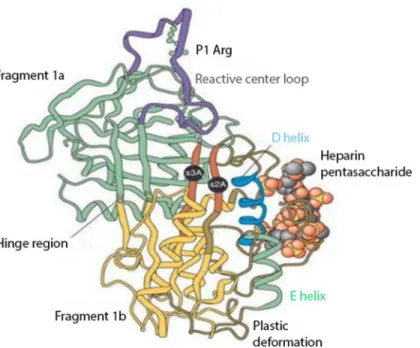
Figure 1 Anti-thrombin III after conformational change induced by heparin binding.Reproduced with permission fromWhisstock et al. (2000).
THE PHYSIOLOGICAL ROLE OF HEPARIN
Heparin is one of the oldest drugs currently in widespread clinical use. Its discovery in 1916 predates the establishment of the Food and Drug Administration of the United States, although it did not enter clinical trials until 1935. It was originally isolated from canine liver cells (Linhardt, 1991), hence its name (hepar or “ηπαρ´ ” is Greek for “liver”).
It is principally employed for its anticoagulation properties. Moreover, its true physiological role in the body remains uncertain, since blood anticoagulation is achieved mostly by heparan sulfate proteoglycans derived from endothelial cells (Marcum et al., 1986). Heparin is usually stored within the mast cells secretory granules and released only into the vasculature at sites of tissue injury. It has been proposed that, in addition to its anticoagulant properties, heparin may play a role in the defense against invading bacteria and other foreign materials (Nader et al., 1999).
Heparin is a glycosaminoglycan composed of chains of alternating residues of d-glucosamine and uronic acid. Its major anticoagulant effect is accounted for in a unique pentasaccharide (GlcNAc/NS(6S)-GlcA-GlcNS(3S,6S)-IdoA(2S)-GlcNS(6S) structure that has a high binding affinity sequence to anti-thrombin III (AT-III) (Rosenberg & Bauer, 1992); however,in-vitrostudies suggest that this structure is present only in about one third of heparin molecules (Rosenberg et al., 1979).
Figure 2 Mechanisms of interaction between heparin, anti-thrombin III, thrombin (A) and factor Xa (B).
Heparin increases the inhibitory effect of AT-III on thrombin and Factor Xa activity by distinct mechanisms (Fig. 2). The acceleration of the inhibition of thrombin by AT-III necessitates the binding of this molecule to the heparin polymer proximally to the pen-tasaccharide units. Heparin has a highly negative charge that is derived from the number of its saccharide units, which contributes to the strong electrostatic interaction of AT-III with thrombin. Thus, heparin’s activity against thrombin is size-dependent, and the ternary complex (including thrombin, AT-III and heparin) requires at least 18 saccharide units for efficient formation and thrombin inactivation (Petitou et al., 1987;Petitou et al., 1997).
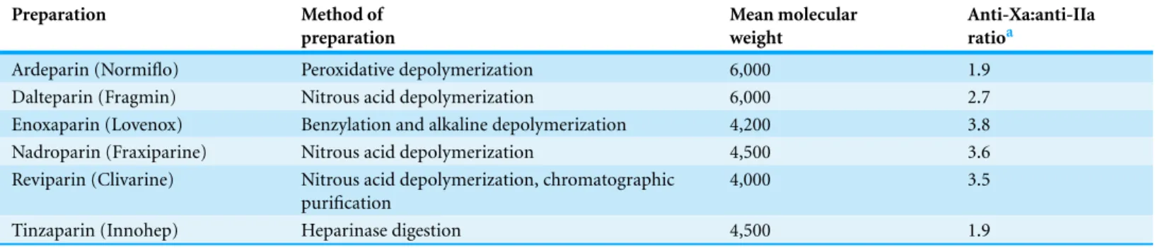
In contrast, the effect of heparin on the inhibition of factor Xa by AT-III is dependent on the conformational change of this molecule at the heparin-binding site; therefore, the size of heparin has no importance in the inhibition of factor Xa by AT-III. This effect has therapeutic implications and led to the development of a new generation of heparin derived anticoagulants including low molecular weight heparins (LMWH) and fondaparinux. LMWH are obtained as fragments of unfractionated heparin as a result of enzymatic or chemical depolymerization, yielding to molecules of mean weight of 5,000 Da (Table 1) (Weitz, 1997) while fondaparinux is a synthetic pentasaccharide based on the heparin antithrombin-binding domain (Chang et al., 2014).
Table 1 Comparison among low molecular weight heparin preparation.Reproduced with permission fromWeitz (1997).
Preparation Method of
preparation
Mean molecular weight
Anti-Xa:anti-IIa ratioa
Ardeparin (Normiflo) Peroxidative depolymerization 6,000 1.9 Dalteparin (Fragmin) Nitrous acid depolymerization 6,000 2.7 Enoxaparin (Lovenox) Benzylation and alkaline depolymerization 4,200 3.8 Nadroparin (Fraxiparine) Nitrous acid depolymerization 4,500 3.6 Reviparin (Clivarine) Nitrous acid depolymerization, chromatographic
purification
4,000 3.5
Tinzaparin (Innohep) Heparinase digestion 4,500 1.9
Notes.
aThe ratios were calculated by dividing the anti–factor Xa (anti-Xa) activity by the antithrombin (anti-IIa) activity. The ratios are based on information provided by the
manufacturers.
improved therapeutic index and less side effects. Indeed, each molecule of fondaparinux binds to one molecule of AT-III at a specific site, and with very high affinity. The binding is rapid, non-covalent, and reversible. It induces a critical conformational change in AT-III, exposing a loop containing an arginine residue that binds factor Xa. Exposure of the arginine-containing loop greatly increases the affinity of AT-III for factor Xa, potentiating the natural inhibitory effect of AT-III against factor Xa by a factor of approximately 300 (Petitou et al., 1987;Petitou et al., 1997).
THE ROLE OF HEPARINS IN IMPLANTATION AND
PLACENTATION
What are the stages of implantation and placentation?
Implantation, a critical step for the establishment of pregnancy, requires complex molecular and cellular events resulting in uterine growth and differentiation, blastocyst adhesion, invasion, and placental formation. Successful implantation necessitates a receptive endometrium, a normal and functional embryo at the blastocyst stage, and a synchronized dialogue between the mother and the developing embryo (Dey et al., 2004). In addition to the well-characterized role of sex steroids, the complexity of blastocyst implantation and placentation is exemplified by the role played by a number of cytokines and growth factors in these processes. Indeed, the process of implantation is orchestrated by hormones like sex steroids and hCG; growth factors such as TGF-B, HB-EGF, IGF-1; cytokines as Leukemia Inhibitory Factor, Interleukin-6 and Interleukin-11; adhesion molecules including L-selectin and E-cadherin; the extracellular matrix (ECM) proteins; and prostaglandins (Dey et al., 2004).
inva-sion process, which involves penetration of the embryo through the luminal epithelium into the endometrial stroma; this activity is mainly controlled by the trophoblast.
The trophoblast lineage is the first to differentiate during human development, at the transition between morula and blastocyst. Initially, at day 6–7 post-conception, a single layer of mononucleated trophoblast cells surrounds the blastocoel and the inner cell mass. At the site of attachment and direct contact to maternal tissues, trophoblast cells fuse to form a second layer of postmitotic multinucleated syncytiotrophoblast (Hoozemans et al., 2004). Once formed, the syncytiotrophoblast grows by means of steady incorporation of new mononucleated trophoblast cells from a proximal subset of stem cells located at the cytotrophoblast layer (Jauniaux, 2000).
Tongues of syncitiotrophoblast cells begin to penetrate the endometrial cells, and gradually the embryo is embedded into the stratum compactum of the endometrium. A plug of fibrin initially seals the defect in the uterine surface, but by days 10–12 the epithelium is restored (Hertig, Rock & Adams, 1956). Only at around the 14th day do mononucleated cytotrophoblasts break through the syncytiotrophoblast layer and begin to invade the uterine stroma at sites called trophoblastic cell columns. Such cells constitute the extravillous trophoblast, and have at least two main subpopulations: interstitial trophoblast, comprising all those extravillous trophoblast cells that invade uterine tissues and that are not located inside vessel walls and lumina; and endovascular trophoblast, located inside the media or lining the spiral artery lumina and partly occluding them (sometimes this subtype is further subdivided into intramural and endovascular trophoblast) (Hertig, Rock & Adams, 1956).
At a molecular level, trophoblast adhesion from the stage of implantation onwards is an integrin-dependent process (Damsky, Fitzgerald & Fisher, 1992;Zhou et al., 1997) that takes place in a chemokine- and cytokine- rich microenvironment analogous to the blood-vascular interface. Of note, uterine expression of chemokines in humans is hormonally regulated and the blastocyst expresses chemokine receptors. In addition, oxygen tension plays an important role in guiding the differentiation process that leads to cytotrophoblast invasion to the uterus (Lash et al., 2006;Zhao et al., 2012).
What is the role of heparin and heparin derived molecules in the process of implantation?
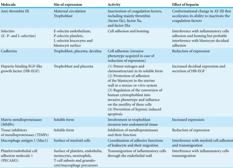
Heparin and heparin derived molecules influence all stages of implantation. This anticoagulant has an effect on the expression of adhesion molecules, matrix degrading enzymes and trophoblast phenotype and apoptosis (seeTable 2).
Selectins and cadherins
Table 2 Overview of molecules involved in the process of implantation, trophoblast development and placentation, and effect of heparin on these molecules.
Molecule Site of expression Activity Effect of heparin
Anti-thrombin III Maternal circulation Trophoblast
Inactivation of coagulation factors, including mainly thrombin (factor IIa), factor Xa, and factor IXa
Conformational change in AT-III that accelerates its ability to inactivate the coagulation factors
Selectins
(E- P- and L-selectins)
E-selectin endothelium, P-selectin platelets, L-selectin leucocytes and blastocyst surface
Cell adhesion and homing Interference with inflammatory cells adhesion and homing but probable interference with blastocyst decidual adhesion
Cadherins Trophoblast, placenta, decidua Cell adhesion (invasive phenotype acquired in case of reduction of expression)
Reduction of expression
Heparin-binding EGF-like growth factor (HB-EGF)
Trophoblast and placenta (1) Potent mitogen and
chemoattractant in its soluble form (2) Promotion of adhesion of the blastocyst to the uterine wall in a mouse-in-vitro-system (3) Regulation of the conversion of human cytotophoblast into invasive phenotype and influence on the motility of these cells (4) Prevention of hypoxic induced apoptosis
Increased decidual expression and secretion of HB-EGF
Matrix metalloproteinases (MMPs)
Soluble form Involvement in trophoblast invasion into endometrial tissue
Increased expression
Tissue inhibitors
of metalloproteinases (TIMPs)
Soluble form Inhibition of metalloproteinases and their function
Reduction of expression
Macrophage antigen 1 (Mac1) Surface of myeloid cells Coordination of adhesive functions of leukocyte and their migration
Interference with myeloid cell adhesion and transmigration
Platelet/endothelial cell adhesion molecule 1 (PECAM1)
Surface of platelets, endothelia, monocytes, neutrophils, T-cell subsets and granulo-cyte/macrophage precursors
Transmigration of inflammatory cells through the endothelial wall
Interference with inflammatory cells transmigration
may constitute an initial step in the implantation process. Indeed, L-selectin is strongly expressed on the blastocyst surface while, during the window of implantation, there is an up-regulation in the decidual expression of the selectin oligosaccharide-based ligands, predominantly on endometrial luminal epithelium (Genbacev et al., 2003). This may assist in the blastocyst decidual apposition during the implantation process.
LMWH on selectin molecules in cancer cell lines. Tinzaparin, with 22% to 36% of frag-ments greater than 8 kDA, significantly impaired L-selectin binding to its ligand; whereas enoxaparin, with 0% to 18% fragments greater than 8 kDa, did not affect L-selectin expression (Stevenson, Choi & Varki, 2005). Thus, heparins with high proportion of fragments longer than 8 kDa may reduce inflammatory cell adhesion and homing; on the other hand, they may affect blastocyst adhesion by blocking selectins ligand binding sites.
Cadherins are a group of cell adhesion proteins that mediate Ca2+-dependent cell–cell adhesion, a fundamental process required for blastocyst implantation and embryonic de-velopment (Frenette & Wagner, 1996). E-cadherin plays an important role in maintaining cell adhesion. In cancer cells, the reduction of E-cadherin expression promotes acquisition of invasive phenotype. Interestingly, gestational trophoblastic diseases (choriocarcinoma and complete hydatidiform mole), that are characterized by invasive trophoblast behavior, has a lower E-cadherin trophoblastic expression than that of first-trimester placenta (Xue et al., 2003). In contrast, the trophoblast expression of E-cadherin is higher in placentas of patients with preeclampsia than in those of normal pregnant women (Li et al., 2003). The effect of heparin on E-cadherin expression was studied byErden et al. (2006), who randomly treated female rats with different heparins (UFH, enoxaparin, and tinzaparin) during the preconceptional period, and examined E-cadherin expression in tissue sections of placenta and decidua from the different groups. The group treated by UFH had a lower E-cadherin placental staining than other study groups. In addition, the decidual staining score of this molecule was lower both in the UFH and Enoxaparin groups in comparison to controls and rats treated with tinzaparin. Therefore, there is evidence to support the effect of heparins on trophoblast invasiveness through E-cadherin expression, providing a possible mechanism by which heparin could promote trophoblast cell differentiation and motility.
Heparin binding EGF-like growth factor
Heparin-binding EGF-like growth factor (HB-EGF) is a 76–86 amino acid glycosylated protein that was originally cloned from macrophage-like U937 cells. It is a member of the epidermal growth factor (EGF) family that stimulates growth and differentiation. HB-EGF utilizes various molecules as its “receptors”. The primary receptors are in the ErbB (also named HER) system, especially ErbB1 and ErbB4, human tyrosine kinase receptors. HB-EGF is initially synthesized as a transmembrane precursor protein, similar to other members of the EGF family of growth factors. The membrane-anchored form of HB-EGF (pro HB-EGF) is composed of a pro domain followed by heparin-binding, EGF-like, juxtamembrane, transmembrane and cytoplasmic domains. Subsequently, proHB-EGF is cleaved at the cell surface by a protease to yield the soluble form of HB-EGF (sHB-EGF) using a mechanism known as ectodomain shedding. sHB-EGF is a potent mitogen and chemoattractant for a number of different cell types. Studies of mice expressing non-cleavable HB-EGF have indicated that the major functions of HB-EGF are mediated by the soluble form (Miyamoto et al., 2006).
cell specific expression during the human endometrial cycle and early placentation, and high levels expression in the first trimester (Yoo, Barlow & Mardon, 1997).
The membrane active precursor functions as a justacrine growth factor and cell-surface receptor. It has been demonstrated to promote adhesion of the blastocyst to the uterine wall in a mouse-in-vitro-system (Raab & Klagsbrun, 1997) suggesting a role for HB-EGF in embryo attachment to the uterine luminal epithelium. As stated above, the majority of its biological functions are mediated by its mature soluble form. A major role in early stages of placentation is represented by cellular differentiation and consequent invasion of the uterine wall and vascular network.
Several changes occur in the expression of adhesion molecules as cytotrophoblast differentiation proceeds, which results in pseudovasculogenesis or the adaptation by cytotrophoblast to a molecular phenotype that mimics endothelium (Zhou et al., 1997). For example, during extravillous differentiationin vivo, integrin expression is altered from predominantlyα6β4 in the villous trophoblast toα1β1 in cytotrophoblasts migrating throughout the decidual stroma (Damsky, Fitzgerald & Fisher, 1992) or engaging in endovascular invasion (Zhou et al., 1997).
Leach et al. (2004)demonstrated the role of HB-EGF in regulating the conversion of human cytotrophoblasts into invasive phenotype and the motility of these cells. This study demonstrated the ability of HB-EGF to induce ‘integrin switching’ through intracellular signaling following ligation of HER tyrosine kinases, altering integrin gene expression to stimulate cytotrophoblast invasion at a molecular level.
In addition to its effect on the invasive trophoblast phenotype, HB-EGF can affect cell motility. Indeed, cytotrophoblast motility was specifically increased by each of the EGF family members examined. The expression by cytotrophoblasts of each growth factor, as well as their receptors, suggests the possibility of an autocrine loop that advances cytotrophoblast differentiation to the extravillous phenotype.
The ability of the HB-EGF molecule to prevent hypoxic induced apoptosis plays a fundamental role in early stages of placentation. During the entire 1st trimester, the organogenesis period, embryonic development takes place in a low O2 tension environment. Oxygen concentration is relatively low (18 mmHg or 2%) at the human implantation site through the first 10 weeks of gestation due to occlusion of the uterine spiral arteries by extravillous trophoblasts. Oxygen availability serves as a developmental cue to regulate trophoblast proliferation. Experimental evidence suggests that this environment is essential for both fetal and placental development, and premature exposure to normal oxygen concentrations is associated with increased rate of pregnancy complications such as preeclampsia, IUGR and miscarriage (Jauniaux et al., 2003).
(HSPG) through its heparin binding domain, and this is followed by receptor homo- or heterodimerization with other members of the HER family. Subsequent transphosphory-lation of HER cytoplasmatic domains at tyrosine residues initiates a downstream signaling that increases proHB-EGF accumulation and inhibits apoptosis. This positive feedback loop upregulates HB-EGF secretion to achieve extracellular HB-EGF levels sufficient to maintain cell survival at 2% O2 (Armant et al., 2006).
As a result, HB-EGF has a fundamental role in successful pregnancies. This molecule mediates a vast number of functions beginning from the earliest stages of pregnancy and up to term, ranging from adhesion, to implantation and invasion, successful placentation, and protection from hypoxic induced aptoptosis. The effect of heparin on this molecule is currently being studied.Di Simone et al. (2012)demonstrated that LMWH induced an increased decidual expression and secretion of HB-EGF in a dose-dependent manner. A different study byD’Ippolito et al. (2012)demonstrated that LMWH induces activation of Activator Protein-1 (AP-1), a DNA-binding transcription factor which regulates the expression of HB-EGF. Activated AP-1 translocates to the nucleus and binds the promoter region of HB-EGF gene, thus enhancing its protein expression.Hills et al. (2006) demonstrated that heparin is capable of activating the EGF receptor in primary villous trophoblast.
Thus, we propose that accumulating evidence suggests that the beneficial effect of heparin in preventing placental mediated pregnancy complications may derive from its effect on HB-EGF expression and concentration, especially during the first trimester.
Matrix metalloproteinases
In addition to the adhesion molecules, matrix metalloproteinases (MMPs) are an important component in the process of blastocyst implantation. MMPs are a group of matrix degrading enzymes which are secreted as inactive zymogen and must be cleaved to become active (Isaka et al., 2003). Among the members of the MMP family, MMP-2 and MMP-9 type IV collagenases were suggested to be involved in trophoblast invasion into endometrial tissue (Librach et al., 1991). Indeed, the profile of proMMP 2 and 9 secretion differs during the stages of trophoblast invasion and implantation, and differences in these zymogens expression were found between 6–8 and 9–12 weeks of gestation in extravillous cytotrophoblast cells (Staun-Ram et al., 2004).Di Simone et al. (2007)investigated the effect of LWMH specifically on placental MMPs, and the degrading capacity of the tro-phoblast cells. This effect is mediated by the action of heparins on both metalloproteinases (MMPs) and their tissue inhibitors (TIMPs). Heparin increased both the MMPs concen-tration and activity by affecting their transcription, the conversion of the proenzyme into the active form, and the reduction of the synthesis of the specific inhibitors TIMPs (both the mRNA and protein levels) in a dose dependent manner (Di Simone et al., 2007).
Immunologic and anti-inflammatory effects of heparins
that anchors the placenta to the decidua, and further differentiate into endovascular trophoblast that invades spiral arteries and remodels the vessel walls, is also in direct contact with the maternal blood (Norwitz, Schust & Fisher, 2001;Bischof & Campana, 1996;Red-Horse et al., 2004). Both innate and adaptive immune responses contribute to a maternal fetal cross-talk that balances the anti- and pro-inflammatory processes in the feto-maternal interface (Norwitz, Schust & Fisher, 2001;Moffett-King, 2002). These processes involve: MHC class I molecules, hormones, complement regulatory proteins, immunoregulatory molecules (i.e., indolamine 2,3-dioxygenase, Fas/Fas- Ligand, IL-10), regulatory T cells (CD4+CD25+Foxp3+), regulatory macrophages, and growth factors expressed at the placental–decidual interface (Redecha et al., 2007;Thangaratinam et al., 2011;Wegmann et al., 1993;Thellin et al., 2000;Li & Huang, 2009;Mj¨osberg et al., 2007; Karimi, Blois & Arck, 2008;Aluvihare, Kallikourdis & Betz, 2004). These mechanisms act in concert to sustain the maternal tolerance to the semi-allogenic placenta and fetus (Kalkunte et al., 2009). In addition to its well-understood anticoagulant activity, heparin also has an impact on the immune system (Martz & Benacerraf, 1973;Sy et al., 1983;Arfors & Ley, 1993). The main known effect of heparin is on the migration and adhesion of leukocytes during an inflammatory response (Stevenson, Choi & Varki, 2005).
The anti-inflammatory effects of heparin are derived from several mechanisms: (1) the molecular structure of heparin is so that, upon its binding to the endothelial cells of blood vessels, it creates a negatively charged surface that is facing the vessel lumen. These negatively charged molecules repulse the negatively charged leukocytes and prevent their adhesion to the endothelium (heparan sulfate molecules that are expressed on leukocyte surfaces are responsible for the negative charge of these cells); (2) heparin is a large molecule that can bind a substantial number of proteins which play an important role in inflammation including selectins (L-selectin (Koenig et al., 1998) and P-selectin molecules (Skinner et al., 1991)) and integrins. The B2-integrin adhesion molecule CD11b/CD18, also known as Macrophage antigen 1 (Mac1), is a member of a subfamily of related cell-surface glycoproteins that coordinate adhesive functions including leukocyte migration (Kishimoto et al., 1989). Mac1 is expressed on myeloid cells and binds to molecules as intercellular adhesion molecule 1 (ICAM1), fibrinogen, iC3b, and factor Xa. The heparin-Mac1 bond interferes with myeloid cell adhesion and transmigration (Diamond et al., 1995). Heparin also binds to platelet/endothelial cell adhesion molecule 1 (PECAM1), a member of the Ig superfamily, expressed on a variety of cells such as platelets, endothelia, monocytes, neutrophils, T-cell subsets and granulocyte/macrophage precursors. This molecule is involved in homotypic and heterotypic cellular adhesion and plays a role in the transmigration of inflammatory cells through the endothelial wall. Heparin is capable of binding PECAM1 and interfering with its action (Watt et al., 1993), thus reducing the effectiveness of the inflammatory response.
of IL-10 mRNA, leading to a down regulation of inflammatory cytokines production. LMWH also imitates the function of Syndecan-1 (a protein which expression is inversely correlated to the mRNA expression of IL-1β in the intestinal mucosa of DSS-induced colitis models). It plays an important role in promoting wound repair, maintaining cell morphogenesis, and mediating inflammatory responses (G¨otte, 2003) by aiding the clearance of pro-inflammatory chemokines. In additionLi et al. (2013)found that treatment with UFH can attenuate inflammatory responses of lypopolisaccharide induced acute lung injury in rats. The mechanisms by which UFH exerts its anti-inflammatory effect seem to correlate with its inhibition on IL-1ß and IL-6 production via inactivation of the NF-κB pathways.
In humans the anti-inflammatory activity of heparin has been evidenced by small clinical trials in patients suffering from a range of inflammatory diseases (Gaffney & Gaffney, 1996), including rheumatoid arthritis and bronchial asthma. Remission of disease has been described in nine out of ten patients with refractory ulcerative colitis treated with combined heparin and sulphasalazine (Gaffney & Gaffney, 1996). A subjective improvement of asthma symptoms using intravenous heparin is described (Fine, Shim & Williams, 1968;Boyle, Smart & Shirey, 1964), while other studies with inhaled heparin demonstrated reduced bronchoconstrictive responses in patients with exercise-induced asthma (Garrigo, Danta & Ahmed, 1996;Ahmed et al., 1993).
The clinical rationale for the use of heparin in the treatment of inflammatory diseases may be based on the fact that many of the molecular mechanisms involved in tumor metastasis are the same responsible for cell recruitment in inflammation; heparin has been successful in treating both conditions (Tyrrell et al., 1999).
CLINICAL APPLICATION OF THE USE OF HEPARINS
DURING THE FIRST HALF OF PREGNANCY
In light of the possible effects of heparins and heparin binding molecules on the blastocyst implantation and placentation, this family of drugs may play a role in the prevention of RIF in IVF patients and in the treatment of patients with RPL.
Is there a benefit of the use of heparins in the prevention of recur-rent implantation failure in IVF patients?
The term RIF has been used since 1983 to describe the failure of embryos to implant following IVF treatments. There is no unanimous definition for RIF in terms of the number of failed cycles or the total number of transferred embryos that have not successfully implanted. The ESHRE PGD consortium document (Thornhill et al., 2005) mentioned that RIF can be considered after more than three high-quality embryo transfers or implantation failure with transfer of≥10 embryos in multiple transfers with exact numbers to be determined by each center. In order to improve pregnancy outcomes in women with RIF, various investigations and treatment adjuncts including heparin have been studied.
the use of LMWH with placebo or no adjuvant treatment in women with RIF undergoing IVF/ICSI. After the process of literature search and selection, one quasi-randomized (Potdar et al., 2013) and two randomized (Amarin et al., 2008;Urman et al., 2009) studies were selected for the meta-analysis and included 243 women with RIF who underwent IVF/ICSI, 127 in the intervention group and 116 in the control/placebo.
All three studies were unclear for detection bias, and none of the studies explicitly stated whether the individuals assessing the outcome were blinded to the trial or not. However, assessment for pregnancy outcome is unlikely to be subjective since implantation, clinical pregnancy, multiple pregnancies and miscarriage are all objectively assessed during the ultrasound scan.
The results of this meta-analysis show that, in women with≥3 RIF, the use of LMW as
an adjunct to IVF treatment significantly improved the life birth rate by 79%.
This result suggests that there could be a potential role of LMWH in improving pregnancy outcomes for women with RIF.
What is the effect of heparins on pregnancy success in women with recurrent pregnancy loss?
A beneficial effect of antithrombotic agents (heparin in particular) in women with RPL was already suggested in 1980 (Langer et al., 1980). Different clinical definitions of this condition were arrived at by the different scientific societies: the Royal College of Obstetricians and Gynecologists (RCOG) considers three or more first trimester miscarriages as RPL, while the American Society for Reproductive Medicine (ASRM) establishes a limit of two or more pregnancy losses.
Many underlying mechanisms have been recognized for RPL, including chromosomal defects (Rajcan-Separovic et al., 2010), endocrinopathies (Ke, 2014) (thyroid diseases and diabetes), uterine malformations (Jaslow, 2014), and autoimmune diseases (Kwak-Kim et al., 2013). In addition, thrombophilic mutations (Lino et al., 2014) have been suggested as leading to alterations in embryonic formation, migration, implantation and placentation; in the past three decades, this area has become a field of extensive study with the goal of increasing the rate of live births in these patients.
to inherited thrombophilia, women with antiphospholipid antibodies, MTHFR 677TT genotype and hyperhomocysteinemia were eligible. The live birth rates in both groups were similar (84.3% and 78.3%, respectively), but as a control group was lacking, the effect of enoxaparin could not be validated; (2) In 2006,Dolitzky et al. (2006)randomized 54 patients with RPL either for treatment with enoxaparin or aspirin, considering subsequent live births or miscarriage as the main outcome. Both groups had a similar live birth rate (RR 0.92, 95% CI [0.58–1.46]). This study showed that, even though there were not statistical difference between the study groups, women who were treated by heparins had a higher live birth rate to that reported in literature on women with RPL; (3) The ALIFE study (Kaandorp et al., 2010) included 364 women with two or more unexplained pregnancy losses that were randomized to nadroparin 2,850 International Unit combined with aspirin 80 mg, aspirin 80 mg only, or a placebo before conception or at a maximum gestational age of 6 weeks. Of these women, 299 became pregnant. The chance of live birth did not differ among the treatment groups. The relative risk of live birth for women who became pregnant was 1.03 (95% CI [0.85–1.25]) for nadroparin combined with aspirin, and 0.92 (95% CI [0.75–1.13]) for aspirin only compared with placebo. The study was not designed to evaluate the beneficial effect of heparin on thrombophilic patients, but the author performed a secondary analysis on the effect of heparins according to the presence of inherited thrombophilia, which showed no significant difference in the primary outcome among the groups; (4) A randomized double-blind (for aspirin) multicenter trial (Visser et al., 2011) was performed among 207 women with three or more consecutive first trimester (<13 weeks) miscarriages, two or more second trimester (13–24 weeks) miscarriages, or one third trimester fetal loss combined with one first trimester mis-carriage. Women underwent workup for thrombophilia and were randomly allocated before seven weeks gestation to either enoxaparin 40 mg+placebo (n=68), enoxaparin
40 mg+aspirin 100 mg (n=63) or aspirin 100 mg(n=76). The primary outcome was the live-birth rate. The trial was stopped prematurely because of slow recruitment. A live birth rate of 71% (RR 1.17, 95% CI [0.92–1.48]) was found for enoxaparin and placebo and 65% (RR 1.17, 95% CI [0.92–1.39]) for enoxaparin and aspirin when compared to aspirin alone (61%, reference group). In the whole study group, the live birth rate was 65% (95% CI [58.66–71.74]) for women with three or more miscarriages (n=204). No difference in pregnancy complications, neonatal outcome or adverse effects was observed. No significant difference in live birth rate was found with enoxaparin treatment versus aspirin or a combination of both versus aspirin in women with recurrent miscarriage.
CONCLUSION
Heparins play a role in embryonic implantation and placentation, and contribute to the development of a normal pregnancy. This effect is gained through the interaction of hep-arins with coagulation factors, anticoagulation proteins, their effect on the expression of adhesion molecules, matrix degrading enzymes and trophoblast phenotype and apoptosis: all important components in the process of embryonic implantation and placentation.
The fact that heparins may play a role in implantation and placentation led to their use in the prevention of RIF and RPL. In RIF heparins demonstrated a beneficial effect that could be attributed to the effects of this molecule on enhancing endometrial receptivity and trophoblast invasion due to the regulation of heparin-binding factors, adhesion molecules or inhibition of complement activation. In contrast, the positive effect of heparin as a treatment for RPL is not that clear. One possible explanation for this lack of conclusive evidence is the syndromic nature of RPL which results from several underlying mechanisms of disease. Thus, heparins may have a role in improving pregnancy outcomes among a subset of patients with RPL regardless to the presence of thrombophilia, but a conclusive statement in this matter awaits further investigation.
ADDITIONAL INFORMATION AND DECLARATIONS
Funding
The authors declare that there was no funding for this work.
Competing Interests
Offer Erez is an Academic Editor for PeerJ.
Author Contributions
• Michela Quaranta, Offer Erez, Salvatore Andrea Mastrolia, Arie Koifman, Elad Leron, Tamar Eshkoli, Moshe Mazor and Gershon Holcberg wrote the paper, prepared figures and/or tables, reviewed drafts of the paper.
REFERENCES
Ahmed T, Garrigo J, Danta I. 1993. Preventing bronchoconstriction in exercise-induced asthma with inhaled heparin.New England Journal of Medicine 329:90–95
DOI 10.1056/NEJM199307083290204.
Aluvihare VR, Kallikourdis M, Betz AG. 2004.Regulatory T cells mediate maternal tolerance to the fetus.Nature Immunology5:266–271DOI 10.1038/ni1037.
Arfors KE, Ley K. 1993.Sulfated polysaccharides in inflammation.Journal of Laboratory and Clinical Medicine121:201–202.
Armant DR, Kilburn BA, Petkova A, Edwin SS, Duniec-Dmuchowski ZM, Edwards HJ,
Bischof P, Campana A. 1996.A model for implantation of the human blastocyst and early placentation.Human Reproduction Update2:262–270DOI 10.1093/humupd/2.3.262. Bj¨ork I, Lindahl U. 1982.Mechanism of the anticoagulant action of heparin.Molecular and
Cellular Biochemistry48:161–182DOI 10.1007/BF00421226.
Boyle JP, Smart RH, Shirey JK. 1964.Heparin in the treatment of chronic obstructive bronchopul-monary disease.American Journal of Cardiology14:25–28DOI 10.1016/0002-9149(64)90100-6. Brenner B, Hoffman R, Blumenfeld Z, Weiner Z, Younis JS. 2000.Gestational outcome in
thrombophilic women with recurrent pregnancy loss treated by enoxaparin.Thrombosis and Haemostasis83:693–697.
Brenner B, Hoffman R, Carp H, Dulitsky M, Younis J, Investigators L-E. 2005.Efficacy and safety of two doses of enoxaparin in women with thrombophilia and recurrent pregnancy loss: the LIVE-ENOX study.Journal of Thrombosis and Haemostasis3:227–229
DOI 10.1111/j.1538-7836.2004.01090.x.
Carp H, Dolitzky M, Inbal A. 2003.Thromboprophylaxis improves the live birth rate in women with consecutive recurrent miscarriages and hereditary thrombophilia.Journal of Thrombosis and Haemostasis1:433–438DOI 10.1046/j.1538-7836.2003.00066.x.
Chang CH, Lico LS, Huang TY, Lin SY, Chang CL, Arco SD, Hung SC. 2014.Synthesis of the heparin-based anticoagulant drug fondaparinux.Angewandte Chemie53:9876–9879
DOI 10.1002/anie.201404154.
Cha J, Sun X, Dey SK. 2012.Mechanisms of implantation: strategies for successful pregnancy.
Nature Medicine18:1754–1767DOI 10.1038/nm.3012.
Clark DA, Arck PC, Chaouat G. 1999. Why did your mother reject you? Immunogenetic determinants of the response to environmental selective pressure expressed at the uterine level.American Journal of Reproductive Immunology41:5–22
DOI 10.1111/j.1600-0897.1999.tb00071.x.
Damsky CH, Fitzgerald ML, Fisher SJ. 1992.Distribution patterns of extracellular matrix components and adhesion receptors are intricately modulated during first trimester cytotrophoblast differentiation along the invasive pathway,in vivo.Journal of Clinical Investigation89:210–222DOI 10.1172/JCI115565.
Dey SK, Lim H, Das SK, Reese J, Paria BC, Daikoku T, Wang H. 2004.Molecular cues to implantation.Endocrine Reviews25:341–373DOI 10.1210/er.2003-0020.
Diamond MS, Alon R, Parkos CA, Quinn MT, Springer TA. 1995.Heparin is an adhesive ligand for the leukocyte integrin Mac-1 (CD11b/CD1).Journal of Cell Biology 130:1473–1482
DOI 10.1083/jcb.130.6.1473.
D’Ippolito S, Di Nicuolo F, Marana R, Castellani R, Stinson J, Tersigni C, Scambia G, Di Simone N. 2012.Emerging nonanticoagulant role of low molecular weight heparins on extravillous trophoblast functions and on heparin binding-epidermal growth factor and cystein-rich angiogenic inducer 61 expression.Fertility and Sterility98:1028–1036.e1-2. Di Simone N, Di Nicuolo F, Castellani R, Veglia M, Tersigni C, Silano M, Tritarelli A,
Scambia G, Marana R. 2012. Low-molecular-weight heparins induce decidual heparin-binding epidermal growth factor-like growth factor expression and promote survival of decidual cells undergoing apoptosis.Fertility and Sterility 97:169–177.e1
DOI 10.1016/j.fertnstert.2011.10.021.
Di Simone N, Di Nicuolo F, Sanguinetti M, Ferrazzani S, D’Alessio MC, Castellani R, Bompiani A, Caruso A. 2007.Low-molecular weight heparin inducesin vitrotrophoblast invasiveness: role of matrix metalloproteinases and tissue inhibitors.Placenta28:298–304
Dolitzky M, Inbal A, Segal Y, Weisss A, Brenner B, Carp H. 2006.A randomized study of thromboprophylaxis in women with unexplained consecutive recurrent miscarriages.Fertility and Sterility86:362–366DOI 10.1016/j.fertnstert.2005.12.068.
Erden O, Imir A, Guvenal T, Muslehiddinoglu A, Arici S, Cetin M, Cetin A. 2006.Investigation of the effects of heparin and low molecular weight heparin on E-cadherin and laminin expression in rat pregnancy by immunohistochemistry.Human Reproduction21:3014–3018
DOI 10.1093/humrep/del262.
Fine NL, Shim C, Williams MH. 1968.Objective evaluation of heparin in the treatment of asthma.
American Review of Respiratory Disease98:886–887.
Frenette PS, Wagner DD. 1996.Adhesion molecules–Part 1.New England Journal of Medicine 334:1526–1529DOI 10.1056/NEJM199606063342308.
Gaffney A, Gaffney P. 1996.Rheumatoid arthritis and heparin.British Journal of Rheumatology 35:808–809DOI 10.1093/rheumatology/35.8.808.
Garrigo J, Danta I, Ahmed T. 1996.Time course of the protective effect of inhaled heparin on exercise-induced asthma.American Journal of Respiratory and Critical Care Medicine 153:1702–1707DOI 10.1164/ajrccm.153.5.8630624.
Genbacev OD, Prakobphol A, Foulk RA, Krtolica AR, Ilic D, Singer MS, Yang ZQ, Kiessling LL, Rosen SD, Fisher SJ. 2003.Trophoblast L-selectin-mediated adhesion at the maternal-fetal interface.Science299:405–408DOI 10.1126/science.1079546.
G¨otte M. 2003.Syndecans in inflammation.FASEB Journal17:575–591
DOI 10.1096/fj.02-0739rev.
Hertig AT, Rock J, Adams EC. 1956.A description of 34 human ova within the first 17 days of development.The American Journal of Anatomy98:435–493DOI 10.1002/aja.1000980306. Hills FA, Abrahams VM, Gonz´alez-Tim ´on B, Francis J, Cloke B, Hinkson L, Rai R, Mor G,
Regan L, Sullivan M, Lam EW, Brosens JJ. 2006.Heparin prevents programmed cell death in human trophoblast.Molecular Human Reproduction12:237–243DOI 10.1093/molehr/gal026. Hoozemans DA, Schats R, Lamblack CB, Homburg R, Hompes PG. 2004.Human embryo
implantation: current knowledge and clinical implications in assisted reproductive technology.
Reproductive Biomedicine Online9:692–715DOI 10.1016/S1472-6483(10)61781-6.
Isaka K, Usuda S, Ito H, Sagawa Y, Nakamura H, Nishi H, Suzuki Y, Li YF, Takayama M. 2003. Expression and activity of matrix metalloproteinase 2 and 9 in human trophoblasts.Placenta 24:53–64DOI 10.1053/plac.2002.0867.
Jaslow CR. 2014.Uterine factors.Obstetrics and Gynecology Clinics of North America41:57–86
DOI 10.1016/j.ogc.2013.10.002.
Jauniaux E. 2000.Design, beauty and differentiation: the human fetus during the first trimester of gestation.Reproductive Biomedicine Online1:107–108DOI 10.1016/S1472-6483(10)61948-7. Jauniaux E, Hempstock J, Greenwold N, Burton GJ. 2003.Trophoblastic oxidative stress in
relation to temporal and regional differences in maternal placental blood flow in normal and abnormal early pregnancies.American Journal of Pathology162:115–125
DOI 10.1016/S0002-9440(10)63803-5.
Kaandorp SP, Goddijn M, Van der Post JA, Hutten BA, Verhoeve HR, Hamuly´ak K, Mol BW, Folkeringa N, Nahuis M, Papatsonis DN, B¨uller HR, Van der Veen F, Middeldorp S. 2010. Aspirin plus heparin or aspirin alone in women with recurrent miscarriage.New England Journal of Medicine362:1586–1596DOI 10.1056/NEJMoa1000641.
cells at the maternal-fetal interface.Journal of Immunology182:4085–4092
DOI 10.4049/jimmunol.0803769.
Karimi K, Blois SM, Arck PC. 2008.The upside of natural killers.Nature Medicine14:1184–1185
DOI 10.1038/nm1108-1184.
Ke RW. 2014.Endocrine basis for recurrent pregnancy loss.Obstetrics and Gynecology Clinics of North America41:103–112DOI 10.1016/j.ogc.2013.10.003.
Kishimoto TK, Jutila MA, Berg EL, Butcher EC. 1989.Neutrophil Mac-1 and MEL-14 adhesion proteins inversely regulated by chemotactic factors.Science245:1238–1241
DOI 10.1126/science.2551036.
Koenig A, Norgard-Sumnicht K, Linhardt R, Varki A. 1998.Differential interactions of heparin and heparan sulfate glycosaminoglycans with the selectins. Implications for the use of unfractionated and low molecular weight heparins as therapeutic agents.Journal of Clinical Investigation101:877–889DOI 10.1172/JCI1509.
Kupferminc MJ, Fait G, Many A, Lessing JB, Yair D, Bar-Am A, Eldor A. 2001. Low-molecular-weight heparin for the prevention of obstetric complications in women with thrombophilias.
Hypertension in Pregnancy20:35–44DOI 10.3109/10641950109152640.
Kupferminc MJ, Fait G, Many A, Lessing JB, Yair D, Bar-Am A, Eldor A. 2007.Reduction of high fetal loss rate by anticoagulant treatment during pregnancy in antithrombin, protein C or protein S deficient women.British Journal of Haematology 136:656–661
DOI 10.1111/j.1365-2141.2006.06480.x.
Kwak-Kim J, Agcaoili MS, Aleta L, Liao A, Ota K, Dambaeva S, Beaman K, Kim JW, Gilman-Sachs A. 2013.Management of women with recurrent pregnancy losses and antiphospholipid antibody syndrome. American Journal of Reproductive Immunology 69:596–607DOI 10.1111/j.1600-0897.2012.01183.x.
Langer R, Schreyer P, Bukovsky I, Caspi E. 1980.Adjuvant anticoagulant therapy in repeated fetal loss.Harefuah99:65–67.
Lash GE, Otun HA, Innes BA, Bulmer JN, Searle RF, Robson SC. 2006. Low oxygen
concentrations inhibit trophoblast cell invasion from early gestation placental explants via alterations in levels of the urokinase plasminogen activator system.Biology of Reproduction 74:403–409DOI 10.1095/biolreprod.105.047332.
Leach RE, Khalifa R, Ramirez ND, Das SK, Wang J, Dey SK, Romero R, Armant DR. 1999. Multiple roles for heparin-binding epidermal growth factor-like growth factor are suggested by its cell-specific expression during the human endometrial cycle and early placentation.
Journal of Clinical Endocrinology and Metabolism84:3355–3363.
Leach RE, Kilburn B, Wang J, Liu Z, Romero R, Armant DR. 2004.Heparin-binding EGF-like growth factor regulates human extravillous cytotrophoblast development during conversion to the invasive phenotype.Developmental Biology266:223–237DOI 10.1016/j.ydbio.2003.09.026. Librach CL, Werb Z, Fitzgerald ML, Chiu K, Corwin NM, Esteves RA, Grobelny D, Galardy R,
Damsky CH, Fisher SJ. 1991.92-kD type IV collagenase mediates invasion of human cytotrophoblasts.Journal of Cell Biology113:437–449DOI 10.1083/jcb.113.2.437. Li HW, Cheung AN, Tsao SW, Cheung AL, O WS. 2003. Expression of e-cadherin and
beta-catenin in trophoblastic tissue in normal and pathological pregnancies.International Journal of Gynecological Pathology22:63–70DOI 10.1097/00004347-200301000-00013. Li M, Huang SJ. 2009.Innate immunity, coagulation and placenta-related adverse pregnancy
Li X, Li Z, Zheng Z, Liu Y, Ma X. 2013.Unfractionated heparin ameliorates lipopolysaccharide-induced lung inflammation by downregulating nuclear factor-κB signaling pathway.
Inflammation36:1201–1208DOI 10.1007/s10753-013-9656-5.
Linhardt RJ. 1991.Heparin: an important drug enters its seventh decade.Chem Indust2:45–50. Lino FL, Traina E, Barreto JA, Moron AF, Mattar R. 2014.Thrombophilic mutations and
polymorphisms, alone or in combination, and recurrent spontaneous abortion.Clinical and Applied Thrombosis/HemostasisEpub ahead of print January 24 2014.
Lopata A, Bentin-Ley U, Enders A. 2002.“Pinopodes” and implantation.Reviews in Endocrine and Metabolic Disorders3:77–86DOI 10.1023/A:1015455709833.
Marcum JA, McKenney JB, Galli SJ, Jackman RW, Rosenberg RD. 1986. Anticoagulantly active heparin-like molecules from mast cell-deficient mice.American Journal of Physiology 250:H879–H888.
Martz E, Benacerraf B. 1973.Inhibition of immune cell-mediated killing by heparin.Clinical Immunology and Immunopathology1:533–546DOI 10.1016/0090-1229(73)90009-3. Miyamoto S, Yagi H, Yotsumoto F, Kawarabayashi T, Mekada E. 2006.Heparin-binding
epidermal growth factor-like growth factor as a novel targeting molecule for cancer therapy.
Cancer Science97:341–347DOI 10.1111/j.1349-7006.2006.00188.x.
Mj¨osberg J, Berg G, Ernerudh J, Ekerfelt C. 2007.CD4+CD25+regulatory T cells in human pregnancy: development of a Treg-MLC-ELISPOT suppression assay and indications of paternal specific Tregs.Immunology120:456–466DOI 10.1111/j.1365-2567.2006.02529.x.
Moffett-King A. 2002.Natural killer cells and pregnancy.Nature Reviews Immunology2:656–663
DOI 10.1038/nri886.
Nader HB, Chavante SF, dos-Santos EA, Oliveira TW, de-Paiva JF, Jerˆonimo SM, Medeiros GF, de-Abreu LR, Leite EL, de-Sousa-Filho JF, Castro RA, Toma L, Tersariol IL, Porcionatto MA, Dietrich CP. 1999.Heparan sulfates and heparins: similar compounds performing the same functions in vertebrates and invertebrates?Brazilian Journal of Medical and Biological Research 32:529–538DOI 10.1590/S0100-879X1999000500005.
Norwitz ER, Schust DJ, Fisher SJ. 2001.Implantation and the survival of early pregnancy.New England Journal of Medicine345:1400–1408DOI 10.1056/NEJMra000763.
Petitou M, Duchaussoy P, Jaurand G, Gourvenec F, Lederman I, Strassel JM, Bˆarzu T, Cr´epon B, H´erault JP, Lormeau JC, Bernat A, Herbert JM. 1997. Synthesis and
pharmacological properties of a close analogue of an antithrombotic pentasaccharide (SR 90107A/ORG 31540).Journal of Medicinal Chemistry40:1600–1607DOI 10.1021/jm960726z. Petitou M, Duchaussoy P, Lederman I, Choay J, Jacquinet JC, Sina¨y P, Torri G. 1987.Synthesis
of heparin fragments: a methyl alpha-pentaoside with high affinity for antithrombin III.
Carbohydrate Research167:67–75DOI 10.1016/0008-6215(87)80268-9.
Potdar N, Gelbaya TA, Konje JC, Nardo LG. 2013.Adjunct low-molecular-weight heparin to improve live birth rate after recurrent implantation failure: a systematic review and meta-analysis.Human Reproduction Update19:674–684DOI 10.1093/humupd/dmt032. Qublan H, Amarin Z, Dabbas M, Farraj AE, Beni-Merei Z, Al-Akash H, Bdoor AN,
Nawasreh M, Malkawi S, Diab F, Al-Ahmad N, Balawneh M, Abu-Salim A. 2008. Low-molecular-weight heparin in the treatment of recurrent IVF-ET failure and
thrombophilia: a prospective randomized placebo-controlled trial.Human Fertility11:246–253
DOI 10.1080/14647270801995431.
Rajcan-Separovic E, Diego-Alvarez D, Robinson WP, Tyson C, Qiao Y, Harvard C, Fawcett C, Kalousek D, Philipp T, Somerville MJ, Stephenson MD. 2010.Identification of copy number variants in miscarriages from couples with idiopathic recurrent pregnancy loss.Human Reproduction25:2913–2922DOI 10.1093/humrep/deq202.
Redecha P, Tilley R, Tencati M, Salmon JE, Kirchhofer D, Mackman N, Girardi G. 2007.Tissue factor: a link between C5a and neutrophil activation in antiphospholipid antibody induced fetal injury.Blood110:2423–2431DOI 10.1182/blood-2007-01-070631.
Red-Horse K, Zhou Y, Genbacev O, Prakobphol A, Foulk R, McMaster M, Fisher SJ. 2004. Trophoblast differentiation during embryo implantation and formation of the maternal-fetal interface.Journal of Clinical Investigation114:744–754DOI 10.1172/JCI200422991.
Romero R, Dekker G, Kupferminc M, Saade G, Livingston J, Peaceman A, Mazor M, Yoon BH, Espinoza J, Chaiworapongsa T, Gomez R, Arias F, Sibai B. 2002.Can heparin prevent adverse pregnancy outcome? Journal of Maternal-Fetal and Neonatal Medicine12:1–8
DOI 10.1080/jmf.12.1.1.8.
Rosen SD. 2004.Ligands for L-selectin: homing, inflammation, and beyond.Annual Review of Immunology22:129–156DOI 10.1146/annurev.immunol.21.090501.080131.
Rosen SD. 2006.Homing in on L-selectin.Journal of Immunology177:3–4DOI 10.4049/jim-munol.177.1.3.
Rosenberg RD, Bauer KA. 1992.The heparin-antithrombin system: a natural anticoagulant mechanism. In: Colman RW, Hirsh V, Marder J, et al., eds.Hemostasis and thrombosis: basic principles and clinical practice. Philadelfia: JB Lippincott.
Rosenberg RD, Jordan RE, Favreau LV, Lam LH. 1979.Highly active heparin species with multiple binding sites for antithrombin.Biochemical and Biophysical Research Communications 86:1319–1324DOI 10.1016/0006-291X(79)90260-2.
Skinner MP, Lucas CM, Burns GF, Chesterman CN, Berndt MC. 1991.GMP-140 binding to neutrophils is inhibited by sulfated glycans.Journal of Biological Chemistry266:5371–5374. Staun-Ram E, Goldman S, Gabarin D, Shalev E. 2004.Expression and importance of matrix
metalloproteinase 2 and 9 (MMP-2 and -9) in human trophoblast invasion.Reproductive Biology and Endocrinology2:59DOI 10.1186/1477-7827-2-59.
Stevenson JL, Choi SH, Varki A. 2005.Differential metastasis inhibition by clinically relevant levels of heparins–correlation with selectin inhibition, not antithrombotic activity.Clinical Cancer Research11:7003–7111DOI 10.1158/1078-0432.CCR-05-1131.
Sy MS, Schneeberger E, McCluskey R, Greene MI, Rosenberg RD, Benacerraf B. 1983.Inhibition of delayed-type hypersensitivity by heparin depleted of anticoagulant activity.Cellular Immunology82:23–32DOI 10.1016/0008-8749(83)90137-5.
Thangaratinam S, Tan A, Knox E, Kilby MD, Franklyn J, Coomarasamy A. 2011.Association between thyroid autoantibodies and miscarriage and preterm birth: meta-analysis of evidence.
BMJ342:d2616DOI 10.1136/bmj.d2616.
Thellin O, Coumans B, Zorzi W, Igout A, Heinen E. 2000.Tolerance to the foeto-placental ‘graft’: ten ways to support a child for nine months.Current Opinion in Immunology12:731–737
DOI 10.1016/S0952-7915(00)00170-9.
Thornhill AR, deDie-Smulders CE, Geraedts JP, Harper JC, Harton GL, Lavery SA, Moutou C, Robinson MD, Schmutzler AG, Scriven PN, Sermon KD, Wilton L. 2005.ESHRE PGD Consortium ‘Best practice guidelines for clinical preimplantation genetic diagnosis (PGD) and preimplantation genetic screening (PGS)’.Human Reproduction20:35–48
Tyrrell DJ, Horne AP, Holme KR, Preuss JM, Page CP. 1999.Heparin in inflammation: potential therapeutic applications beyond anticoagulation.Advances in Pharmacology46:151–208. Urman B, Ata B, Yakin K, Alatas C, Aksoy S, Mercan R, Balaban B. 2009.Luteal phase empirical
low molecular weight heparin administration in patients with failed ICSI embryo transfer cycles: a randomized open-labeled pilot trial.Human Reproduction24:1640–1647
DOI 10.1093/humrep/dep086.
Visser J, Ulander VM, Helmerhorst FM, Lampinen K, Morin-Papunen L, Bloemenkamp KW, Kaaja RJ. 2011.Thromboprophylaxis for recurrent miscarriage in women with or without thrombophilia. HABENOX: a randomised multicentre trial.Thrombosis and Haemostasis 105:295–301DOI 10.1160/TH10-05-0334.
Wang XF, Li AM, Li J, Lin SY, Chen CD, Zhou YL, Wang X, Chen CL, Liu SD, Chen Y. 2013. Low molecular weight heparin relieves experimental colitis in mice by downregulating IL-1β and inhibiting syndecan-1 shedding in the intestinal mucosa.PLoS ONE8:e66397
DOI 10.1371/journal.pone.0066397.
Watt SM, Williamson J, Genevier H, Fawcett J, Simmons DL, Hatzfeld A, Nesbitt SA,
Coombe DR. 1993.The heparin binding PECAM-1 adhesion molecule is expressed by CD34+
hematopoietic precursor cells with early myeloid and B-lymphoid cell phenotypes.Blood 82:2649–2663.
Wegmann TG, Lin H, Guilbert L, Mosmann TR. 1993.Bidirectional cytokine interactions in the maternal-fetal relationship: is successful pregnancy a TH2 phenomenon?Immunology Today 14:353–356DOI 10.1016/0167-5699(93)90235-D.
Weitz JI. 1997.Low-molecular-weight heparins.New England Journal of Medicine337:688–698
DOI 10.1056/NEJM199709043371007.
Whisstock JC, Pike RN, Jin L, Skinner R, Pei XY, Carrell RW, Lesk AM. 2000.Conformational changes in serpins: II. The mechanism of activation of antithrombin by heparin.Journal of Molecular Biology301(5):1287–1305DOI 10.1006/jmbi.2000.3982.
Xue WC, Feng HC, Tsao SW, Chan KY, Ngan HY, Chiu PM, Maccalman CD, Cheung AN. 2003.Methylation status and expression of E-cadherin and cadherin-11 in gestational trophoblastic diseases. International Journal of Gynecological Cancer 13:879–888
DOI 10.1111/j.1525-1438.2003.13400.x.
Yoo HJ, Barlow DH, Mardon HJ. 1997. Temporal and spatial regulation of expression of heparin-binding epidermal growth factor-like growth factor in the human endometrium: a possible role in blastocyst implantation. Developmental Genetics 21:102–108
DOI 10.1002/(SICI)1520-6408(1997)21:1<102::AID-DVG12>3.0.CO;2-C.
Zhao H, Jiang Y, Cao Q, Hou Y, Wang C. 2012.Role of integrin switch and transforming growth factor Beta 3 in hypoxia-induced invasion inhibition of human extravillous trophoblast cells.
Biology of Reproduction87(2):1–7DOI 10.1095/biolreprod.112.099937.
Zhou Y, Fisher SJ, Janatpour M, Genbacev O, Dejana E, Wheelock M, Damsky CH. 1997. Human cytotrophoblasts adopt a vascular phenotype as they differentiate. A strategy for successful endovascular invasion? Journal of Clinical Investigation99:2139–2151