203
Radiol Bras. 2013 Jul/Ago;46(4):203–208
Prevalence of simple liver cysts and hemangiomas
in cirrhotic and non-cirrhotic patients submitted to magnetic
resonance imaging
*
Prevalência de cistos simples e hemangiomas hepáticos em pacientes cirróticos e não cirróticos submetidos a exames de ressonância magnética
Breno Victor Tomaz Galvão1, Lucas Rios Torres2, Patrícia Prando Cardia2, Thiago Franchi Nunes2, Priscila Silveira Salvadori1, Giuseppe D’Ippolito3
Objective: To determine the prevalence of liver cysts and hemangiomas in the general population and in cirrhotic patients.
Materials and Methods: Retrospective, observational, and cross-sectional study selecting consecutive magnetic resonance imaging studies performed in the period from February to July 2011. A total of 303 patients (187 women and 116 men) with mean age of 53.3 years were included in the present study. Patients with previously known liver lesions were excluded. The images were consensually analyzed by two observers in the search for simple liver cysts and typical liver hemangiomas, according to universally accepted imaging criteria. Lesions prevalence, diameters and location were determined in both cirrhotic and non-cirrhotic individuals. Results: The authors observed prevalence of 8.6% for hemangiomas and 14.5% for simple cysts. No statistically significant difference was observed in relation to prevalence of hemangiomas and cysts among cirrhotic and non-cirrhotic patients (p = 0.954; p = 0.472). Conclusion: In the present study, the prevalence of cysts and hemangiomas was higher than the prevalence reported by autopsy series. No influence of cirrhosis was observed on the prevalence and appearance of such incidental lesions.
Keywords: Hemangioma; Cysts; Magnetic resonance imaging; Prevalence; Liver.
Objetivo: Determinar a prevalência de cistos e hemangiomas hepáticos na população geral e em pacientes cirróticos.
Materiais e Métodos: Estudo retrospectivo, observacional e transversal selecionando exames consecutivos de resso-nância magnética de abdome realizados entre fevereiro e julho de 2011. Foram incluídos 303 pacientes (187 mulheres e 116 homens) com idade média de 53,3 anos, excluindo-se os com lesão hepática previamente conhecida. Os exa-mes foram lidos por dois examinadores em consenso, procurando caracterizar cistos hepáticos simples e hemangiomas típicos segundo critérios de imagem universalmente aceitos. Foram medidos a prevalência das lesões, seus diâmetros e localização em pacientes com e sem cirrose hepática. Resultados: Encontrou-se prevalência de 8,6% para giomas e 14,5% para cistos simples. Não houve diferenças estatisticamente significativas nas prevalências de heman-giomas e cistos entre pacientes cirróticos e não cirróticos (p = 0,954; p = 0,472). Conclusão: As prevalências encon-tradas de cistos e hemangiomas foram mais altas que as das séries de autópsias. Não houve influência da presença de cirrose na prevalência e aspecto dessas lesões incidentais.
Unitermos: Hemangioma; Cistos; Ressonância magnética; Prevalência; Fígado.
Abstract
Resumo
* Study developed in the Department of Diagnostic Imag-ing, Escola Paulista de Medicina – Universidade Federal de São Paulo (EPM-Unifesp), São Paulo, SP, Brazil.
1. MDs, Residents, Department of Diagnostic Imaging, Escola Paulista de Medicina – Universidade Federal de São Paulo (EPM-Unifesp), São Paulo, SP, Brazil.
2. MDs, Radiologists, Department of Diagnostic Imaging, Escola Paulista de Medicina – Universidade Federal de São Paulo (EPM-Unifesp), São Paulo, SP, Brazil.
3. Associate Professor, Department of Diagnostic Imaging, Escola Paulista de Medicina – Universidade Federal de São Paulo (EPM-Unifesp), São Paulo, SP, Brazil.
Mailing Address: Dr. Giuseppe D’Ippolito. Departamento de Diagnóstico por Imagem – EPM-Unifesp. Rua Napoleão de Bar-ros, 800, Vila Clementino. São Paulo, SP, Brazil, 04508-011. E-mail: giuseppe_dr@uol.com.br.
Received April 21, 2012. Accepted after revision March 19, 2013.
Galvão BVT, Torres LR, Cardia PP, Nunes TF, Salvadori PS, D’Ippolito G. Prevalence of simple liver cysts and hemangiomas in cirrhotic and non-cirrhotic patients submitted to magnetic resonance imaging. Radiol Bras. 2013 Jul/Ago;46(4):203–208.
signs and symptoms presented by the pa-tient(1).
Because of its frequency, special atten-tion has been paid to such type of lesion in the literature, leading medical societies to develop consensus and treatment and fol-low-up protocols(2).
As regards liver incidentalomas, cysts and hemangiomas are most frequently found. Although in most cases the diagno-sis of such lesions, as well as the determi-nation of their benign nature by means of the available imaging methods are
per-INTRODUCTION
formed with no major difficulties, their prevalence at imaging studies in the general population is still to be accurately esti-mated. The references utilized in most stud-ies on the theme rely on autopsy studstud-ies or surgical series including quite divergent data, ranging from 0.4% to 20% for heman-giomas and from 1% to 14% for simple cysts, with such data sometimes having been acquired several decades ago(3–5).
Furthermore, the increase in the number of cirrhotic patients, either for viral or toxic causes, has led to the increase in the number of screening investigations(6). For
these patients, the differentiation between benign and malignant lesions is crucial to determine the therapeutic approach. For that reason, a system for the characteriza-tion of hepatic lesions, called LI-RADS®,
was recently developed(7).
Some recent studies have attempted to establish the frequency of pancreatic cysts incidentally found at MRI(8), but there is a
necessity of similar studies approaching the detection of FLLs.
A theme that has been widely discussed in the literature, generating some contro-versy is related to the frequency of heman-giomas in cirrhotic livers, which would be lower than that in the general population(9– 11). MRI is considered to be a highly
sensi-tive and specific method for the diagnosis of hepatic cysts and hemangiomas(9). The
possibility of acquiring images in different planes, the high contrast between soft tis-sue structures and the analysis of the vas-cular behavior of lesions and surrounding organs potentialize its utilization as a diag-nostic tool optimized with the adoption of new techniques and sequences(12–15), thus
improving the capability of the method to detect and characterize focal lesions. For these reasons, the prevalence of FLLs
iden-tified at MRI could be higher than the pre-viously observed prevalence.
Based on such considerations, the present study was proposed in order to pro-vide updated data on the frequency of cysts and hemangiomas incidentally found at MRI both on cirrhotic and non-cirrhotic pa-tients.
MATERIALAS AND METHODS
Population
A retrospective, cross-sectional and ob-servational study was undertaken by means of survey on the digital Picture Archiving and Communication System – PACS (Synapse®; FujiFilm, USA) of all
patients who had undergone upper abdo-men MRI in the period between February 1st, and July 10, 2011, comprising a total
of 363 patients. Exclusion criteria were the following: patients under the age of 18, indication for investigation of hepatic nod-ules in non-oncologic cases, images con-sidered technically unsatisfactory, and images acquired under directed protocols which did not comprise the whole liver parenchyma (adrenal and MRI urography, for example). The final study sample in-cluded 303 patients (187 women and 116 men), with mean age of 53.3 years (± 16.5 years), 57 of them with cirrhosis. The pa-tients were considered cirrhotic in the pres-ence of a combination of clinical/labora-tory findings (for example: esophageal varices, splenomegaly, thrombocytopenia, increased bilirubin and liver enzymes lev-els), with imaging findings (37 patients), or with percutaneous biopsy findings (20 patients).
The patients’ clinical data were col-lected from the digital records system of the institution, comprising information on
vis-its, previous imaging studies, laboratory tests results and surgical data, if available. The clinical indications for the reviewed studies varied, with the most common be-ing evaluation of obstructive bile duct dis-eases and cancer staging.
The application of a term of free and formed consent was waived, according in-structions and approval by the Committee for Ethics in Research of the institution (CEP 1067/11).
Imaging technique
All patients were submitted to upper ab-domen MRI according the institution’s standard protocol, in an Magnetom Sonata Maestro Class® equipment operating with
a 1.5 tesla high magnetic field (Siemens; Erlangen, Germany), with synergy coil and with breath hold technique.
T1-weighted in-phase and out-phase, gradient-echo sequences were acquired as follows: single shot fast spin echo (HASTE®); steady state fast field echo
(TRUFI®); and 3D turbo field echo fat-sat
(VIBE®), before and after contrast medium
injection (0.5 mmol/ml gadoteric acid – Dotarem®, Guerbet), on arterial, portal and
equilibrium phases (respectively 30, 60 and 180 seconds after the intravenous contrast medium injection) at an infusion rate of 2.0 ml/s and dose of 0.2 ml/kg. The technical parameters of the sequences are repre-sented on Table 1.
Images interpretation
The acquired images were available for visualization on a digital workstation (Syn-apse®; FujiFilm, USA), where they were
consensually evaluated by two radiologists, among the five radiologists who comprised the observers team, with experience in ra-diology ranging from 2 to 20 years. In the
Table 1 Technical parameters of upper abdomen magnetic resonance imaging.
Sequence
T1 in/out phase Single shot fast spin echo*
Single shot fast spin echo Steady state fast field echo*
3D turbo field echo fat-sat†
Commercial name HASTE HASTE TRUFI VIBE Slice plane Axial Axial Coronal Coronal Axial Slice thickness (mm) 5.0 5.0 5.0 5.0 2.5 Matrix
256 × 179 256 × 179
256 × 179 256 × 179
256 × 179
FOV (mm) 350–400 350–400 350–400 350–400 350–400 Gap 10% 10% 0 0 0 Flip angle (°) 70 180 180 75 30 Band (Hz/pxl) 380 390 390 500 320 NA 1 1 1 1 1 AT (s) 24 29/36 27 19/21 TR (ms) 132 900 900 4.3 4,66 6900 TE (ms) 2.3/4.7 86 86 2.15 2.15 86
* Sequences with and without fat saturation. †
cases where more than one study from a single patient was available over the stud-ied period, only the most recent one was considered.
The observers actively sought for liver hemangiomas and simple hepatic cysts with characteristic features, as widely de-scribed in the literature(8–10). Thus,
heman-giomas were those nodules with hyper-signal on T2-weighted images with long echo time (TE > 130 ms) and with globuli-form, discontinuous, progressive and cen-tripetal enhancement (progressive type) or with fast, early and persistent enhancement (flash-filling type)(9,10), and simple cysts,
those homogeneous nodules hypointense on T1-weighted and hyperintense on T2-weighted, with a thin capsule, without septa, vegetations or enhancement areas after intravenous paramagnetic contrast medium injection(8). Focal lesions of
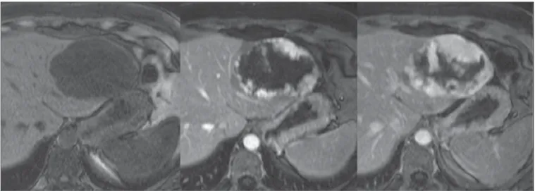
unde-termined nature or of other natures were not taken into consideration. The measurement of the largest axial diameter of the lesion was made on the sequence with highest conspicuity. The number and location of lesions were defined, as well as the en-hancement pattern for the hemangiomas (progressive or flash-filling) (Figures 1 and 2)(10). In cases of more than five lesions in
a single patient, only the largest five lesions were considered. Patients with innumerable small cysts (< 1.5 cm) widely disseminated throughout the parenchyma and with ap-pearance suggestive of biliary hamartomas, were not included in the study sample.
The liver lesions were characterized ac-cording to their imaging features as simple cysts, typical hemangiomas and solid nod-ules, including in the latter group any lesion which did not meet the criteria for the two first groups. The cysts which were
consid-ered atypical(16) were not included in any of
the groups. Additionally, those patients with cirrhotic liver features were also cat-egorized following the criteria widely adopted in literature(6,17).
Statistical analysis
The associations between qualitative variables (gender, type of hemangioma en-hancement and presence or absence of liver cirrhosis) were made by means of the chi-squared test (χ2). For the quantitative
vari-ables (patients’ age and lesion dimensions) parametric tests were utilized whenever normal distribution was observed, and non-parametric tests whenever normal distribu-tion was not observed.
The comparison of quantitative vari-ables between the several groups was made by means of the t test in the case of two groups, or by variance analysis in the case
Figure 2. Hepatic heman-gioma with flash-filling en-hancement pattern.
of more than two groups. In the case of sig-nificant variance analysis, a multiple com-parisons test was subsequently applied (Turkey’s test).
The adopted significance level was 5% and the statistical tests were carried out by means of the SPSS 11.0 software.
RESULTS
Hepatic cysts
Among all individuals, 44 had 95 he-patic cysts, making up a prevalence of 14.5% in the studied population (in 10 cir-rhotic and 34 non-circir-rhotic patients). Out the total of 44 patients with cysts, 23
sented single cysts, while 21 patients pre-sented multiple cysts. The most commonly affected hepatic segments were the IVa (23%) and VI (17%) segments. The mean cyst size was 1.0 cm (ranging from 0.2 cm to 8.8 cm). Out the total of 95 cysts, 66 were < 1.0 cm.
As the prevalence of simple hepatic cysts in the cirrhotic population was deter-mined (10/57; 17.5%) and compared with the prevalence in the non-cirrhotic popula-tion (34/246; 13.8%) no statistically signifi-cant difference was observed (p = 0.472). The dimensions and distribution of the cysts in these two populations did not present any statistically significant differ-ence either. No differdiffer-ence was observed in the prevalence of hepatic cysts with respect to gender and age.
Hemangiomas
In the present study, 47 hemangiomas were detected in 26 patients (prevalence of 8.6%), 7 of them cirrhotic, while 19 were non-cirrhotic. Out of the total of 26 patients with hemangiomas, 17 had single heman-giomas, while 9 had multiple hemangio-mas. The most commonly affected hepatic segments were the VI segment (21%) and II segment (19%), and the mean diameter of the hemangiomas was 2.13 cm (ranging from 0.4 cm to 13.5 cm), with no statisti-cally significant difference between the group of cirrhotic patients (2.51 cm) and the non-cirrhotic group (2.05 cm).
The most common enhancement pat-tern was the progressive type, with a fre-quency of 89.4%, and the flash-filling pat-tern was observed in only 10.6% of the he-mangiomas in the sample. In only two pa-tients out of the total of 26 papa-tients with hemangiomas, the coexistence of two en-hancement patterns was observed (one patient had two hemangiomas, one of each type, and the other had five hemangiomas, with only one with the flash-filling en-hancement pattern.
No statistically significant difference was observed in the prevalence of hepatic hemangiomas among cirrhotic (5/57; 8.8%) and non-cirrhotic patients (21/246; 8.5%), with p = 0.954. The same was observed for the dimensions, distribution and enhance-ment pattern of such lesions. Patients’ age and gender did not influence the prevalence
of hepatic hemangiomas in the study sample, i.e., the presence or absence of hemangiomas was not associated with pa-tients’ gender (χ2 = 0.695;
p = 0.659; non-significant), occurring in 8% of the women and in 9.5% of the men. Mean sizes of the hemangiomas in women and in men were, respectively, 2.29 cm and 1.75 cm.
DISCUSSION
Cysts and hemangiomas are the most common benign focal hepatic lesions(18).
Simple hepatic cysts may be either con-genital or acquired and are related to devel-opmental anomalies and/or conformation of the biliary tree(19). Abdominal traumas
determining bile extravasation outside of the biliary tree have been regarded as a plausible cause of hepatic cysts(20). At MRI,
the cysts are seen as well-defined lesions with hypersignal on T2-weighted se-quences and hyposignal on T1-weighted sequences. Depending upon the presence of hemorrhage, there may be variation in signal intensity. No enhancement is ob-served after paramagnetic contrast medium injection(16).
Reports in the literature indicate that the prevalence and number of cysts increase with age because of obstruction and biliary stasis in small biliary ducts(21). An
Austra-lian study evaluating obstetric ultrasonog-raphy images over a period of more than 10 years has found only three hepatic cysts in fetuses(22). There is a reported
predomi-nance in female individuals, with a ratio ranging between 1.5:1 and 5.5:1(23). The
data in the present study, however, do not demonstrate any relationship between age or gender and the occurrence of cysts.
The first reports on prevalence of simple hepatic cysts were observed in autopsy studies. In a series of 20,000 autopsies, a prevalence of 0.14% (28 cysts)(24) was
found. Other similar studies have reported values of 0.17% and 0.53%(25,26). More
cent data derived from imaging studies re-veal slightly higher values. In 1989, an European study about the prevalence at ultrasonography recorded a prevalence of 2.5% (43 cases in a total of 1,695 pa-tients)(27). Another study, in 1993, found
755 patients with hepatic cysts in a total of 26,000 sonographic images, corresponding
to a prevalence of 2.9%(28). On the other
hand, such prevalence may reach 5%-14%, as demonstrated in at least one publica-tion(29). A tomographic series developed in
2003 points out a simple hepatic cyst preva-lence of 18% in 617 patients(30). More
re-cent studies focused on the therapeutic approach for such lesions report values ranging from 0.1% to 7%, frequently mak-ing reference to decades-old data(31,32).
There is no report in the literature about the prevalence of hepatic cysts found at MRI. The results in the present study (14.5%) demonstrate a higher prevalence than the average found in previous studies, but simi-lar to the prevalence in the studies which relied on computed tomography(30),
sup-posedly related to the high detection capa-bility of these diagnostic methods. Patients with biliary hamartomas were not included in the present study, as the authors under-stand that such entities are different from hepatic cysts(29). However, on the 363 MRI
studies initially evaluated, no case resem-bling imaging findings suggestive of biliary hamartomas was observed.
Hemangiomas correspond to vascular spaces filled with slow flowing blood. They present hypersignal at T2-weighted sequences, which demonstrates to become more conspicuous as the echo time of the sequences increases, because of the de-crease in signal from the surrounding he-patic parenchyma(33,34). Three enhancement
patters can be observed after contrast agent injection(34). The first pattern refers to
le-sions with intense enhancement at the ear-lier phases, which remains unchanged at delayed phases, and is called flash-filling. The second pattern is the most common one, and is characterized by peripheral, discontinued and progressive enhance-ment. The third pattern comprises the le-sions with progressive enhancement, how-ever with their center persistently without enhancement. Like other authors(10), the
de-termination of such diagnosis(10,35) and this
was the utilized strategy to define the typi-cal hemangiomas found in the present sample.
The references cited by authors who de-scribe the prevalence of hemangiomas commonly refer to decades-old studies ei-ther with autopsy or surgical series. For example, a 1999 study reported that in 508 hepatectomies in cirrhotic individuals only 9 hemangiomas were found, corresponding to a prevalence of 1.7%(11). In 1997, another
study found a prevalence of 1.2% in a se-ries of 596 autopsies(3). Higher values are
reported by a 1986 study(4), and like the
references cited by Semelka et al. in 1997 reach 20%(36). Finally, some studies still
rely on decades-old references, such as a 1958 North-American atlas, which points out a prevalence of hemangiomas in 0.4% of the individuals(37).
Although this is not a population study, no previous attempts have been made to es-tablish the prevalence of such lesions by means of MRI. The wide variation in data presented in the literature – from 0.4% to 20% for hemangiomas, and from 0.1% to 18% for simple cysts –, in association with the attention that has been devoted to inci-dental liver lesions have motivated the au-thors to seek more updated data utilizing modern and noninvasive diagnostic tools. The results from the present study allow the conclusion that the frequency of cysts and hemangiomas found at MRI is high and above that observed in autopsy studies, perhaps because of the diligent survey for such findings at MRI and because of the facility in characterizing such type of le-sion.
Because of vascular alterations ob-served in cases of chronic liver disease, one has suggested that, in such patients, there might be differences in the presentation of hepatic hemangiomas(10,38). Reports have
suggested that hemangiomas could degen-erate and decrease in size as cirrhosis progresses(11,39). On the other hand, at least
one study has demonstrated that size, num-ber, location and enhancement pattern of hemangiomas are not different in cirrhotic patients(10). Similarly, in the present study,
no difference was observed in relation to frequency or presentation of hemangiomas in cirrhotic and non-cirrhotic livers.
Additionally, it was suggested that sup-posed alterations of hemangiomas in cir-rhotic patients might decrease their detect-ability and, therefore, reduce their preva-lence at imaging studies(9,11). The present
study results, however, contradict such a possibility, demonstrating similar preva-lence in both groups.
Although hemangiomas and hepatic cysts are easily diagnosed and differenti-ated from other liver lesions at ultrasonog-raphy, computed tomography and MRI, it is known that small hepatocarcinomas may present homogeneous and early enhance-ment similar to the flash-filling pattern which may occur in some hemangio-mas(40,41) and like in approximately 10% of
the present study sample.
The main limitation in the present study lies in the inclusion criteria of the sample. One can argue that the population referred to a tertiary health center is not representa-tive of the general population, for being knowingly composed of unhealthy indi-viduals, which might interfere in the veri-fication of FLLs frequency. However, con-sidering the lack of recent prevalence stud-ies relying on diagnostic imaging methods, the authors believe that the present study brings a significant contribution to under-standing the behavior of cysts and heman-giomas. Future population studies will cer-tainly clarify such questions.
Additionally, anatomopathological studies were not utilized as a reference stan-dard in the present study. However, the uti-lization of imaging parameters for the di-agnosis of cysts and hemangiomas is widely diffused in the literature, thus elimi-nating the necessity of utilizing the gold standard, considering the questionable cost-benefit ratio in utilizing invasive pro-cedures instead, to obtain a diagnosis of little prognostic repercussion for the pa-tients under investigation. On the other hand, the analysis of liver explants in cir-rhotic patients might contribute for the es-tablishment of the actual frequency of cysts and hemangiomas in such group of pa-tients. Finally, the diagnostic criteria uti-lized for inclusion of patients in the cir-rhotic group considered clinical/laboratory parameters in a part of the sample (65%), and anatomopathological study in the other (35%) with less advanced stages of the
dis-ease, which, in a certain manner, reflects the characteristics of the population as-sisted in the authors’ institution.
CONCLUSIONS
In the present study the values for preva-lence of cysts and hemangiomas are higher than values reported by previously pub-lished autopsy series, a fact which the au-thors attribute to the high diagnostic capa-bility of MRI for the described FLLs. Val-ues that may be extrapolated to the general population are still to be obtained by means of population studies.
The prevalence and behavior of such lesions in cirrhotic patients do not seem to be different from that in the non-cirrhotic population, contrary to what initial reports suggested.
REFERENCES
1. Berland LL, Silverman SG, Gore RM, et al. Man-aging incidental findings on abdominal CT: white paper of the ACR incidental findings committee. J Am Coll Radiol. 2010;7:754–73.
2. Megibow AJ. Preface imaging of incidentalomas. Radiol Clin North Am. 2011;49:xi–xii.
3. Ruiz Guinaldo A, Martín Herrera L, Roldán Cuadra R. Hepatic tumors in patients with cirrho-sis: an autopsy study. Rev Esp Enferm Dig. 1997; 89:771–80.
4. Karhunen PJ. Benign hepatic tumours and tu-mour-like conditions in men. J Clin Pathol. 1986; 39:183–8.
5. Ishak KG, Rabin L. Benign tumors of the liver. Med Clin North Am. 1975;59:995–1013. 6. Kudo M, Zheng RQ, Kim SR, et al. Diagnostic
accuracy of imaging for liver cirrhosis compared to histologically proven liver cirrhosis. A multi-center collaborative study. Intervirology. 2008;51 Suppl 1:17–26.
7. Liver Imaging Reporting and Data System [homepage on the Internet]. Reston: American College of Radiology; c2011 [updated 2011 March; cited 2011 Aug 18]. Available from: http:/ /www.acr.org/SecondaryMainMenuCategories/ quality_safety/LI-RADS.aspx.
8. Lee KS, Sekhar A, Rofsky NM, et al. Prevalence of incidental pancreatic cysts in the adult popu-lation on MR imaging. Am J Gastroenterol. 2010; 105:2079–84.
9. Brancatelli G, Federle MP, Blachar A, et al. He-mangioma in the cirrhotic liver: diagnosis and natural history. Radiology. 2001;219:69–74.
10. Mastropasqua M, Kanematsu M, Leonardou P, et al. Cavernous hemangiomas in patients with chronic liver disease: MR imaging findings. Magn Reson Imaging. 2004;22:15–8. 11. Dodd GD 3rd, Baron RL, Oliver JH 3rd, et al.
Spectrum of imaging findings of the liver in end-stage cirrhosis: Part II, focal abnormalities. AJR Am J Roentgenol. 1999;173:1185–92.
Hyperacute stroke: evaluation with combined multisection diffusion-weighted and hemody-namically weighted echo-planar MR imaging. Radiology. 1996;199:391–401.
13. Moseley ME, Kucharczyk J, Mintorovitch J, et al. Diffusion-weighted MR imaging of acute stroke: correlation with T2-weighted and magnetic sus-ceptibility-enhanced MR imaging in cats. AJNR Am J Neuroradiol. 1990;11:423–9.
14. Gourtsoyianni S, Papanikolaou N, Yarmenitis S, et al. Respiratory gated diffusion-weighted imag-ing of the liver: value of apparent diffusion coef-ficient measurements in the differentiation be-tween most commonly encountered benign and malignant focal liver lesions. Eur Radiol. 2008; 18:486–92.
15. Koike N, Cho A, Nasu K, et al. Role of diffusion-weighted magnetic resonance imaging in the dif-ferential diagnosis of focal hepatic lesions. World J Gastroenterol. 2009;15:5805–12.
16. Vuillemin-Bodaghi V, Zins M, Vullierme MP, et al. Imaging of atypical cysts of the liver. Study of 26 surgically treated cases. Gastroenterol Clin Biol. 1997;21:394–9.
17. Gupta AA, Kim DC, Krinsky GA, et al. CT and MRI of cirrhosis and its mimics. AJR Am J Roentgenol. 2004;183:1595–601.
18. Washington K. Masses of the liver. In: Odze R, Goldblum JR, editors. Surgical pathology of the GI tract, liver, biliary tract and pancreas. 2nd ed. New York: Elsevier; 2009. p. 657–789. 19. Jones WL, Mountain JC, Warren KW.
Symptom-atic non-parasitic cysts of the liver. Br J Surg. 1974;61:118–23.
20. Cowles RS, Mulholland MW. Solitary hepatic cysts. J Am Coll Surg. 2000;191:311–21. 21. Kim JY, Kim SH, Eun HW, et al. Differentiation
between biliary cystic neoplasms and simple cysts
of the liver: accuracy of CT. AJR Am J Roent-genol. 2010;195:1142–8.
22. Foley PT, Sithasanan N, McEwing R, et al. En-teric duplications presenting as antenatally de-tected abdominal cysts: is delayed resection ap-propriate? J Pediatr Surg. 2003;38:1810–3. 23. Seo JK, Kim SH, Lee SH, et al. Appropriate
di-agnosis of biliary cystic tumors: comparison with atypical hepatic simple cysts. Eur J Gastroenterol Hepatol. 2010;22:989–96.
24. Eliason EL, Smith DC. Solitary nonparasitic cyst of the liver: case report. Clinics. 1944;3:607–21.
25. Sanfelippo PM, Beahrs OH, Weiland LH. Cystic disease of the liver. Ann Surg. 1974;179:922–5. 26. Feldman M. Polycystic disease of the liver. Am J
Gastroenterol. 1958;29:83–6.
27. Gaines PA, Sampson MA. The prevalence and characterization of simple hepatic cysts by ultra-sound examination. Br J Radiol. 1989;62:335–7.
28. Caremani M, Vincenti A, Benci A, et al. Ecographic epidemiology of non-parasitic hepatic cysts. J Clin Ultrasound. 1993;21:115–8.
29. Craig JR, Peters RL, Edmonson HA. Tumors of the liver and intrahepatic bile ducts. In: Atlas of human pathology. 2nd ed. Washington, DC: Armed Forced Institute of Pathology; 1989. p. 56–62. 30. Carrim ZI, Murchison JT. The prevalence of simple
renal and hepatic cysts detected by spiral com-puted tomography. Clin Radiol. 2003;58:626–9.
31. Faulds JM, Scudamore CH. Technical report of a novel surgical technique: laparoscopic cyst fen-estration and falciform ligament pedicle graft for treatment of symptomatic simple hepatic cysts. J Laparoendosc Adv Surg Tech A. 2010;20:857–61. 32. Ramia JM, de La Plaza R, Figueras J, et al. Tu-mores hepáticos quísticos benignos no parasita-rios. Cir Esp. 2011;89:565–73.
33. Itoh K, Saini S, Hahn PF, et al. Differentiation between small hepatic hemangiomas and me-tastases on MR images: importance of size-spe-cific quantitative criteria. AJR Am J Roentgenol. 1990;155:61–6.
34. Semelka RC, Brown ED, Ascher SM, et al. He-patic hemangiomas: a multi-institutional study of appearance on T2-weighted and serial gado-linium-enhanced gradient-echo MR images. Ra-diology. 1994;192:401–6.
35. Tung GA, Vaccaro JP, Cronan JJ, et al. Cavern-ous hemangioma of the liver: pathologic correla-tion with high-field MR imaging. AJR Am J Roentgenol. 1994;162:1113–7.
36. Semelka RC, Sofka CM. Hepatic hemangiomas. Magn Reson Imaging Clin N Am. 1997;5:241–53. 37. Edmondson HA. Tumors of the liver and intrahe-patic bile ducts. In: Atlas of tumor pathology. Washington, DC: Armed Forces Institute of Pa-thology; 1958. p. 24–8.
38. Oliver JH 3rd, Baron RL, Dodd GD 3rd, et al. Does advanced cirrhosis with portosystemic shunting affect the value of CT arterial portography in the evaluation of the liver? AJR Am J Roentgenol. 1995;164:333–7.
39. Yamashita Y, Ogata I, Urata J, et al. Cavernous hemangioma of the liver: pathologic correlation with dynamic CT findings. Radiology. 1997;203: 121–5.
40. Namimoto T, Yamashita Y, Sumi S, et al. Focal liver masses: characterization with diffusion-weighted echo-planar MR imaging. Radiology. 1997;204:739–44.