PONTIFÍCIA UNIVERSIDADE CATÓLICA DO RIO GRANDE DO SUL FACULDADE DE ODONTOLOGIA
LUCIANO MAYER
ANÁLISE DO EFEITO SISTÊMICO DA LLLT EM REGIÃO
PERIMPLANTAR: ESTUDO EM COELHOS
Orientador: Prof. Dr. João Batista Blessmann Weber
LUCIANO MAYER
ANÁLISE DO EFEITO SISTÊMICO DA LLLT EM REGIÃO
PERIMPLANTAR: ESTUDO EM COELHOS
Tese apresentada ao Programa de Pós-Graduação da Faculdade de Odontologia da Pontifícia Universidade Católica do Rio Grande do Sul, como parte dos requisitos obrigatórios para a obtenção do grau de Doutor em Odontologia, área de
concentração em Cirurgia e
Traumatologia Bucomaxilofacial.
Orientador: Prof. Dr. João Batista Blessmann Weber
ANÁLISE DO EFEITO SISTÊMICO DA LLLT EM REGIÃO PERIMPLANTAR: ESTUDO EM COELHOS
Tese apresentada ao Programa de Pós-Graduação da Faculdade de Odontologia da Pontifícia Universidade Católica do Rio Grande do Sul, como parte dos requisitos obrigatórios para a obtenção do grau de Doutor em Odontologia, área de
concentração em Cirurgia e
Traumatologia Bucomaxilofacial.
BANCA EXAMINADORA
Orientador: Prof. Dr. João Batista Blessmann Weber
Profa. Dra. Marília Gerhardt de Oliveira
Prof. Dr. José Antônio Poli de Figueiredo
Profa. Dra. Deise Ponzoni
Prof. Dr. Carlos Eduardo Baraldi
A minha família.
A minha esposa Simone e meus filhos Arthur e Thales pelo amor verdadeiro e, principalmente, pela compreensão nos momentos de ausência.
Ao meu orientador Prof. Dr. João Batista Blessmann Weber, a quem tenho grande respeito e admiração, agradeço pelo apoio durante toda a minha jornada nesta Universidade.
A minha eterna orientadora, Profa. Dra. Marília Gerhardt de Oliveira, maior responsável pelo meu ingresso neste PPG, pela dedicação com que me orientou no início desse trabalho, incentivou-me e acreditou no desenvolvimento desta pesquisa. Muito obrigado por toda a paciência e colaboração. Sua história de conquistas na Odontologia e a sua determinação motivam aqueles que trabalham com pesquisa. Saiba que foi uma honra ter sido seu orientado.
Ao Prof. Edgar Eduardo Erdmann, grande amigo e incentivador desde a graduação, pela amizade ao longo dos últimos 15 anos.
A Profa. Dra. Edela Puricelli pelo apoio imprescindível para a realização deste estudo junto à Unidade de Experimentação Animal (UEA) do Hospital de Clínicas da Universidade Federal do Rio Grande do Sul (HCPA-UFRGS) e principalmente por ter acreditado e apostado no desenvolvimento desse trabalho.
Aos colegas Fernando Vacilotto Gomes e Fabrício Poletto Massotti. Amigos que compartilharam todos os momentos desta pesquisa.
Ao meu colega e grande amigo, Frederico Mattis. Sua ajuda nas fases preliminares deste trabalho e seu incentivo ao longo dos últimos anos foram essenciais para que eu cumprisse mais essa etapa da minha vida acadêmica. Você é uma pessoa de grande coração. Muito obrigado por tudo.
Aos meus pais e irmã, pelo apoio incondicional e por confiarem em mim e no meu trabalho.
A minha colega de consultório Luciana Paiva, minha secretária Cristiane e minha assistente Karina pelo inestimável apoio e pela paciência.
À Médica Veterinária do LACVet – UFRGS Dra. Viviane Marques Guyoti. Sua competência e profissionalismo foram determinantes para a realização dessa pesquisa.
À Médica Veterinária Dra. Fabíola Schons Meyer e a Enfª. Marta Justina Giotti Cioato da UEA – UFRGS pelo apoio durante a fase experimental do trabalho.
Aos colegas de Pós-Graduação, Marcus Woltmann, Juliana Gonçalves Göelzer, e Wâneza Dias Borges Hirsch. Amigos que compartilharam esta caminhada.
Aos Professores do Programa de Pós-Graduação em Cirurgia e Traumatologia Bucomaxilofacial da PUCRS, obrigado pelo empenho e dedicação ao transmitirem seus ensinamentos.
Ao Prof. Dr. João Feliz Duarte de Moraes, Coordenador do Departamento de Estatística – PUCRS, pela contribuição na análise estatística deste trabalho.
À Pontifícia Universidade Católica do Rio Grande do Sul, na pessoa do Reitor, Prof. Dr. Joaquim Clotet.
À Faculdade de Odontologia da Pontifícia Universidade Católica do Rio Grande do Sul, na pessoa do seu Diretor, Prof. Marcos Túlio Mazzini Carvalho.
Ao Coordenador do Programa de Pós-Graduação em Odontologia da PUCRS, Prof. Dr. José Antônio Poli de Figueiredo.
Aos funcionários, da Secretaria de Pós-Graduação da Faculdade de Odontologia da PUCRS: Ana, Davenir, Paulo, Cléber e Cláudia pela atenção e gentileza com que sempre me atenderam.
Aos meus colegas, professores do curso de Especialização em Implantodontia da AGOR/RS, Cláudio Chedid, Ricardo Vadenal, Renato Almeida, Felipe Volkart, Gislaine Denck, Jorge Puhl e Giancarlo Soarez pela amizade e pelo apoio.
A mente que se abre a uma nova ideia
jamais voltará ao seu tamanho original.
RESUMO
Este estudo é parte de um projeto que teve por objetivo avaliar o efeito da terapia com laser de baixa intensidade pós-implante dentário osseointegrável no funcionamento da glândula tireoide e, consequentemente, na regulação do cálcio, através da mensuração dos níveis hormonais de Triiodotironina (T3), Tiroxina (T4) e dos níveis de Cálcio e Albumina no soro sanguíneo de coelhos. Foi avaliado o efeito de 3 doses terapêuticas distintas de LLLT utilizadas para aceleração do processo de osseointegração de implantes dentários. Para tanto, foram utilizados 40 coelhos da ordem Lagomorpha, raça Nova Zelândia, machos, pesando entre 3 e 4 kg, clinicamente sadios, distribuídos aleatoriamente em cinco grupos, com oito animais cada, sendo dois grupos designados como controle: o grupo controle CI (animais não irradiados e não operados) e o grupo controle CII (animais não irradiados); e três grupos designados como experimentais: EI, EII e EIII – animais irradiados com três doses distintas de laser – grupo experimental EI (dose total - 70J/cm²), grupo experimental EII (dose total - 35J/cm²) e grupo experimental EIII (dose total - 140J/cm²). Para padronizar os experimentos, todos os animais dos grupos CII, EI, EII e EIII foram submetidos ao procedimento cirúrgico de exodontia do incisivo inferior esquerdo e colocação imediata de um implante osseointegrável com superfície nanotexturizada (Nanotite® - Biomet 3iTM) no respectivo alvéolo, criando uma condição clínica inicial de igualdade entre os quatro grupos operados. Os animais do grupo controle CI participaram da mesma rotina dos demais; no entanto, não foram submetidos a nenhum dos procedimentos clínicos/cirúrgicos, servindo como controle absoluto nos testes imunológicos para contagem de T3, T4, Cálcio e Albumina. Os animais dos grupos experimentais foram irradiados com o laser de diodo infravermelho com meio ativo GaAlAs (Arseneto de Gálio e Alumínio), com comprimento de onda de 830nm, de forma pontual, com potência de 50mW, no modo de emissão contínua, a cada 48 horas, num total de sete sessões de aplicação, durante o período de 13 dias. O protocolo de irradiação foi iniciado imediatamente após o procedimento cirúrgico. As coletas de sangue para dosagens laboratoriais de T3, T4, Cálcio e Albumina foram realizadas por meio de punção venosa da veia jugular nos cinco grupos em quatro momentos distintos: 72 horas antes do procedimento cirúrgico, imediatamente após a primeira aplicação de laser, 72 horas após a primeira aplicação de laser e 72 horas após a última aplicação de laser. Os resultados obtidos demonstraram diferenças estatisticamente significativas para os valores de T3 e Cálcio entre os grupos estudados e para os valores de T3, T4, Cálcio e Albumina nos diferentes tempos de coleta ao longo do experimento. Conclui-se que a LLLT, no protocolo de irradiação utilizado neste estudo, apesar de ter alterado significativamente os níveis hormonais de T3 e T4 e os níveis de Cálcio e Albumina circulantes no soro de coelhos, não comprometeu definitivamente o funcionamento da glândula tireoide dos mesmos, pois na etapa final do controle hormonal percebe-se o restabelecimento da função glandular.
Palavras-chave1: Glândula Tireoide; LLLT; Hormônios Tireoideos, Implantes Dentários, Laser.
1Descritores em Ciência da Saúde (DeCS); disponível em: <http://decs.bvs.br/>.Acesso em: 06 nov.
This study is part of a larger project that sought to assess the effect of low-level laser therapy (LLLT) after placement of an osseointegrated dental implant on thyroid gland function – and, consequently, calcium regulation – and its potential interference with the osseointegration process. Toward this end, circulating serum levels of triiodothyronine (T3), thyroxine (T4), calcium, and albumin were measured in rabbits. The effects of three therapeutic doses of LLLT, used to accelerate the dental implant osseointegration process, were assessed. Forty healthy male New Zealand rabbits (order Lagomorpha), weight 3–4 kg, were allocated randomly across five groups of eight each: two control groups, CI (no LLLT, no surgery) and CII (no LLLT), and three experimental groups, EI, EII, EIII, exposed to three different doses of laser radiation (EI, total dose 70 J/cm²; EII, total dose 35 J/cm²; and EIII, total dose 140 J/cm²). For standardization purposes, all animals in groups CII, EI, EII, and EIII underwent surgical extraction of the mandibular left incisor and immediate placement of a nanoparticle-coated osseointegrated implant (NanoTite® – Biomet 3iTM) into the prepared socket, creating a condition of equality between groups at baseline. Animals in group CI were exposed to the same handling conditions, but did not undergo any clinical or surgical procedures, thus serving as an absolute control for T3, T4, calcium, and albumin measurements. Animals in the experimental groups received spot irradiation with a GaAlAs (gallium aluminium arsenide) infrared diode laser, wavelength 830 nm, power 50 mW, in continuous emission mode, over two points, every 48 hours over 13 days, for a total of seven sessions; the irradiation protocol was started immediately after the surgical procedure. Blood was collected for measurement of T3, T4, calcium, and albumin levels, by puncture of the external jugular vein, at four points in time: 72 hours before surgery, immediately after the first LLLT session, 72 hours after the first LLLT session and 72 hours after the last LLLT session. The results showed significant differences in T3 and calcium levels between study groups, as well as significant within-group differences in T3, T4, calcium, and albumin levels over time. We conclude that, despite a significant effect on circulating serum levels of T3, T4, calcium and albumin, the LLLT protocol used in this study did not lead to impairment of thyroid function in a rabbit model, because in the final stage of the hormonal control it is noticed the reestablishment of glandular function.
Keywords2: Thyroid Gland; LLLT; Thyroid Hormones, Dental Implants, Laser.
2Medical Subject Headings (MeSH), disponível em: <http://www.nlm.nih.gov/mesh/MBrowser.html>.
ARTIGO II - Figure 01. Surgical procedure: A) Local anesthesia with 0.5 mL lidocaine 2% with epinephrine 1:100,000; B) Extraction of the mandibular left incisor with #5 pediatric extraction forceps; C) Placement of a 3.25øx11.5mm osseointegrated implant (NanoTite®) into the fresh extraction socket; D) Occlusal view after implant placement; E) Wound closure with 4-0 monofilament nylon sutures; F) Long axis of the implant tattooed onto skin with surgical marker to guide LLLT... 80
ARTIGO II - Figure 02. A) Administration of LLLT. B) Collection of venous blood (3 mL) by puncture of the external jugular vein... 80
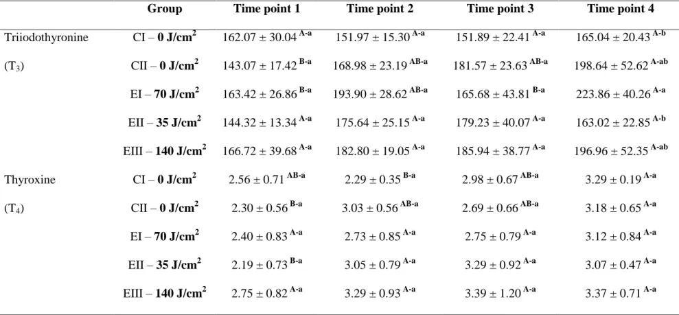
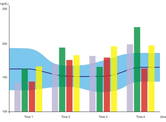
ARTIGO II - Figure 03. Variation in triiodothyronine (T3) levels (ng/dL) over time. The blue-shaded horizontal area represents control group CI, with the central line corresponding to the mean and the shaded area itself representing the standard deviation from said mean (Time 1, 162.07 ± 30.04; Time 2, 151.97 ± 15.30; Time 3, 151.89 ± 22.41; Time 4, 165.04 ± 20.43). Bars represent, respectively, control group CII (gray bar; Time 1, 143.07 ± 17.42; Time 2, 168.98 ± 23.19; Time 3, 181.57 ± 23.63; Time 4, 198.64 ± 52.62) and each of the experiment groups: EI = 70 J/cm² (green bar; Time 1, 163.42 ± 26.86; Time 2, 193.90 ± 28.62; Time 3, 165.68 ± 43.81; Time 4, 223.86 ± 40.26); EII = 35 J/cm² (red bar; Time 1, 144.32 ± 13.34; Time 2, 175.64 ± 25.15; Time 3, 179.23 ± 40.07; Time 4, 163.02 ± 22.85); and EIII = 140 J/cm² (yellow bar; Time 1, 166.72 ± 39.68; Time 2, 182.80 ± 19.05; Time 3, 185.94 ± 38.77; Time 4, 196.96 ± 52.35)... 81
standard deviation from said mean (Time 1, 2.56 ± 0.71; Time 2, 2.29 ± 0.35; Time 3, 2.98 ± 0.67; Time 4, 3.29 ± 0.19). Bars represent, respectively, control group CII (gray; Time 1, 2.30 ± 0.56; Time 2, 3.03 ± 0.56; Time 3, 2.69 ± 0.66; Time 4, 3.18 ± 0.65) and each of the experiment groups: EI = 70 J/cm² (green bar; Time 1, 2.40 ± 0.83; Time 2, 2.73 ± 0.85; Time 3, 2.75 ± 0.79; Time 4, 3.12 ± 0.84); EII = 35 J/cm² (red bar; Time 1, 2.19 ± 0.73; Time 2, 3.05 ± 0.79; Time 3, 3.29 ± 0.92; Time 4, 3.07 ± 0.47); and EIII = 140 J/cm² (yellow bar; Time 1, 2.75 ± 0.82; Time 2, 3.29 ± 0.93; Time 3, 3.39 ± 1.20; Time 4, 3.37 ± 0.71)... 82
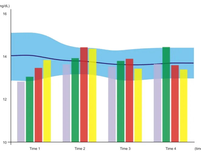
ARTIGO II - Figure 05. Variation in calcium levels (mg/dL) over time. The blue-shaded horizontal area represents control group CI, with the central line corresponding to the mean and the shaded area itself representing the standard deviation from said mean (Time 1, 14.01 ± 1.02; Time 2, 13.82 ± 0.62; Time 3, 13.67 ± 0.72; Time 4, 13.72 ± 0.70). Bars represent, respectively, control group CII (gray bar; Time 1, 12.82 ± 0.27; Time 2, 13.63 ± 0.75; Time 3, 13.53 ± 0.19; Time 4, 13.65 ± 0.62) and each of the experiment groups: EI = 70 J/cm² (green bar; Time 1, 13.05 ± 0.32; Time 2, 13.95 ± 0.55; Time 3, 13.79 ± 0.27; Time 4, 14.46 ± 0.47); EII = 35 J/cm² (red bar; Time 1, 13.47 ± 0.34; Time 2, 14.41 ± 0.55; Time 3, 13.91 ± 0.31; Time 4, 13.59 ± 0.70); and EIII = 140 J/cm² (yellow bar; Time 1, 13.81 ± 0.26; Time 2, 14.39 ± 0.28; Time 3, 13.43 ± 0.30; Time 4, 13.41 ± 0.73)... 83
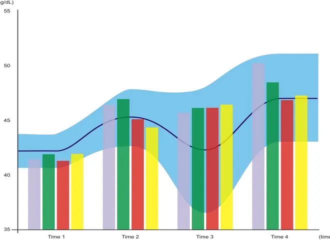
J/cm² (yellow bar; Time 1, 41.93 ± 1.39; Time 2, 44.36 ± 2.13; Time 3, 46.43 ± 2.09; Time 4, 47.28 ± 2.51)... 84
APÊNDICE B – Resultados das dosagens hormonais de Triiodotironina (T3) e Tiroxina (T4) no soro sanguíneo dos coelhos... 141
APÊNDICE C – Resultados das dosagens dos níveis de Cálcio no soro sanguíneo dos coelhos... 142
ARTIGO I - Table 1. Studies of the effects of LLLT on bone healing... 54
ARTIGO I - Table 2. Studies of the effects of LLLT on osseointegration... 55
ARTIGO II - Table 01. Low-level laser therapy (LLLT) protocols used in
previous studies... 76
ARTIGO II - Table 02. Study parameters... 77
ARTIGO II - Table 03. Statistical analysis: results of serum T3, T4, calcium, and albumin measurement. Baseline levels expressed as mean ± standard
deviations. Means followed by different uppercase superscript letters across the same row denote significant differences. Means followed by different lowercase superscript letters across the same column denote significant differences on repeated measures. Analysis of variance (ANOVA) with Tukey’s multiple
LISTA DE ABREVIATURAS, SIGLAS E SÍMBOLOS
CEUA Comissão de Ética no Uso de Animais
cm centímetro
cm2 centímetro quadrado
CTBMF Cirurgia e Traumatologia Bucomaxilofacial
DE Densidade de energia
dL decilitro
et al. e colaboradores
GaAlAs Arseneto de Gálio e Alumínio
GaAs Arseneto de Gálio
HILT Hight Intensity Laser Treatment
HLLT Hight reactive - Level Laser Treatment
HeNe Hélio-Neônio
InGaAlP Fosfeto de Índio-Gálio-Alumínio
J Joule
J/cm2 Joule por centímetro quadrado
J/s Joule por segundo
kg quilograma
LASER Light Amplification by Stimulated Emission of Radiation LILT Low Intensity Level Treatment
LLLT Low reactive-Level Laser Treatment ou Low Level Laser Therapy
mg miligrama
mg/dL miligrama por decilitro
ml mililitro
mm milímetro
mW miliwatts
mW/cm2 miliwatts por centímetro quadrado
n° número
ng nanograma
ng/dL nanograma por decilitro
p probabilidade de erro
P potência
PUCRS Pontifícia Universidade Católica do Rio Grande do Sul
RIE Radioimunoensaio
rpm Rotações por minuto
s segundos
SPSS Statistical Package for Social Science TM do inglês Trademark – marca registrada
TRH Tireotropina
TSH Hormônio estimulador da tireoide
T3 Triiodotironina
T4 Tiroxina
W Watt
W/cm2 Watts por centímetro quadrado
λ lambda = comprimento de onda
β beta
µg micrograma
µg/dL micrograma por decilitro
® marca registrada
% porcento, porcentagem / percentagem
≤ menor ou igual
1 ANTECEDENTES E JUSTIFICATIVA ... 29 2 ARTIGO I ... 39 3 ARTIGO II ... 57 4 DISCUSSÃO GERAL ... 86 5 CONCLUSÕES ... 93 REFERÊNCIAS ... 95 ANEXO A - Protocolo da Comissão Científica e de Ética da Faculdade de
Odontologia da PUCRS... 106 ANEXO B - Protocolo da Comissão de Ética no Uso de Animais da PUCRS .. 107 ANEXO C - Lei 11.794 de 08 de outubro de 2008 (Lei Arouca) ... 108 ANEXO D - Princípios Éticos na Pesquisa Experimental... 113 ANEXO E - Protocolo da Comissão de Ética no Uso de Animais (CEUA) do HCPA – Hospital de Clínicas de Porto Alegre (UFRGS) ... 115 ANEXO F - Autorização do Laboratório de Análises Clínicas Veterinárias
(LACVet – UFRGS) para realização da pesquisa ... 116 ANEXO G - Justificativa do cálculo do tamanho da amostra ... 117 ANEXO H – Instruções para colocação dos implantes Nanotite®– Biomet 3i® (conexão externa – HE – 3,25mm de diâmetro – 11,5mm de comprimento) .... 118 ANEXO I – Normas para publicação - periódico Clinical Implant Dentistry and Related Research ... 119 ANEXO J –Comprovante de submissão do artigo intitulado “Effect of low-level laser therapy on peri-implant bone healing: a review of the literature” para o
periódico Clinical Implant Dentistry and Related Research ... 125 ANEXO K – Normas para publicação - periódico International Journal of Oral & Maxillofacial Surgery ... 126 ANEXO L –Comprovante de submissão do artigo intitulado “Systemic effects of LLLT on thyroid function after titanium dental implant placement: an
APÊNDICE B – Resultados das dosagens hormonais de Triiodotironina (T3) e
Tiroxina (T4) no soro sanguíneo dos coelhos ... 141
APÊNDICE C – Resultados das dosagens dos níveis de Cálcio no soro
1 ANTECEDENTES E JUSTIFICATIVA
As idéias fundamentais para o desenvolvimento do laser foram apresentadas por Albert Einstein em 1916 com a formulação da Teoria da Emissão Estimulada de Radiação, onde se discutia o relacionamento da quantidade de energia liberada pelo processo atômico. Neste estudo, Einstein discorreu sobre a interação de átomos, íons e moléculas com a radiação eletromagnética em termos de absorção e emissão espontânea de radiação e concluiu que o terceiro processo de interação, a emissão estimulada, deveria existir, e nela, a radiação eletromagnética deveria ser produzida por um processo atômico. Deste modo, Theodore H. Maiman e colaboradores construíram e apresentaram, em 16 de Maio de 1960, o primeiro equipamento capaz de amplificar a radiação eletromagnética, este ficou conhecido como Laser (FREIRE Jr.; CARVALHO NETO, 1997).
A palavra LASER é um acrônimo composto pelas iniciais de Light Amplification by Stimulated Emission of Radiation, ou seja, “amplificação da luz por emissão estimulada de radiação”. (CONLAN; RAPLEY; COOB, 1996; BRUGNERA JÚNIOR; PINHEIRO, 1998; BRUGNERA JÚNIOR et al., 2003).
Existem diferenças marcantes entre o Laser e a luz comum; Entre elas, as principais consistem em: direção, coerência e cromaticidade. Enquanto a luz laser apresenta uma unidirecionalidade (paralelismo), a luz comum apresenta uma dispersão de seus feixes em várias direções. A luz laser é coerente, já a luz comum é incoerente, pois apresenta vários comprimentos de onda e seus fótons viajam sem sincronismo. Por fim, a luz laser é monocromática, pois apresenta um único comprimento de onda, portanto, cor pura, enquanto que a luz comum apresenta mais de um comprimento de onda (ALMEIDA-LOPES, 1998).
Para entender os princípios da luz Laser, é importante entender que a luz pode ser descrita como uma onda eletromagnética e, como tal, apresenta características ondulatórias específicas que a identificam (MELLO; MELLO, 2001). Uma característica importante da radiação laser é chamada de comprimento de onda (distância medida entre dois picos consecutivos de sua trajetória). É o comprimento de onda que define a cor da luz. A unidade utilizada para a medição deste comprimento é o nanômetro (nm), uma fração do metro que equivale a 10-9 metro. Essa característica, juntamente com a frequência e a amplitude, define a luz dentro do espectro de ondas eletromagnéticas (DEDERICH, 1993).
Os Laseres têm sido classificados de acordo com inúmeros critérios. A mais ampla classificação é aquela que os divide em dois grandes grupos, de acordo com a intensidade de energia do feixe, podendo esta ser alta intensidade – chamados de ablativos ou Hight Intensity Laser Treatment (HILT) – ou baixa intensidade – denominados terapêuticos ou Low Intensity Level Treatment (LILT). O comprimento de onda do laser é determinado pelo meio ativo que o nomeia, podendo, por sua vez, variar no espectro do infravermelho ao ultravioleta. O tipo de meio ativo (sólido, gasoso ou semicondutor) também determina a afinidade ou não do laser com o tecido alvo. De acordo com a reação celular, a terapia com laser é classificada em Hight reactive-Level Laser Treatment (HLLT) ou laser cirúrgico, usado em procedimentos cirúrgicos; e Low reactive-Level Laser Treatment (LLLT) ou laser não cirúrgico, também chamado de laser terapêutico, usado em procedimentos terapêuticos clínicos (BRUGNERA JÚNIOR et al., 2003).
A vantagem da luz dos laseres, quando comparada a outros tipos de luz, é que trata-se de um feixe monocromático de alta energia, passível de ser focado e facilmente manipulado, além de ser capaz de promover efeitos fotobiológicos específicos. Parâmetros da irradiação como comprimento de onda, eixo energético, diâmetro do feixe, duração do pulso e tempo de exposição podem ser combinados com as propriedades físicas, químicas e biológicas do tecido alvo para que se obtenham resultados desejáveis (MISERENDINO; PICK, 1995).
Quando se discute a interação do laser com um tecido biológico, deve-se levar em consideração, tanto as propriedades e os parâmetros da radiação laser, quanto as propriedades ópticas dos tecidos (GUTKNECHT; EDUARDO, 2004).
Energia: é definida como a quantidade de luz depositada no tecido tratado, sendo que a sua unidade de media é o Joule (J).
Energia total: pode ser calculada multiplicando a potência de saída – em Watts (W) –, pelo tempo de irradiação – em segundos (s) –; ou seja, utilizar a energia total (J) apenas informa a quantidade total de energia depositada no tecido ao final da irradiação.
Fluência, Dose ou Densidade de Energia: é a quantidade de energia aplicada no tecido com relação à área sobre a qual esta energia é aplicada. Em outras palavras, é a distribuição da energia por unidade de área. A unidade, portanto, é Joule por centímetro quadrado (J/cm²).
Potência: é a taxa com que uma quantidade de energia é transmitida ao tecido, ou seja, a relação entre a energia aplicada e o tempo que leva para que ela seja aplicada. A unidade é Watt (W ou J/s).
Irradiância, Intensidade ou Densidade de Potência: é a razão com que a potência é dissipada numa certa área do tecido ou a quantidade de energia por segundo aplicada numa certa área. A unidade utilizada é Watts por centímetro quadrado (W/cm²).
Segundo Karu (1989), a luz laser interage com os tecidos biológicos basicamente de quatro formas: parte da luz é refletida; parte da luz pode ser espalhada dentro do tecido; parte da luz pode ser absorvida pelos componentes do tecido; e, finalmente, parte da luz pode ser transmitida através do tecido, sem atenuação podendo ocasionar interação em locais distantes da região de aplicação da luz.
A absorção e a difusão do laser no tecido resultam numa resposta fotorreativa. Esta é conseqüência de uma fotodestruição ou uma fotoativação tecidual, determinadas pelo efeito fotoquímico, fotoelétrico ou fototérmico do laser (OHSHIRO; FUJINO, 1993). Dentre os efeitos fotoquímicos, pode-se incluir a biomodulação tecidual, a qual corresponde a ação da luz laser sobre os processos moleculares e bioquímicos nos tecidos (BRUGNERA JÚNIOR; PINHEIRO, 1998).
onda. A energia pode transmitir-se até alcançar um tecido específico, no qual as características de absorção ocorram. Só então, algum efeito poderá ser observado (MELLO; MELLO, 2001).
Os laseres não-cirúrgicos são usados para fins terapêuticos, desde a década de 1960, por suas características de baixa intensidade de energia e comprimento de onda capaz de penetrar nos tecidos (BRUGNERA JÚNIOR; VILLA; GENOVESE, 1991). Essa terapia tem ação biomoduladora importante no processo de reparo tecidual e é empregada amplamente nas diversas áreas da saúde, sendo a Odontologia uma das ciências que mais faz uso dessa tecnologia (BRUGNERA JÚNIOR et al., 2003).
Segundo Niccoli Filho et al. (1993), a terapia com laser de baixa intensidade na Odontologia teve início com Benedicenti, em 1982, com um aparelho laser diodo de Arseneto de Gálio (GaAs λ=904 nm). Esta terapia tem sido utilizada com grande sucesso na clínica odontológica, podendo ser empregada isoladamente ou como coadjuvante em tratamentos convencionais (BRUGNERA JÚNIOR et al., 2003; CATÃO, 2004).
A LLLT (Low Level Laser Therapy) tem sido indicada para promover a regeneração tecidual, tendo como vantagens: a estimulação da cicatrização, a redução da inflamação e o controle da dor pós-operatória (PINHEIRO; FRAME, 1992). Essa terapia promove mudanças de caráter metabólico, energético e funcional nos corpos submetidos à radiação, permitindo a evolução para a cura em um curto período de tempo (BRUGNERA JÚNIOR et al., 2003; CATÃO, 2004). A LLLT atua na estimulação do reparo tecidual, melhorando a regeneração e a cicatrização de tecidos, através da promoção da proliferação celular (KARU, 1989; DA SILVA; CAMILLI, 2006); da aceleração na formação de tecidos de granulação (KOLÁVORÁ; DITRICHOVÁ; WAGNER, 1999); do estímulo na síntese do colágeno, com formação das fibras pro-colágenas tipo I e tipo III (PINHEIRO; GERBI, 2006).
A LLLT deve seguir os seguintes parâmetros: escolha do comprimento de onda, densidade de energia, densidade de potência, tipo de regime de operação do laser, frequência do pulso, número de sessões e características ópticas do tecido como os coeficientes de absorção e espalhamento (CATÃO, 2004).
O comprimento de onda do laser é um dos fatores que influenciam na sua capacidade de penetração nos tecidos, uma vez que cada cromóforo tecidual possui um grau de absorção específico para cada comprimento de onda, promovendo diferentes efeitos biológicos, de acordo com o tipo de laser utilizado (THEODORO; GARCIA; MARCANTONIO JÚNIOR, 2002). Os laseres Hélio-Neônio (HeNe) com λ=632,8nm penetram em torno de 0,5mm, os laseres com maior comprimento de onda, como os infravermelhos, podem penetrar uma maior profundidade, sendo pouco absorvidos pela epiderme e derme (SCHINDL et al., 2000).
Outro fator a ser considerado, é a densidade de energia (DE), também chamada de fluência ou dosimetria, a qual estabelece a relação entre a energia administrada por um emissor laser e a superfície de radiação do raio de luz laser ou spot, sendo expressa em J/cm2. Diversos equipamentos dispõem de cálculo direto, sendo o tempo de exposição determinado automaticamente através da inserção da DE, potência de emissão e da área do spot (RIGAU I MAS, 1998; ALMEIDA-LOPES; 2003).
Os laseres mais utilizados na LLLT são o Hélio-Neônio (HeNe) e os diodos (BASFORD, 1995; PINHEIRO et al., 1998). O HeNe foi o primeiro laser gasoso desenvolvido e, também, o primeiro a emitir, de forma contínua, raios com dois comprimentos de onda: λ=632,8 nm (vermelho) e λ=543,5 nm (verde), com potência variável de 1mW a dezenas de miliwatts (BASFORD, 1995).
De acordo com Brugnera Júnior et al. (2003), o meio ativo determina afinidade ou não do laser com o tecido alvo, o que é muito relevante, pois apenas a indicação correta do laser para determinado tecido resultará no objetivo esperado.
Os laseres de GaAlAs são muito utilizados na biomodulação, sendo que a composição do cristal semicondutor de luz pode variar consideravelmente. Dependendo da porcentagem de cada substância utilizada, o comprimento de onda da luz emitida pode variar de 660 a 940nm. Os mais utilizados são os laseres com comprimento de onda de 820 a 830nm (infravermelhos) e 670nm (vermelho), os quais emitem radiação tanto no modo contínuo quanto no modo pulsado (FREDDO et al, 2009).
Os aparelhos de laser de GaAlAs mais modernos são apresentados em unidades portáteis, compactas e que se mostram confiáveis. Estes foram desenvolvidos com considerações econômicas e ergonômicas, que reduziram seu custo em comparação a outros equipamentos de laser (ROMANOS; NENTWIG, 1999).
O fenômeno dos bioefeitos do laser está relacionado com a dose e a afinidade do tecido com o comprimento de onda específico (BELKIN; SCHWARTZ, 1989). Segundo Brugnera Júnior et al. (2003), não se pode definir a exata profundidade que o laser alcança nem a dose que atinge os alvos abaixo do ponto de incidência, o que dificulta o conhecimento da dosimetria correta.
Muitos benefícios estão associados à biomodulação através da terapia com laser de baixa intensidade (Low Level Laser Therapy – LLLT). Dependendo do efeito desejado é calculada a fluência, ou dose de energia, necessária. O limiar de exposição é específico e cada tecido responde de maneira diferente a um determinado comprimento de onda (λ). De acordo com a fluência, é possível ter estimulação ou inibição de atividades bioquímicas, fisiológicas e proliferativas. Altas doses de energias, dentro de limites específicos para cada combinação tecido-laser, podem gerar prejuízo; uma vez que os efeitos são dose-dependentes (BELKIN; SCHWARTZ, 1989).
ponto de irradiação (ROCHKIND et al., 1989). Portanto, quando se realiza a LLLT, pode-se atingir órgãos distantes do local de irradiação.
Estudos sugerem que o laser apresenta efeitos sistêmicos, por meio de mediadores metabólicos liberados na circulação, atingindo áreas distantes do local de aplicação (OHSHIRO, 1991; MARINO, 2003).
Embora a LLLT tenha se tornado uma prática comum nas áreas biomédicas, ainda não se tem um entendimento claro sobre seus possíveis efeitos colaterais. Estudos com relação à utilização do laser de emissão infravermelha sobre a glândula tireóide têm demonstrado um aumento na atividade mitótica de células foliculares, hiperatividade transitória em alguns folículos (PÉREZ DE VARGAS et al.,1987; PARRADO et al., 1990) e alterações dos níveis hormonais de triiodotironina (T3) e tiroxina (T4) circulantes no soro sangüíneo (AZEVEDO et al., 2005).
A tireóide e a paratireóide são glândulas importantes do organismo humano e tem como uma das suas principais funções a produção de hormônios que regulam o metabolismo corpóreo. Quando a função dessas glândulas não é adequada, podem ocorrer repercussões no organismo em graus variáveis de severidade, desde sintomas que muitas vezes podem passar despercebidos até formas extremamente graves que podem trazer risco de vida (SONIS; FAZIO; FANG, 1996; JUNQUEIRA; CARNEIRO, 2004; GUYTON; HALL, 2006).
O controle da produção e secreção dos hormônios tireoidianos é mediado pelo hipotálamo que produz o hormônio de liberação de tireotropina (TRH) e pela adeno-hipófise que estimula a produção do hormônio estimulador da tireóide (TSH). O TSH irá estimular a produção de triiodotironina e tiroxina pela glândula tireóide. Esses hormônios podem realimentar negativamente a hipófise e o hipotálamo para interromper a secreção de TSH, mantendo o organismo com quantidades adequadas de T3 e T4 (SONIS; FAZIO; FANG, 1996; JUNQUEIRA; CARNEIRO, 2004).
Cerca de 93% dos hormônios circulantes secretados pela tireóide consistem em T4, e 7% em T3. Porém, a triiodotironina é mais potente e exerce a sua ação com maior rapidez. Em torno de 50% da quantidade de tiroxina é convertida em triiodotironina, sendo o T3 o principal hormônio tireoidiano utilizado pelos tecidos (FISHER, 1996; GUYTON; HALL, 2006).
valores variam de não mais que um picograma (bilionésimo de miligrama) por mililitro de sangue até, no máximo, alguns microgramas (milionésimos de um grama) (GUYTON; HALL, 2006).
A medida de T3 e T4 total inclui tanto as frações ligadas à proteínas como as frações livres. Alguns laboratórios oferecem a medida dos hormônios tireoidianos totais e outros oferecem a medida dos hormônios tireoidianos livres, porém ambas fornecem a mesma informação sobre o funcionamento normal, hipo ou hiperativo da tireóide. (CZEPIELEWSKI, 2003a, 2003b).
Nos últimos 40 anos, houve uma melhora substancial na sensibilidade e especificidade dos testes tireoidianos principalmente com o desenvolvimento dos métodos imunométricos não isotópicos. O método radioimunoensaio (RIE) foi desenvolvido para a dosagem de insulina por Yallow e Berson, em 1959, sendo posteriormente utilizado para a quantificação de vários outros hormônios (MOURA et al., 2001).
Anormalidades estruturais ou funcionais das glândulas tireóide e paratireóide podem levar a uma deficiência de produção dos hormônios (tiroxina, triiodotironina e paratormônio) afetando o metabolismo de todos os órgãos e sistemas (SONIS; FAZIO; FANG, 1996; GUYTON; HALL, 2006).
Qualquer lesão óssea (fraturas, defeitos, fixação de implantes, interrupção do suprimento sangüíneo) ativa a regeneração óssea local pela liberação de hormônios de crescimento. A formação óssea necessita de dois requisitos indispensáveis: amplo suprimento vascular e suporte mecânico (a união desses fatores irá ativar os osteoblastos na produção de tecido ósseo). Sistemicamente, a remodelagem óssea é ativada pelos hormônios de crescimento e pela tireóide e paratireóide, e inibida pela calcitonina e cortisona. Localmente, a remodelagem óssea é ativada por qualquer traumatismo ao osso, isto é, fraturas, procedimentos cirúrgicos, ou fixação de implantes (HOLLINGER et al., 1999).
da inflamação e edema; a modulação e atenuação da sintomatologia dolorosa; e a analgesia pós-operatória (TRELLES; MAYAYO, 1987; RIGAU I MAS et al., 1991; PINHEIRO; FRAME, 1992; DO NASCIMENTO et al., 2004; WEBER et al., 2006; MOHAMMED et al., 2007; SOARES et al., 2008).
O uso potencial dos laseres na biomodulação do reparo ósseo através de suas propriedades fotoquímicas e fotobiológicas tem sido estudado por diversos pesquisadores com o objetivo de proporcionar ao paciente submetido à cirurgia uma maior rapidez na cicatrização óssea, menor desconforto pós-operatório, menor quadro de edema pós-cirúrgico e melhor cicatrização tecidual (TAKEDA, 1988).
Os protocolos para a utilização dos laseres em diversos procedimentos clínicos estão sendo investigados, pois nem todas as suas atuações e efeitos sistêmicos estão esclarecidos. A revista de literatura revela que a LLLT pode atuar nas funções endócrinas, comprovando os possíveis efeitos do laser sobre as glândulas secretoras (SMITH-AGREDA et al., 1985; PARRADO et al., 1988).
Segundo Pinheiro (2009) a dose é o parâmetro mais importante da fototerapia com laser, no entanto, um protocolo definitivo com relação à dosimetria da LLLT administrada e o uso nas diferentes situações clínicas ainda é alvo de discussões entre os pesquisadores.
Com o propósito de verificar a ação sistêmica do laser de diodo infravermelho com meio ativo GaAlAs (Arseneto de Gálio e Alumínio), com comprimento de onda de 830nm (λ= 830nm) e três diferentes doses/fluências (35J/cm2, 70J/cm2 e 140J/cm2), aplicado após inserção de implantes com superfície nanotexturizada, realizou-se esta pesquisa, a qual teve por objetivos:
2 ARTIGO I
Effect of low-level laser therapy on peri-implant bone healing: a review of the literature
Luciano Mayer1– Fernando Vacilotto Gomes2– Marília Gerhardt de Oliveira3– João Batista Blessmann Weber4
1
Specialist, Implant Dentristry; PhD Candidate, Oral and Maxillofacial Surgery and Traumatology – Department of Oral and Maxillofacial Surgery, Pontifícia Universidade Católica do Rio Grande do Sul (PUCRS) School of Dentistry, Rio Grande do Sul, Brazil. 2
Specialist, Oral and Maxillofacial Surgery and Traumatology; Master's Candidate, Oral and Maxillofacial Surgery and Traumatology – Department of Surgery, Universidade Federal do Rio Grande do Sul (UFRGS) School of Dentistry, Rio Grande do Sul, Brazil.
3
Research Fellow, National Council for Scientific and Technological Development (CNPq); Oral and Maxillofacial Surgery Service, Hospital Cristo Redentor – Grupo Hospitalar
Conceição (GHC), Rio Grande do Sul, Brazil. 4
Professor and Coordinator of the Department of Preventive Dentistry, Pontifícia
Universidade Católica do Rio Grande do Sul (PUCRS) School of Dentistry, Rio Grande do Sul, Brazil.
Conflict of interest: none to declare
Corresponding author:
João Batista Blessmann Weber Faculdade de Odontologia - PUCRS Av. Ipiranga, 6681 – Prédio 6, Partenon
Porto Alegre, RS, Brazil Phone: +55 51 3320-3562 Fax: + 55 51 3320-3626
jbbweber@terra.com.br
ABSTRACT
The use of low-level laser therapy (LLLT) as an adjunct to a variety of dental procedures is now a reality. In implant dentistry, LLLT has been increasingly studied and employed as an aid to bone repair and osseointegration. Research suggests that the biomodulatory action of laser can optimize bone repair and remodeling and, consequently, accelerate the
osseointegration process. The aim of the present study was to conduct a review of the existing literature on LLLT and its effects on bone tissue repair and osseointegration of dental
implants. The reviewed literature was consistent with wide variability in clinical use of LLLT in terms of dose, number of sessions, power, and wavelength. Nevertheless, most studies suggested positive effects of laser irradiation in their respective clinical settings. We conclude that further research on this topic is required, as is development of safe and predictable
protocols to enable routine use of laser therapy in clinical implant dentistry. However, the positive biomodulatory effects of LLLT on the osseointegration process appear indisputable.
INTRODUCTION
The advent of dental implants was a major advancement in rehabilitation of partially and fully edentulous arches. The replacement of lost teeth by implant-supported bridges has enabled improved physical and psychosocial rehabilitation for patients in whom the only option available previously was the use of removable prostheses.1
Osseointegration implies a direct structural and functional connection between vital, organized bone and the surface of a titanium implant capable of functional loading. This phenomenon depends on a variety of factors and can be achieved with proper surgical implant installation technique. However, if osseointegration is to be maintained, the surgeon must respect the bone healing time required for adequate bone–implant integration.2
The effects of low-level laser therapy (LLLT) on the bone repair process and on peri-implant bone healing have been the object of extensive research. The aims of LLLT are to reduce postoperative discomfort, speed the peri-implant bone healing process and, potentially, exert a bactericidal effect.3, 4
Dose is one of the key parameters of LLLT. However, definitive dosage protocols for LLLT in different clinical settings are still the subject of debate.5
Some preclinical studies have suggested that LLLT has favorable effects on bone healing,6-8 particularly peri-implant bone healing (osseointegration) after placement of titanium dental implants.3, 9-13 Investigations of the effect of laser therapy on the peri-implant bone healing process have shown greater evidence of bone maturation3, 10, 12-14 and greater bone-to-implant contact15 after laser irradiation than in non-irradiated control groups.
Within this context, the objective of the present article was to review the literature on LLLT and the effects of its use on bone repair and osseointegration of dental implants.
LITERATURE REVIEW
Effects of LLLT on Bone Repair
Non-ablative lasers have been used for therapeutic purposes since the 1960s due to their characteristic interactions with tissues. Among other actions, low-level laser therapy has a major biomodulatory effect on the tissue healing process (Table 1). Its use is increasingly common in all areas of health care, but in the various fields of dentistry in particular. One clinical setting where laser irradiation can be beneficial is the bone tissue repair process, with good outcomes in terms of improvement in post-repair bone quality and speeding the healing process as a whole.6, 8, 9, 17-26
In an experimental mouse study, scanning electron microscopy was used for
histological analysis of tibial fracture repair after exposure to 632.8-nm HeNe laser at a dose of 2.4 J/cm² over one point, in a series of 12 treatments, the first shortly after fracture. The authors observed improvement in fracture healing, with an important increase in
vascularization and faster formation of bone callus, with a dense trabecular net, as compared with the control group, which presented only chondroid tissue and poor vascularization consistent with the initial stage of bone consolidation. The authors believe laser might modulate osteocyte function, promoting faster metabolism and reaction of bone callus.17
Application of low-level laser directly over the surgical wound is now a widely used technique. LLLT exerts a stimulatory effect on bone remodeling by dampening the initial inflammatory reaction and enabling rapid reconstitution of injured tissues. Histological assessment has shown that, after preparation of bone defects in the rat mandible, bone neoformation occurred in animals treated with LLLT and in non-irradiated controls as well, but animals in the irradiated group exhibited a more advanced tissue response compared to the control group, shortening the initial inflammatory reaction and promoting rapid bone matrix formation.21
In addition to facilitating the bone repair process, formation of a callus with increased density and volume, and increasing the bone mineral density of injured bone, laser therapy has been shown by some authors, using a rabbit tibial fracture model, to not only improve bone quantity and quality but also increase the speed of bone remodeling.27
Effects of LLLT on Osseointegration of Dental Implants
Osseointegration has been an established fact since the 1960s studies of Per-Ingvar Brånemark, which advocated a specific protocol involving a waiting period of 4–6 months before rehabilitation with dental prosthetics, in an attempt to increase the success rate of the surgical procedure.28 The search for a means of speeding the peri-implant healing process so as to decrease time between implant surgery and definitive restoration stimulated significant progress in implant dentistry, both concerning implant macrostructure and surface
One study sought to assess the effects of low-level laser irradiation on osteocytes and bone resorption at implant sites. Five baboons were used as an animal model. Four holes were drilled in each iliac crest to receive implants. Those on the left side were irradiated with laser (690 nm, 100 mW, 1 min, 6 Joules) immediately after implant insertion. Five days after surgery, the bone was removed en bloc and evaluated histomorphometrically. The osteocyte count per unit area was higher in the irradiated group (109.8 vs. 94.8), as was osteocyte viability (41.7% vs. 34.4%). There were no between-group differences in bone resorption. The authors concluded that osteocyte viability was higher at irradiated sites in comparison with control sites. These findings suggest a positive effect on implant osseointegration.16 Another study, with similar findings, sought to evaluate the influence of LLLT on peri-implant bone healing in the tibiae of 12 rabbits. Analysis of the bone-peri-implant contact (BIC) surface and of the bone-filled area (BA) around the implants was performed. Examination of histologic slides using the toluidine blue method showed that laser therapy increased BIC as compared with the control group, although the total bone area was similar in all groups.4
Another experimental study33 using a rabbit model assessed the effects of LLLT with a GaAlAs laser (780 nm) on cylindrical hydroxyapatite implants placed into the femurs of 12 rabbits. Three and six weeks after implantation, histomorphometric and microhardness measurements were obtained, showing high affinity at the bone–implant interface. The
authors also observed a significant difference in microhardness between the irradiated and control groups, which suggests that LLLT is able to enhance union at the bone–implant
interface.
Other authors15 investigated the effects of LLLT with a 780-nm GaAlAs laser on peri-implant healing by using coin-shaped peri-implants inserted into the tibiae of 12 rabbits.
phosphorus were significantly higher in the irradiated group, suggesting faster bone maturation. The same authors investigated the effect of LLLT on the attachment,
proliferation, differentiation and production of transforming growth factor-β1 (TGF-β1) by
human osteoblast-like cells. The results showed that, after exposure to LLLT, human
osteoblast-like cells cultured on titanium had a tendency toward increased cellular attachment, proliferation, differentiation and production of TGF- β1, indicating that in vitro laser therapy can modulate the activity of peri-implant cells and tissues.
Other investigations have shown that diode laser irradiation can increase bone regeneration and peri-implant bone healing after maxillary sinus grafting. One such study found a positive effect of LLLT on the osseointegration of maxillary implants, with higher bone-implant contact.10
By analyzing the effects of laser therapy on peri-implant repair in poor-quality bone and in implants with poor primary stability (that is, poor locking at the time of placement), some authors have found that LLLT can promote osseointegration of implants with poor initial stability, particularly when administered at the early stages of bone repair, usually within the first 2 weeks after surgery.12 In another study using similar methods, in which implants were placed in the tibiae of 24 Wistar rats with poor initial stability, animals in the experimental group underwent six sessions of low-level infrared laser therapy (795 nm). Fourteen days after surgery, implant removal torque was indicative of greater bone-implant mechanical locking in the irradiated group.
DISCUSSION
pain, edema and inflammation 34 providing greater comfort in the postoperative period, and can facilitate peri-implant bone healing, encouraging osseointegration.10
Analysis of the existing literature showed a lack of homogeneity in methods for LLLT irradiation,9, 11-14, 16, 31 mostly due to divergences in parameters such as wavelength, irradiation time, number of sessions, energy density, and power (Tables 1 and 2). Therefore, some basic protocols must be defined so as to ensure standardization of research and enable improved analysis, comparison, and reproducibility of results.
Of the various studies analyzed herein, some claim that the major effect seen in investigations of bone-implant contact occurs when primary stability is poor.12, 14
Therefore, when initial stability is inadequate and thus endangers potential
osseointegration of the implant during the months following surgery, laser therapy would be indicated to speed healing and prevent implant loss.
The studies analyzed in this review reported good peri-implant bone healing outcomes with LLLT in the preclinical setting. Most found positive physiological effects as determined by the use of techniques such as histomorphometric analysis, histology, and scanning electron microscopy.9-11, 15, 29, 31 On the other hand, few clinical studies have demonstrated the real effectiveness of laser on osseointegration, due to the ethical challenges of conducting such evaluations. One study of human subjects assessed the survival of implants placed in the posterior mandible of eight patients using a split-mouth design (one side irradiated, one side control). The implant stability quotient (ISQ) on both sides was measured during 14 days of LLLT. The authors found no macroscopic changes in stability in the control side as compared with the experimental side, and suggested that the main determinants of implant survival are good bone quality and adequate primary stability.14
the biomodulatory effects of laser.9-12, 15, 16 Conversely, in human studies, in which microscopic assessment is highly challenge and macroscopic, non-invasive assessment is required, more marked changes are not observed.14 This stresses the need for more specific assessments in human experimental models, with a view to analysis of the effects of LLLT on peri-implant bone healing at the cellular level, as these effects are positive in experimental models.
Lasers used for irradiation of bone tissue are in the infrared wavelength range, as their tissue-penetrating abilities are appropriate for this purpose. Conversely, red laser is used for treatment of more superficial structures, such as the skin, subcutaneous tissue and muscles. Another important issue is the systemic effect of LLLT, which is a subject of considerable research interest in the current literature, although many investigators still use experimental models in which one side acts as a control and the other is irradiated. Some authors have shown that local irradiation with a combination of red and infrared-range LLLT can produce systemic actions on the repair process of skin wounds produced on the back of rats,
particularly during the first days of the healing process. These results strengthen the
possibility that laser not only acts on the site of irradiation, but has systemic effects as well.35
CONCLUSION
REFERENCES
1. Adell R, Lekholm U, Rockler B, Branemark PI. A 15-year study of osseointegrated implants in the treatment of the edentulous jaw. Int J Oral Surg. 1981 Dec;10(6):387-416. 2. Branemark PI, Hansson BO, Adell R, Breine U, Lindstrom J, Hallen O, et al.
Osseointegrated implants in the treatment of the edentulous jaw. Experience from a 10-year period. Scand J Plast Reconstr Surg Suppl. 1977;16:1-132.
3. Shibli JA, Theodoro LH, Haypek P, Garcia VG, Marcantonio E, Jr. The effect of CO(2) laser irradiation on failed implant surfaces. Implant Dent. 2004 Dec;13(4):342-51. 4. Pereira CL, Sallum EA, Nociti FH, Jr., Moreira RW. The effect of low-intensity laser therapy on bone healing around titanium implants: a histometric study in rabbits. Int J Oral Maxillofac Implants. 2009 Jan-Feb;24(1):47-51.
5. Pinheiro ALB, Oliveira MG, Martins PPM, Ramalho LMP, Oliveira MAM, Silva Junior AN, et al. Biomodulatory effects of LLLT on bone regeneration. Laser Therapy. 2001;13:73-9.
6. Karu T. Photobiology of low-power laser effects. Health Phys. 1989 May;56(5):691-704.
7. Pinheiro AL, Limeira Junior Fde A, Gerbi ME, Ramalho LM, Marzola C, Ponzi EA, et al. Effect of 830-nm laser light on the repair of bone defects grafted with inorganic bovine bone and decalcified cortical osseus membrane. J Clin Lase Med Surg. 2003 Oct;21(5):301-6. 8. Weber JB, Pinheiro AL, de Oliveira MG, Oliveira FA, Ramalho LM. Laser therapy improves healing of bone defects submitted to autologous bone graft. Photomed Laser Surg. 2006 Feb;24(1):38-44.
10. Jakse N, Payer M, Tangl S, Berghold A, Kirmeier R, Lorenzoni M. Influence of low-level laser treatment on bone regeneration and osseointegration of dental implants following sinus augmentation. An experimental study on sheep. Clin Oral Implants Res. 2007
Aug;18(4):517-24.
11. Lopes CB, Pinheiro AL, Sathaiah S, Da Silva NS, Salgado MA. Infrared laser photobiomodulation (lambda 830 nm) on bone tissue around dental implants: a Raman spectroscopy and scanning electronic microscopy study in rabbits. Photomed Laser Surg. 2007 Apr;25(2):96-101.
12. Campanha BP, Gallina C, Geremia T, Loro RC, Valiati R, Hubler R, et al. Low-level laser therapy for implant without initial stability. Photomed Lase Surg. 2010 Jun;28(3):365-9. 13. Maluf AP, Maluf RP, Brito Cda R, Franca FM, de Brito RB, Jr. Mechanical evaluation of the influence of low-level laser therapy in secondary stability of implants in mice
shinbones. Lasers Med Sci. 2010 Sep;25(5):693-8.
14. Garcia-Morales JM, Tortamano-Neto P, Todescan FF, de Andrade JC, Jr., Marotti J, Zezell DM. Stability of dental implants after irradiation with an 830-nm low-level laser: a double-blind randomized clinical study. Lasers Med Sci. 2012 Jul;27(4):703-11.
15. Khadra M, Ronold HJ, Lyngstadaas SP, Ellingsen JE, Haanaes HR. Low-level laser therapy stimulates bone-implant interaction: an experimental study in rabbits. Clin Oral Implants Res. 2004 Jun;15(3):325-32.
16. Dortbudak O, Haas R, Mailath-Pokorny G. Effect of low-power laser irradiation on bony implant sites. Clin Oral Implants Res. 2002 Jun;13(3):288-92.
18. Pinheiro AL, Limeira Junior Fde A, Gerbi ME, Ramalho LM, Marzola C, Ponzi EA. Effect of low level laser therapy on the repair of bone defects grafted with inorganic bovine bone. Braz Dent J. 2003;14(3):177-81.
19. Lino MDMC, de Carvalho FB, Moraes MF, Cardoso JA, Pinheiro ALB, Ramalho LMP, editors. The effects of photobiomodulation on healing of bone defects in streptozotocin induced diabetic rats. Progress in Biomedical Optics and Imaging - Proceedings of SPIE 2011.
20. Gerbi ME, Pinheiro AL, Marzola C, Limeira Junior Fde A, Ramalho LM, Ponzi EA, et al. Assessment of bone repair associated with the use of organic bovine bone and membrane irradiated at 830 nm. Photomed Laser Surg. 2005 Aug;23(4):382-8.
21. Pretel H, Lizarelli RF, Ramalho LT. Effect of low-level laser therapy on bone repair: histological study in rats. Lasers Surg Med. 2007 Dec;39(10):788-96.
22. Blaya DS, Guimaraes MB, Pozza DH, Weber JB, de Oliveira MG. Histologic study of the effect of laser therapy on bone repair. J Contemp Dent Pract. 2008;9(6):41-8.
23. Freddo AL, Rodrigo SM, Massotti FP, Etges A, de Oliveira MG. Effect of low-level laser therapy after implantation of poly-L-lactic/polyglycolic acid in the femurs of rats. Lasers Med Sci. 2009 Sep;24(5):721-8.
24. Kreisner PE, Blaya DS, Gaiao L, Maciel-Santos ME, Etges A, Santana-Filho M, et al. Histological evaluation of the effect of low-level laser on distraction osteogenesis in rabbit mandibles. Med Oral Patol Oral Cir Bucal. 2010 Jul;15(4):e616-8.
25. Nascimento SB, Cardoso CA, Ribeiro TP, Almeida JD, Albertini R, Munin E, et al. Effect of low-level laser therapy and calcitonin on bone repair in castrated rats: a
26. Korany NS, Mehanni SS, Hakam HM, El-Maghraby EM. Evaluation of socket healing in irradiated rats after diode laser exposure (histological and morphometric studies). Arch Oral Biol. 2012 Jul;57(7):884-91.
27. Liu X, Lyon R, Meier HT, Thometz J, Haworth ST. Effect of lower-level laser therapy on rabbit tibial fracture. Photomed Laser Surg. 2007 Dec;25(6):487-94.
28. Branemark PI, Adell R, Breine U, Hansson BO, Lindstrom J, Ohlsson A. Intra-osseous anchorage of dental prostheses. I. Experimental studies. Scand J Plast Reconstr Surg.
1969;3(2):81-100.
29. Khadra M. The effect of low level laser irradiation on implant-tissue interaction. In vivo and in vitro studies. Swed Dent J Suppl. 2005(172):1-63.
30. Carlsson L, Rostlund T, Albrektsson B, Albrektsson T, Branemark PI. Osseointegration of titanium implants. Acta Orthop Scand. 1986 Aug;57(4):285-9.
31. Kim YD, Kim SS, Hwang DS, Kim SG, Kwon YH, Shin SH, et al. Effect of low-level laser treatment after installation of dental titanium implant-immunohistochemical study of RANKL, RANK, OPG: an experimental study in rats. Lasers Surg Med. 2007 Jun;39(5):441-50.
32. Pinheiro ALB, Sathaiah S, Lopes CB, Duarte J, Martins MC, editors. Laser biomodulation in bone implants: a Raman spectral study. Proceedings of SPIE; 2003; California: The International Society for Optical Engineering.
33. Guzzardella GA, Torricelli P, Nicoli-Aldini N, Giardino R. Osseointegration of endosseous ceramic implants after postoperative low-power laser stimulation: an in vivo comparative study. Clin Oral Implants Res. 2003 Apr;14(2):226-32.
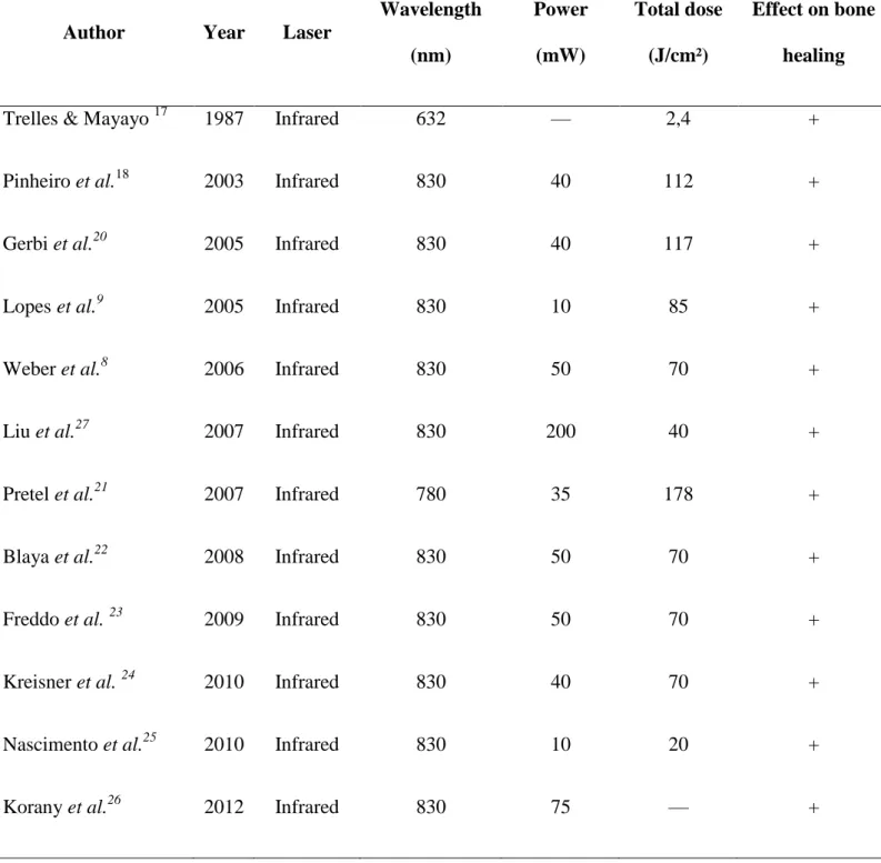
Table 1. Studies of the effects of LLLT on bone healing.
Author Year Laser
Wavelength
(nm)
Power
(mW)
Total dose
(J/cm²)
Effect on bone
healing
Trelles & Mayayo 17 1987 Infrared 632 — 2,4 +
Pinheiro et al.18 2003 Infrared 830 40 112 +
Gerbi et al.20 2005 Infrared 830 40 117 +
Lopes et al.9 2005 Infrared 830 10 85 +
Weber et al.8 2006 Infrared 830 50 70 +
Liu et al.27 2007 Infrared 830 200 40 +
Pretel et al.21 2007 Infrared 780 35 178 +
Blaya et al.22 2008 Infrared 830 50 70 +
Freddo et al. 23 2009 Infrared 830 50 70 +
Kreisner et al. 24 2010 Infrared 830 40 70 +
Nascimento et al.25 2010 Infrared 830 10 20 +
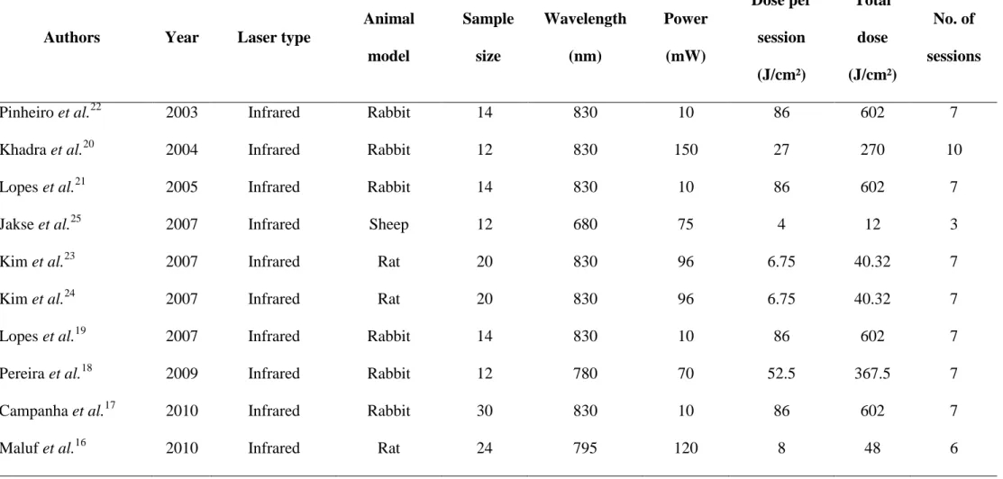
Table 2. Studies of the effects of LLLT on osseointegration.
Author Years Laser
Wavelength
(nm)
Power
(mW)
Total
dose
(J/cm²)
Effect on
osseointegration
Dörtbudak et al. 16 2002 Infrared 690 100 30 +
Pinheiro et al. 18 2003 Infrared 830 10 602 +
Lopes et al. 9 2005 Infrared 830 10 602 +
Jakse et al. 10 2007 Infrared 680 75 12 +
Kim et al.31 2007 Infrared 830 96 40.32 +
Lopes et al.11 2007 Infrared 830 10 602 +
Pereira et al.4 2009 Infrared 780 70 367.5 +
Campanha et al.12 2010 Infrared 830 10 602 +
Maluf et al.13 2010 Infrared 795 120 48 +
Garcia-Morales et al.14 2012 Infrared 830 86 92.1 -
3 ARTIGO II
Systemic effects of LLLT on thyroid function after titanium dental implant placement: an experimental rabbit model
Luciano Mayer1 – Fernando Vacilotto Gomes2 – Carlos Eduardo Baraldi2 – Fabrício Poletto Massotti2– Deise Ponzoni2– Marília Gerhardt de Oliveira3– João Batista Blessmann Weber1
1
Department of Oral and Maxillofacial Surgery, Pontifícia Universidade Católica do Rio Grande do Sul (PUCRS) School of Dentistry, Rio Grande do Sul, Brazil.
2
Department of Surgery, Universidade Federal do Rio Grande do Sul (UFRGS) School of Dentistry, Rio Grande do Sul, Brazil.
3
National Council for Scientific and Technological Development (CNPq); Oral and Maxillofacial Surgery Service, Hospital Cristo Redentor - Grupo Hospitalar Conceição (GHC), Rio Grande do Sul, Brazil.
This study should be attributed to the Department of Oral and Maxillofacial Surgery, Pontifícia Universidade Católica do Rio Grande do Sul (PUCRS)
School of Dentistry, Rio Grande do Sul, Brazil Faculdade de Odontologia - PUCRS
Av. Ipiranga, 6681 - Prédio 6, Partenon 90619-900 Porto Alegre, RS, Brazil Phone: + 55 (51) 3320-3562
Fax: +55 (51) 3320-3626
Corresponding author:
Av. Ipiranga, 6681 - Prédio 6, Partenon 90619-900 Porto Alegre, RS, Brazil
Phone: +55 (51) 3320-3562 / Fax: +55 (51) 3320-3626
jbbweber@terra.com.br
Keywords: Thyroid gland; laser therapy; low level; thyroid hormones; dental implant.
ABSTRACT
This study sought to assess the systemic effect of low-level laser therapy (LLLT) on thyroid function and, consequently, on calcium regulation by measurement of triiodothyronine (T3), thyroxine (T4), calcium, and albumin levels in rabbits. Forty male New Zealand rabbits were allocated randomly across five groups of eight each: CI (no LLLT, no surgery), CII (no LLLT), EI (70 J/cm²), EII (35 J/cm²) and EIII (140 J/cm²). In all groups except CI, animals underwent extraction of the mandibular left incisor followed by immediate placement of an osseointegrated implant. All experiment groups received infrared laser irradiation (GaAlAs, λ=830 nm, 50mW, CW) every 48h over 13 days, for a total of 7 sessions. T3, T4, calcium and
INTRODUCTION
Laser-emitting devices are widely used by health professionals, mostly for therapeutic and diagnostic purposes. In dentistry in particular, the utility of lasers of different wavelengths for distinct indications in the field of oral health has enabled incorporation of laser technology into a wide range of clinical and surgical procedures.1-3
Clinical use of low-level laser therapy (LLLT) is based on the ability of this treatment modality to exert stimulating effects on the molecular and biochemical processes that occur during tissue repair at the cellular level. Its therapeutic effects include increased epithelial proliferation, increased fibroblast proliferation and increased collagen synthesis, which can speed the healing process; improvement of bone remodeling and repair potential; restoration of neural function after injury; normalization of hormone function; regulation of the immune system; reduction of inflammation and edema; and modulation and relief of pain, including postoperative pain.4-11
Many in vivo and in vitro studies have reported the beneficial effects of LLLT on the repair process in animal models and tissue cultures.10, 12-14 However, even though dose is the most important parameter of laser therapy,15 there is no definitive protocol for its application in different clinical settings; this remains a point of debate in the literature16-25 (Table 01).
Studies on the effect of infrared laser irradiation on the thyroid gland have shown increased mitotic activity in follicular cells, transient hyperactivity of some thyroid follicles,29, 30
and changes in circulating serum levels of the thyroid hormones triiodothyronine (T3) and thyroxine (T4).31
The objective of this study was to assess the systemic effect of LLLT on thyroid gland function by measurement of circulating levels of triiodothyronine (T3), thyroxine (T4),
calcium and albumin in rabbit serum after low-level laser irradiation as an adjunctive therapy for osseointegration of implants placed immediately into fresh extraction sockets.
MATERIALS AND METHODS
Sample
Forty New Zealand male rabbits (Oryctolagus cuniculus), aged 3 months and weighing 3–4 kg, were used. Animals were allocated randomly into five distinct groups of
eight rabbits: three experiment groups, EI, EII and EIII, and two control groups, CI (no LLLT and no surgery) and CII (no LLLT). Animals were fed solid chow (Purina®, Nestlé Purina Petcare, St. Louis, MO, USA) and water ad libitum throughout the experiment and were kept in a climate-controlled environment, under normal lighting, humidity, and temperature conditions. Rabbits in the experiment groups (EI, EII and EIII) and control group CII underwent surgical extraction of the mandibular left incisor and immediate placement of an osseointegrated implant, creating a condition of equality between groups at baseline. Animals in group CI did not undergo any procedures other than blood collection and sham
surgery/sham irradiation, and thus served as an absolute control for triiodothyronine (T3), thyroxine (T4), calcium and albumin measurements (Table 02).
Operative technique
General anesthesia was induced with ketamine hydrochloride (Dopalen®, Ceva Santé Animale, Libourne, France) 40 mg/kg and xylazine hydrochloride (Anasedan®, Ceva Santé Animale, Libourne, France) 3 mg/kg by intramuscular injection (IM). The mandibular left incisor region was disinfected with chlorhexidine digluconate 2% (FGM Produtos
Odontológicos, Joinvile, SC, Brazil) and anesthetized by local infiltration of 0.5 mL lidocaine 2% with epinephrine 1:100,000 (Fig. 01-A), and the tooth extracted with the aid of #5
The surgical wound was closed with 4-0 nylon monofilament (Ethicon®, Johnson & Johnson, Somerville, NJ, USA) (Fig. 01-E). At the end of the procedure, animals were shaved and tattooed with a surgical marker (Codman®, Johnson & Johnson, New Jersey, USA) on the body of the mandible, over the region corresponding to the long axis of the dental implant, creating a target for laser irradiation (Fig. 01-F) and it was made a control RX to confirm the implant position (Fig. 02-A). Immediately after the procedure and 24 hours later, animals received tramadol (5 mg/kg IM) for analgesia and enrofloxacin (5 mg/kg IM, once daily for 3 days) for antibiotic prophylaxis.
Laser irradiation
Laser therapy was performed with a GaAlAs infrared diode laser, wavelength 830 nm, power 50 mW, in continuous emission mode (Thera Lase®, DMC Equipamentos, São Carlos, SP, Brazil). In the experiment groups, the irradiation protocol was started immediately after extraction of the mandibular left incisor and placement of the osseointegrated implant. Non-irradiated animals (control groups CI and CII) underwent sham irradiation with the laser device unpowered. Irradiation was performed every 48 hours for a total of seven sessions over 13 days. The total dose per session was divided across two points – one medial and one lateral to the long axis of the implant as marked on the overlying skin. The laser handpiece was held perpendicular to the basal mandibular bone.
Animals in group EI received a total dose of 70 J/cm2 at an energy density of 5 J/cm2 per point (index dose), over an irradiation time of 101 s (irradiation time is adjusted