INTRODUCTION
Titanium (Ti) implants have been widely used in dentistry to restore mastication, phonation and esthetics. The purposes of surgical procedures are to control, guide, and accelerate healing, which leads to the implant integration into bone. To achieve osseointegration, however, an adequate interaction between implant surface and bone is necessary (1).
Several treatments have been proposed to improve and accelerate bone formation onto implant surface, among which low-level laser therapy (LLLT). The laser is a source of non-ionizing radiation, highly concentrated, which, in contact with different tissues, produces thermal, photochemical and non-linear effects, depending on the type of radiation (2). In dentistry, LLLT is effectively used to accelerate recovery in cases of recurrent aphthous stomatitis, oral mucositis,
Effects of Low-Level Laser Therapy on Human
Osteoblastic Cells Grown on Titanium
Alice Dias PETRI Lucas Novaes TEIXEIRA Grasiele Edilaine CRIPPA Marcio Mateus BELOTI Paulo Tambasco de OLIVEIRA
Adalberto Luiz ROSA
Cell Culture Laboratory, Ribeirão Preto Dental School, University of São Paulo, Ribeirão Preto, SP, Brazil
The aim of this study was to investigate the effects of low-level laser therapy (LLLT) by using gallium aluminum arsenide (GaAlAs) diode laser on human osteoblastic cells grown on titanium (Ti). Osteoblastic cells were obtained by enzymatic digestion of human alveolar bone and cultured on Ti discs for up to 17 days. Cells were exposed to LLLT at 3 J/cm2 (wavelength of 780 nm) at days 3 and 7 and non-irradiated cultures were used as control. LLLT treatment did not influence culture growth, ALP activity, and mineralized matrix formation. Analysis of cultures by epifluorescence microscopy revealed an area without cells in LLLT treated cultures, which was repopulated latter with proliferative and less differentiated cells. Gene expression of ALP, OC, BSP, and BMP-7 was higher in LLLT treated cultures, while RUNXures, while R 2, OPN, and OPG were lower. These results indicate that LLLT modulates cell responses in a complex way stimulating osteoblastic differentiation, which suggests possible benefits on implant osseointegration despite a transient deleterious effect immediately after laser irradiation.
Key Words: Bone, laser, osteoblast, osseointegration, titanium.
traumatic ulcers, herpetic lesions (3,4), and treatment of temporomandibular disorders (5). Concerning bone tissue, LLLT has been applied in several clinical situations, such as orthodontic treatment, alveolar repair after tooth extraction, bone fracture healing, and osseointegration of dental implants as an adjuvant therapy (6-9). However, there are only few studies on its use in oral implantology (10,11). In addition, studies using osteoblastic cell cultures grown on Ti, which mimic
in vitro the process of in vivo bone formation in contact with implant surfaces did not evaluate all phases of the osteoblastic phenotype development (12,13).
Considering evidence that LLLT has positive effects on bone healing, it is possible to hypothesize that this therapy can enhance and/or accelerate the events related to osseointegration of Ti implants. Thus, the aim of this study was to investigate the effects of LLLT by using a gallium aluminum arsenide (GaAlAs) diode
laser at a wavelength of 780 nm on human alveolar bone-derived cells cultured on Ti.
MATERIAL AND METHODS
Ti Discs
Two hundreds and thirty-four discs of commercially pure Ti (12 mm diameter and 2 mm thick) were polished with 600-grit silicon carbide abrasive paper, cleaned in an ultrasonic bath, and autoclaved before using in the cell culture experiments.
Culture of Human Alveolar Bone-Derived Cells
Human alveolar bone fragments (explants) were obtained from 3 healthy donors, using the research protocols approved by the local Research Ethics Committee (protocol #2003.1.1271.58). Osteoblastic cells were obtained from these explants by enzymatic digestion using collagenase type II (Gibco Life Technologies, Inc., Grand Island, NY, USA). These cells were cultured in α-Minimum Essential Medium (Gibco), supplemented with 10% fetal bovine serum (Gibco), 50 μg/mL gentamicin (Gibco), 0.3 μg/mL fungizone (Gibco), 10-7 M dexamethasone (Sigma, St.
Louis, MO, USA), 5 μg/mL ascorbic acid (Gibco), and 7 mM β-glycerophosphate (Sigma). Subconfluent cells in primary culture were harvested after treatment with 1 mM ethylenediaminetetraacetic acid (EDTA) (Gibco) and 0.25% trypsin (Gibco) and subcultured in 24-well culture plates (Falcon, Franklin Lakes, NJ, USA) on Ti discs at a cell density of 2×10⁴ cells/disc. During
the culture period, cells were incubated at 37ºC in a humidified atmosphere of 5% CO2 and 95% air; the
medium was changed every 3 or 4 days.
Laser Irradiation
A photon-plus, GaAlAs diode laser device (MM Optics Twin Laser, São Paulo, SP, Brazil), was used in this study. This system operates in the near infrared spectrum at a continuous wavelength of 780 nm and a power output of 70 mW, and spot diameter of 0.2 cm. The cultures were irradiated at 3 and 7 days at dosage of 3 J/cm2 for 9 min. During the irradiation the probe was
kept at 12.63 cm from the cultures, a distance calculated considering the area of the cone base to guarantee that the whole culture on Ti disc would be irradiated.
Cell Growth
C e l l g r o w t h w a s e v a l u a t e d b y 3-[4,5-dimethylthiazol-2-yl]-2,5- diphenyl tetrazolium bromide (MTT; Sigma) assay at 10 and 14 days. Cells were incubated with 10% of MTT (5 mg/mL) in culture medium at 37°C for 4 h. The MTT solution was then aspirated from the well and 1 mL of acid isopropanol (0.04 N HCl in isopropanol) was added to each well. The plates were then agitated on a plate shaker for 5 min, and 150 μL of this solution were transferred to a 96-well format using opaque-walled transparent-bottomed plates (Fisher Scientific, Pittsburgh, PA, USA). The optical density was read at 570-650 nm on the plate reader (μQuant; Biotek, Winooski, VT, USA) and data were expressed as absorbance.
ALP Activity
ALP activity was assayed by the release of thymolphthalein from thymolphthalein monophosphate using a commercial kit (Labtest Diagnostica SA, Lagoa Santa, MG, Brazil) at 10, 14 and 17 days. Briefly, the culture medium was removed and the wells were washed 3 times with phosphate-buffered saline (Gibco) at 37°C and filled with 2 mL of deionized water, and the cultures were submitted to 5 cycles of thermal-shock (alternating temperature between 15 min at 37ºC and 20 min at -20ºC). Then, 50 μL of thymolphthalein monophosphate was mixed with 0.5 mL of diethanolamine buffer, 0.3 mmol/mL, pH 10.1, and left for 2 min at 37ºC. After this period, it was added 50 μL of the sample from each well. This stood for 10 min at 37ºC and then 2 mL of a solution of Na2CO3 0.09 mmol/mL and NaOH 0.25
mmol/mL was added to allow color development. After 30 min, absorbance was measured at 590 nm and ALP activity was calculated from a standard curve. Results were calculated and data were expressed as ALP activity normalized by the total protein content measured at 10, 14 and 17 days.
Mineralized Matrix Formation
for 10 min. The calcium content was evaluated using a colorimetric method. Briefly, 280 μL of 10% acetic acid was added to each well containing Ti samples stained with Alizarin Red S, and the plate was incubated at RT for 30 min under shaking. This solution was transferred to a microcentrifuge tube and after vortexing for 1 min, the slurry was overlaid with 100 μL of mineral oil (Sigma), heated to exactly 85ºC for 10 min, and transferred to ice for 5 min. The slurry was then centrifuged at 20,000 g for 15 min and 100 μL of supernatant was transferred to a new microcentrifuge tube. Then, 40 μL of 10% ammonium hydroxide was added to neutralize the acid and this solution containing 140 μL was read at 405 nm in 96-well format using opaque-walled transparent-bottomed plates (Fisher Scientific) on the plate reader μQuant (Biotek). Data were expressed as absorbance.
Immunolabeling
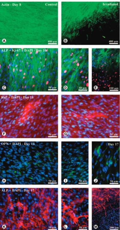
At days 8, 10, 14, and 17, cells were fixed at room temperature with 4% paraformaldehyde in 0.1 M phosphate buffer (PB), pH 7.2 or methanol p.a. (Merck, Darmstadt, Germany) at -20°C for 10 min. After washing in PB, cells were routinely processed for immunofluorescence labeling to detection of alkaline phosphatase (ALP), bone sialoprotein (BSP), osteopontin (OPN), and the nuclear antigen Ki-67. Cells fixed with paraformaldehyde were permeabilized with 0.5% Triton X-100 in PB for 10 min. Then, the samples were blocked with 5% skimmed milk in PB for 30 min. Primary antibodies to ALP (monoclonal B4-78, 1:100; Developmental Studies Hybridoma Bank, Iowa City, IA, USA), BSP (polyclonal LF100, 1:1000, Dr Larry W. Fischer, National Institutes of Health, Bethesda, MD, USA), OPN (monoclonal MPIIIB10, 1:800, Developmental Studies Hybridoma Bank, Iowa City, IA), and Ki-67 antigen (polyclonal, 1:70; Diagnostic Biosystems, Pleasanton, CA, USA) were used, followed by a mixture of goat anti-mouse or goat anti-rabbit secondary antibody (1:200; Molecular Probes, Eugene, OR, USA), conjugated with Alexa Fluor 594 (red fluorescence) or Alexa Fluor 488 (green fluorescence). Actin cytoskeleton was labeled with Alexa Fluor 488-conjugated phalloidin (1:200; Molecular Probes). All antibody incubations were performed in a humidified environment for 60 min at room temperature. Between each incubation step, the samples were washed 3 times (5 min each) in PB. Before mounting for microscope observation, cell nuclei were stained with 300 nM
4’,6-diamidino-2-phenylindole, dihydrochloride (DAPI; Molecular Probes) for 5 min and samples were briefly washed with dH2O. Ti discs were placed face up on glass
slides, while a Fisherbrand 12 mm-round glass coverslip (Fisher Scientific) was mounted with an antifade kit (Prolong; Molecular Probes) on the surface containing cells. The samples were examined under epifluorescence using a Leica DMLB light microscope, with N Plan (310/0.25, 320/0.40) and HCX PL Fluotar (340/0.75) objectives, outfitted with a Leica DC 300F digital camera (Leica; Bensheim, Germany). The acquired digital images were processed with Adobe Photoshop software (Adobe Systems, San Jose, CA, USA).
RNA Extraction and Quantitative Real-Time Reverse Transcriptase-Polymerase Chain Reaction (RT-PCR)
Gene expression of runt-related transcription factor 2 (RUNX
factor 2 (R
factor 2 (R 2), collagen type I (COL I), alkaline phosphatase (ALP), osteocalcin (OC), osteopontin (OPN), bone sialoprotein (BSP), bone morphogenic protein-7 (BMP-7), receptor activator of NF-kB ligand (RANKL), osteoprotegerin (OPG), and intercellular adhesion molecule (ICAM) were evaluated by RT-PCR at day 14. Gene-specific primers were designed with Primer Express 2.0 (Applied Biosystems, Foster City, CA, USA) and are presented in Table 1.
The total RNA from cells was extracted using the Promega RNA extraction kit (Promega, Madison, WI, USA), according to the manufacturer`s instructions. The concentration of RNA was determined by optical density at a wavelength of 260 nm, using the Biomate 3 spectrophotometer (Thermospectronic, Rochester, NY, USA). Complementary DNA (cDNA) was synthesized using 2 μg of RNA through a reverse transcription reaction (M-MLV reverse transcriptase, Promega). RT-PCR was performed in an ABI Prism 7000 Sequence Detection System using the SybrGreen system (Applied Biosystems, Warrington, UK, USA). SybrGreen PCR MasterMix (Applied Biosystems), specific primers and 2.5 ng cDNA were used in each reaction. The standard PCR conditions were 50°C (2 min), 95°C (10 min) and 40 cycles of 95°C (15 s), 60°C (1 min), followed by the standard denaturation curve. To mRNA analysis, the relative level of gene expression was calculated in reference to β-Gus expression and normalized by the gene expression of control cultures (calibrator) using the cycle threshold (Ct) method.
All experiments were carried out in quadruplicates with exception of RT-PCR, which was conducted in triplicates. Data were subjected to analysis of variance (two way MANOVA) followed by Tukey’s test, when appropriated. Data from RT-PCR were analyzed by the Mann-Whitney test. For both tests
significance level was set at 5%.
RESULTS
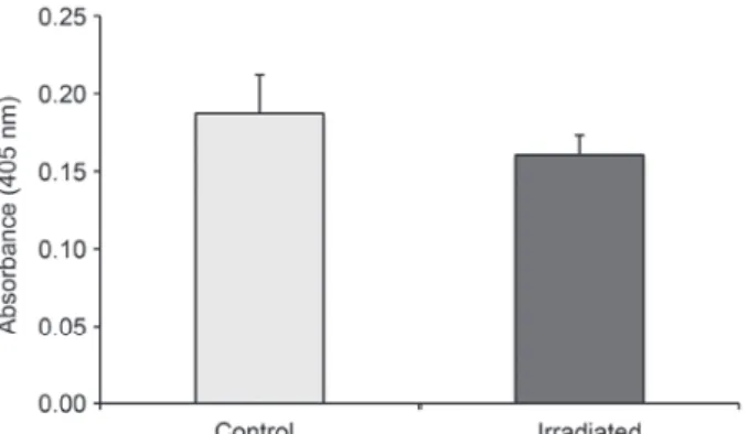
MTT assay indicated that cell growth was affected by time only in LLLT group (Fig. 1). From day 10 to 14 LLLT treated cultures showed an increase of cell growth (p<0.05), whereas cell population was maintained in control cultures in the same period (p>0.05). However, no significant difference (p>0.05) was observed either at 10 or 14 days between LLLT treated and control cultures. ALP activity was affected by time, irrespective of treatment. Higher levels of ALP activity were observed at day 10 (p<0.05) compared to days 14 and 17. However, LLLT treatment did not influence (p>0.05) ALP activity in any time point (Fig. 2). Calcium content measured by the extraction of Alizarin Red S from mineralized matrix was not affected (p>0.05) by LLLT treatment at day 17 (Fig. 3). At 24 h after irradiation procedures, analysis of cultures by epifluorescence microscopy revealed that all cultures were confluent on Ti discs (Fig. 4A and B). However, it was observed an area without cells in LLLT treated cultures (Fig. 4B). At day 10, ALP labeling of both cultures showed punctuate pattern mainly at cell borders (Fig. 4C and D). At this time point, a relevant number of cycling cells, Ki-67 positive, was noticed in areas previously without cells in LLLT treated cultures. These cells did not express ALP compared to the neighboring ones (Fig. 4E). Both cultures exhibited extracellular accumulation of BSP (Fig. 4F and G) and were positive for OPN labeling at
day 14 (Fig. 4H and I), with control cultures showing more intense OPN labeled areas (Fig. 4H). At day 17, cells in areas with lower density of cells on LLLT group were strongly labeled for OPN (Fig. 4J). The same ALP punctuate pattern labeling observed at day 10 was present in both cultures at day 17 (Fig. 4K and
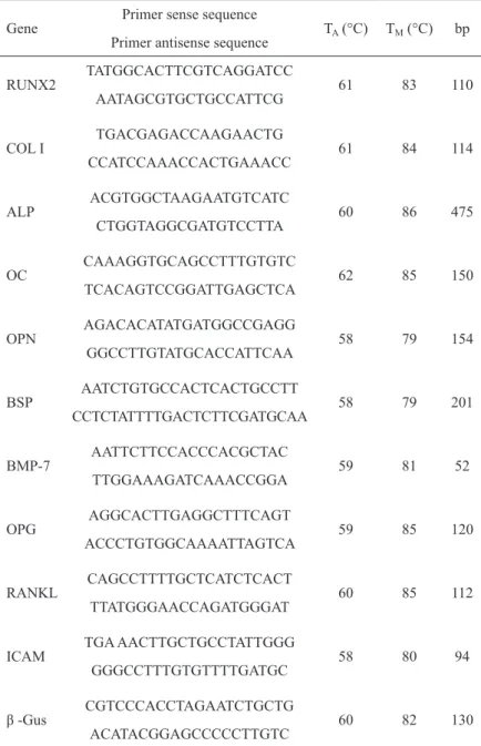
Table 1. Primer sequences and reaction properties.
Gene Primer sense sequence TT (°C)AA TM (°C) bp Primer antisense sequence
RUNX
R 2 TATGGCACTTCGTCAGGATCC 61 83 110
AATAGCGTGCTGCCATTCG
COL I
TGACGAGACCAAGAACTG
61 84 114
CCATCCAAACCACTGAAACC
ALP
ACGTGGCTAAGAATGTCATC
60 86 475
CTGGTAGGCGATGTCCTTA
OC
CAAAGGTGCAGCCTTTGTGTC
62 85 150
TCACAGTCCGGATTGAGCTCA
OPN
AGACACATATGATGGCCGAGG
58 79 154
GGCCTTGTATGCACCATTCAA
BSP AATCTGTGCCACTCACTGCCTT 58 79 201
CCTCTATTTTGACTCTTCGATGCAA
BMP-7
AATTCTTCCACCCACGCTAC
59 81 52
TTGGAAAGATCAAACCGGA
OPG
AGGCACTTGAGGCTTTCAGT
59 85 120
ACCCTGTGGCAAAATTAGTCA
RANKL
CAGCCTTTTGCTCATCTCACT
60 85 112
TTATGGGAACCAGATGGGAT
ICAM
TGA AACTTGCTGCCTATTGGG
58 80 94
GGGCCTTTGTGTTTTGATGC
β -Gus CGTCCCACCTAGAATCTGCTG 60 82 130
ACATACGGAGCCCCCTTGTC
RUNX R
R 2: Runt-related transcription factor-2; COL I: Type I collagen; ALP: alkaline phosphatase; OC: osteocalcin; OPN: osteopontin; BSP: Bone sialoprotein; BMP-7: bone morphogenetic protein-7; OPG: osteoprotegerin; RANK-L: receptor activator
of nuclear factor kappa B ligand; ICAM: intercellular adhesion molecules; β-Gus:
beta-glucuronidase; TA beta-glucuronidase; T
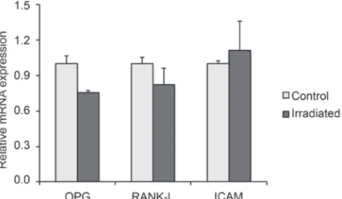
L). For that reason, in some regions it was possible to visualize the cell morphology (Fig. 4L). At this period, cells of the LLLT group were uniformly distributed on the whole Ti discs. Although the region without cells had been repopulated, an area of ALP-negative labeling was noticed (Fig. 4M). Osteoblastic phenotype of LLLT treated and control cultures was confirmed by the expression of genes encoding RUNX2, COL I, ALP, OC, OPN, BSP and BMP-7 at 14 days. The results showed that gene expression of ALP, OC, BSP, and BMP-7 was higher (p<0.05) in LLLT treated cultures, while RUNXs, while Rs, while R 2 and OPN were lower (p<0.05). The LLLT did not affect (p>0.05) COL I expression (Fig. 5). The gene expression of OPG, RANKL and ICAM, genes related to bone remodeling, were expressed by both cultures. LLLT reduced OPG expression (p<0.05) without affecting the expression of RANKL and ICAM (Fig. 6).
DISCUSSION
The present study evaluated the effect of LLLT using a GaAlAs laser on osteoblastic cells cultured on Ti discs. The dosage, 3 J/cm2, and wavelength, 780 nm
used were similar to previous works that evaluated the effects of LLLT on osteoblastic cells using a dosage between 0.48 and 3.84 J/cm2 and a wavelength of 830
nm (14,15). Biochemical, immunolabeling, and RT-PCR assays confirmed that, irrespective of treatment, cells derived from alveolar bone cultured in osteogenic medium express the osteoblastic phenotype (16,17). Parameters assessed in this work indicated that LLLT affects cell behavior in a complex way suggesting that osteoblastic differentiation was stimulated despite a transient deleterious effect immediately after laser irradiation.
MTT assay revealed that cell growth was maintained in control cultures from day 10 to day 14, whereas in LLLT treated ones it was increased. Considering that in this culture system proliferation occurs up to day 10 (12,13), it suggests a stimulatory effect of laser on cell growth. This mitogenic effect was also described for different cell lines such as osteoblasts derived from fetal rat calvariae (14). On the other hand, 24 h after LLLT treatment at day 8 of culture it was observed areas without cells in Ti discs contrasting with control cultures where Ti discs were fully covered by cells. In spite of this, cell growth at day 10 was not affected by LLLT treatment. This finding suggests that the reduced cell population induced by laser either presented a magnitude that was not detectable by MTT assay or there was a prompt recovery of cell number 3 days after LLLT treatment. Supporting this latter explanation, at day 10 of culture cells strongly Figure 1. Effect of LLLT on growth of osteoblastic cells cultured
on Ti at 10 and 14 days. Data are presented as mean ± standard deviation (n=4).
Figure 3. Effect of LLLT on mineralized matrix formation by osteoblastic cells cultured on Ti at 17 days. Data are presented as mean ± standard deviation (n=4).
Ki-67-positive, which indicates a high proliferation rate, repopulated those areas previously without cells. Interestingly, it seems that these cells were not committed to osteoblastic differentiation as they no longer presented immunolabeling for ALP even at day 17 of culture. This transient damage to osteoblastic cells was not reported by Khadra et al. (11) most probably due to the lack of microscopic observation of the cultures following laser irradiation.
In the present study, for control cultures ALP immunolabeling parallels its activity. At 10 days of culture most of the cells were strongly ALP-positive with a noticeable decrease observed at 17 days. The same pattern was found by assaying ALP activity in control cultures. However, for LLLT treated cultures a slightly different behavior was observed. Despite the lack of ALP immunolabeling in many cells both at 10 and 17 days, LLLT treated cultures displayed the same level of ALP activity compared to control ones. It suggests a stimulatory effect of LLLT on osteoblastic cells that is corroborated by the increase in the expression of gene enconding ALP. Our data are in line with Ueda and Shimizu (14) that, using rat calvaria-derived cells, observed a raising in gene expression and ALP activity, but are opposite to those reported by Khadra et al. (11), that reported a lack of effect of laser irradiation on human cultures derived from mandible bone and osteosarcoma cell lines, respectively.
The amount of calcium deposition assayed by Alizarin Red S extraction was alike in both groups. Nevertheless, considering that cells in repopulated areas did not contribute to calcium deposition as at that time point they did not express the osteoblastic phenotype as
evidenced by ALP labeling, it is possible to speculate that the reduced osteoblastic cell population in irradiated cultures produced the same amount of mineralized matrix compared to the control ones. Such interpretation suggests that the LLLT stimulates the formation of mineralized matrix. Agreeing with this assumption, regarding gene expression, we observed an increase in OC and BSP, both related to mineralization process, a reduction in RUNX2, usually associated with the progress of osteoblast differentiation, and a significant increase in BMP-7 a potent inductor of osteogenesis.
The OPG/RANKL/RANK system plays a central role in the paracrine regulation and osteoclast function. The OPG binds to RANKL, both secreted by osteoblasts, and prevent the RANKL binding to RANK, that is present in the osteoclast membrane, inhibiting bone resorption (18). The ICAM is responsible for osteoblast-osteoclast coupling, which allows the connection RANK/RANKL (19). In this study we observed a reduction in the expression of OPG while no change in the expression of RANKL and ICAM was noticed. This result suggests that osteoblastic cells irradiated with laser could increase the osteoclastic activity. However, it should not be interpreted as a possible increase in bone resorption, since several cytokines released by osteoblastic cells exert regulatory functions on osteoclasts. In addition, other researchers showed opposite effect, in which laser irradiation resulted in an increase in the expression of OPG and reduction in the expression of RANKL in rat calvarial cells (20). In addition to irradiation parameters, different cell lines and culture conditions could explain the impaired results between studies. In this way, further investigations should be done to understand the influence of LLLT on osteoblast-osteoclast coupling
Figure 5. Effect of LLLT on the expression of genes related to osteoblastic phenotype of osteoblastic cells cultured on Ti at 14 days. The results are presented as the expression of the target
mRNAs normalized to β-Gus and to the control group. Data are
presented as mean ± standard deviation (n=3).
and consequently on bone remodeling.
In conclusion, the present results indicate that LLLT stimulates the expression of osteoblastic phenotype in cells cultured on Ti suggesting possible benefits on implant osseointegration. Despite this, deleterious effects associated with laser irradiation cannot be discarded. Therefore, the use of LLLT to promote implant osseointegration demands further in vitro and in vivo studies, which should focus on the mechanisms involved in desirable and undesirable effects of irradiation.
RESUMO
Este estudo teve como objetivo investigar o efeito do laser diodo de gálio-alumínio-arsênio (GaAlAs) em células osteoblásticas humanas cultivadas sobre discos de Ti. Para tanto, células osteoblásticas foram obtidas por digestão enzimática de osso alveolar humano e cultivadas sobre discos de Ti por 17 dias. As células foram submetidas à irradiação no 3º e 7º dias na dose de 3 J/cm2 e comprimento de onda de 780 nm e células não irradiadas foram usadas como controle. A irradiação não alterou a proliferação celular, atividade de ALP e formação de matriz mineralizada. Microscopia por epifluorescência indicou que após 24 h da aplicação do laser, as culturas irradiadas apresentaram áreas sem células, que mais tarde foram repovoadas por células em fase de proliferação e menos diferenciadas. O laser aumentou a expressão gênica relativa da ALP, OC, BSP e BMP-7 e reduziu a de RUNX2, OPN e OPG. Os resultados indicam que a terapia com laser modula de forma complexa as respostas celulares, estimulando a diferenciação osteoblástica. Assim, é possível sugerir possíveis benefícios do laser na osseointegração de implantes de Ti apesar do efeito deletério às células imediatamente após a irradiação.
ACKNOWLEDGEMENTS
The authors would like to thank the São Paulo State Research Foundation (FAPESP, Brazil), and the National Council of Scientific and Technological Development (CNPq, Brazil) for funding. The authors also wish to express their gratitude to Mr. Roger Rodrigo Fernandes, Ms. Junia Ramos for technical assistance, and Dr. Larry W. Fisher (NIH, Bethesda, MD, USA) for providing the anti-human BSP (LF-100) antibody.
REFERENCES
1. Albrektsson T, Johansson C. Osteoinduction, osteoconduction and osseointegration. Eur Spine 2001;10:S96-S101.
2. Peplow PV, Chung TY, Baxter GD. Laser photobiomodulation of wound healing: a review of experimental studies in mouse and rat animal models. Photomed Laser Surg 2010;28:291-325.
3. Lima AG, Antequera R, Peres MPSM, Snitcosky IML, Federico MHH, Villar RC. Efficacy of low-level laser therapy and aluminum hydroxide in patients with chemotherapy and radiotherapy-induced oral mucositis. Braz Dent J 2010;21:186-192.
4. Colvard M, Kuo P. Managing apthous ulcers: laser treatment applied. J Am Dent Assoc 1991;122:51-53.
5. Mazzetto MO, Hotta TH, Pizzo RCA.Measurements of jaw movements and TMJ pain intensity in patients treated with GaAlAs laser. Braz Dent J 2010;21:356-360.
6. Kawasaki K, Shimizu N. Effects of low-energy laser irradiation on bone remodeling during experimental tooth movement in rats. Laser Surg Med 2000;26:282-291.
7. Luger EJ, Rochkind S, Wollman Y. Effect of low power laser irradiation on the mechanical properties of bone fracture healing in rats. Laser Surg Med 1998;22:97-102.
8. Takeda Y. Irradiation effect of low-energy laser on alveolar bone after tooth extraction. Experimental study in rats. Int J Oral Maxillofac Surg 1988;17:388-391.
9. Yaakobi T, Maltz L, Andoron U. Promotion of bone repair in the cortical bone of the tibia in rats by low energy laser (He-Ne) irradiation. Calcif Tissue Int 1996;59:297-300.
10. Dörtbudak O, Haas R, Mailath-Pokorny G. Effect of low-power laser irradiation on bony implant sites. Clin Oral Implants Res 2002;13:288-292.
11. Khadra M, Lyngstadaas SP, Haanæsa HR, Mustafac K. Effect of laser therapy on attachment, proliferation and differentiation of human osteoblast-like cells cultured on titanium implant material. Biomaterials 2005;26:3503-3509.
12. Franco RD, Chiesa R, Beloti MM, de Oliveira PT, Rosa AL. Human osteoblastic cell response to a Ca- and P-enriched titanium surface obtained by anodization. J Biomed Mater Res 2008;19:15-20.
13. Rosa AL, Crippa GE, de Oliveira PT, Taba Jr M, Lefebvre LP, Beloti MM. Human alveolar bone cell proliferation, expression of osteoblastic phenotype, and matrix mineralization on porous titanium produced by powder metallurgy. Clin Oral Impl Res 2009;20:472-481.
14. Ueda Y, Shimizu N. Effects of pulse frequency of low-level laser therapy (LLLT) on bone nodule formation in rat calvarial cells. J Clin Laser Med Surg 2003;21:271-277.
15. Fujimoto K, Kiyosaki T, Mitsui N, Mayahara K, Omasa S, Suzuki N, et al.. Low-intensity laser irradiation stimulates mineralization via increased BMPs in MC3T3-E1 cells. Lasers Surg Med 2010;42:519-526.
16. Carvalho DR, Carvalho PS, Magro Filho O, de Mello JD, Beloti MM, Rosa AL. Characterization and in vitro cytocompatibility of an acid-etched titanium surface. Braz Dent J. 2010;21:3-11.
17. Hassan MQ, Javed A, Morasso MI, Karlin J, Montecino M, Van
Wijnen AJ, et al.. Dlx3 transcriptional regulation of osteoblast differentiation: temporal recruitment of Msx2, Dlx3, and Dlx5 homeodomain proteins to chromatin of the osteocalcin gene. Mol Cell Biol 2004;24:9248-9261.
18. Khosla S. Minireview: the OPG/RANKL/RANK system. Endocrinology 2001;142:5050-5055.
19. Matsuo K, Irie N. Osteoclast-osteoblast communication. Arch Biochem Biophys 2008;15:201-209.
20. Xu M, Tietao D, Feizhi M, Bin D, Wingho L, Pingxiang D, et al.. Low-intensity pulsed laser irradiation affects RANKL and OPG mRNA expression in rat calvarial cells. Photomed Laser Surg 2009;27:309-315.