Campus de São José do Rio Preto
Mariana Nogueira Batista
Avaliação
in vitro
do efeito da cafeína na inibição do vírus da
hepatite C
Mariana Nogueira Batista
Avaliação
in vitro
do efeito da cafeína na inibição do vírus da
Hepatite C
Dissertação apresentada como parte dos requisitos para obtenção do título de Mestre em Microbiologia, junto ao Programa de Pós-Graduação em Microbiologia, Área de Concentração - Virologia, do Instituto de Biociências, Letras e Ciências Exatas da Universidade Estadual Paulista “Júlio de Mesquita Filho”, Campus de São José do Rio Preto.
Orientador: Profª. Drª. Paula Rahal
Co-orientador: Dr. Bruno Moreira Carneiro
Mariana Nogueira Batista
Avaliação
in vitro
do efeito da cafeína na inibição do vírus da
Hepatite C
Dissertação apresentada como parte dos requisitos para obtenção do título de Mestre em Microbiologia, junto ao Programa de Pós-Graduação em Microbiologia, Área de Concentração - Virologia, do Instituto de Biociências, Letras e Ciências Exatas da Universidade Estadual Paulista “Júlio de Mesquita Filho”, Campus de São José do Rio Preto.
Comissão Examinadora
Prof. Dr. Bruno Moreira Carneiro
UNESP
–
São José do Rio Preto
Co-orientador
Prof. Drª. Laura Cristina Sichero Vetorazzo
ICESP
–
São Paulo
Prof. Dr. Maurício Lacerda Nogueira
FAMERP
–
São José do Rio Preto
DEDICATÓRIA
Dedico este trabalho..
À Maria Rosa Nogueira Batista e Vidercílio Batista da Silva, meus guias, meus espelhos, meus amores, meus companheiros, meus pais. Obrigada por toda
força e todo empenho por minha educação. Obrigada por me ensinarem os valores reais na vida, pela luz, pelo amor, pelo caráter, pela calma, enfim por
tudo. Obrigada por sonharem meus sonhos, por lutarem em minhas lutas. Todas as conquistas em minha vida, não são minhas, são nossas. Obrigada
AGRADECIMENTOS
A Deus, em primeiro lugar, por sempre ter traçado caminhos iluminados para minha vida e por todas as oportunidades maravilhosas.
À minha orientadora, Profª Drª Paula Rahal, pelo voto de confiança, e por me acolher em seu grupo tão conciso e com tanto a oferecer. Pelos conhecimentos científicos e em especial agradeço pelo crescimento pessoal.
Ao meu co-orientador, Dr. Bruno Moreira Carneiro, agradeço por toda paciência, dedicação, ajuda e conhecimentos transmitidos. Em especial um agradecimento para os momentos de sufoco.
À amiga e companheira de trabalho, Ana Cláudia Silva Braga, por dividir comigo não só os conhecimentos adquiridos, mas os bons e maus momentos. Agradeço também o apoio.
Às queridas, Bru e Bia pelo auxílio no desenvolvimento dos experimentos, com destaque para o auxílio da manutenção da saúde mental. Vocês ficam para sempre.
Aos amigos de laboratório e de almoço pelos momentos de choque de realidade, mas especialmente pelos momentos de descontração.
A todos os companheiros de trabalho do LEGO pelas ideias, discussões e convívio diário. E a Lê, por “segurar a barra” durante tempos difíceis.
À CAPES e FAPESP pelo apoio financeiro, sem o qual não seria possível o desenvolvimento deste trabalho.
Aos meus pais agradeço imensamente por existirem em minha vida. Por todo esforço, amor e coragem. Pelo estímulo e ponderação, mas em especial pelo equilíbrio transmitido. Amo imensamente vocês!
A meu namorado Cecílio, agradeço por toda dedicação, paciência e carinho. Agradeço também pelo estímulo constante, compreensão e amor incondicionais. Mas acima de tudo, pelo companheirismo e até experimentos realizados no desenvolvimento desse projeto.
A meus irmãos, Allan e Simoni, agradeço pela presença constante, ombro amigo, pelas “loucuras” e momentos de descontração.
Às minhas sobrinhas, por mostrarem em seus pequenos gestos o quão maravilhoso cada simples momento pode ser.
Aos demais familiares, agradeço pelo amor, paciência e por simplesmente apoiarem meus anseios.
A minhas amigas Carol e Té, agradeço por todo apoio mesmo corridinhos ou de longe. Obrigada por fazerem parte da minha vida.
EPÍGRAFE
“Quer vencer os desafios? - Confie em Deus! Quer ser bom no que faz? - Pratique!
Quer alcançar o objetivo? - Jamais desista!
Quer crescer? - Tenha raízes.
Quer ver resultados? Persevere.
Quer ser feliz? - Esqueça o passado.
Quer falar bem? - Escute melhor.
Quer aprender? - Persista em ler.
Quer realização pessoal? - Sirva!
Quer fazer diferença? - Pague o preço.
Aqueles que nada fazem e esperam algum tipo de vitória estão enganados.
A vitória é dos que lutam, dos que agem, dos que "saem do porto". A vitória é dos que se arriscam para alcançar o alto da montanha”
RESUMO
A hepatite C é a inflamação do fígado decorrente da infecção pelo vírus da hepatite C (HCV), frequentemente evolui para quadros crônicos, sendo considerada mundialmente a maior causa de cirrose e carcinoma hepatocelular. O tratamento padrão com PEG-IFN e ribavirina não é efetivo contra alguns genótipos do HCV, possui alto custo e efeitos colaterais severos. Portanto, novos tratamentos vêm sendo buscados. A cafeína vem sendo associada a um efeito benéfico sobre várias doenças hepáticas incluindo a melhora da bioquímica anormal do fígado, cirrose e carcinoma hepatocelular. A cafeína atua diretamente desacelerando a progressão da fibrose, além de melhorar a função de vias celulares hepáticas, dentre elas vias utilizadas durante o ciclo replicativo do HCV. Embora a cafeína tenha demonstrado efetividade no controle de doenças hepáticas e interação direta com vias celulares utilizadas pelo HCV, não há na literatura correlação direta entre o efeito da cafeína e as etapas do ciclo replicativo do HCV. Assim, o presente estudo propôs estabelecer a relação direta entre a cafeína e sua capacidade inibitória sobre as diferentes etapas do ciclo replicativo completo do HCV. Para esse estudo foram utilizados o replicon subgenômico SGR-JFH-FEO, os replicons completos FL-J6/JFH-5′C19Rluc2AUbi e JFH-1 e a linhagem celular Huh-7.5. A expressão viral foi avaliada por ensaios de Luciferase, Western Blotting, Imunofluorescencia indireta e qPCR. A cafeína demonstrou inibição da replicação viral em todos os níveis avaliados, apresentando IC50 de 0.7263 mM e atingindo em concentrações seguras, inibição máxima da replicação de HCVcc em torno de 79 %. A cafeína demonstrou ainda inibição de 30 % sobre a entrada quando aplicada em conjunto ao sobrenadante infeccioso. Entretanto, essa inibição dobra quando há a exposição das partículas à cafeína previamente à introdução em cultura de células, possivelmente havendo interação entre a cafeína e alguma proteína viral. Por outro lado, não houve influência da cafeína sobre o processo de liberação viral
ABSTRACT
Hepatitis C is the liver inflammation arising from hepatitis C virus (HCV) infection, often evolves to chronic conditions and has been considered the major world cause of cirrhosis and hepatocellular carcinoma. Standard treatment using PEG-IFN and ribavirin is not effective against some HCV genotypes, besides that it has high cost and severe side-effects. Therefore, new treatments have been sought. Caffeine has been found to have beneficial effect in several liver disorders, including the improvement of abnormal liver biochemistry, cirrhosis and hepatocellular carcinoma. Caffeine acts directly by delaying fibrosis, beyond improving the function of liver cellular pathways and interfering with pathways used by the HCV replication cycle. Although, the caffeine showed positive effects for liver disorders and a direct interaction with cell pathways used by HCV, there is no evidence of a direct correlation between caffeine and HCV replication cycle. Thus, the current study proposed to establish the direct relationship between caffeine and different steps of HCV replication cycle. To this study, it was used the subgenomic replicon SGR-JFH-FEO, the full-length replicons FL-J6/JFH-5′C19Rluc2AUbi and JFH-1; and Huh-7.5 cell line. The viral expression was evaluated by Luciferase, Western blotting, Indirect immunofluorescence and qPCR. The caffeine demonstrated to be able to inhibit viral replication on different stages of viral replication, demonstrating an IC50 value of 0.7263 mM and reaching on safe concentrations, HCVcc maximal replication inhibition around 79 %. Caffeine demonstrated also 30 % of inhibition on viral entry on host cells when tested in combination with infectious supernatant. Moreover, this inhibition increased two fold when particles were exposed to caffeine before introduction on cell culture, possibly, indicating an interaction between caffeine and viral proteins. On the other hand, there is no influence of caffeine on viral secretion process.
SUMÁRIO
Capítulo I
1 INTRODUÇÃO ... 17
1.1 Aspectos gerais da Hepatite C ... 17
1.2 Vírus da hepatite C ... 17
1.2.1 Descoberta, Classificação e Organização do Genoma ... 17
1.2.2 Heterogeneidade genética e distribuição ... 23
1.2.3 Ciclo replicativo ... 26
1.2.4 Modelos para o estudo do HCV in vitro ... 27
1.2.5 Vias de transmissão e tratamentos disponíveis ... 30
1.2 Fitoquímicos de ação terapêutica e cafeína ... 32
2 JUSTIFICATIVA ... 35
3 OBJETIVOS ... 36
4 REFERÊNCIAS BIBLIOGRÁFICAS ... 37
Capítulo II 1 ARTIGO CIENTÍFICO 1 ... 45
Capítulo III 1 ARTIGO CIENTÍFICO 2 ... 45
Capítulo IV 1 DISCUSSÃO ... 85
2 CONCLUSÃO ... 89
3 PROBLEMAS E PERSPECTIVAS ... 89
LISTA DE FIGURAS
Capítulo I ... 16
Figura 1: Estrutura da partícula viral e do genoma do HCV ... 23
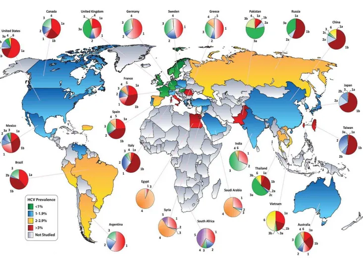
Figura 2: Prevalência mundial do HCV e distribuição geográfica dos genótipos .... 25
Figura 3: Ciclo Replicativo do HCV ... 27
Figura 4: Estrutura esquemática dos replicons do HCV ... 29
Capítulo II ... 44
Figura 1: Análise da viabilidade de células Huh-7.5 sob tratamento com cafeína .. 61
Figura 2: Efeito inibitório da cafeína sobre a replicação do HCV em 48 h ... 62
Figura 3: Análise do efeito da cafeína sobre o padrão de ciclo celular da Huh-7.5 63 Figura 4: Análise do perfil apoptótico de Huh-7.5 tratadas com cafeína por 48 h .. 64
Figura 5: Análise do perfil autofágico de Huh-7.5 tratadas com cafeína por 48 h .. 65
Figura S1: Efeito inibitório da cafeína sobre a replicação do HCV em 24 h ... 66
Figura S2: Análise do perfil apoptótico de Huh-7.5 tratadas com cafeína por 24 h ... 68
Capítulo III ... 69
Figura 1: Perfil citotóxico da cafeína sobre a linhagem Huh-7.5/SGR-JFH-FEO ... 81
Figura 2: Efeito inibitório da cafeína sobre a entrada do HCV ... 82
LISTA DE TABELAS
Capítulo III ... 69
LISTA DE ABREVIAÇÕES
ALT - Alanina aminotransferase, do Inglês Alanine aminotransferase ANOVA - Análise de Variância, do Inglês Analysis of Variance
ASP - Aspartato aminotransferase, do Inglês Aspartate aminotransferase BCA - Ácido bicincônico, do Inglês Bicinchoninic acid
C - Proteína do capsídeo do vírus da Hepatite C, do Inglês Core protein CAPES - Coordenação de Aperfeiçoamento de Pessoal de Nível Superior CD81 - Cluster de Diferenciação 81, do Inglês Cluster of Diferenciation 81 CLDN - Claudina, do Inglês Claudin
CLDN1 - Claudina 1, do Inglês Claudin 1
COX-2 - Ciclooxigenase 2, do Inglês Cyclooxygenase-2 CsA - Ciclosporina A, do Inglês Cyclosporine A
DAAs - Antivirais de Ação direta, do Inglês Direct-acting antiviral agents
DMEM - Meio Eagle Modificado por Dulbeco, do Inglês Dulbecco's Modified Eagle's Medium
E1 - proteína do envelope 1 do vírus da hepatite C, do Inglês Envelope protein 1
E2 - proteína do envelope 2 do vírus da hepatite C, do Inglês Envelope protein 2
EGCG - Epigalocatequina-3-galato, do Inglês Epigallocatechin-3-Gallate
EGFR - Receptor do Fator de Crescimento Epidérmico, do Inglês Epidermal growth factor receptor
EphA2 - Precursor do receptor 2 de efrina tipo-A, do Inglês Ephrin type-A receptor 2 precursor
ERK - Quinase regulada por sinal extracelular, do Inglês Extracellular signal-regulated kinase
FAPESP - Fundação de Amparo à Pesquisa do Estado de São Paulo FBS - Soro Fetal Bovino, do Inglês Fetal Bovine Serum
FEO - Fusão de Firefly e Neomicina, do Inglês Firefly and Neomicin FFU - Unidade Formadora de Foco, do Inglês Focus-Forming Units FL-J6/JFH-5′C19Rluc2AUbi – Quimera replicon completo J6/JFH1 G418 – Geneticina
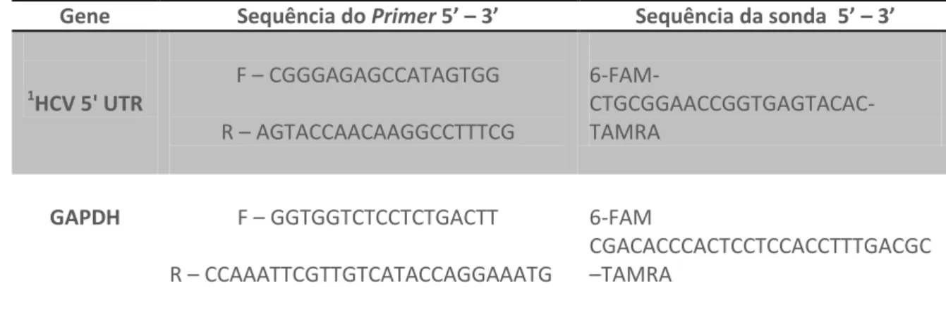
GAPDH - Gliceraldeído-3-fosfato dehidrogenase, do Inglês glyceraldehyde-3-phosphate dehydrogenase
HCV - Virus da Hepatite C, do Inglês Hepatitis C virus
HCVcc - Virus da Hepatite C derivado de cultura de células, do Inglês cell‐culture‐derived HCV
HEPES - tampão de Ácido 4-(2-HidroxiEtil)-1-PiperazinEtanolSulfônico, do Inglês 4-(2-hydroxyethyl)-1-piperazineethanesulfonic acid
HSP90 - proteína de choque térmico 90, do inglês Heat Shock Protein 90 Huh - Hepatoma humano, do Inglês Human hepatoma
HVR1 - Região hipervariável 1 , do Inglês Hypervariable region 1 HVR2 - Região hipervariável 1 , do Inglês Hypervariable region 2
IC50 - Concentração inibitória de 50 %, do Inglês half-maximal inhibitory concentration
ICTV - Comitê Internacional em Taxonomia de Virus, do Inglês International Committee on Taxonomy Viruses
IFN-α - Interferon alpha
IRES - Sítio interno de entrada ribossomal, do Inglês Internal ribosome entry site
ISG - Genes estimulados por Interferon, do Inglês Interferon stimulated gene JFH-1 - Replicon completo do virus da Hepatite C “Hepatite Japonesa Fulminante 1”, do Inglês “Japanese Fulminant Hepatitis1”
LDL-R - Receptor de Lipoproteina de baixa densidade, do Inglês Low-Density Lipoprotein Receptor
M.O.I - Multiplicidade de Infecção, do Inglês Multiplicity of Infection
MEK - Quinase da Proteína quinase mitogeno-ativada, do Inglês Mitogen-activated protein kinase kinase
MTT - Dimetil tiazolio 3-(4,5-dimethylthiazol-2-yl)-2,5-diphenyltetrazolium bromide
NK - Matadoras naturais, do Inglês Natural Killer
NPT - Neomicina fosfotransferase, Neomicin phosphotransferase NS - Proteínas Não estruturais, do Inglês Non-structural proteins NTP -Nucleotídeo Trifosfato, do Inglês NucleotideTriphosphate
NTPase - Nucleotídeo trifosfatase, do Inglês Nucleotide triphosphatase
NTRs - Regiões não traduzíveis, do Inglês non-translated regions (sinônimo de UTRs)
OCLN - Ocludina, do Inglês Occludin
ORF - Quadro aberto de leitura, do Inglês Open Reading Frame P7 - Proteína 7
PEG - Polietilenoglicol
PEG-IFN - Interferon Peguilado pH - Potencial Hidrogeniônico
PHH - Hepatócitos humanos primários, do Inglês Primary human hepatocytes PKA - Proteína quinase A, do Inglês Protein Kinase A
qPCR - PCR em tempo Real
Ras – Proteína Sarcoma de rato, do Inglês Rat Sarcoma protein RBV - Ribavirina
RE - Retículo Endoplasmático RLuc - Renilla Luciferase
RNA - Ácido Ribonucleico, do Inglês Ribonucleic Acid SDS - Dodecil sulfato de sódio
SGR-JFH-FEO ou SGR-Feo JFH-1 - Replicon subgenomico do JFH-1 SGR-Neo-Con1 - Replicon Subgenomico do Con1
SR-BI - Receptor Scavenger Classe B tipo I, do Inglês Scavenger Receptor Class B Type I
1 INTRODUÇÃO
1.1 Aspectos gerais da Hepatite C
A hepatite C é a inflamação do fígado decorrente da infecção pelo vírus da Hepatite C (HCV). Essa infecção causa inicialmente hepatite aguda, a qual é frequentemente subclínica podendo evoluir para um quadro crônico. A evolução para a cronicidade ocorre em aproximadamente 80 % dos casos (LAVANCHY, 2011) e dentre esses pacientes crônicos, 70 % desenvolvem algum tipo de patologia no fígado das quais 5 a 20 % corresponde à cirrose hepática (ASHFAQ et al., 2011) sendo que de 1 a 5 % morrem de cirrose ou hepatocarcioma (WHO, 2013). A evolução para hepatocarcinoma ocorre em cerca de 30-50 % dos pacientes cirróticos após aproximadamente 10 anos da infecção (GIANNINI; BRECHOT, 2003).
Essa doença é de grande importância para a saúde pública visto que, a cada ano, mais de 350 mil óbitos são relacionados à cirrose, doenças hepáticas em estágio terminal e do carcinoma hepatocelular (HCC) associadas a esta infecção (CHEVALIEZ; PAWLOTSKY, 2007; WHO, 2013). Estima-se que cerca de 150 milhões de pessoas ao redor do mundo estejam infectadas cronicamente pelo vírus (WHO, 2013) e que cerca de três a quatro milhões de novos casos surjam a cada ano (WHO, 2013).
1.2 Vírus da hepatite C
1.2.1 Descoberta, Classificação e Organização do Genoma
BARTENSCHLAGER, 2012). O HCV é classificado como membro da família
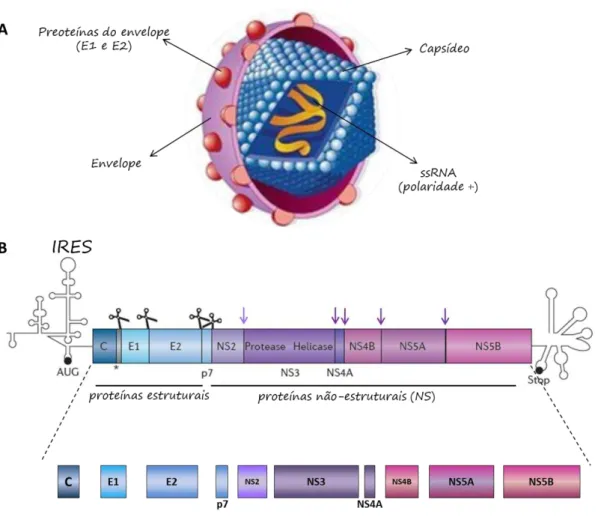
Flaviviridae e do gênero Hepacivirus (ICTV, 2013). São vírus envelopados, com capsídeo icosaédrico e aproximadamente 50 nm de diâmetro (Figura 1a), cujo genoma de cerca de 9.6 Kb corresponde a uma cadeia simples e linear de RNA com polaridade positiva contendo uma única fase aberta de leitura (ORF - open
reading frame); que codifica uma poliproteína precursora de 3.011 aminoácidos, flanqueada por duas regiões terminais não codificantes (BARTENSCHLAGER; LOHMANN; PENIN, 2013). Esta poliproteína é co - e pós-traducionalmente processada por proteases virais e celulares em dez proteínas, estruturais (C, E1, E2/p7) e não-estruturais (NS2, NS3, NS4A, NS4B, NS5A e NS5B) (BARTENSCHLAGER; LOHMANN; PENIN, 2013).
As regiões não traduzíveis (non-translated regions - NTRs) são necessárias tanto para a tradução da proteína quanto para a replicação do vírus. A região 5’NTR é uma porção altamente estruturada do genoma viral. Composta por
elemento invariável de 98 nucleotídeos designado cauda X (LINDENBACH; RICE, 2005).
As proteínas estruturais encontram-se presentes na região amino-terminal da poliproteína e são os principais constituintes das partículas virais infecciosas (THIMME; BINDER; BARTENSCHLAGER, 2012). Segundo Penin et al. (2004), a proteína do capsídeo (C) é uma proteína básica constituída pelos primeiros 191 aminoácidos da poliproteína, sendo a proteína viral mais conservada e correspondendo à segunda região mais conservada do genoma. Unidades repetitivas dessa proteína compõem o nucleocapsídeo viral, encapsulando o genoma e sendo essencial para a montagem das partículas virais. A proteína do capsídeo se localiza no citoplasma, onde se encontra ligada ao Retículo endoplasmático, podendo ainda apresentar-se, em menor proporção, no núcleo. Já foi demonstrado que esta proteína pode estar envolvida na modulação da transcrição de genes, proliferação celular, apoptose e sinalização celular, podendo interferir no metabolismo de lipídeos e na supressão da resposta imune (PENIN et al., 2004).
encontradas duas regiões hipervariáveis (HVR1 e HVR2) envolvidas na subversão do sistema imune (WEINER et al., 1991) além de possivelmente realizar uma ligação cruzada com CD81 e inibir células Natural Killer (NK) (THIMME; BINDER; BARTENSCHLAGER, 2012). Acredita-se que a E1 seja responsável por mediar a fusão com a membrana plasmática, enquanto têm sido demonstrado que a E2 é responsável pela ligação direta aos receptores celulares da célula hospedeira (ROTHWANGL; RONG, 2009).
A região p7 codifica uma proteína integral de membrana com 63 aminoácidos que é composta por duas α-hélices. O arranjo dessa proteína é
realizado em hexâmeros ou heptâmeros com propriedades indicativas de canal iônico, sendo associadas ao grupo proteico das viroporinas (STEINMANN et
al., 2007; CHANDLER et al., 2012). A função exata da proteína p7 ainda não é conhecida, mas existem evidências de que esta atue, assim como a NS2 na montagem do vírion e também na liberação viral (THIMME; BINDER; BARTENSCHLAGER, 2012; BARTENSCHLAGER; LOHMANN; PENIN, 2013). Tem se aceitado a hipótese de que a proteína p7 controle a permeação de H+ e possa atuar evitando a acidificação precoce das vesículas de transporte contendo o vírus ainda imaturo, visto que vírions maduros são ácido-estáveis, entretanto vírus intracelulares podem ser inativados por pH ácido (WOZNIAK et
al., 2010). Essa proteína também tem sido elucidada na prevenção de modificações precoces na conformação das proteínas do envelope para a entrada viral (WOZNIAK et al., 2010).
NS3, formando uma cisteíno-protease NS2/NS3 autocatalítica (Figura 1b), sendo responsável pela clivagem da junção NS2/NS3 e pela maturação das outras proteínas não-estruturais (LINDENBAC; RICE, 2005; SCHREGEL et al., 2009).
A NS3 é uma proteína multifuncional constituída de um domínio N-terminal serino-protease e um domínio C-terminal RNA helicase/NTPase. A serino protease forma um complexo estável com o co-fator NS4A e catalisa a clivagem da poliproteína nos sítios NS3-4A; NS4A-B, NS4B-5A e NS5A-5B (THIMME; BINDER; BARTENSCHLAGER, 2012; BARTENCHSCHLAGER; LOHMAN; PENIN, 2013) (Figura 1b). A helicase/NTPase usa a energia da hidrólise de NTP possivelmente para desenrolar a dupla fita de RNA intermediária na direção 3’→ 5’ formada durante a replicação, para eliminar
estruturas secundárias de RNA ou para separar o ácido nucleico associado a proteínas (ASHFAQ et al., 2011) . Esta função do HCV não é bem conhecida, mas pode estar envolvida na iniciação da síntese de RNA durante a replicação (LINDENBACH; RICE, 2005).
O polipeptídeo NS4A funciona como um co-fator para a proteína NS3, formando um complexo NS3/NS4A (BARTENCHSCHLAGER; LOHMAN; PENIN, 2013). Outra função atribuída a esta proteína é a participação na hiperfosforilação de NS5A (MACDONALD; HARRIS, 2004).
rede membranosa, em adição todas as proteínas virais foram encontradas nesse local, sugerindo um sítio para a formação do complexo replicativo (EGGER et al., 2002). Recentemente foi sugerido que a formação da rede membranosa não é mediada exclusivamente pela NS4B e que possivelmente necessite de todas as replicases (ROMERO et al., 2012).
NS5A é uma fosfoproteína hidrofílica organizada em três domínios (I; II e III). Entre as proteínas não estruturais do HCV, a NS5A é a que apresenta maior interação com o hospedeiro e existem evidências para um papel desta proteína em antagonizar a resposta imune inata do hospedeiro, interagindo com inúmeras vias de transdução de sinal (HWANG et al., 2010). Além dessas diversas influências sobre a célula hospedeira, a NS5A está envolvida na replicação e montagem do HCV, sendo capaz de interagir independentemente com todas as proteínas não-estruturais do vírus (MACDONALD et al., 2004).
A proteína NS5B é uma RNA polimerase dependente de RNA. A estrutura do centro ativo dessa enzima pode ser comparada a uma mão, com “palma”,
“polegar” e os “outros dedos”. O sitio ativo da polimerase é altamente
Figura 1: A) Representação esquemática da morfologia da partícula viral demonstrando as proteínas do core arranjadas em unidades repetitivas formando um capsídeo icosaédrico, bem como as proteínas E1 e E2 associadas ao envelope viral (constituintes estruturais do vírus) e o RNA viral como uma fita simples linear - Adaptado de JAMES, 2001. B) Estrutura do genoma viral e do processamento da poliproteína - Adaptado de BARTENCHSCHLAGER; LOHMAN; PENIN, 2013.
1.2.2 Heterogeneidade genética e distribuição
de nucleotídeos e cerca de 30 % na sequência de aminoácidos quando considerado o genoma completo (SUZUKI et al., 2005). Cada um desses genótipos pode ser subdividido em vários subtipos, identificados por letras minúsculas (a, b, c, etc.), que diferem entre si em 20 – 25 % na sequência de nucleotídeos (PAWLOTSKY, 2003; SIMMONDS, 2004). Durante a replicação, devido à infidelidade inerente da polimerase viral, cada progênie de RNA contém mutações que levam a uma contínua diversificação da população viral, consequentemente o HCV circula in vivo na forma de quasispécies (FARCI et
al., 2011), genomas que diferem entre si de 1 a 5 % (DAVIS, 1999). Algumas regiões do genoma estão mais ou menos propensas à presença de variações como as regiões gênicas E1 e E2 com maior variabilidade e regiões como 5’
NTR e core que aparecem como as regiões mais conservadas do genoma (DI BISCEGLIE; HOOFNAGLE, 2002).
A grande diversidade na sequência do genoma do HCV é decorrente dos altos níveis de replicação do vírus e a baixa fidelidade da RNA polimerase codificada pelo vírus, a qual não possui atividade corretiva e acumula cerca de 104 modificações nucleotídicas por sítio ao ano (BARTENSCHLAGER; LOHMANN, 2000).
1.2.3 Ciclo replicativo
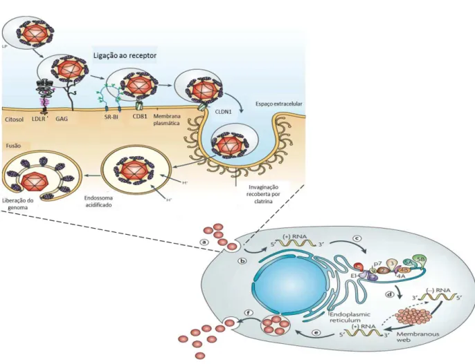
empacotada para constituir novos vírus (DE FRANCESCO et al., 2003). O esquema do ciclo replicativo do HCV está representado na figura 3.
Figura 3: Ciclo Replicativo do HCV. (a) Internalização do vírus na célula. (b) Liberação citoplasmática. (c) Tradução e processamento da poliproteína. (d) Replicação do RNA viral. (e) Montagem da partícula viral. (f) Maturação e liberação do vírions. Fonte: Adaptada de MORADPOUR, D.; PENIN, F.; RICE, C.M., 2007.
1.2.4 Modelos para o estudo do HCV in vitro
sistema de replicação in vitro com a construção de replicons sub-genômicos capazes de se auto-replicar em culturas de células de hepatoma humano (Huh-7) (LOHMANN et al., 1999). Replicons são construções genômicas e sub-genômicas que expressam o complexo de replicase viral e são capazes autonomamente de realizar a replicação viral (DUVERLIE; WYCHOWSKI, 2007). Os primeiros replicons subgenômicos eram derivados do clone CON1, genótipo 1b, e, eram moléculas de RNA bicistrônicas que possuíam o gene da neomicina fosfotransferase (NPT) e os genes não estruturais, codificantes das proteína requeridas para a replicação do RNA do HCV (NS3-NS5B) (SGR-Neo-Con1) (LOHMANN et al., 1999).
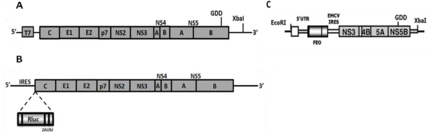
Com o avanço do conhecimento no desenvolvimento de replicons, foram desenvolvidas algumas variações do genoma original do clone CON1 e mais tarde clones provenientes do genótipo 2 foram desenvolvidos. O replicon subgenômico SGR-JFH-FEO é um replicon subgenômico bicistrônico que contém os genes não estruturais do clone do HCV genótipo 2a (JFH-1) (WAKITA et al., 2005) e que possui o gene Firefly Luciferase associado a um gene de resistência à neomicina (FEO) (WYLES et al., 2009) (Figura 4a).
(SIMISTER et al., 2009). Existem ainda quimeras associando genomas completos do HCV, a exemplo do FL-J6/JFH-5′C19Rluc2AUbi, um genoma monocistrônico completo do HCV que corresponde a uma quimera dos clones J6 e JFH-1 derivado de um sistema infeccioso previamente descrito do genótipo 2a denominado J6/JFH (LINDENBACH et al., 2005) associado ao gene de Renilla Luciferase (RLuc) (Figura 4b) (TSCHERNE et al., 2006)
A linhagem celular que apresenta-se atualmente mais permissiva à infecção por partículas de HCV derivada de cultura de células (HCVcc), é a linhagem Huh-7.5. Essa corresponde a uma linhagem de hepatoma humano derivada da linhagem Huh-7, a qual expressava estavelmente um replicon genótipo 1b e que foi tratada e curada da infecção por IFN-α. Estudos posteriores ao tratamento demonstraram que essa linhagem era defectiva do gene RIG-1, um dos responsáveis pela ativação da via do IFN (SUMPTER et
al., 2005) e é mais permissiva a reinfecção por RNA do HCV (BLIGHT, MCKEATING; RICE, 2002).
Figura 4: Representação esquemática dos sistemas de replicação utilizados como modelos de estudo in vitro do HCV. A) JFH-1: sistema de genoma completo do HCV genótipo 2a, primeiro replicon completo desenvolvido para o HCV (Fonte: Adaptada de WAKITA et al., 2005). B) FL-J6/JFH-5′C19Rluc2AUbi: sistema de genoma completo do HCV genótipo 2a, associado ao
1.2.5 Vias de transmissão e tratamentos disponíveis
O vírus da hepatite C infecta principalmente os hepatócitos e, diferente dos demais vírus que causam hepatite, não gera resposta imune adequada, tornando a doença aguda menos sintomática (HOOFNAGLE, 2002). Esta característica particular do HCV faz com que a grande maioria dos indivíduos infectados evolua para um quadro crônico, que leva ao desenvolvimento de cirrose hepática e carcinoma hepatocelular (WHO, 2013).
O vírus é transmitido eficientemente por exposição percutânea direta ao sangue contaminado e a via de transmissão mais comum antes da identificação do vírus eram os eventos de transfusões sanguíneas (CDC, 1998). Atualmente a principal via de transmissão do vírus relaciona-se ao compartilhamento de seringas entre usuários de drogas injetáveis (WHO, 2013; CDC, 2014).
O tratamento convencional baseia-se na utilização de interferon alfa (IFN-α)
tratamento é evidente, contudo os efeitos da ribavirina só são efetivos na terapia conjunta com o INF-α (ROFFI et al.,2008). Acredita-se que a ribavirina possa agir induzindo genes ativados pelo interferon (Interferon-stimulated
genes –ISG) como os fatores 7 e 9, atuando sinergicamente durante a terapia convencional (THOMAS et al., 2011).
de mutações virais que conferem resistência a esses medicamentos (HALFON; LOCARNINI, 2011).
1.3 Fitoquímicos de ação terapêutica e cafeína
O uso de produtos naturais na fabricação de drogas é uma prática antiga e bem estabelecida, e o princípio ativo de várias drogas comuns no dia-a-dia foram identificadas a partir de produtos naturais (TZIVELEKA; VAGIAS; ROUSSIS, 2003). Dentre vários produtos naturais com ação antiviral ainda em fase de teste destacam-se contra o vírus da hepatite C, o extrato da raiz e de folhas do gênero Phyllanthus, que interagem com a NS3 e NS5B do HCV respectivamente (RAVIKUMAR et al., 2011). Algumas substâncias provenientes de plantas já demonstraram possuir algum efeito na inibição do HCV in vitro, dentre eles estão: o polifenol EGCG proveniente do chá verde que atua efetivamente em etapas da entrada viral (CIESEK et al., 2011), bem como epicatequinas fenólicas atuando na replicação viral (LIN et al., 2013), o flavonóide BJ486K proveniente de Marrubium peregrinum L (Lamiaceae) que demonstrou efetividade contra diversos genótipos do vírus, atuando no bloqueio da entrada viral (HAID et al., 2012), bem como os flavonoides provenientes de extratos de Embelia ribes os quais interagem diretamente com a NS3 do HCV (BACHMETOV et al., 2011).
Em relação às várias terapêuticas em análise, o consumo do café (Coffea
consomem cerca de três xícaras de café diariamente (FREEDMAN et al., 2009), aproximadamente 408 mg de cafeína; (COSTENTIN et al., 2011). Além disso, o consumo de café foi significativamente associado à diminuição de morte por carcinoma (WAKAI et al., 2007), à redução de mortes em geral e um crescente número de estudos vem sugerindo que a cafeína possa ter diretamente um efeito hepatoprotetor (FREEDMAN et al., 2009; MODI et al., 2010; COSTENTIN et al., 2011; CHEN et al., 2014).
Modi et al. (2010) estabeleceram uma associação direta entre o consumo da cafeína, e a redução do desenvolvimento de fibrose hepática. Os resultados mostraram que o aumento do consumo de cafeína estava diretamente relacionado à diminuição do desenvolvimento de fibrose em pacientes portadores de doenças crônicas no fígado. Estes resultados foram particularmente expressivos para pacientes com infecção crônica pelo vírus da hepatite C.
proteína não-estrutural 5 (NS5A) (OKAMOTO et al., 2006; NAKAGAWA et al., 2007; BERAN et al., 2012); redução da fosforilação de algumas proteínas da via Ras-ERK, que interagem diretamente com a proteína NS5B e com a região 5’NTR viral (OKANO et al., 2008; YI et al., 2011; ZEITLIN et al., 2011); bem como já foi demonstrado que a cafeína é capaz de reduzir a fosforilação da ciclooxigenase 2 (COX-2), sendo esta associada frequentemente à capacidade replicativa do HCV (GRETTON; HUGHES; HARRIS, 2010; KANG et al., 2011; LEE et al., 2011; KANG et al., 2012; LIN et al., 2013).
A cafeína é um alcalóide identificado como 1,3,7,trimetilxantina, cuja estrutura contém um esqueleto de purina (DE MARIA; MOREIRA, 2007). Este alcalóide é encontrado em vários tipos vegetais como a semente do café (Coffea sp.), onde aparece em grande quantidade; nas folhas de chá verde (Camilla sinensis) e em outros produtos vegetais como o guaraná (Paullinia cupana), a erva-mate (Ilex paraguayensis) (BUCCI, 2000) e o cacau (Theobroma cocoa) (CAUDLE et al., 2001). Possui efeito geralmente associado a alterações no sistema nervoso central sendo altamente psicoestimulante (FREEDHOLM et al., 1999). Apesar de terem sido estabelecidas relações inversamente proporcionais entre a cafeína e a evolução de algumas doenças hepáticas como carcinoma hepatocelular (HOSAKA et al., 2001), cirrose (RUHL; EVERHART, 2005) e fibrose (MODI et
2 JUSTIFICATIVA
A hepatite C é uma infecção que frequentemente se apresenta na forma
crônica e atinge cerca de 3 % da população mundial. Comumente evolui para uma
doença hepática progressiva causando em última instância cirrose e
hepatocarcinoma, sendo um dos principais promotores de transplante hepático no
mundo. O tratamento atual é feito com interferon e ribavirina, porém a resposta
virológica sustentada é de apenas 40 % para portadores do genótipo 1, o mais
prevalente no Brasil. As novas estratégias terapêuticas como boceprevir e
telaprevir tem um alto custo e efeitos colaterais graves. Esse quadro demonstra a
urgente necessidade de terapias mais eficientes contra o HCV.
A cafeína mostra nesse contexto alto potencial, seja do ponto do bem estar
paciente/tratamento, seja do ponto de vista econômico uma vez, que afeta vias
celulares que melhoram a função de enzimas hepáticas e já demonstrou efeitos na
desaceleração da evolução de doenças hepática. Além disto seu consumo foi
relacionado a quadros mais brandos de doenças hepáticas frequentemente
associadas ao HCV. Também, como um fitoquímico encontrado em diversos tipos
vegetais e de isolamento simples e rápido, poderia representar um tratamento
mais econômico e ainda podendo ser complementado como enriquecimento na
dieta dos pacientes.
Embora já tenha sido relacionada à melhora nas vias metabólicas
hepáticas, com destaque para pacientes portadores do HCV, não existem relações
diretas entre este fitoquímico e o ciclo replicativo do HCV. Além disso, por
apresentar interferência em vias celulares utilizadas na entrada e liberação viral, o
estudo do efeito da cafeína no ciclo completo do vírus poderia ampliar o potencial
3 OBJETIVOS
3.1 Objetivos gerais
Avaliar o efeito da cafeína em diferentes etapas do ciclo replicativo do HCV em um sistema permissível a infecção pelo replicon completo do HCV e determinar a eficiência da droga.
3.2 Objetivos específicos
- Estabelecer uma linhagem estável expressando o replicon subgenômico do HCV, SGR-JFH-FEO;
- Estabelecer cultura de células infectadas com o vírus da Hepatite C genótipo 2a e estoque de sobrenadante infeccioso;
- Avaliar o efeito da cafeína na replicação do vírus da hepatite C
- Avaliar a influência da cafeína sobre diferentes mecanismos de morte celular
- Determinar o IC50 do tratamento com cafeína para fins comparativos com outras drogas
4 REFERÊNCIAS BIBLIOGRÁFICAS
ASHFAQ, U. A. et al. An overview of HCV molecular biology, replication and immune responses. Virol J, v. 8, p. 161, 2011.
BACHMETOV L, GAL-TANAMY M, SHAPIRA A, VOROBEYCHIK M, GITERMAN-GALAM T, SATHIYAMOORTHY P, GOLAN-GOLDHIRSH A, BENHAR I, TUR-KASPA R, ZEMEL R. Suppression of hepatitis C virus by the flavonoid quercetin is mediated by inhibition of NS3 protease activity. J. Viral
Hepat. v.19, p. e81–e88, 2012.
BARTENSCHLAGER, R.; LOHMANN, V. Replication of hepatitis C virus. J Gen Virol, v. 81, n. Pt 7, p. 1631-48, Jul 2000.
BARTENSCHLAGER, R.; LOHMANN, V.; PENIN, F. The molecular and structural basis of advanced antiviral therapy for hepatitis C virus infection. Nat Rev Microbiol, v. 11, n. 7, p. 482-96, Jul 2013.
BERAN, R. K. et al. Cellular growth kinetics distinguish a cyclophilin inhibitor from an HSP90 inhibitor as a selective inhibitor of hepatitis C virus. PLoS One, v. 7, n. 2, p. e30286, 2012.
BLIGHT, K. J.; MCKEATING, J. A.; RICE, C. M. Highly permissive cell lines for subgenomic and genomic hepatitis C virus RNA replication. J Virol, v. 76, n. 24, p. 13001-14, Dec 2002.
BRASIL. Agência Nacional de Vigilância Sanitária/ ANVISA. Disponível em < http://portal.anvisa.gov.br>. Acessado em 07/04/2012.
BUCCI, L. R. Selected herbals and human exercise performance. Am J Clin Nutr, v. 72, n. 2 Suppl, p. 624S-36S, Aug 2000
BUKH, J. et al. Challenge pools of hepatitis C virus genotypes 1-6 prototype strains: replication fitness and pathogenicity in chimpanzees and human liver-chimeric mouse models. J Infect Dis, v. 201, n. 9, p. 1381-9, May 1 2010. CADDEN, I. S.; PARTOVI, N.; YOSHIDA, E. M. Review article: possible beneficial effects of coffee on liver disease and function. Aliment Pharmacol Ther, v. 26, n. 1, p. 1-8, Jul 1 2007.
CAMPIOTTO, S. et al. Geographic distribution of hepatitis C virus genotypes in Brazil. Braz J Med Biol Res, v. 38, n. 1, p. 41-9, Jan 2005.
CAUDLE, A. G.; YIFANG, G.; BELL, L. N. Improved analysis of theobromine and caffeine in chocolate food products formulated with cocoa powder. Food Res. Int., v. 34, p. 599 - 603, 2001.
CDC. Centers for Disease Control And Prevention, 2014
Disponível em: < http://www.cdc.gov/hepatitis/ChooseC.htm > Acesso em 13/03/14
CHANDLER DE, PENIN F, SCHULTEN K, CHIPOT C. The p7 protein of hepatitis C virus forms structurally plastic, minimalist ion channels. PLoS
Comput Biol., v. 8, n. 9, p. e1002702. doi: 10.1371/journal.pcbi.1002702.
Epub, 2012.
CHEN, S. et al. Coffee and non-alcoholic fatty liver disease: Brewing evidence for hepatoprotection? J Gastroenterol Hepatol, v. 29, n. 3, p. 435-41, Mar 2014.
CHEVALIEZ, S.; PAWLOTSKY, J. M. Hepatitis C virus: virology, diagnosis and management of antiviral therapy. World J Gastroenterol, v. 13, n. 17, p. 2461-6, May 7 2007.
CHOO, Q.L.; KUO, G.; WEINER, A.J.; OVERBY, L.R; BRADLEY, D.W.; HOUGHTON, M. Isolation of a cDNA clone derived from a bloodborne non-A, non-B viral hepatitis genome. Science, v.244, p.359-62, 1989.
CHOO, Q.L.; RICHMAN, K.H.; HAN, J.H.; BERGER, K.; LEE, C.; DONG, C.; GALLEGOS, C.; COIT, D.; MEDINA-SELBY, A.; BARR, P.J.; WEINER, A.J. BRADLEYT, D.W.; KUO, G.; HOUGHTON, M. Genetic organization and diversity of hepatitis C virus. Proc Natl Acad Sci USA, v.88, p.2451-5, 1991. CIESEK, S. et al. The green tea polyphenol, epigallocatechin-3-gallate, inhibits hepatitis C virus entry. Hepatology, v. 54, n. 6, p. 1947-55, Dec 2011.
COSTENTIN, C. E. et al. Association of caffeine intake and histological features of chronic hepatitis C. J Hepatol, v. 54, n. 6, p. 1123-9, Jun 2011. DAVIS, G. L. Hepatitis C virus genotypes and quasispecies. Am J Med, v. 107, n. 6B, p. 21S-26S, Dec 27 1999.
DE MARIA, C.A; MOREIRA, R.F. Cafeína: revisão sobre métodos de análise. Quim Nova, v.31, n.1, p. 99-105, 2007.
DE FRANCESCO, R. et al. Approaching a new era for hepatitis C virus therapy: inhibitors of the NS3-4A serine protease and the NS5B RNA-dependent RNA polymerase. Antiviral Res, v. 58, n. 1, p. 1-16, Mar 2003. DI BISCEGLIE, A. M.; HOOFNAGLE, J. H. Optimal therapy of hepatitis C. Hepatology, v. 36, n. 5 Suppl 1, p. S121-7, Nov 2002.
DUVERLIE, G.; WYCHOWSKI, C. Cell culture systems for the hepatitis C virus. World J Gastroenterol, v. 13, n. 17, p. 2442-5, May 7 2007.
distinct membrane alterations including a candidate viral replication complex. J Virol, v. 76, p.5974-84, 2002.
FARCI D. New insights into the HCV quasispecies and compartmentalization. Semin Liver Dis., v. 31, n.4, p. 356-74 doi: 10.1055/s-0031-1297925. Epub, 2011.
FAUVELLE, C.; FELMLEE, D. J.; BAUMERT, T. F. Unraveling hepatitis C virus structure. Cell Res, Mar 14 2014.
FARCI et al. New Insights into the HCV Quasispecies and Compartmentalization. Semin Liver Dis., Dec 21 2011.
FELD, J. J.; HOOFNAGLE, J. H. Mechanism of action of interferon and ribavirin in treatment of hepatitis C. Nature, v. 436, n. 7053, p. 967-72, Aug 18 2005. FREEDHOLM, B.B.; BATTIG, K.; HOLMEN, J.; NEHLIG, A.; ZVARTAU, E.E. Actions of Caffeine in the Brain with Special Reference to Factors That Contribute to Its Widespread Use. Pharmacological Reviews, v. 51, n.1, p. 83-133, 1999.
FREEDMAN, N. D. et al. Coffee intake is associated with lower rates of liver disease progression in chronic hepatitis C. Hepatology, v. 50, n. 5, p. 1360-9, Nov 2009.
FRIEBE, P.; LOHMANN, V.; KRIEGER, N.; BARTENSCHLAGER, R.Sequences in the 5′nontranslated region of hepatitis C virus required for RNA replication. J. Virol, v. 75, p. 12047-57, 2001.
GIANNINI, C.; BRECHOT, C. Hepatitis C virus biology. Cell Death Differ, v. 10 Suppl 1, p. S27-38, Jan 2003.
GRETTON, S.; HUGHES, M.; HARRIS, M. Hepatitis C virus RNA replication is regulated by Ras-Erk signalling. J Gen Virol, v. 91, n. Pt 3, p. 671-80, Mar 2010.
HAID S, NOVODOMSKÁ A, GENTZSCH J, GRETHE C, GEUENICH S, BANKWITZ D, CHHATWAL P, JANNACK B, HENNEBELLE T, BAILLEUL F, KEPPLER OT, POENISCH M,BARTENSCHLAGER R, HERNANDEZ C, LEMASSON M, ROSENBERG AR, WONG-STAAL F, DAVIOUD-CHARVET E, PIETSCHMANN T. A plant-derived flavonoid inhibits entry of all HCV genotypes into human hepatocytes. Gastroenterology, v. 143, n.1, p. 213-22.e5. doi: 10.1053/j.gastro.2012.03.036. 2012.
HALFON, P.; LOCARNINI, S. Hepatitis C virus resistance to protease inhibitors. J Hepatol, v. 55, n. 1, p. 192-206, Jul 2011.
HOSAKA, S.; KAWA, S.; AOKI, Y.; TANAKA, E.; YOSHIZAWA, K.; KARASAWA, Y.; HOSAKA, N.; KIYOSAWA, K. Hepatocarcinogenesis inhibition by caffeine in ACI rats treated with acetylaminofluorene. Food Chem Toxicol, v. 39, p. 557-561, 2001.
HWANG, J. et al. Hepatitis C virus nonstructural protein 5A: biochemical characterization of a novel structural class of RNA-binding proteins. J Virol, v. 84, n. 24, p. 12480-91, Dec 2010.
ICTV – International Committee on Taxonomy Viruses, 2013. Disponível em: <http://ictvonline.org/virusTaxonomy.asp> acessado em: 31/03/14
JAMES, A. Ilustração do vírus da Hepatite C. Disponível em: <http://www.rit.edu/~japfaa/hcv.jpg>. Acessado em outubro, 2010.
KANG, C. H. et al. Caffeine suppresses lipopolysaccharide-stimulated BV2 microglial cells by suppressing Akt-mediated NF-kappaB activation and ERK phosphorylation. Food Chem Toxicol, v. 50, n. 12, p. 4270-6, Dec 2012.
KANG, N. J. et al. Coffee phenolic phytochemicals suppress colon cancer metastasis by targeting MEK and TOPK. Carcinogenesis, v. 32, n. 6, p. 921-8, Jun 2011.
KAWANO Y, NAGATA M, KOHNO T, ICHIMIYA A, IWAKIRI T, OKUMURA M, ARIMORI K. Caffeine increases the antitumor effect of Cisplatin in human hepatocellular carcinoma cells. Biol Pharm Bull.; n. 35, v.3, p. 400-7, 2012. KERSHENOBICH, D. et al. Trends and projections of hepatitis C virus epidemiology in Latin America. Liver Int, v. 31 Suppl 2, p. 18-29, Jul 2011. KIM, A. I.; SAAB, S. Treatment of Hepatits C. Am J Med, v.118, n.8, p. 808-15, 2005.
KUIKEN, C.; SIMMONDS, P. Nomenclature and numbering of the hepatitis C virus. Methods Mol Biol, v. 510, p. 33-53, 2009.
LAVANCHY, D. Evolving epidemiology of hepatitis C virus. Clin Microbiol Infect, v. 17, n. 2, p. 107-15, Feb 2011.
LEE, J. C. et al. Anti-hepatitis C virus activity of Acacia confusa extract via suppressing cyclooxygenase-2. Antiviral Res, v. 89, n. 1, p. 35-42, Jan 2011. LIN, Y. T. et al. Green tea phenolic epicatechins inhibit hepatitis C virus replication via cycloxygenase-2 and attenuate virus-induced inflammation. PLoS One, v. 8, n. 1, p. e54466, 2013.
LINDENBACH, B. D.; RICE, C. M. Unravelling hepatitis C virus replication from genome to function. Nature, v. 436, n. 7053, p. 933-8, Aug 18 2005.
LOHMANN, V. et al. Replication of subgenomic hepatitis C virus RNAs in a hepatoma cell line. Science, v. 285, n. 5424, p. 110-3, Jul 2 1999.
MACDONALD, A.; CROWDER, K.; STREET, A.; MCCORMICK, C.; HARRIS, M. The hepatitis C virus NS5A protein binds to members of the Src family of tyrosine kinases and regulates kinase activity. Journal of General Virology,v. 85, p.721-729, 2004.
MACDONALD, A.; HARRIS, M. Hepatitis C virus NS5A: tales of a promiscuous protein. J Gen Virol, v. 85, n. Pt 9, p. 2485-502, Sep 2004.
MANNS, M. P. et al. Peginterferon alfa-2b plus ribavirin compared with interferon alfa-2b plus ribavirin for initial treatment of chronic hepatitis C: a randomised trial. Lancet, v. 358, n. 9286, p. 958-65, Sep 22 2001.
MATSUURA, Y.; MIYAMURA, T. The molecular biology of hepatitis C virus. Seminars in VIROLOGY, v.4, p. 297-304, 1993.
MAUSS, BERG, ROCKSTROH, SARRAZIN,WEDEMEYER, et al., Hepatology: A clinical textbook. Fifth Edition, 2014.
MODI, A. A. et al. Increased caffeine consumption is associated with reduced hepatic fibrosis. Hepatology, v. 51, n. 1, p. 201-9, Jan 2010.
MORADPOUR, D.; PENIN, F.; RICE, C. M. Replication of hepatitis C virus. Nat Rev Microbiol, v. 5, n. 6, p. 453-63, Jun 2007.
NAKAGAWA, S. et al. Hsp90 inhibitors suppress HCV replication in replicon cells and humanized liver mice. Biochem Biophys Res Commun, v. 353, n. 4, p. 882-8, Feb 23 2007.
NEGRO, F.; ALBERTI, A. The global health burden of hepatitis C virus infection. Liver Int, v. 31 Suppl 2, p. 1-3, Jul 2011.
OKAMOTO, T. et al. Hepatitis C virus RNA replication is regulated by FKBP8 and Hsp90. EMBO J, v. 25, n. 20, p. 5015-25, Oct 18 2006.
OKANO, J. et al. Caffeine inhibits the proliferation of liver cancer cells and activates the MEK/ERK/EGFR signalling pathway. Basic Clin Pharmacol Toxicol, v. 102, n. 6, p. 543-51, Jun 2008.
PAWLOTSKY, J. M. Hepatitis C virus genetic variability: pathogenic and clinical implications. Clin Liver Dis, v. 7, n. 1, p. 45-66, Feb 2003.
PENIN, F. et al. Structural biology of hepatitis C virus. Hepatology, v. 39, n. 1, p. 5-19, Jan 2004.
RAVIKUMAR, Y. S. et al. Inhibition of hepatitis C virus replication by herbal extract: Phyllanthus amarus as potent natural source. Virus Res, v. 158, n. 1-2, p. 89-97, Jun 2011.
ROTHWANGL, K. B.; RONG, L. Analysis of a conserved RGE/RGD motif in HCV E2 in mediating entry. Virol J, v. 6, p. 12, 2009.
RUHL, C.E. EVERHART, J.E. Coffee and tea consumption are associated with a lower incidence of chronic liver disease in the United States. Gastroenterology; v.129, p.1928 1936, 2005.
SCHREGEL, V. et al. Hepatitis C virus NS2 is a protease stimulated by cofactor domains in NS3. Proc Natl Acad Sci U S A, v. 106, n. 13, p. 5342-7, Mar 31 2009.
SIMISTER, P. et al. Structural and functional analysis of hepatitis C virus strain JFH1 polymerase. J Virol, v. 83, n. 22, p. 11926-39, Nov 2009.
SIMMONDS, P. Genetic diversity and evolution of hepatitis C virus--15 years on. J Gen Virol, v. 85, n. Pt 11, p. 3173-88, Nov 2004.
STEINMANN, E.; PENIN, F.; KALLIS, S.; PATEL, A.H.; BARTENSCHLAGER, R.; PIETSCHMANN, T. Hepatitis C virus p7 protein is crucial for assembly and release of infectious virions. PLoS Pathog, v.3 p.e103, 2007.
SUMPTER, R., JR. et al. Regulating intracellular antiviral defense and permissiveness to hepatitis C virus RNA replication through a cellular RNA helicase, RIG-I. J Virol, v. 79, n. 5, p. 2689-99, Mar 2005.
SUZUKI, R. et al. Molecular determinants for subcellular localization of hepatitis C virus core protein. J Virol, v. 79, n. 2, p. 1271-81, Jan 2005.
TANG, H.; GRISE, H. Cellular and molecular biology of HCV infection and hepatitis. Clin Sci (Lond), v. 117, n. 2, p. 49-65, Jul 2009.
TAO, Z. et al. Quantitative measure of cytotoxicity of anticancer drugs and other agents. Anal Biochem, v. 381, n. 1, p. 43-52, Oct 1 2008.
THIMME, R.; BINDER, M.; BARTENSCHLAGER, R. Failure of innate and adaptive immune responses in controlling hepatitis C virus infection. FEMS Microbiol Rev, v. 36, n. 3, p. 663-83, May 2012.
TSCHERNE, D. M. et al. Time- and temperature-dependent activation of hepatitis C virus for low-pH-triggered entry. J Virol, v. 80, n. 4, p. 1734-41, Feb 2006.
TZIVELEKA, L.A; VAGIAS, C.; ROUSSIS, V. Natural products with anti-HIV activity from marine organisms. Curr Top Med Chem, v. 3, p. 1512-35, 2003 VIEYRES, G.; DUBUISSON, J.; PIETSCHMANN, T. Incorporation of hepatitis C virus e1 and e2 glycoproteins: the keystones on a peculiar virion. Viruses, v. 6, n. 3, p. 1149-87, 2014.
WAKAI, K. et al. Liver cancer risk, coffee, and hepatitis C virus infection: a nested case-control study in Japan. Br J Cancer, v. 97, n. 3, p. 426-8, Aug 6 2007.
WAKITA, T. et al. Production of infectious hepatitis C virus in tissue culture from a cloned viral genome. Nat Med, v. 11, n. 7, p. 791-6, Jul 2005.
WEINER, A. J. et al. Sequence variation in hepatitis C viral isolates. J Hepatol, v. 13 Suppl 4, p. S6-14, 1991.
WHO, world Health Organization, 2013.
Disponível em: < http://www.who.int/mediacentre/factsheets/fs164/en/> Acesso em 13/03/14
WOHNSLAND, A.; HOFMANN, W. P.; SARRAZIN, C. Viral determinants of resistance to treatment in patients with hepatitis C. Clin Microbiol Rev, v. 20, n. 1, p. 23-38, Jan 2007.
WOZNIAK, A. L. et al. Intracellular proton conductance of the hepatitis C virus p7 protein and its contribution to infectious virus production. PLoS Pathog, v. 6, n. 9, p. e1001087, 2010.
WYLES, D. L. et al. The octadecyloxyethyl ester of (S)-9-[3-hydroxy-2-(phosphonomethoxy) propyl]adenine is a potent and selective inhibitor of hepatitis C virus replication in genotype 1A, 1B, and 2A replicons. Antimicrob Agents Chemother, v. 53, n. 6, p. 2660-2, Jun 2009.
YI, Z. et al. Hepatitis C virus co-opts Ras-GTPase-activating protein-binding protein 1 for its genome replication. J Virol, v. 85, n. 14, p. 6996-7004, Jul 2011.
ZEITLIN, R. et al. Caffeine induces beneficial changes in PKA signaling and JNK and ERK activities in the striatum and cortex of Alzheimer's transgenic mice. Brain Res, v. 1417, p. 127-36, Oct 12 2011.
1 ARTIGO CIENTÍFICO 1
Caffeine inhibits Hepatitis C virus replication in vitro
Mariana N. Batistaa, Bruno M. Carneiroa, Ana Cláudia S. Bragaa, Paula Rahala*
aDepartment of Biology, Institute of Bioscience, Language & Literature and Exact
Science, São Paulo State University, IBILCE - UNESP, São José do Rio Preto, SP, Brazil
* Corresponding author at: Departamento de Biologia, Instituto de Biociências, Letras e Ciências Exatas - Universidade Estadual Paulista, Rua Cristóvão Colombo 2265, 15054-000 São José do Rio Preto-SP, Brazil. Tel.: +55 17 32212390; fax: +55 17
32212390. E-mail address:prahal@ibilce.unesp.br; brunocopo@yahoo.com.br
Abstract
Hepatitis C is considered the major cause of cirrhosis and hepatocellular carcinoma. Conventional treatment is not effective against some hepatitis C virus (HCV) genotypes; therefore, new treatments are needed. Coffee and more recently, caffeine, have been found to have a beneficial effect in several disorders of the liver, including those manifesting abnormal liver biochemistry, cirrhosis and hepatocellular carcinoma. Caffeine acts directly by delaying fibrosis, thereby improving the function of liver cellular pathways and interfering with pathways used by the HCV replication cycle. In the current study, the direct relationship between caffeine and viral replication was evaluated. The Huh-7.5 cell line was used for transient infections with FL-J6/JFH-5′C19Rluc2AUbi and to establish a cell line stably expressing SGR-Feo JFH-1. Caffeine efficiently inhibited HCV replication in a dose-dependent manner at non-cytotoxic concentrations and demonstrated an IC50 value of 0.7263 mM after 48 h of incubation. These data demonstrate that caffeine may be an important new agent for anti-HCV therapies due to its efficient inhibition of HCV replication at non-toxic concentrations.
Keywords:
Introduction
Hepatitis C is a liver infection caused by hepatitis C virus (HCV) [5]. Usually, infection does not generate an adequate host immune response [13] and it progresses to a chronic infection in approximately 80 % of patients [18]. HCV infection is a global public health problem and has been associated with 350,000 annual deaths related to cirrhosis and hepatocellular carcinoma [4]. Pegylated interferon alpha (PEG-INF) in association with ribavirin (RBV) has been the standard treatment for patients with chronic HCV infection in last decade [35]. The new standard treatment includes the protease inhibitors, boceprevir and telaprevir, in association with PEG-IFN and RBV [28]. However, severe side effects are observed, the treatment is costly and viral resistance has been demonstrated for all classes of directly acting antivirals [17]. Therefore, new treatments are needed.
Coffee and caffeine consumption has been associated with beneficial effects in patients with abnormal liver function, cirrhosis and hepatocellular carcinoma [3, 6, 10]. The daily consumption of approximately 3 cups of coffee [10], which is approximately 408 mg of caffeine [6], may reduce the risk of chronic liver disease progression. Caffeine has also been demonstrated to have an antiproliferative effect on liver cancer cells [27]. Moreover, caffeine consumption may limit liver fibrosis, especially in patients with chronic HCV infection (14 % milder than other fibrosis groups) [23].
Materials and Methods
HCV Replicons
The replicons used in the study were as follows: FL-J6/JFH-5′C19Rluc2AUbi, a monocistronic full-length HCV genome that expresses Renilla luciferase (Rluc) [33], derived from the previously described infectious genotype 2a of the HCV genome, J6/JFH1 [21]; and SGR-Feo JFH-1, a bicistronic subgenomic replicon based on the JFH-1 sequence that possesses the firefly luciferase gene fused to a neomycin resistance gene [37]. RNA was transcribed from the plasmids (pFL-J6/JFH-5′C19Rluc2AUbi J6-JFH1 RLUC and pSGR-Feo JFH-1) after linearization with the XbaI enzyme and Mung bean treatment followed by phenol/chloroform extraction [43]. The purified product was used as a template for transcription with the T7 RiboMAXTM Express Large Scale RNA Production System (Promega, Madison – WI, USA).
Cell Culture
The human hepatoma cell line (Huh-7.5) was maintained in DMEM medium supplemented with 10 % FBS (Cutilab, Campinas – SP, BR), 1 % (v/v) non-essential amino acids (Gibco – Life Technologies, USA), 100 U/mL penicillin (Gibco – Life Technologies, USA), 100 mg/mL streptomycin (Gibco – Life Technologies, USA) and 1 % HEPES (Gibco – Life Technologies, USA) in a humidified 5 % CO2 incubator. The Huh-7.5 cell line stably expressing SGR-Feo JFH-1 was maintained in DMEM supplemented with 500 µg/mL G418 (Sigma-Aldrich, St. Louis - MO, USA).
HCV RNA Transfection
obtained after G418 selection of electroporated cells (Sigma-Aldrich, St. Louis - MO, USA) after at least three weeks of culture.
Caffeine Cytotoxicity Profile
The cytotoxicity of caffeine (Sigma-Aldrich, St. Louis - MO, USA) was determined by the MTT (3-(4,5-dimethylthiazol-2-yl)-2,5-diphenyl tetrazolium bromide) assay (Sigma-Aldrich, St. Louis - MO, USA). Cytotoxicity was assessed after the cells were incubated for 24 h, 48 h and 72 h with caffeine. Cell cycle analysis by flow cytometry
Prior to start this assay, cells were synchronized in G0 by FBS deprivation for 24 h. To determine cell cycle pattern of caffeine-treated cells, synchronized Huh-7.5 cells stably expressing SGR-Feo JFH-1 were seeded on a 96-well plate (5x10³cells/well) 24 h before treatment. Caffeine was then added at the following concentrations: 0; 0.250 mM; 0.5 mM; 1 mM; 2 mM and 4 mM. One set of cells was maintained without serum to serve as positive control of cell cycle arrest in G0. Forty eight hours post-treatment, cells were resuspended, counted and fixed overnight with cold ethanol 75 %. On the following day, cells were stained with Guava Cell Cycle Reagent following the manufacturer’s instructions (EMD Millipore Corporation, Hayward,CA - EUA). Subsequently, cells were analyzed using easyCyte 5HT flow cytometry (Guava Easycheck, Millipore Corporation, Hayward,CA - EUA) with 10,000 events acquired from each sample.
Apoptosis fluorescence staining
microscope (Zeiss Axio Vert A1). All cells from 20 different fields were counted and cell count was compared with negative control (zero caffeine).
Autophagy fluorescence staining
To analyze caffeine influence on nonapoptotic programmed cell death, Huh-7.5 cells stably expressing SGR-Feo JFH-1 were seeded on 24-well plate (1x10⁴ cells/well) 24 h before treatment. Caffeine was then added at the following concentrations: 0; 0.250 mM; 0.5 mM; 1 mM; 2 mM and 4 mM. Curcumin (20 µM) was used as positive control of autophagy [7, 29]. Forty eight hours post treatment, cells were incubated with Acridine Orange (500 ug/mL) for 15 minutes. The analysis was performed during 45 minutes after acridine addition to avoid acridine-induced cell death. All cells from 20 different fields were counted and cell count was compared with negative control (no caffeine).
Indirect Immunofluorescence assay
Cells were fixed with 4 % paraformaldehyde and permeabilized with 0.1 % triton-X-100. The cells were then incubated with a primary polyclonal sheep antibody against NS5A protein (kindly provided by Professor Mark Harris, University of Leeds) at a dilution of 1:4,000 followed by incubation with a 1:500 dilution of an Alexa Fluor® 594-conjugated donkey anti-sheep IgG (H+L) (Life Technologies, Carlsbad – CA, USA) both for 2 h at room temperature.
Virus Titration
Cell supernatants were serially diluted 10-fold in complete DMEM and used to infect 5x10³ Huh-7.5 cells per well in 96-well plates. The inoculum was incubated with the cells for 48 h at 37°C and then fixed with 4 % paraformaldehyde. HCV infection was determined by immunofluorescent staining for the HCV NS5A. The viral titer was expressed as focus-forming units per milliliter of supernatant (FFU/mL), determined by calculating the average number of NS5A-positive foci present at the highest dilutions.
Luciferase reporter assay
96-well plate (5x10 cells/well) 24 h before treatment. Caffeine was then added at the following concentrations: 10 mM; 3.17 mM; 1 mM; 0.317 mM; 0.1 mM; 0.0317 mM; 0.01 mM; 0.00317 mM; 0.001 mM; 0.000317 mM. The cells were incubated at 37ºC in a humidified atmosphere with 5 % CO2 for 48 h. Cyclosporine A (Sigma-Aldrich, St. Louis - MO, USA) was used as a positive control for inhibition, and milli-Q water (diluent of caffeine solutions) was used as a negative control for inhibition. After treatment, the cells were disrupted with 30 µL of passive lysis buffer (Promega, Madison - WI, USA) and exposed to a luciferase substrate (Promega, Madison - WI, USA). Luciferase activity was measured using a luminometer (FLUOstar Omega/BMG Labtech, Offenberg - BWL, DE), and a BCATM protein assay kit (Thermo-Scientific, Rockford - IL, USA) was used to normalize protein concentrations.
Protein expression analysis
Statistical methods
The half-maximal inhibitory concentration of caffeine (IC50) was calculated using a linear regression. The results of the inhibition of SGR-Feo JFH-1 were calculated as the percentage of the negative control (medium, no drug). All statistical analyses were performed by one-way ANOVA with Tukey’s post test using GraphPad Prism 5.0 software (GraphPad Software, San Diego - CA, USA).
Results
MTT
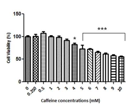
Caffeine cytotoxicity against Huh-7.5 cells containing SGR-Feo JFH-1 was determined by MTT assay. The solubility of caffeine in water is 16 mg/mL at room temperature, and we used this point of reference to test different concentrations in cell culture. We observed that cell viability was affected by caffeine in a time- and dose-dependent manner (Fig. 1a). When the cells were treated with caffeine concentrations above 2 mM for 48 h, the cell viability was close to 80 %. After 72 h of treatment, the cells reached this arbitrary cell viability cutoff at concentrations below 1 mM. Thus, we observed maximum viral inhibition at safe concentrations after 48 h; this time point was therefore selected for all subsequent experiments. Caffeine concentrations equal to or below 1 mM were safely tolerated by the cells, but a 2-fold increase (2 mM) reduced cell viability to approximately 80 % after 48 h. Therefore, 2 mM was the highest concentration utilized for further experiments.
Inhibitory effect of Caffeine on HCV replication
the luciferase activity by 82 ± 2 % (n=9) of the mock control values. Protein expression levels were also verified by western blot analysis of the NS3 virus protein (Fig. 2b). After 48 h of treatment, caffeine reduced viral protein expression by at least 70 % at the highest drug concentration.
HCVcc inhibition by caffeine
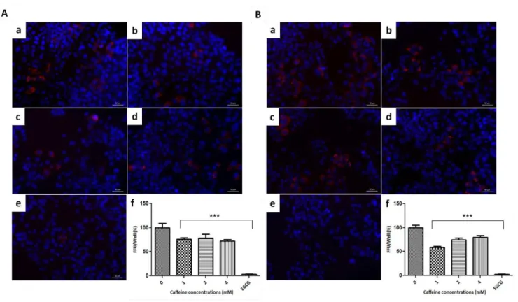
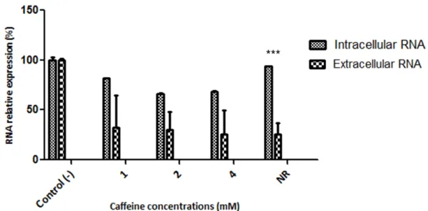
To verify whether caffeine could also interfere with full-length HCV replication, Huh-7.5 cells were infected with cell-culture derived HCV (HCVcc) at a multiplicity of infection of 0.1 and treated with different concentrations of caffeine. Similar to what was observed in the stable cell line, caffeine reduced viral replication in a dose-dependent manner (Fig. 2c). Using the drug at 2 mM, the intracellular levels of luciferase were reduced by 79.3 ± 9 % of the mock control, whereas at 0.5 mM, luciferase levels were reduced by 40.6 ± 13 % of the mock control. Analysis of the NS3 virus protein by western blot showed that after 48 h of treatment, viral protein expression was reduced by at least 60 % at the highest drug concentration (Fig. 2d).
Physiologic state analysis
To evaluate the influence of caffeine on the cell cycle, caffeine-treated cells were submitted to cell cycle assay by flow cytometry. We observed that both cells treated with or without caffeine kept the in division process, ranging between S or G2/M phases. The highest concentration used in inhibition assays (2 mM) presented 74,15 % of cells in division (S or G2/M) and caffeine free cells (negative control) presented 78 % of cells in division, thus there was no statistical significant differences among frequency of cell division. Cells maintained without serum were used as positive control of cell cycle arrest, and presented 52.45 % cell cycle arrest in G0/G1 while negative control presented 22 % of cells at this stage (Fig. 3).
apoptotic bodies was also considered as apoptosis. The percentage of apoptotic cells in the overall population upon caffeine treatment showed no significant increase of apoptosis related to negative control (zero caffeine). At Caffeine concentration of 2 mM, it was observed 9.91 % (relative to total analyzed cells) of apoptotic cells, while negative control presented 6.82 % of apoptosis (no statistical significant differences among groups were observed). Curcumin-treated cells (50 µM) were used as positive control of apoptosis and presented 62.2 % of apoptotic cells while negative control presented 6.82 % of total cells (Fig. 4).
Autophagy fluorescence staining
Aiming to clarify caffeine-induced cell death, the mechanism of autophagy was evaluated on different caffeine concentrations used to virus inhibition tests. Cells were considered in autophagy process when presented red corpuscles indicative of acid compartments [38] as mature autophagosomes or autophagolysosomes. The percentage of autophagic cells in the overall population in caffeine treatments showed no significant increase of autophagy related to negative control (zero caffeine). At Caffeine concentration of 2 mM, it was observed 69.7 % of autophagic cells; while negative control presented 51.8 % of autophagy (no statistical significant differences among groups were observed). Curcumin-treated cells (20 µM) were used as positive control of autophagy and presented 68.7 % of apoptotic cells while negative control presented 51.8 % of total cells (Fig. 5).
Discussion