Chronic caffeine intake decreases circulating catecholamines and prevents
diet-induced insulin resistance and hypertension in rats
Silvia V. Conde
1, Tiago Nunes da Silva
1,2, Constancio Gonzalez
3,4,5, Miguel Mota Carmo
2,
Emilia C. Monteiro
1and Maria P. Guarino
2*
1
Centro de Estudos de Doenc¸as Cro´nicas (CEDOC), Departamento de Farmacologia, Faculdade de Cieˆncias Me´dicas, Universidade Nova de Lisboa, Campo Ma´rtires da Pa´tria, 130, 1169-056 Lisbon, Portugal
2Centro de Estudos de Doenc¸as Cro´nicas (CEDOC), Departamento de Fisiopatologia, Faculdade de Cieˆncias Me´dicas,
Universidade Nova de Lisboa, Campo Ma´rtires da Pa´tria, 130, 1169-056 Lisbon, Portugal
3Departamento de Bioquı´mica y Biologı´a Molecular y Fisiologı´a, Universidad de Valladolid, Facultad de Medicina, 47005
Valladolid, Spain
4Instituto de Biologı´a y Gene´tica Molecular, Consejo Superior de Investigaciones Cientı´ficas (CSIC), 47005 Valladolid, Spain 5Ciber de Enfermedades Respiratorias, Centro de Investigacio´n Biome´dica en Red de Enfermedades Respiratorias (CIBERES),
Instituto de Salud Carlos III, 47005 Valladolid, Spain
(Received 5 October 2010 – Revised 17 February 2011 – Accepted 31 March 2011 – First published online 23 June 2011)
Abstract
We tested the hypothesis that long-term caffeine intake prevents the development of insulin resistance and hypertension in two pathologi-cal animal models: the high-fat (HF) and the high-sucrose (HSu) diet rat. We used six groups of animals: control; caffeine-treated (Caff; 1 g/l in drinking water during 15 d); HF; caffeine-treated HF (HFCaff); HSu; caffeine-treated HSu (HSuCaff). Insulin sensitivity was assessed using the insulin tolerance test. Blood pressure, weight gain, visceral fat, hepatic glutathione, plasma caffeine, insulin and NO, and serum NEFA and catecholamines were measured. Caffeine reversed insulin resistance and hypertension induced by both the HF and HSu diets. In the HF-fed animals caffeine treatment restored fasting insulin levels to control values and reversed increased weight gain and visceral fat mass. In the HSu group, caffeine reversed fasting hyperglycaemia and restored NEFA to control values. There were no changes either in plasma NO or in hepatic glutathione levels. In contrast, caffeine totally prevented the increase in serum catecholamines induced by HF and HSu diets. To test the hypothesis that inhibition of the sympathetic nervous system prevents the development of diet-induced insulin resistance we administered carvedilol, an antagonist ofb1,b2 and alsoa1 adrenoceptors, to HF and HSu rats. Carvedilol treatment fully prevented diet-induced insulin resistance and hypertension, mimicking the effect of caffeine. We concluded that long-term caffeine intake prevented the development of insulin resistance and hypertension in HF and HSu models and that this effect was related to a decrease in circulating catecholamines.
Key words:Caffeine: Insulin resistance: Hypertension: Catecholamines
Insulin resistance and hypertension are core metabolic abnormalities in highly prevalent diseases such as type 2 dia-betes, the metabolic syndrome and obstructive sleep apnoea(1). Drug development for these diseases has focused on reversal strategies, while preventive interventions are mainly related to lifestyle changes. One of the lifestyle modi-fications advised is to limit caffeine intake, based on several studies describing that caffeine acutely increases blood press-ure(2)and lowers insulin sensitivity(3,4). Recently, the benefits
of caffeine withdrawal have been questioned, since a number of epidemiological studies showed no association between long-term coffee consumption, type 2 diabetes risk(5,6) and high blood pressure(7 – 10), which confirms that acute and chronic caffeine intakes have opposite effects(11,12). Besides the discussion on the protective v. deleterious effects of coffee in type 2 diabetes and hypertension, there is also controversy concerning the nature of the compound involved in it. Some authors claim that the beneficial effects
*Corresponding author:Dr Maria Pedro Guarino, faxþ351 218803078, email [email protected]
Abbreviations:Caff, caffeine; CVD, carvedilol; HF, high-fat; HFCaff, high-fat dietþ caffeine; HFCVD, high-fat dietþcarvedilol; HFMC, high-fat dietþ methlycellulose vehicle; Hsu, high-sucrose; HSuCVD, high-sucrose dietþ carvedilol; HsuCaff, high-sucrose dietþcaffeine; HsuMC, high-sucrose diet þ methlycellulose vehicle; ITT, insulin tolerance test; KITT, constant rate for glucose disappearance; MAP, mean arterial pressure; MC, 0·5 % methylcellulose in drinking water (vehicle).
qThe Authors 2011
British
Journal
of
of coffee are mediated by caffeine(5,13,14)while others support that coffee components, apart from caffeine, are responsible for the protection against type 2 diabetes and cardiovascular disease(7,15,16). Regarding the protective role of caffeine intake in the development of type 2 diabetes, the primary mechanism proposed is weight loss attributed to increased thermogenesis, lipolysis and fat oxidation induced by the drug(5,13,14,17); still this hypothesis remains debatable since it has also been observed that long-term coffee consumption does not cause significant weight reduction(18).
Sporadic caffeine intake has also been correlated with elev-ated blood pressure(2,19 – 21)due to sympathetic over-activation by increased catecholamine levels and down-regulation of b-receptors(22), antagonism of adenosine receptors(23), increased noradrenaline release via direct effects on the adre-nal medulla and activation of the renin – angiotensin system(9). However, though caffeine administration raises blood press-ure acutely, tolerance to this effect develops rapidly, and heavy coffee drinkers are less likely to show an increase in blood pressure after caffeine intake(24,25). In fact, recent prospective studies even suggest a protective role of high coffee intake against hypertension(9).
In the present study, we tested the effects of long-term caffeine intake, at a dose equivalent to moderate human consumption that was previously shown to produce plasma caffeine levels comparable with those in moderate consumers of caffeinated beverages(26,27) in the prevention of diet-induced insulin sensitivity and hypertension in two pathologi-cal animal models, the high-sucrose diet rat and the high-fat diet rat. We found that chronic caffeine consumption prevented the development of insulin resistance and hyper-tension and led to a decrease in circulating catecholamines in the pathological animal models tested. The present results also showed that the pharmacological inhibition of the sym-pathetic nervous system with carvedilol (CVD), an antagonist of a1, b1 and b2 adrenoceptors, prevented diet-induced insulin resistance in high-sucrose diet and high-fat diet rats similarly to caffeine. We propose that long-term caffeine administration has a protective effect on the development of diet-induced insulin resistance and hypertension through a decrease in circulating catecholamines.
Methods
Animals and experimental procedures
Experiments were performed in male and female Wistar rats (weight 200 – 420 g), aged 3 months, obtained from the vivarium of the Faculdade de Cieˆncias Me´dicas. The animals were kept under temperature and humidity control (21^18C; 55^10 % humidity) with a 12 h light – 12 h dark cycle.
Rats were divided into six groups: (1) control group; (2) caffeine group (Caff); (3) high-fat diet group (HF), a model of the metabolic syndrome(28); (4) high-fat diet combined with caffeine intake group (HFCaff); (5) high-sucrose diet group (HSu), a lean model of combined insulin resistance
and hypertension(29); (6) the high-sucrose diet combined with caffeine intake group (HSuCaff).
The control group was fed standard chow (7·4 % fatþ75 % carbohydrate (4 % sugar) þ17 % protein; SDS diets RM1; Probiolo´gica, Sintra, Portugal); the HF group was fed a lipid-rich diet (45 % fat þ35 % carbohydrate þ20 % protein; Mucedola, Settimo Milanese, Italy) for 21 d; the HSu group was fed standard chow and drank 35 % sucrose (Panlab, Lisbon, Portugal) for 28 d. We chose to submit the HSu rats to a 28 d high-sucrose treatment, since at 21 d the animals were neither insulin resistant nor hypertensive (data not shown).
Caffeine (1 g/l) was administered in drinking water during the last 15 d of the experimental protocol(30). In the HSuCaff group, sucrose and caffeine were administered in the same bottle.
A second set of experiments was performed to test if the effect of sympathetic blockade on insulin sensitivity was simi-lar to the effect of caffeine in the pathological animal models tested. CVD, an antagonist ofa1, b1 and b2 adrenoceptors, was the drug of choice and it was administered at a dose of 0·2 g/l in a 0·5 % methylcellulose solution in the drinking bottles, during the last 15 d of the experimental protocol. Methylcellulose was used to facilitate dissolution of CVD and absorption of the drug(31).
Rats were also divided into six groups: (1) the control group administered the vehicle, 0·5 % methylcellulose in drinking water (MC); (2) the CVD group; (3) the HF group administered the vehicle (HFMC); (4) the HF group treated with CVD (HFCVD); (5) the HSu group administered the vehicle (HSuMC); (6) the HSu group treated with CVD (HSuCVD). Individual daily liquid intake was measured to monitor caf-feine and CVD ingestion. Body weight was assessed twice per week. On the last day of the experimental protocol, rats were fasted overnight and allowed free access to caffeine solutions or water. Afterwards, the animals were anaesthetised with intraperitoneal sodium pentobarbital (60 mg/kg) and transferred to a heating pad to maintain body temperature at 37·5^0·58C throughout the experiment. Animals were eutha-nised by an intracardiac overdose of pentobarbital.
Principles of laboratory care were followed in accordance with the European Union Directive for Protection of Ver-tebrates Used for Experimental and Other Scientific Ends (86/609/CEE). Experimental protocols were previously approved by the Ethics Committee for Animal Care and Use at the Faculty of Medical Sciences, New University of Lisbon (FCM-UNL; Faculdade de Cieˆncias Me´dicas, Universidade Nova de Lisboa).
Measurement of insulin sensitivity
The insulin tolerance test (ITT) was used to measure insulin sensitivity. The ITT is one of the earliest methods developed to assess insulin sensitivityin vivo and provides an estimate of overall insulin sensitivity, correlating well with the ‘gold standard’ hyperinsulinaemic – euglycaemic clamp(32). The ITT consists of the administration of an intravenous insulin bolus of 0·1 U (4·5mg)/kg body weight, after an overnight fast, fol-lowed by the measure of the decline in plasma glucose con-centration over 15 min at 1 min intervals. The constant rate
British
Journal
of
for glucose disappearance (KITT) was calculated using the formula 0·693/t1/2. Glucose half-time (t1/2) was calculated from the slope of the least-square analysis of plasma glucose concentrations during the linear decay phase. Blood samples were collected by tail tipping and glucose levels were measured with a Glucose Analyzer (1500 YSI Sport; Yellow Springs Instruments, Yellow Spring, OH, USA).
Measurement of blood pressure
To measure mean arterial pressure (MAP), the femoral artery was cannulated under a dissection microscope and the cath-eter was connected to a pressure transducer and amplifier (model 603; HSE-HA GmgH, Harvard Apparatus, Madrid, Spain). MAP data were acquired with HSE-Harvard PULM-ODYN W software (Harvard Apparatus).
Plasma caffeine quantification
In the caffeine-treated groups, plasma samples, collected by heart puncture into EDTA precoated tubes, were pre-treated for protein precipitation with perchloric acid (30 %) and neutralised with KOH (4M) –2-amino-2-hydroxymethyl-propane-1,3-diol (Tris; 0·4M). Caffeine and its metabolites theobromine, theophyline and paraxanthine were quantified by reverse-phase HPLC with low-pressure gradient with UV detection at 274 nm. The HPLC system consisted of an LC 10-A solvent delivery pump (Shimadzu, Kyoto, Japan), an automatic injector SIL-20AC (Shimadzu), a SPD-6 AV UV-VIS wavelength detector (Shimadzu) and Shimadzu Class VP soft-ware to analyse the chromatograms. A low-pressure gradient was used: the initial mobile phase consisted of a solution of 50 mM-KH2PO4 (pH 4·58), with 9 % of methanol and 2 % of acetonitrile running at a flux of 1·5 ml/min. Concentrations of the organic solvents changed throughout the sample run. Standards of caffeine, theophylline, theobromine and para-xanthine were prepared in 5 % of bovine serum albumin and submitted to the same pre-treatment as the biological samples. Identification of the peaks in the biological samples was made by comparison with the retention times of the standards.
Quantification of blood glucose, insulin, NEFA, nitric oxide and catecholamines
In all the experiments, plasma and serum were collected by heart puncture. Insulin, caffeine and NO were determined in plasma; NEFA and catecholamines were determined in serum. Insulin was quantified with an ELISA kit (Mercodia Ultrasensi-tive Rat Insulin ELISA kit; Mercodia AB, Uppsala, Sweden) and NEFA with a colorimetric assay (Zen-Bio Inc., Research Triangle Park, NC, USA). For NO/NO32determination, plasma samples obtained were deproteinised by adding two volumes of ethanol (08C). NO/NO32 concentration was determined by using a selective and sensitive NO/ozone chemiluminescence technique (NO-Analyzer 280; Sievers Research Inc., Boulder, CO, USA). For the quantification of catecholamines in serum, 500ml of serum samples were purified and catecholamines
were extracted using 30 mg OASIS Hlb Wat cartridges (Waters, Waltham, MA, USA) and eluted in 500ml of mobile phase. A quantity of 100ml of the samples was directly injected into an HPLC system composed of a Waters 600 controller pump, a Waters C18 (particle size 4mm) column, a Waters 717 plus autosampler and a Bioanalytical Systems LC-4A electrochemical detector (set at a holding potential of 0·65 mV and a sensitivity of 1 nA). An isocratic elution was used: the mobile phase consisted of a solution of 25 mM-Na2HPO4with 6 % of methanol (pH 3·55), running at a flux of 1·0 ml/min. The signal coming out of the detector was fed to an analogue to digital converter controlled by Peak Sample Chromatography System Software (Buck Scientific, East Norwalk, CT, USA). Identification and quantification of catecholamines were done against external standards.
Measurement of total hepatic glutathione
Glutathione is the major intracellular non-protein thiol and an important protector against free radical damage. We tested the effects of caffeine on hepatic reduced glutathione stores since it is known that total glutathione is significantly depleted after exposure to oxidative stress and increases in the presence of antioxidant compounds. A sample of the medium lobe of the liver was collected after an abdominal laparotomy and stored at 2808C in an ultra-low freezer (Heraeus, Madrid, Spain). Approximately 100 mg of the tissue was weighed and homogenised in metaphosphoric acid (5 %) using a Teflon pestle. The homogenate was centrifuged at 12 000gfor 10 min at 48C and the supernatant fraction was collected and frozen at2808C for future use. Total hepatic glutathione was quantified using an enzymic method (HT Glutathione Assay Kit, no. 7511-100-K; Trevigen European, Amsbio, Abingdon, Oxon, UK).
Collection and weight of abdominal fat
Renal, genital and visceral fat was collected after an abdominal laparotomy and weighed.
Drugs and chemicals
Caffeine, paraxanthine, theobromine, theophyline, bovine serum albumin, KH2PO4, KOH, metaphosphoric acid, Na2HPO4, noradrenaline, adrenaline, perchloric acid and Tris were obtained from Sigma-Aldrich (Madrid, Spain). Sucrose was obtained from Panlab. Insulin was commercially available as Humulinw
Regular (Lilly, Alge´s, Portugal) in a concentration of 100 UI/ml (4·5 mg/ml). CVD was kindly provided by Labesfal (Lisbon, Portugal).
Data analysis
Data were evaluated using Graph Pad Prism Software, version 4 (GraphPad Software Inc., San Diego, CA, USA) and was presented as mean values with their standard errors. The significance of the differences between the mean values was calculated by one- and two-way ANOVA with Dunnett’s and Bonferroni multiple comparison tests, respectively. Potential
British
Journal
of
relationships between the variables under analysis were explored using Pearson’s correlation coefficient. Differences were considered significant atP#0·05.
Results
The ingestion of caffeine in drinking water (1 g/l) was stable throughout the experiments in all the groups since there were no significant changes in individual daily liquid intake per kg. Liquid intake in the Caff group was 6·78 (SEM 0·50) ml/kg per d, which was not significantly different from control (7·23(SEM0·27) ml/kg per d). Liquid intake in the HF and HSu groups was 5·83 (SEM0·25) and 8·31 (SEM0·40) ml/kg per d, respectively. Liquid intake in the HFCaff and HSuCaff groups was 6·01 (SEM0·19) and 8·08 (SEM0·68) ml/kg per d, respect-ively. No significant differences were observed within the groups. Food intake was also not significantly different (con-trol, 57·78 (SEM 2·05); Caff, 55·03 (SEM 5·25); HF, 62·56 (SEM 1·99); HFCaff, 60·58 (SEM2·79); HSu, 51·22 (SEM4·51); HSuCaff, 52·78 (SEM3·81) mg/kg per d).
As expected we did not detect the presence of caffeine or its metabolites in control, HF and HSu groups. Plasma caffeine concentration in the Caff group was 3·73 (SEM 1·08) mg/ml, in the HFCaff group 3·23 (SEM 1·09) mg/ml and 4·34 (SEM 1·15) mg/ml in the HSuCaff group. These values were not significantly different and are within the range of 0·37– 5·95mg/ml previously described by Gasior et al. for doses of 0·25– 1 mg/ml of caffeine in drinking water in rats(30). Although we did not monitor the physical activity of the animals we did not observe any behavioural changes during the period of caffeine ingestion.
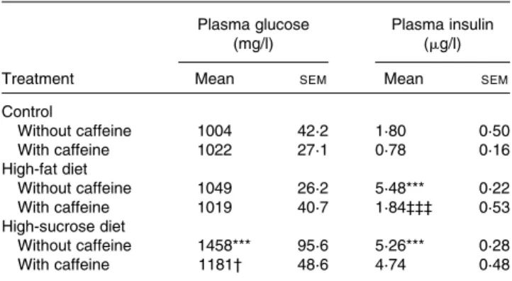
Fasting glycaemia was not significantly different in control and HF groups although the HSu diet significantly increased fast-ing glycaemia in comparison with the control group (P,0·001) (Table 1). Chronic caffeine intake did not modify fasting glycae-mia both in the control and HF rats but it reversed hyperglycae-mia induced by the HSu diet (P,0·05) (Table 1).
Fasting insulinaemia was 1·80 (SEM0·50)mg/l in control rats. Chronic caffeine intake did not significantly change basal insu-lin concentrations in control rats. Both the HF and HSu diets caused a significant increase in plasma insulin levels (P,0·001; Table 1) in comparison with the control group. This effect was reduced to control values by chronic caffeine intake in HF (P,0·001) but not in HSu rats (Table 1).
Effect of chronic caffeine intake on insulin sensitivity
Fig. 1 represents the effect of chronic caffeine intake (1 g/l) on insulin sensitivity in control, HF and HSu groups. The HF diet induced a decrease in KITTfrom a control value of 4·49 (SEM 0·43) % glucose/min to 3·02 (SEM 0·34) % glucose/min (P,0·05). The HSu diet decreased KITTto 2·68 (SEM0·32) % glu-cose/min (P,0·01). Chronic caffeine intake did not modify insulin sensitivity in control animals (KITT Caff¼4·52 (SEM 0·23) % glucose/min). In both the HF and HSu diet groups, chronic caffeine intake completely reversed the insulin resist-ance induced by the diets (KITTHFCaff ¼4·26 (SEM0·16) % glucose/min; KITTHSuCaff ¼4·07 (SEM0·26) % glucose/min).
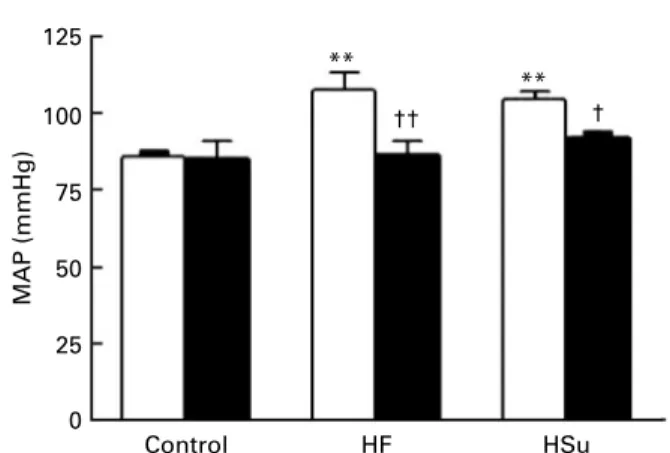
Effect of chronic caffeine intake on mean arterial pressure
Fig. 2 displays the effect of chronic caffeine consumption on MAP in control, HF and HSu animals. HF and HSu administration induced a significant increase in MAP compared with controls (MAP control¼85·99 (SEM 2·15) mmHg; MAP HF¼108·04 (SEM 5·30) mmHg; MAP HSu¼104·69 (SEM2·72) mmHg). Chronic caffeine intake did not modify MAP in control animals; however, when admini-strated to HF and HSu rats, it completely prevented the hypertension induced by both diets (MAP Caff¼85·53 (SEM 5·32) mmHg; MAP HFCaff¼86·92 (SEM 3·96) mmHg; MAP HSuCaff¼92·13 (SEM1·82) mmHg).
5
KITT
(% glucose/min)
4
3
2
1
0
Control
* †
††
**
HF HSu
Fig. 1.Effect of chronic caffeine intake (1 g/l) on insulin sensitivity in control, high-fat (HF) and high-sucrose (HSu) diet rats, determined by the insulin tolerance test, expressed as constant rate for glucose disappearance (KITT). (A), Without caffeine (n9 – 12); (B), with caffeine (n 12 – 15). Values are means, with standard errors represented by vertical bars. Mean value was significantly different from that of the control rats not given caffeine: *P,0·05, **P,0·01 (two-way ANOVA with Bonferroni multiple comparison test). Mean value was significantly different from that of the rats within the same diet group not given caffeine: †P,0·05, ††P,0·01 (two-way ANOVA with Bonferroni multiple comparison test).
Table 1.Effect of chronic caffeine intake (1 g/l) on glycaemia and insuli-naemia in control rats and in rats fed high-fat and high-sucrose diets (Mean values with their standard errors for all the individual samples obtained from 9 – 15 rats)
Plasma glucose (mg/l)
Plasma insulin (mg/l)
Treatment Mean SEM Mean SEM
Control
Without caffeine 1004 42·2 1·80 0·50 With caffeine 1022 27·1 0·78 0·16 High-fat diet
Without caffeine 1049 26·2 5·48*** 0·22 With caffeine 1019 40·7 1·84‡‡‡ 0·53 High-sucrose diet
Without caffeine 1458*** 95·6 5·26*** 0·28 With caffeine 1181† 48·6 4·74 0·48
*** Mean value was significantly different from that of the control rats (P,0·001; two-way ANOVA with Bonferroni multiple comparison test).
† Mean value was significantly different from that of the rats fed the high-sucrose diet without caffeine (P,0·05; two-way ANOVA with Bonferroni multiple compari-son test).
‡‡‡ Mean value was significantly different from that of the rats fed the high-fat diet without caffeine (P,0·001; two-way ANOVA with Bonferroni multiple com-parison test).
British
Journal
of
Effect of chronic caffeine intake on body weight, visceral fat and NEFA
Fig. 3 represents the effects of chronic caffeine intake on weight gain (Fig. 3(a)), visceral fat (Fig. 3(b)) and serum NEFA (Fig. 3(c)). The HF diet caused a significant increase in weight gain per d compared with control animals, whereas the HSu diet did not (control¼1·53 (SEM0·26) g/d; HF¼4·32 (SEM0·45) g/d; HSu: 2·66 (SEM0·25) g/d). Caffeine intake did not significantly modify weight gain either in control or in HSu animals (Caff¼1·14 (SEM 0·16) g/d; HSuCaff¼2·51 (SEM 0·29) g/d). In contrast, caffeine administration signifi-cantly reduced weight gain to 2·39 (SEM 0·36) g/d in HF animals (Fig. 3(a)).
The HF diet significantly increased visceral fat compared with control animals (control¼8·48 (SEM 0·63) g/kg; HF¼12·70 (SEM 0·64) g/kg) while the HSu diet did not (10·99 (SEM 0·86) g/kg). Caffeine consumption in control and HSu animals did not significantly modify visceral fat (Caff¼6·91 (SEM 0·40) g/kg; HSuCaff¼9·91 (SEM 0·97) g/kg) but the effect was significant in the HFCaff group
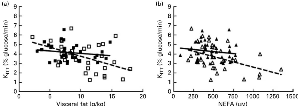
(7·80 (SEM 0·90) g/kg), a value similar to control (Fig. 3(b)). Fig. 3(c) shows that while the HF diet did not alter serum NEFA (NEFA control¼389·14 (SEM 40·45) mM; NEFA HF¼436·45 (SEM 36·23) mM), the HSu diet significantly increased NEFA to 940·62 (SEM 89·66) mM. Chronic caffeine intake did not modify NEFA either in control or in HF animals (NEFA Caff¼406·57 (SEM 29·55) mM; NEFA HFCaff¼391·67 (SEM 38·23)mM); however, it significantly decreased NEFA in the HSu group to 610·24 (SEM 41·06) mM. The correlations between both KITTand visceral fat and KITTand NEFA, with and without caffeine treatment, are depicted in Fig. 4. We observed a statistically significant negative correlation between KITT and visceral fat (r20·467;P¼0·0071) that is lost upon chronic caffeine treatment (r20·181; P¼0·314; Fig. 4(a)). Similarly, a statistically significant correlation was observed between KITT and serum NEFA (r20·444;
P¼0·014) and this correlation was lost in the caffeine-treated groups (r20·148;P¼0·338; Fig. 4(b)).
Effect of chronic caffeine intake on total hepatic glutathione and plasma NO/NO32concentration
Fig. 5(a) shows that the HF and HSu diets significantly decreased the total hepatic glutathione content by 35·37 and 70·40 %, respectively, from a control value of 75·3 (SEM3·85) nmol/mg tissue. Chronic caffeine administration did not significantly modify hepatic glutathione þ glutathione disul-fide (GSSG) levels (Caff¼83·6 (SEM 7·46) nmol/mg tissue; HFCaff¼59·1 (SEM 7·55) nmol/mg tissue; HSuCaff¼35·3 (SEM3·28) nmol/mg tissue).
Fig. 5(b) represents the effect of chronic caffeine intake on plasma NO/NO32. The HF diet caused a significant increase in plasma NO/NO32compared with control animals, whereas the HSu diet did not (control, 15·4 (SEM1·0)mM; HF, 24·1 (SEM2·4)
mM; HSu, 12·9 (SEM 1·0)mM). Caffeine intake did not modify plasma NO/NO32in any of the groups tested (Caff, 17·3 (SEM 0·7)mM; HFCaff, 22·0 (SEM1·8)mM; HSuCaff, 14·8 (SEM1·2)mM).
Effect of chronic caffeine intake on serum catecholamines
The effect of chronic caffeine intake on serum catecholamines (adrenaline plus noradrenaline) in control, HSu and HF rats is 125
MAP (mmHg)
100
75
50
25
0
Control
† ††
** **
HF HSu
Fig. 2.Effect of chronic caffeine intake (1 g/l) on mean arterial pressure (MAP) in control, high-fat (HF) and high-sucrose (HSu) diet rats. (A), Without caffeine (n9 – 12); (B), with caffeine (n9 – 15). Values are means, with stan-dard errors represented by vertical bars. ** Mean value was significantly different from that of the control rats not given caffeine (P,0·01; two-way ANOVA with Bonferroni multiple comparison test). Mean value was significantly different from that of the rats within the same diet group not given caffeine: †P,0·05, ††P,0·01 (two-way ANOVA with Bonferroni multiple comparison test).
5
(a) (b) (c)
∆
Rat weight (g/d)
4
3
2
†††
Control HF HSu 1
0
15
Visceral fat (g/kg)
10
5
††† **
Control HF HSu 0
1250
NEFA (
µ
M
)
1000
750
500
250
††† ***
Control HF HSu 0
Fig. 3.Effect of chronic caffeine intake (1 g/l) on body-weight increment, calculated as total weight variation during the experimental period (a), visceral fat, weighed post-mortem and corrected to body weight (b) and serum NEFA (c) in control, high-fat (HF) and high-sucrose (HSu) diet rats. (A), Without caffeine (n 10 – 12,n9 – 12 andn9 – 12 for body-weight increment, visceral fat and NEFA, respectively); (B), with caffeine (n12 – 16,n9 – 13 andn10 – 15 for body-weight increment, visceral fat and NEFA, respectively). Values are means, with standard errors represented by vertical bars. Mean value was significantly different from that of the control rats not given caffeine: **P,0·01, ***P,0·001 (two-way ANOVA with Bonferroni multiple comparison test). ††† Mean value was significantly different from that of the rats within the same diet group not given caffeine (P,0·001; two-way ANOVA with Bonferroni multiple comparison test).
British
Journal
of
represented in Fig. 6(a). The HSu and HF diets significantly increased plasma catecholamines by 211·9 and 124·1 %, respectively, from a control value of 50·03 (SEM 6·91) mM. Chronic caffeine intake did not modify plasma catecholamines in control animals; however, when administered together with the HSu and HF diets, caffeine prevented the increase in circu-lating catecholamines, suggesting that the metabolic and hae-modynamic effects of chronic caffeine intake are mediated by a decrease in sympathetic activation. Moreover, KITT was negatively correlated with plasma catecholamines both in the absence (r 20·399;P¼0·0484) and presence (r 20·419;
P¼0·009) of caffeine treatment (Fig. 6(b)).
Effect of carvedilol on insulin sensitivity and mean arterial pressure
There were no significant changes in individual daily liquid intake per kg in any of the groups tested. Food intake was also not significantly different (data not shown). In the HFMC group there was a decrease in KITT from a control
value of 4·90 (SEM0·26) % glucose/min to 3·50 (SEM0·23) % glucose/min. The HSuMC diet decreased KITT to 3·35 (SEM 0·19) % glucose/min. CVD did not modify insulin sensitivity in control animals (KITT CVD¼4·68 (SEM 0·30) % glucose/ min) but it completely reversed the insulin resistance induced by the diets (KITT HFCVD¼4·64 (SEM 0·27) % glucose/min; KITT HSuCVD ¼5·20 (SEM 0·12) % glucose/min) (Fig. 7(a)). CVD significantly decreased fasting glycaemia in the HSuMC group from 1165 (SEM 45·6) to 919 (SEM27·5) mg/l. Both the HFMC and HSuMC diets caused a significant increase in plasma insulin levels in comparison with the MC group and this effect was reversed by CVD administration (MC¼2·45 (SEM0·26) mg/l; HFMC¼4·88 (SEM0·30)mg/l; HSuMC¼4·36 (SEM0·37)mg/l; HFCVD¼2·42 (SEM0·42)mg/l; HSuCVD¼1·76 (SEM0·79)mg/l). We also observed that CVD completely restored MAP to control values in HF and HSu rats, as expected due to its anti-hypertensive action (MC¼95·6 (SEM 3·79) mmHg; CVD¼97·2 (SEM 3·35) mmHg; HFMC¼113·0 (SEM 4·07) mmHg; HFCVD¼99·4 (SEM4·59) mmHg; HSuMC¼113·6 (SEM 3·19) mmHg; HSuCVD¼99·0 (SEM5·17) mmHg).
9
(a) (b)
8
7
KITT
(% glucose/min)
6
5
4 3
2
1
0
0 5
Visceral fat (g/kg)
10 15 20
9
8
7
KITT
(% glucose/min)
6
5
4 3
2
1
0
0 250 500
NEFA (µM)
750 1000 1250 1500
Fig. 4.Effect of chronic caffeine intake (1 g/l) on the correlations between insulin sensitivity, visceral fat and NEFA in all the animals tested. (a) Caffeine eliminates the correlation between constant rate for glucose disappearance (KITT) and visceral fat mass. (b) Caffeine eliminates the correlation between KITTand serum NEFA. Pearson correlation coefficient was in (a):r20·467,P¼0·0071 without caffeine (A, - - -) andr20·181,P¼0·314 with caffeine (B, – – ); in (b):r20·444, P¼0·014 without caffeine (D, - - -) andr20·148,P¼0·338 with caffeine (O, - - -). Data represent all the individual samples obtained from control, high-fat and high-sucrose diet rats.
100
(a) (b)
75
50
25
0
Control
GSH+GSSG (nmol/mg tissue)
HF HSu
**
***
** 30
20
10
0
Control
NO (
µ
M
)
HF HSu
Fig. 5.Effect of chronic caffeine intake (1 g/l) on biomarkers of oxidative status. (a) Effect of caffeine on total hepatic glutathione in control rats and in rats sub-mitted to high-fat (HF) and high-sucrose (HSu) diets. (b) Effect of caffeine on plasma NO/NO32in control, HF and HSu rats. (A), Without caffeine (n8 – 11 and n8 – 13 for glutathione and NO, respectively); (B), with caffeine (n8 – 12 andn6 – 12, for glutathione and NO, respectively). Values are means, with standard errors represented by vertical bars. Mean value was significantly different from that of the control rats not given caffeine: **P,0·01, ***P,0·001 (two-way ANOVA with Bonferroni multiple comparison test).
British
Journal
of
Effect of carvedilol on serum catecholamines
The HSuMC and HFMC groups showed a significant increase in plasma catecholamines of 112·4 and 97·9 %, respectively, from a control value of 58·53 (SEM 8·88) mM in the MC group. In contrast with caffeine, CVD treatment did not modify plasma catecholamines in any of the groups tested (CVD, 59·2 (SEM 17·80) mM; HFCVD, 108·6 (SEM 33·14) mM; HSuCVD, 88·8 (SEM32·26)mM; Fig. 7(b)). CVD administration did not significantly modify weight gain, visceral fat, NEFA, hepatic glutathione or NO/NO32 in any of the disease models tested (data not shown).
Discussion
We demonstrated in the present study that chronic caf-feine intake prevents the development of insulin resistance and hypertension in high-fat- and high-sucrose-fed rats and induces a pronounced decrease in circulating adrenaline and
noradrenaline. Also, the effect of CVD, an antagonist of b1, b2 and a1 adrenoceptors, on insulin sensitivity mimicked the effect of caffeine, supporting the hypothesis that caffeine’s beneficial effects are mediated by the inhibition of the sym-pathetic nervous system.
Chronic caffeine treatment did not significantly change fast-ing glycaemia both in the control and HF groups, which were normoglycaemic without caffeine treatment(33), but it totally reversed the hyperglycaemia induced by the HSu diet. Also, in contrast with the acute effects of caffeine on insulin secretion described in the literature(12), the present results show that plasma insulin levels significantly decreased in the HFCaff group and trend towards a decrease in the HSuCaff group com-pared with the HF and HSu groups, respectively. In the HFCaff group the reduction of fasting insulinaemia, accompanied by an increase in KITT, indicates that chronic caffeine not only increased insulin sensitivity but also normalised insulin secretion in the HF animals. We do not know at this point if the decrease in plasma insulin levels is a direct effect of caffeine
(a)
KITT
(% glucose/min)
6
5
4
3
2
1
0
Control HF HSu
(b)
200 †††
†
**
* 150 * *
Catecholamines (
µ
M
)
100
50
0
Control HF HSu
Fig. 7.Effect of carvedilol (CVD, 0·2 g/l) and its vehicle, methylcellulose (MC, 0·5 %), on insulin sensitivity and plasma catecholamines in control, high-fat (HF) and high-sucrose (HSu) diet rats. (a) The effect of CVD on insulin sensitivity was determined by the insulin tolerance test and is expressed as constant rate for glucose disappearance (KITT). (A), MC (n10 – 17); (B), CVD (n7 – 12). Values are means, with standard errors represented by vertical bars. Mean value was significantly different from that of the control rats given MC: *P,0·05, **P,0·01 (two-way ANOVA with Bonferroni multiple comparison test). Mean value was significantly different from that of the rats within the same diet group given MC: †P,0·05, †††P,0·001 (two-way ANOVA with Bonferroni multiple comparison test). (b) Effect of CVD on catecholamine (adrenaline plus noradrenaline) plasma levels. (A), MC (n10 – 36); (B), CVD (n5 – 7). Values are means, with standard errors represented by vertical bars. * Mean value was significantly different from that of the control rats given MC (P,0·05; two-way ANOVA with Bonferroni multiple comparison test).
9 (b)
8
7
KITT
(% glucose/min)
6
5
4
3
2
1
0
0 100
Catecholamines (µM)
200 300
(a)
200
**
***
†† †††
150
Catecholamines (
µ
M
)
100
50
0
Control HF HSu
Fig. 6.Effect of chronic caffeine intake (1 g/l) on plasma catecholamine concentration in control, high-sucrose (HSu) and high-fat (HF) diet rats. (a) Effect of caffeine on catecholamine (adrenaline plus noradrenaline) plasma levels. (A), Without caffeine (n10 – 16); (B), with caffeine (n12 – 16). Values are means, with standard errors represented by vertical bars. Mean value was significantly different from that of the control rats not given caffeine: **P,0·01, ***P,0·001 (two-way ANOVA with Bonferroni multiple comparison test). Mean value was significantly different from that of the rats within the same diet group not given caffeine: ††P,0·01, †††P,0·001 (two-way ANOVA with Bonferroni multiple comparison test). (b) Caffeine treatment does not change the correlation between constant rate for glucose disappearance (KITT) and circulating catecholamine (adrenaline plus noradrenaline) plasma levels. Pearson correlation coefficient wasr20·399, P¼0·0484 without caffeine (W) andr20·419,P¼0·009 with caffeine (X). Data represent all the individual samples obtained from control, HF and HSu rats.
British
Journal
of
on the pancreas, or simply a negative-feedback mechanism induced by improved peripheral insulin sensitivity at the b-cell. However, previous studies in human subjects have described that caffeinated coffee consumption leads to a reduction of plasma C-peptide concentrations in healthy and in obese women, suggesting that coffee components decrease insulin secretion(16)andin vitrostudies also showed that adeno-sine regulates insulin secretion by theb-cell(34).
In contrast with the HFCaff animals, in the HSuCaff group caffeine restored insulin sensitivity and normoglycaemia but the animals remained hyperinsulinaemic compared with con-trols. This suggests that in the caffeine-treated sucrose-fed model, insulin action improved sufficiently to maintain glu-cose homeostasis but not enough to normalise endogenous insulin secretion.
In the HF rats both visceral fat and weight gain increased sig-nificantly and caffeine reversed these effects, in agreement with the literature(12). Weight loss and visceral fat mass decrease induced by long-term caffeine consumption may have been due to increased lipolysis accompanied by increased NEFA oxi-dation, since circulating NEFA were not elevated compared with control animals. We observed no changes in weight gain or visceral fat mass in the HSu rat treated with caffeine. These results indicate that increased adipose tissue mass is not contri-buting to insulin resistance in this animal model, which is in accordance with previous results by Kanazawa’s group(35).
In contrast to the HF diet, the HSu diet increased circulating NEFA, indicating augmented lipolysis or decreased NEFA oxi-dation in the sucrose-fed rats as previously described by others(36). These results may also be due to an increased
de novo lipogenesis in adipocytes and liver in this model
which, combined with insulin resistance, may lead to enhanced lipolysis and systemic NEFA(37). We have observed that chronic caffeine intake reversed the increase in NEFA induced by the HSu diet. This effect may be related to an augmented activity of 50-AMP activated protein kinase (AMPK) induced by caffeine
that promotes an enhancement in NEFA uptake and oxidation in skeletal muscle(38,39). Recently, it has also been shown that caf-feine can increase GLUT-4 mRNA in skeletal muscle cells and insulin-independent glucose transport mediated by AMPK(38), which may explain the return to normal glycaemia observed in the caffeine-treated rats. Despite the beneficial effects of caf-feine on visceral fat in the HF-fed rat and in circulating NEFA in the HSu rat, the correlation analyses show that insulin action loses its dependence of these variables after caffeine treatment. The insulin-sensitising effect of chronic caffeine treatment may also be directly related to antagonism of adenosine receptors on insulin target tissues. The effect of adenosine on glucose homeostasis is currently a hot-topic area with con-troversial results: while positive effects of A1receptor partial agonists are well documented on adipose tissue glucose uptake(40), studies using adenosine receptor antagonists have shown increases(41), decreases(42) and no effect(43) of these drugs on skeletal muscle glucose uptake. Also, A2B receptor antagonists have been used to increase plasma insulin levels
in vivo(44)and more recently to reduce insulin resistance(45).
Clearly, more studies are required to clarify the role of adeno-sine receptors on glucose homeostasis.
In the present study we also demonstrated that chronic caffeine intake prevents the development of hypertension in HF- and HSu-fed animals. Recently, it has been shown that endothelium-dependent vasodilatation after long-term caffeine administration is enhanced in young healthy men(46), a mechanism by which caffeine treatment could lower blood pressure. However, we did not find any effect of chronic caffeine consumption on NO production, sug-gesting that NO is involved neither in the prevention of hypertension nor in insulin resistance induced by caffeine. It has also been suggested that caffeine may prevent oxidative damage(47,48) which could improve endothelium-dependent vasodilation and insulin action. However, in our experimental setting we observed no changes in total hepatic glutathione, both in HF- and HSu-treated animals. Hepatic glutathione stores are significantly depleted after exposure to oxidative stress(49) and if administration of caffeine produced signifi-cant antioxidant effects, we would expect intracellular hepatic glutathione levels to rise, which did not happen.
Chronic caffeine treatment caused a decrease in circulating catecholamines, suggesting that decreased sympathetic ner-vous system activation, either at the adrenal medulla or at the sympathetic nerves innervating blood vessels, is involved in the protective effects of caffeine. Adenosine receptor ago-nists inhibit sympathetic neurotransmission and catecholamine release in several tissues, including the hippocampus(50) and adrenal chromaffin cells(51), when administered acutely. Adenosine receptor antagonists, such as caffeine, increase plasma renin activity, adrenaline and noradrenaline(52) after acute administration; however, in accordance with the present results, it has been shown that the increase in adrenaline levels induced by a single dose of caffeine is lost with sustained consumption(53).
The present results also show that the adrenergic antago-nist, CVD, mimicked the effects of caffeine. As observed with caffeine treatment, CVD totally reversed the hyperglycaemia induced by the HSuMC diet. Plasma insulin levels significantly decreased after CVD treatment in the HFCVD and HSuCVD groups, in contrast to the hypoinsulinaemic effect of caffeine that was observed in the HFCaff but not in the HSuCaff group. Others have also reported that CVD, among other third-generation b-blockers, lack adverse metabolic effects and may actually produce favourable effects on insulin sensi-tivity(54,55). The mechanisms behind these findings are not yet fully understood but the role of thea1 andb2 adrenoceptor blocking effects of CVD appears to be crucial(56). While caffeine significantly decreased weight gain and visceral fat mass in HF rats and circulating NEFA in HSu rats, CVD treatment did not change any of these factors in the experimental groups, although the effect of both drugs on insulin sensitivity was very similar. These results are consistent with the hypo-thesis that the insulin-sensitising properties of both caffeine and CVD are not directly related with changes in adipose tissue homeostasis. Finally, CVD has also been reported to increase endogenous NO production(57,58), affecting endo-thelium-dependent vasodilation, a mechanism by which this drug could increase insulin sensitivity(59,60). We did not find any effect of CVD on NO production in our experimental
British
Journal
of
conditions, suggesting that NO is involved neither in the pre-vention of hypertension nor in insulin sensitisation induced by CVD.
In conclusion, chronic caffeine administration prevents the development of insulin resistance and hypertension in high-fat- and high-sucrose-fed animals due to a decrease in circulating catecholamines. Treatment with an adrenergic antagonist mimics the protective effects of caffeine against diet-induced insulin resistance by blockade of adrenergic receptors without changes in serum catecholamine concen-tration. The exact biological mechanism underlying the decrease in circulating catecholamines induced by caffeine remains to be clarified and may involve alterations in the expression of adenosine receptors as well as in the expression of other neurotransmitter receptors and ionic channels(61,62).
In conclusion, we have shown that long-term caffeine administration has a protective effect on the development of diet-induced insulin resistance and hypertension in rats. Our work strengthens the conclusions of recent epidemiological studies concerning the protective role of coffee consumption in type 2 diabetes and the metabolic syndrome and helps to support the need for a paradigm shift on advised lifestyle changes, like coffee withdrawal, in insulin-resistant and hyper-tensive patients.
Acknowledgements
We are very grateful to Elena Gonzalez Mun˜oz for catechol-amine quantification and to Ineˆs Faustino, Ana Carvalho and Lu´cia Mirrado for technical support and ‘L’Ore´al-Unesco Awards for Women in Science 2009 – Portugal’.
The present study was financially supported by CEDOC/ FCT, Portuguese Society of Diabetology (SPD)/Lilly Portugal, BFU2007-61848 and JCyL-GR242, and by L’Ore´al-Unesco Awards for Women in Science 2009 – Portugal.
S. V. C. and M. P. G. designed and conducted the study and also wrote the manuscript. T. N. da S. participated in data collection and analysis, M. M. C. and E. C. M. contributed in data interpretation. Catecholamine quantification was per-formed in the laboratory of C. G.
There are no conflicts of interest.
References
1. Kashyap SR & Defronzo RA (2007) The insulin resistance syndrome: physiological considerations. Diab Vasc Dis Res
4, 13– 19.
2. Riksen NP, Rongen GA & Smits P (2009) Acute and long-term cardiovascular effects of coffee: implications for coronary heart disease.Pharmacol Ther121, 185 – 191.
3. Moisey LL, Kacker S, Bickerton AC,et al.(2008) Caffeinated coffee consumption impairs blood glucose homeostasis in response to high and low glycemic index meals in healthy men.Am J Clin Nutr87, 1254 –1261.
4. Keijzers GB, De Galan BE, Tack CJ,et al.(2002) Caffeine can decrease insulin sensitivity in humans. Diabetes Care 25, 364 –369.
5. van Dam RM, Willett WC, Manson JE,et al. (2006) Coffee, caffeine, and risk of type 2 diabetes: a prospective cohort
study in younger and middle-aged U.S. women. Diabetes Care29, 398 – 403.
6. van Dam RM & Feskens EJ (2002) Coffee consumption and risk of type 2 diabetes mellitus.Lancet360, 1477– 1478. 7. Noordzij M, Uiterwaal CS, Arends LR, et al. (2005) Blood
pressure response to chronic intake of coffee and caffeine: a meta-analysis of randomized controlled trials.J Hypertens
23, 921 – 928.
8. Jee SH, He J, Whelton PK,et al.(1999) The effect of chronic coffee drinking on blood pressure: a meta-analysis of con-trolled clinical trials.Hypertension33, 647 –652.
9. Geleijnse JM (2008) Habitual coffee consumption and blood pressure: an epidemiological perspective.Vasc Health Risk Manag4, 963 –970.
10. Conde SV, Pinto P, Ba´rbara C,et al.(2009) Should caffeine be avoided in obstructive sleep apnea? InEuropean Respi-ratory Society Annual Congress, Vienna, Austria: 2009. Abstracts book P3729.
11. Arnlov J, Vessby B & Riserus U (2004) Coffee consumption and insulin sensitivity.JAMA291, 1199 –1201.
12. Greenberg JA, Boozer CN & Geliebter A (2006) Coffee, dia-betes, and weight control.Am J Clin Nutr84, 682 –693. 13. Zheng G, Sayama K, Okubo T, et al. (2004) Anti-obesity
effects of three major components of green tea, catechins, caffeine and theanine, in mice.In Vivo18, 55 –62.
14. Lopez-Garcia E, van Dam RM, Rajpathak S, et al. (2006) Changes in caffeine intake and long-term weight change in men and women.Am J Clin Nutr83, 674 – 680.
15. Greenberg JA, Axen KV, Schnoll R,et al.(2005) Coffee, tea and diabetes: the role of weight loss and caffeine. Int J Obes (Lond)29, 1121 –1129.
16. Wu T, Willett WC, Hankinson SE,et al. (2005) Caffeinated coffee, decaffeinated coffee, and caffeine in relation to plasma C-peptide levels, a marker of insulin secretion, in U.S. women.Diabetes Care28, 1390– 1396.
17. Cheung WT, Lee CM & Ng TB (1988) Potentiation of the anti-lipolytic effect of 2-chloroadenosine after chronic caffeine treatment.Pharmacology36, 331 – 339.
18. Astrup A, Breum L, Toubro S,et al. (1992) The effect and safety of an ephedrine/caffeine compound compared to ephedrine, caffeine and placebo in obese subjects on an energy restricted diet. A double blind trial.Int J Obes Relat Metab Disord16, 269 – 277.
19. Sung BH, Whitsett TL, Lovallo WR,et al.(1994) Prolonged increase in blood pressure by a single oral dose of caffeine in mildly hypertensive men.Am J Hypertens7, 755 – 758. 20. Sung BH, Lovallo WR, Whitsett T,et al.(1995) Caffeine
elev-ates blood pressure response to exercise in mild hyperten-sive men.Am J Hypertens8, 1184 – 1188.
21. Pincomb GA, Lovallo WR, McKey BS, et al. (1996) Acute blood pressure elevations with caffeine in men with border-line systemic hypertension.Am J Cardiol77, 270 –274. 22. Benowitz NL (1990) Clinical pharmacology of caffeine.Annu
Rev Med41, 277 – 288.
23. Fredholm BB, Battig K, Holmen J,et al.(1999) Actions of caffeine in the brain with special reference to factors that contribute to its widespread use.Pharmacol Rev51, 83– 133. 24. Robertson D, Wade D, Workman R,et al.(1981) Tolerance to the humoral and hemodynamic effects of caffeine in man.
J Clin Invest67, 1111– 1117.
25. Myers MG (1988) Effects of caffeine on blood pressure.Arch Intern Med148, 1189 –1193.
26. Holtzman SG & Finn IB (1988) Tolerance to behavioral effects of caffeine in rats.Pharmacol Biochem Behav29, 411 – 418. 27. Gasior M, Shoaib M, Yasar S,et al.(1999) Acquisition of
nic-otine discrimination and discriminative stimulus effects of
British
Journal
of
nicotine in rats chronically exposed to caffeine.J Pharmacol Exp Ther288, 1053 – 1073.
28. Shearer J, Severson DL, Su L,et al.(2009) Partial A1 adeno-sine receptor agonist regulates cardiac substrate utilization in insulin-resistant ratsin vivo.J Pharmacol Exp Ther328, 306 –311.
29. Ribeiro RT, Lautt WW, Legare DJ,et al.(2005) Insulin resist-ance induced by sucrose feeding in rats is due to an impair-ment of the hepatic parasympathetic nerves. Diabetologia
48, 976 – 983.
30. Gasior M, Jaszyna M, Munzar P,et al.(2002) Caffeine potenti-ates the discriminative-stimulus effects of nicotine in rats.
Psychopharmacology (Berl)162, 385 –395.
31. Rodriguez-Perez JC, Losada A, Anabitarte A, et al. (1997) Effects of the novel multiple-action agent carvedilol on severe nephrosclerosis in renal ablated rats. J Pharmacol Exp Ther283, 336 – 344.
32. Monzillo LU & Hamdy O (2003) Evaluation of insulin sensi-tivity in clinical practice and in research settings.Nutr Rev
61, 397 – 412.
33. Kim SP, Ellmerer M, Van Citters GW,et al.(2003) Primacy of hepatic insulin resistance in the development of the meta-bolic syndrome induced by an isocaloric moderate-fat diet in the dog.Diabetes52, 2453– 2460.
34. Ismail NA, El Denshary EE & Montague W (1977) Adenosine and the regulation of insulin secretion by isolated rat islets of Langerhans.Biochem J164, 409 –413.
35. Kanazawa M, Xue CY, Kageyama H,et al.(2003) Effects of a high-sucrose diet on body weight, plasma triglycerides, and stress tolerance.Nutr Rev61, S27 – S33.
36. Delarue J & Magnan C (2007) Free fatty acids and insulin resistance.Curr Opin Clin Nutr Metab Care10, 142 –148. 37. Lewis GF, Carpentier A, Adeli K,et al.(2002) Disordered fat
storage and mobilization in the pathogenesis of insulin resistance and type 2 diabetes.Endocr Rev23, 201 – 229. 38. Egawa T, Hamada T, Kameda N, et al. (2009) Caffeine
acutely activates 50adenosine monophosphate-activated protein kinase and increases insulin-independent glucose transport in rat skeletal muscles.Metabolism58, 1609 – 1617. 39. Raney MA & Turcotte LP (2008) Evidence for the
involve-ment of CaMKII and AMPK in Ca2þ
-dependent signaling pathways regulating FA uptake and oxidation in contracting rodent muscle.J Appl Physiol104, 1366 –1373.
40. Dhalla AK, Chisholm JW, Reaven GM, et al. (2009) A1 adenosine receptor: role in diabetes and obesity. Handb Exp Pharmacol193, 271 – 295.
41. Espinal J, Challiss RA & Newsholme EA (1983) Effect of adenosine deaminase and an adenosine analogue on insulin sensitivity in soleus muscle of the rat.FEBS Lett158, 103 – 106. 42. Han DH, Hansen PA, Nolte LA, et al. (1998) Removal of adenosine decreases the responsiveness of muscle glucose transport to insulin and contractions. Diabetes47, 1671– 1675.
43. Vergauwen L, Hespel P & Richter EA (1994) Adenosine receptors mediate synergistic stimulation of glucose uptake and transport by insulin and by contractions in rat skeletal muscle.J Clin Invest93, 974 –981.
44. Rusing D, Muller CE & Verspohl EJ (2006) The impact of adenosine and A(2B) receptors on glucose homoeostasis.
J Pharm Pharmacol58, 1639 –1645.
45. Figler RA, Wang G, Srinivasan S,et al.(2011) Links between insulin resistance, adenosine A2B receptors, and inflamma-tory markers in mice and humans.Diabetes60, 669 –679. 46. Umemura T, Ueda K, Nishioka K, et al. (2006) Effects of
acute administration of caffeine on vascular function. Am J Cardiol98, 1538 – 1541.
47. Scharf G, Prustomersky S & Huber WW (2001) Elevation of glutathione levels by coffee components and its potential mechanisms.Adv Exp Med Biol500, 535 – 539.
48. Varma SD & Hegde KR (2010) Prevention of oxidative damage to lens by caffeine. J Ocul Pharmacol Ther 26, 73– 77.
49. Mytilineou C, Kramer BC & Yabut JA (2002) Glutathione depletion and oxidative stress. Parkinsonism Relat Disord
8, 385 – 387.
50. Jackisch R, Fehr R & Hertting G (1985) Adenosine: an endogenous modulator of hippocampal noradrenaline release.Neuropharmacology24, 499 – 507.
51. Tseng CJ, Chan JY, Lo WC, et al. (2001) Modulation of catecholamine release by endogenous adenosine in the rat adrenal medulla.J Biomed Sci8, 389 –394.
52. Robertson D, Frolich JC, Carr RK, et al. (1978) Effects of caffeine on plasma renin activity, catecholamines and blood pressure.N Engl J Med298, 181 – 186.
53. Debrah K, Haigh R, Sherwin R, et al. (1995) Effect of acute and chronic caffeine use on the cerebrovascular, cardi-ovascular and hormonal responses to orthostasis in healthy volunteers.Clin Sci (Lond)89, 475 – 480.
54. Carella AM, Antonucci G, Conte M, et al. (2010) Antihy-pertensive treatment with b-blockers in the metabolic syndrome: a review.Curr Diabetes Rev6, 215 – 221. 55. Bhatt P, Makwana D, Santani D,et al. (2007) Comparative
effectiveness of carvedilol and propranolol on glycemic con-trol and insulin resistance associated withL-thyroxin-induced hyperthyroidism – an experimental study. Can J Physiol Pharmacol85, 514 – 520.
56. Kveiborg B, Christiansen B, Major-Petersen A,et al. (2006) Metabolic effects ofb-adrenoceptor antagonists with special emphasis on carvedilol.Am J Cardiovasc Drugs6, 209 – 217. 57. Corsetti G, Pasini E, Assanelli D,et al.(2007) Acute caffeine administration decreased NOS and Bcl2 expression in rat skeletal muscles.Pharmacol Res55, 96 –103.
58. Ofluoglu E, Pasaoglu H & Pasaoglu A (2009) The effects of caffeine on l-arginine metabolism in the brain of rats.
Neurochem Res34, 395 – 399.
59. Guarino MP & Macedo MP (2006) Co-administration of glutathione and nitric oxide enhances insulin sensitivity in Wistar rats.Br J Pharmacol147, 959 –965.
60. Guarino MP, Correia NC, Raposo J,et al.(2001) Nitric oxide synthase inhibition decreases output of hepatic insulin sensi-tizing substance (HISS), which is reversed by SIN-1 but not by nitroprusside.Proc West Pharmacol Soc44, 25– 26. 61. Shi D & Daly JW (1999) Chronic effects of xanthines on
levels of central receptors in mice.Cell Mol Neurobiol 19, 719 – 732.
62. Stonehouse AH, Adachi M, Walcott EC,et al.(2003) Caffeine regulates neuronal expression of the dopamine 2 receptor gene.Mol Pharmacol64, 1463 – 1473.