Research Article
Antiageing Mechanisms of a Standardized Supercritical
CO
2
Preparation of Black Jack (
Bidens pilosa
L.) in Human
Fibroblasts and Skin Fragments
Gustavo Dieamant,
1Maria Del Carmen V. Pereda,
1Cecília Nogueira,
1Samara Eberlin,
1Gustavo Facchini,
1Juliana Tibério Checon,
2Camila Kappke Cesar,
1Lilian Mussi,
1Márcio Antonio Polezel,
1Divino Martins-Oliveira Jr.,
1and Luiz Claudio Di Stasi
21Chemyunion Quimica Ltda, Avenida Independˆencia 1501, 18087-101 Sorocaba, SP, Brazil
2Laboratory of Phytomedicines, Pharmacology and Biotechnology (PhytoPharmaTech), Department of Pharmacology,
Institute of Biosciences, Universidade Estadual Paulista (UNESP), 18618-000 Botucatu, SP, Brazil
Correspondence should be addressed to Luiz Claudio Di Stasi; ldistasi@ibb.unesp.br
Received 8 December 2014; Revised 23 February 2015; Accepted 15 March 2015
Academic Editor: Alessandra Guerrini
Copyright © 2015 Gustavo Dieamant et al. This is an open access article distributed under the Creative Commons Attribution License, which permits unrestricted use, distribution, and reproduction in any medium, provided the original work is properly cited.
The use of topical retinoids to treat skin disorders and ageing can induce local reactions, while oral retinoids are potent teratogens and produce several unwanted effects. This way, efforts to explore complementary care resources should be supported. Based on
this, we evaluate the antiageing effects of a supercritical CO2extract fromBidens pilosaL. (BPE-CO2A) containing a standardized
multicomponent mixture of phytol, linolenic, palmitic, linoleic, and oleic acids. BPE-CO2A was assessed for its effects on
human dermal fibroblasts (TGF-𝛽1 and FGF levels using ELISA; collagen, elastin, and glycosaminoglycan by colorimetric assays,
and mRNA expression of RXR, RAR, and EGFr by qRT-PCR) and human skin fragments (RAR, RXR, collagen, elastin, and
glycosaminoglycan by immunohistochemical analysis). Levels of extracellular matrix elements, TGF-𝛽1 and FGF, and EGFr gene
expression were significantly increased by BPE-CO2A. The modulation of RXR and RAR was positively demonstrated after the
treatment with BPE-CO2A or phytol, a component of BPE-CO2A. The effects produced by BPE-CO2A were similar to or better
than those produced by retinol and retinoic acid. The ability to stimulate extracellular matrix elements, increase growth factors,
and modulate retinoid and rexinoid receptors provides a basis for the development of preparation containing BPE-CO2A as an
antiageing/skin-repair agent.
1. Introduction
Retinoids are classically defined as compounds that exhibit vitamin A-like effects or bind to nuclear retinoid receptors, exerting their pharmacological effects on gene expression by activating retinoic acid receptors (RARs) or retinoid X receptors (RXRs). RARs are ligand-controlled transcription factors that function as heterodimers with RXRs to regulate
cell growth, differentiation, survival, and death [1]. Activation
of RAR and RXR has been associated with several diseases, and their ligands are widely used to treat skin disorders, mainly aging and photoaging, acne, and psoriasis, and have
been used for cancer therapy and chemoprevention [2,3].
The beneficial effects of retinoids on ageing skin are attributed to increased dermal function, mainly through the increased production of extracellular matrix components, reduced inflammatory response, and antioxidative proper-ties. These effects are related to epidermal growth factor (EGF) and fibroblast growth factor (FGF), both acting as
mediators of the retinoid response in the skin [4]. In addition,
the beneficial effects of retinoids on ageing and photoageing include improved fine wrinkling, diminished tactile rough-ness, improved actinic keratosis, and reduced hyperpigmen-tation, which are histopathologically associated with epider-mal hyperplasia, the compaction of the stratum corneum, the thickening of the granular layer, reduced melanocytic
hypertrophy, the restoration of cell polarity, increased angio-genesis, increased new collagen formation, and the
normal-ization of the appearance of elastic tissue [5].
In contrast, the use of topical retinoids to treat skin disorders can induce local reactions including erythema, burning, dryness, desquamation, stinging, and photosen-sitivity reactions to ultraviolet radiation, whereas the use of oral retinoids is limited because these compounds are potent teratogens and produce several unwanted effects such as cheilitis, cutaneous photosensitivity, conjunctivitis, photophobia, and increased susceptibility to staphylococcal
infections [4]. This way, efforts to explore alternative and
complementary care resources should be supported [6],
and the use of herbal preparations and natural products as a source of new active compounds for complementary and alternative therapies could be an important therapeutic strategy to obtain new products with retinoid-like effects and better management of skin diseases and ageing.
Black Jack (Bidens pilosaL. (Asteraceae)) is an annual and
ruderal plant originating in South America and is also found
in tropical and subtropical regions around the world [7]. This
plant is a cosmopolitan herb with high distribution in dis-turbed areas and is widely used in the traditional medicine of different countries to treat internal and topical inflammatory processes, wounds, insect bites, fungal infections, diabetic ulcers, fever, malaria, inflammation, hepatitis, hemorrhoids,
and cancer [7–11]. Recent review showed thatB. pilosahas
anti-inflammatory, immunomodulatory, antimalarial, antitu-mor, antioxidative, antiulcerogenic, antibacterial,
hepatopro-tective, and antihypertensive activities [7,8]. Phytochemical
studies indicated that approximately 200 different
com-pounds have been identified and isolated inB. pilosa, and
among them mainly flavonoids, polyacetylenes, terpenoids,
phenylpropanoids, and hydrocarbons were reported [7,8]. It
has been demonstrated that nonpolar chemical constituents ofBidens pilosasuch as docosahexaenoic acid, phytanic acid,
phytol, 𝛼-tocopherol, ricinoleic acid, and esters of caffeic
acid have similar retinoid chemical structures, and some of these compounds have been considered to be natural RXR ligands and modulators of the retinoic acid signalling pathway, directly influencing their transcriptional regulatory
activity [12–16].
The study of new products from natural sources, mainly plant species, can be performed through several approaches; however, the supercritical carbon dioxide extraction and
fractionation (SC-CO2) of natural matter is one of the early
and most studied applications with immediate advantages
over traditional extraction techniques [17]. It is a flexible
pro-cess because it allows for the continuous modulation of the solvent power/selectivity and elimination of polluting organic solvents, thereby eliminating the expensive postprocessing of extracts traditionally required for solvent elimination. More-over, it is more economical and simple than other methods; it has diffusivity that reduces mass transfer limitations and a low surface tension, which allows for the penetration and wetting of pores smaller than those accessible with liquid solvents; it allows extraction at low to moderate temperatures, leaving no
solvent residues; and it is environmentally acceptable [17,18].
Based on the pharmacological properties of B. pilosa,
such as antioxidative, anti-inflammatory and retinoid-like effects, and its phytochemical composition of mainly nonpo-lar compounds, the aim of this study was to evaluate a
SC-CO2 nonpolar extract from the aerial parts ofB. pilosato
develop an herbal preparation with retinoid-like activity and to potentially use this preparation as an antiageing or skin-repair agent.
2. Materials and Methods
2.1. Plant Collection, Plant Extraction, and Gas Chromatogra-phy Analysis. B. pilosawas cultivated using organic agricul-tural methods as certified by Ecocert Brazil (Santa Rosa de Lima/Santa Catarina/Br) and submitted to taxonomic iden-tification at Herbarium Irina Gemtchujnikov (Department of Botany, Institute of Biosciences, Universidade Estadual Paulista (UNESP), Botucatu, SP), where a voucher specimen was deposited. The aerial parts were collected, dehydrated in hothouse with air circulation and renewal, and triturated in an industrial mill. A supercritical extraction system (Auto-clave Engineers) under the following conditions of 300 bar,
40∘C, and CO2 flux of 5 L/min was used to generate a
nonpolar extract named BPE-CO2A.
The extraction procedures of BPE-CO2A by GC/MS
analysis were made according to following conditions: 100 mg
of BPE-CO2-A extract with 5 mL of NaOH 0.5 M in methanol
was heated in a water bath for 10 min at 37∘C. After cooling,
5 mL of NH4Cl was added in methanol and heated in a water
bath for 5 min at 37∘C. 1 mL of BPE-CO2A was extracted with
5 mL hexane. After cooling, 1𝜇L of sample was injected in GC
equipment.
GC/MS analysis was performed according to the follow-ing conditions: the gas chromatography-mass spectroscopy analysis system consisted of a Gas Chromatograph Focus (Thermo Scientific) equipped with an automatic Triplus sampler and coupled to an ISQ 230ST (Thermo Scientific) mass selective detector. The GC was fitted with an OV-5MS
fused silica capillary column (30 m×0.25 mm×0.24𝜇m) and
helium was used as the carrier gas at 1 mL/min. Injection was made in splitless mode with injection volume of 1.0 mL/min following a split mode (1 : 50). The injector temperature was
260∘C and detector temperature was 200∘C. Temperature
of column was initially 35∘C and then increased to 195∘C
at 25∘C/min, to 205∘C at 3∘C/min, and finally to 230∘C at
8∘C/min. Xcalibur software, version 2.1 (Thermo Finigan),
was used to acquire and process spectrometric data. Identi-fication of compounds was based on the retention time using NIST08 libraries and comparing with mass spectrum from scientific literature.
2.2. Cell Culture. Primary human adult keratinocytes (Invit-rogen) and normal human dermal fibroblasts (Cambrex/ Lonza) were commercially obtained, grown in specific cul-ture media (keratinocyte growth medium, Epilife-Invitrogen; fibroblast basal medium, Cambrex/Lonza), and cultured in a
humidified environment at 37∘C and 5% CO2. After reaching
cells was counted using a Neubauer camera. After counting, the cells were seeded in culture plates of 24 or 96 wells at
den-sities of 2×10⁵cells/well and 1×10⁴cells/well, respectively.
The apolar extract ofBidens pilosa(BPE-CO2A) obtained
using supercritical fluid extraction was added to the cell cultures at concentrations of 0.05, 0.10, and 0.20 mg/mL. The selection of these doses was based on preliminary cytotoxicity tests using the XTT method (data not shown). As a positive control for comparison with retinoids, we used retinol and retinoic acid at a concentration of 10 mM, according to
previ-ous data [19,20]. After 48-hour incubation, the supernatant
and cell lysate were collected for the subsequent measurement of the proposed parameters.
2.3. Specimen Skin Collection and Sample Preparation.
Human eyelids were surgically removed, dipped in 70% ethanol for 15 seconds, and rinsed 2 times with saline solu-tion. The skin fragments were transferred to a petri dish containing culture medium RPMI-1640 supplemented with foetal bovine serum-Lonza, a combination of antibiotics containing gentamicin and amphotericin-B (gentamicin sulfate/amphotericin-B, Lonza), bovine insulin (Lonza), and growth factor (rhFGF b-r-human fibroblast growth factor-B, Lonza) for a maximum of 24 hours before treatment with
BPE-CO2A.
BPE-CO2A was dissolved in culture medium at 0.5% (a
commonly used concentration for retinol) and applied to the skin fragment homogeneously over the entire surface. As positive controls for comparison, we used retinol and retinoic
acid, 30𝜇M and 0.5%, respectively. The samples were kept
in contact with the cultures for 48 hours in a humidified
atmosphere at 37∘C and 5% CO2.
2.4. TGF-𝛽and FGF Evaluation. Transforming growth
fac-tor beta (TGF-𝛽) and fibroblast growth factor (FGF) were
measured in fibroblast culture supernatant according to the instructions for the commercially obtained ELISA kits (R&D Systems), and the concentrations of both growth factors were calculated with reference to a standard curve generated by known concentrations.
2.5. Collagen, Elastin, and Glycosaminoglycan Evaluation.
Collagen, elastin, and glycosaminoglycan were quantified in the supernatant of fibroblast cultures using commercial colorimetric kits (Biocolor). All extracellular matrix elements were quantified using a standard curve.
2.6. RXR, RAR, and EGFr Gene Expression. After human fibroblasts were incubated with predetermined
concentra-tions of BPE-CO2A for 48 hours, total RNA was extracted
using TriReagent Solution (Applied Biosystems) and quan-tified using a Quant-iT RNA Assay Kit (Invitrogen) and a Quibit Fluorometer (Invitrogen). The tests were conducted in a StepOnePlus sequence detection system (Applied Biosys-tems).
The gene expression of RAR, RXR, and EGFR was eval-uated using a commercially available kit (TaqMan RNA-to-CT 1-Step, Applied Biosystems) and probes (TaqMan Gene
Expression Assays: RAR: Hs0023097 m1; RXR: Hs01067635 m1; EGFR: Hs01075087 m1; B2M: Hs00984230 m1, Applied Biosystems). The B2M (beta-2-microglobulin) gene was used as a reference (endogenous control). The RT-PCR conditions
were 48∘C for 15 min for reverse transcription and 95∘C for
10 minutes for the activation of the Ultra-Pure AmpliTaq
Gold DNA Polymerase, followed by 40 cycles of 94∘C for
15 seconds and 60∘C for 1 minute for denaturation and
annealing, respectively.
The relative amount of mRNA was calculated with the
2-ΔΔCT method. Gene expression was considered significant
when the expression values were greater than 1.5 times compared to the control. For expression inhibition, values less than 0.5-fold were considered relevant.
2.7. Immunohistochemical Assessment in Human Skin Frag-ments. After 48 hours of incubation with concentrations
of BPE-CO2A and phytol, ex vivo skin fragments were
fixed in 4% paraformaldehyde (pH 7.4) for 24 hours and cryoprotected in a 30% sucrose solution for 72 hours. Then,
serial sections of 10𝜇m were collected directly on silanised
slides with a cryostat (Leica CN1850).
After the sections were collected, they were washed with PBS. Endogenous peroxidase activity and nonspecific sites were blocked as directed by the Histostain-SP Kit (Zymed Laboratories). Then, the primary RAR, RXR, anti-collagen, anti-elastin, and anti-glycosaminoglycan antibodies were diluted in PBS buffer pH 7.4 containing BSA (5%) and incubated overnight.
The incubation with secondary antibodies, the amplifica-tion of immunoreactivity using an avidin-biotin complex, the revelation of the staining using a hydrogen peroxide reaction, and the mounting of the slides were performed following the Histostain-SP Kit instructions.
2.8. Histological Sections and Haematoxylin-Eosin Staining.
Skin samples were fixed in 10% buffered formalin. After fixation, the material was embedded in paraffin, and serial
sections approximately 5𝜇m thick were made and placed on
glass slides. The sections were stained with hematoxylin and eosin (HE). The parameters evaluated were visual qualitative epidermal thickness, the number of viable keratinocytes (basal layer, granular layer, and spinous layer), and barrier condition using the integrity of the stratum corneum.
2.9. Ethical Aspects. This study was conducted in accordance with Brazilian law, based on the “Normative Resolution 196/96 CNS/MS,” with the consent of the Committee of Ethics in Research of the School of Medical Sciences, State Univer-sity of Campinas (UNICAMP). For these experiments, we used skin fragments obtained from blepharoplasty performed at the UNICAMP Hospital.
2.10. Statistical Analysis. A parametric method of analysis of variance (ANOVA) was used for statistical analysis followed by Tukey’s multiple comparison test. In all groups studied,
only those with 𝑃 values less than 0.05 were considered
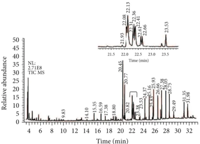
Re la ti ve a b un da n ce Time (min) NL: 2.71E8 TIC MS Time (min) 0
4 6 8
21.5 22.0 22.5 23.0 23.5
10 9.83 21.93 22.08 22.13 22.31 22.36 22.41
22.61 22.66 23.53
14.10
15.35 16.59 17.38 18.80
20.77
20.82 23.3823.53 24.37 25.16
25.83
25.93
26.66 27.38 28.05 28.75
29.49
31.35 31.98
20.45
12 14 16 18 20 22 24 26 28 30 32 5 10 15 20 25 30 35 40 45 50
Figure 1: Total ion chromatogram of BPE-CO2A extract. Identified
compounds are described inTable 1.
3. Results
3.1. Chemical Analysis of BPE-CO2A by GC/MS. In the total
ion chromatogram of BPE-CO2A (Figure 1) the presence of
several compounds such as phytol (0.139%) and fatty acids such as palmitic (30%), oleic (27%), linoleic (24.3%), and linolenic (3.8%) acids was observed. In addition the following
was detected in these plant extracts: thirteen alkanes (C11,
C14, C18, C20, C22, C23, C24, C25, C26, C27, C28, C29, and
C30), two ethyl esters of fatty acids (ethyl hexadecanoate and
ethyl 9,12-octadecadienoate), two sterols (stigmasterol and
sitosterol), and six unidentified compounds (Table 1).
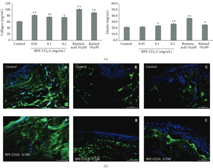
3.2. BPE-CO2A Increases Extracellular Matrix Elements. To
assess the retinoid-like activity of theB. pilosaextract, we
analysed the synthesis of the collagen, elastin, and gly-cosaminoglycans in cultured human fibroblasts treated with
various concentrations of B. pilosa extract, retinoic acid
(10𝜇M), or retinol (10𝜇M). After treatment with BPE-CO2A,
collagen levels were elevated by 32.5% at 0.05 mg/mL, 23.3% at 0.1 mg/mL, and 21.6% at 0.2 mg/mL. For comparison,
retinoic acid (10𝜇M) and retinol (10𝜇M) were able to increase
collagen levels by 62.9% and 45.6%, respectively (Figure 2).
BPE-CO2A was also able to significantly increase the
synthe-sis of elastinin vitro. At 0.2 mg/mL, the extract caused a 25.2%
increase in elastin levels (Figure 2). As expected, retinoic acid
(10𝜇M) and retinol (10𝜇M) increased elastin production by
66.7% and 16.7%, respectively. In contrast, BPE-CO2A was
unable to significantly alter the levels of glycosaminoglycans
(data not shown). Corroborating with the in vitro results,
BPE-CO2A increased the immunofluorescent staining for
collagen and elastin (Figures2(b)(A) and2(b)(B)). In
addi-tion, we also observed an increase in immunofluorescent staining for glycosaminoglycan when skin fragments were
treated with BPE-CO2A (Figure 2(b)(C)).
3.3. BPE-CO2A Stimulates Growth Factors. In another set of
experiments, we evaluated the effects of BPE-CO2A on
TGF-𝛽1 and FGF levels in cultured human fibroblasts as well as
the modulation of epidermal growth factor receptor (EGFr).
At the three concentrations tested, BPE-CO2A significantly
increased TGF-𝛽1 levels, with its highest stimulatory effect
(53.2%) at a concentration of 0.2 mg/mL (Figure 3). In the
same assay, retinoic acid (10𝜇M) increased TGF-𝛽1 synthesis
by 101.3%, and retinol was ineffective (Figure 3). Similar to its
TGF-𝛽1 induction, BPE-CO2A had a significant stimulatory
effect on FGF levels (Figure 3) with an increase of 188.3% after
the treatment of cultured human fibroblasts with BPE-CO2A
at 0.2 mg/mL. Retinoic acid (10𝜇M) and retinol (10𝜇M) also
caused significant FGF increases, but these effects were lower
than those produced by BPE-CO2A (Figure 3). In addition,
we evaluated the modulation of epidermal growth factor
receptor (EGFr) gene expression. BPE-CO2A produced a
2.6-, 2.3-, and 2-fold increase in EGFr gene expression at concentration of 0.05 mg/mL, 0.10 mg/mL, and 0.2 mg/mL, respectively. Retinoic acid increased EGFr mRNA levels by 52.3%, and retinol was not able to significantly increase the
expression of EGFr (Figure 3).
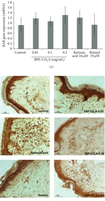
3.4. BPE-CO2A Modulates Retinoic Receptors. To evaluate
whether the effects induced by BPE-CO2A have similar
effects to retinoic acid and retinol in the modulation of receptors RXR and RAR, we evaluated the ability of the extract to modulate the gene expression of these receptors
in human fibroblasts (Figures4and 5). Gene expression of
RXR increased 3.6-, 3.7-, and 1.6-fold in the group treated
with BPE-CO2A at the concentration of 0.05, 0.10, and
0.20 mg/mL, respectively, whereas retinoic acid and retinol
increased gene expression 1.5-fold (Figure 4(a)). An increase
in receptor staining in the immunohistochemical evaluation of RXR performed on human skin fragments corroborates thein vitroresults (Figure 4(b)).
In contrast, the expression of RAR was not significantly
changed after treatment with BPE-CO2A, retinoic acid, or
retinol (Figure 5(a)). However, when we evaluated RAR
protein synthesis using immunohistochemistry, we found that treatment with retinoic acid markedly increased RAR
staining, whereas treatment with BPE-CO2A and retinol
promoted only subtle stimulation (Figure 5(b)).
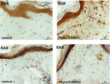
We also evaluated the effect of phytol, an acyclic
iso-prenoid compound present in BPE-CO2A, on RAR and RXR
production in skin fragments. Phytol (0.0002%) had similar
effects to BPE-CO2A, inducing RXR synthesis (Figure 6) and
not altering RAR production (Figure 6). It is thus possible
that the effect of BPE-CO2A on retinoid receptors might be
due to the presence of phytol.
3.5. BPE-CO2A Improves Histological Characteristics. Hae-matoxylin-eosin staining of human skin fragments was per-formed after various treatments with retinoic acid, retinol,
and BPE-CO2A (Figure 7). A comparative analysis showed
Table 1: Chemical compounds identified in the BPE-CO2extract by gas chromatography and mass spectroscopy (GC/MS) according to
chromatogram fromFigure 1.
Retention time (min) Compounds Molecular formula
9.83 Undecane C11
14.10 Tetradecane C14
15.35 Unidentified
16.59 Unidentified
17.38 Unidentified
18.80 Octadecane C18
20.45 Hexadecanoic acid (palmitic acid) C16H32O2
20.77 Ethyl hexadecanoate C18H36O2
20.82 Eicosane C20
21.93 3,7,11,15-Tetramethyl-2-hexadecen-1-ol (phytol) C20H40O
22.08 9,12-Octadecadienoic acid (linoleic acid) C18H32O2
22.13 9-Octadecenoic acid (oleic acid) C18H34O2
22.31 Octadecanoic acid (stearic acid) C18H36O2
22.36 Ethyl 9,12-octadecadienoate C20H36O2
22.41 9,12,15-Octadecatrienoic acid (linolenic acid) C18H30O2
22.61 Unidentified
22.66 Docosane C22
23.53 Tricosane C23
24.37 Tetracosane C24
25.16 Pentacosane C25
25.83 Unidentified
25.93 Hexacosane C26
26.66 Heptacosane C27
27.38 Octacosane C28
28.05 Nonacosane C29
28.75 Triacontane C30
29.49 Unidentified
31.35 Stigmasterol C29H48O
31.98 Sitosterol C29H50O
after a single application. The group treated with retinol had characteristics similar to those observed after treatment with retinoic acid, but with a smaller disruption of the horny layer, due to less aggressive behaviour towards retinoic
acid and retinol. BPE-CO2A, in turn, had a slight effect in
increasing the thickness of the epidermal basal layer and
dermis (Figure 7).
4. Discussion
The ageing of human skin is a complex biological phe-nomenon consisting of two components: intrinsic ageing (passage of time and individual genetic features) and extrinsic ageing caused by cumulative exposure to environmental
factors such as ultraviolet radiation [21]. Intrinsic ageing is a
slow, cumulative, progressive, and degradative process affect-ing mainly elastic fibre, while in extrinsic ageaffect-ing the slow
evolution can be enzymatically accelerated [22]. Both skin
ageing processes are associated with structural and functional changes that occur in the dermal extracellular matrix, where
fibrillar collagens, elastic fibres, and glycosaminoglycans are necessary to confer tensile strength, resilience, and hydration
[23]. In both intrinsic and extrinsic ageing, there is evidence
for the degradation of fibrous extracellular matrix compo-nents including elastin, collagens, and the oligosaccharide
fraction [23,24]. Our results showed that BPE-CO2A was able
to significantly increase the synthesis of collagen and elastin
in vitro.Although thein vitrostimulation promoted by
BPE-CO2A on extracellular matrix components is quantitatively
lower than the effect exerted by retinoic acid, it is evident
in the ex vivo experiments that BPE-CO2A stimulates the
production of collagen and elastin in the skin.
The fundamental mechanism for age-related collagen
synthesis involves the transforming growth factor-𝛽
(TGF-𝛽) signalling pathway, which stimulates collagen synthesis
in cultured fibroblasts [25–28]. Moreover, it has been
sug-gested that reactive oxygen species can activate different surface receptors, including those for epidermal growth factor, and this effect may be mediated in part by the
C
o
llag
en (m
g/mL)
Control
120 100 80 60 40
20 0
0.2 0.1 0.05
∗
∗∗ ∗∗ ∗∗
∗∗
BPE-CO2A (mg/mL)
Retinoic 10 𝜇M Retinol
60.0 50.0 40.0 30.0
20.0 10.0 0.0
∗ ∗
∗∗ ∗∗
Control
E
lastin (m
g/mL)
0.2 0.1
0.05
BPE-CO2A (mg/mL)
Retinoic 10 𝜇M Retinol
acid10 𝜇M acid10 𝜇M
(a)
(b)
Figure 2: Extracellular matrix elements evaluation. (a) Collagen and elastin production in human fibroblast cultures after 48 hours of
incubation with BPE-CO2A, retinoic acid, or retinol. (b) Collagen (A), elastin (B), and glycosaminoglycan (C) in human skin fragments
labelled with anti-collagen (green), anti-elastin (green), and anti-glycosaminoglycan (green) antibodies, respectively, after a 48-hour
incubation. The nuclei were counterstained with DAPI (blue) (40x magnification). The data represent the mean±SD of three individual
experiments.∗𝑃 < 0.05,∗∗𝑃 < 0.01.
The activation of several MAPK members, particularly c-Jun and c-Fos, is related to the transcription complex AP-1 that directly inhibits the production of procollagen by blocking
the TGF-𝛽 receptor [30, 31]. Topical TGF-𝛽 treatment in
wounds promotes collagen synthesis, increases tensile wound strength, stimulates granulation tissue formation, enhances the thickness of regenerate dermal tissue, and stabilizes the
dermoepidermal junction [32,33]. It has been related that the
reduction of collagen in photoaged human skin occurs due to
blocking the TGF-𝛽receptor [30]. BPE-CO2A, retinoic acid,
and retinol enhanced the production of TGF-𝛽in fibroblast
cultures, and this effect was in synergy with the reduction in collagen production that we detected in our experiments. Our results clearly showed that the increase in collagen synthesis
is closely related to the enhanced production of TGF-𝛽.
In addition to TGF-𝛽, fibroblast growth factor-1 has a
wide activity spectrum, mainly increasing synthesis of matrix
macromolecules, notably the main dermal
glycosaminogly-can, hyaluronic acid [34], which contributes to the
wound-healing process by stimulating fibroblast proliferation and
inhibits the expression of collagenase-1 in keratinocytes [35].
In our study, BPE-CO2A exerted a potent stimulatory effect
on FGF-𝛽levels in cultured human fibroblasts that was higher
than the effects produced by both retinoic acid and retinol. Another representative member of the growth factor family is epidermal growth factor (EGF), which can bind to and activate members of the EGF receptor (EGFR) family, leading to the initiation of several MAPKs signal trans-duction cascades and playing an important role in many cellular functions, such as proliferation, apoptosis, migration,
inflammation, and immunity [36–39]. In fact, EGF induces
collagenase expression in human dermal fibroblasts with a
consequent reduction in collagen synthesis [40, 41]. Our
Control 0.1 0.2 0
20000 40000 60000 80000 100000 120000 140000 160000 180000 200000
0.05
∗∗
∗∗ ∗∗ ∗
BPE-CO2A (mg/mL)
Retinoic 10 𝜇M Retinol
TG
F
-𝛽1
(pg/mL)
acid10 𝜇M
(a)
2500
2000
1500
1000
500
0
∗∗ ∗∗
∗∗
Control
FGF (pg/mL)
0.2 0.1 0.05
BPE-CO2A (mg/mL)
Retinoic 10 𝜇M Retinol
acid10 𝜇M
(b)
3.5
3
2.5
2
1.5
1
0.5
0
∗∗ ∗∗
∗∗
∗
EGF g
ene exp
re
ssio
n (mRN
A)
Control 0.05 0.1 0.2
BPE-CO2A (mg/mL)
Retinoic 10 𝜇M Retinol
acid10 𝜇M
(c)
Figure 3: Effects of BPE-CO2A, retinoic acid, and retinol on the TGF-𝛽1 (a) and FGF (b) levels and EGFr mRNA expression in human
fibroblast cultures. The data represent the mean±SD.∗𝑃 < 0.05,∗∗𝑃 < 0.01.
gene expression (2.6-fold), although retinoic acid produced a smaller increase (53.6%), and retinol was not able to increase gene expression. Thus, it is clear that the protective effect of
BPE-CO2A on collagen and elastin degradation was closely
related to the increase in the expression of growth factors,
particularly TGF-𝛽 and FGF-𝛽, and EGFR expression in
human fibroblasts. This effect was similar to that produced by retinoic acid and was sometimes more marked than effects produced by both retinoic acid and retinol.
In a second set of experiments, we evaluated the effects
of BPE-CO2A on both the gene expression and synthesis
of the retinoic acid and retinoid X receptors. BPE-CO2A,
retinoic acid, and retinol were not able to significantly alter the gene expression of RAR. However, retinoic acid markedly increased RAR protein synthesis as evidenced
using immunohistochemistry, whereas BPE-CO2A treatment
produces a subtle stimulation, similar to that produced by
retinol. In contrast, BPE-CO2A treatment increased RXR
gene expression significantly (3.6-fold compared to control) and to a greater extent than retinoic acid and retinol, both of which increased RXR gene expression approximately 1.5-fold. This effect was corroborated by an increase in RXR protein synthesis.
There is evidence that RAR and RXR mRNA levels decrease with age and that compounds able to increase
the expression of these genes have neuroprotective effects in
ageing [42, 43]. Thus, expression of RXRs was reduced in
healthy elderly men suggesting that RXRs reduction might
be a common feature of physiological senescence [44]. In
addition, a single dose of all-transretinoic acid to old rats
was found to increase RAR expression in the liver after 24 h, indicating that reversible alterations in retinoid receptors may
also occur during ageing [44,45]. This way, it is possible that
the protective effects produced by BPE-CO2A are also related
to RXR mRNA expression.
Finally, BPE-CO2A contains a mixture of phytol and
several long chain fatty acids, mainly palmitic, oleic, linoleic, and linolenic acids. Phytol, a branched fatty alcohol, is a carbon side-chain of chlorophylls and their metabolites, such as phytanic acid. Phytol has been shown to be a natural ligand for RXR, able to mimic various effects of conjugated linolenic acids, which activate the peroxisome
proliferator-activated receptor (PPAR) and the RXRs [46,47]. There is
evidence that PPAR-𝛾 mRNA levels decrease with age [41]
and that phytol is functional as a PPAR-𝛼ligand, stimulating
the expression of PPAR-𝛼-target genes in intact cells [47].
Moreover, phytol metabolites are compelling candidates for physiological effectors because their RXR binding affinities and activation potencies match their micromolar circulating
∗∗ ∗∗
Control
R
X
R g
ene exp
re
ssio
n (mRN
A)
0.2 0.1 0.05
BPE-CO2A (mg/mL)
Retinoic 10 𝜇M Retinol ∗
∗ ∗
0 1 2 3 4 5 6
acid10 𝜇M
(a)
(b)
Figure 4: Effects of BPE-CO2A, retinoic acid, and retinol on
RXR mRNA expression in human fibroblast cultures (a) and RXR synthesis (brown precipitate) in human skin fragments labelled with anti-RXR antibodies (b) after a 48-hour incubation (40x
magnification). The data represent the mean±SD. ∗𝑃 < 0.05,
∗∗𝑃 < 0.01.
of long-chain fatty acids have been correlated with normal
ageing and neurodegeneration [48], and some of these
long-chain fatty acids are also RXR ligands or increase RXR gene
expression [12–16,49]. In our study, phytol was able to induce
RXR synthesis but was not able to influence RAR production.
Because phytol is exclusively of dietary origin, aBidens pilosa
extract containing phytol may represent an essential dietary product to control cellular metabolism through the RXR signalling pathways.
Control
RAR g
ene exp
re
ssio
n (mRN
A
)
0.2 0.1 0.05
BPE-CO2A (mg/mL)
Retinoic 10 𝜇M Retinol 0
0.4 0.2 0.6 0.8 1.0 1.2 1.4 1.6 1.8
acid10 𝜇M
(a)
(b)
Figure 5: Effects of BPE-CO2A, retinoic acid, and retinol on RAR
mRNA expression in human fibroblasts (a) and RAR synthesis (brown precipitate) in human skin fragments labelled with anti-RXR antibodies (b) after a 48-hour incubation (40x magnification). The
data represent the mean±SD.∗𝑃 < 0.05,∗∗𝑃 < 0.01.
5. Conclusion
In conclusion, our results showed that BPE-CO2A from
Bidens pilosa enhanced extracellular matrix components, particularly collagen and elastin fibres through the
mainte-nance of TGF-𝛽, FGF-𝛽, and EGF levels and, simultaneously,
affected RXR gene expression and synthesis. It is an innova-tive acinnova-tive ingredient with several clinical applications and pharmacological activities that control intrinsic and extrinsic
Figure 6: Effects of phytol on RXR and RAR synthesis (brown pre-cipitate) in human skin fragments labelled with RXR and anti-RAR antibodies after a 48-hour incubation (40x magnification).
Figure 7: Photomicrographs of human skin fragments stained with haematoxylin-eosin and treated with retinoic acid, retinol, or
BPE-CO2A (40x magnification). Sc: stratum corneum, ep: epidermis, de:
dermis, and va: vacuoles.
is an innovative active ingredient with potential clinical applications to control intrinsic and extrinsic human skin ageing and to develop an herbal preparation with retinoid-like activity useful as a skin-repair and antiageing agent.
Disclaimer
The content of this publication is solely the responsibility of the authors and does not necessarily represent the official views of the financial support agencies.
Conflict of Interests
The authors declare no conflict of interests.
Acknowledgments
Research in the Di Stasi Lab (PhytoPharmaTech) was sup-ported by the S˜ao Paulo Research Foundation (FAPESP) and Chemyunion Quimica Ltda. Fellowships are as follows: LCDS from the National Council for Scientific and Technological Development (CNPq, Brazilian Ministry of Science and Technology).
References
[1] A. le Maire, S. ´Alvarez, P. Shankaranarayanan, A. R. de Lera,
W. Bourguet, and H. Gronemeyer, “Retinoid receptors and
therapeutic applications of RAR/RXR modulators,” Current
Topics in Medicinal Chemistry, vol. 12, no. 6, pp. 505–527, 2012. [2] R. E. B. Watson, J. A. Ratnayaka, R. C. C. Brooke, S.
Yee-Sit-Yu, P. Ancian, and C. E. M. Griffiths, “Retinoic acid receptor
𝛼expression and cutaneous ageing,”Mechanisms of Ageing and
Development, vol. 125, no. 7, pp. 465–473, 2004.
[3] P. Germain, P. Chambon, G. Eichele et al., “International union
of pharmacology. LXIII. Retinoid X receptors,”Pharmacological
Reviews, vol. 58, no. 4, pp. 760–772, 2006.
[4] L. Ritti´e, G. L. Fisher, and C. E. M. Griffiths, “Anti-aging
effects of retinoids and mechanisms of actions,” inRetinoids and
Carotenoids in Dermatology, A. Vahlquist and M. Duvic, Eds., pp. 77–102, Informa Healthcare, New York, NY, USA, 2007. [5] C. Stefanaki, A. Stratigos, and A. Katsambas, “Topical retinoids
in the treatment of photoaging,”Journal of Cosmetic
Dermatol-ogy, vol. 4, pp. 130–134, 2005.
[6] S. Knutsen-Larson, A. L. Dawson, C. A. Dunnick, and R. P. Dellavalle, “Acne vulgaris: pathogenesis, treatment, and needs
assessment,”Dermatologic Clinics, vol. 30, no. 1, pp. 99–106,
2012.
[7] F. L. Silva, D. C. H. Fischer, J. F. Tavares, M. S. Silva, P. F. De Athayde-Filho, and J. M. Barbosa-Filho, “Compilation of
secondary metabolites fromBidens pilosaL,”Molecules, vol. 16,
no. 2, pp. 1070–1102, 2011.
[8] A. P. Bartolome, I. M. Villase˜nor, and W.-C. Yang, “Bidens pilosa
L. (Asteraceae): botanical properties, traditional uses,
phyto-chemistry, and pharmacology,”Evidence-Based Complementary
and Alternative Medicine, vol. 2013, Article ID 340215, 51 pages, 2013.
[9] L. C. Di Stasi, C. A. Hiruma, E. M. Guimaraes, and C. M. Santos, “Medicinal plants popularly used in Brazilian Amazon,”
Fitoterapia, vol. 65, no. 6, pp. 529–540, 1994.
[10] L. C. Di Stasi, G. P. Oliveira, M. A. Carvalhaes et al., “Medicinal plants popularly used in the Brazilian Tropical Atlantic Forest,”
Fitoterapia, vol. 73, no. 1, pp. 69–91, 2002.
GAP-43/neuromodulin and RC3/neurogranin in the rat brain,”
British Journal of Nutrition, vol. 103, no. 12, pp. 1720–1729, 2010. [12] P. K. Lemotte, S. Keidel, and C. M. Apfel, “Phytanic acids is a
retinoid X receptor ligand,”European Journal of Biochemistry,
vol. 236, no. 1, pp. 328–333, 1996.
[13] J. Lengqvist, A. Mata de Urquiza, A.-C. Bergman et al., “Polyunsaturated fatty acids including docosahexaenoic and
arachidonic acid bind to the retinoid X receptor 𝛼
ligand-binding domain,”Molecular and Cellular Proteomics, vol. 3, no.
7, pp. 692–703, 2004.
[14] S. A. Moore, E. Hurt, E. Yoder, H. Sprecher, and A. A. Spector, “Docosahexaenoic acid synthesis in human skin fibroblasts involves peroxisomal retroconversion of tetracosahexaenoic
acid,”Journal of Lipid Research, vol. 36, no. 11, pp. 2433–2443,
1995.
[15] F. Zapata-Gonzalez, F. Rueda, J. Petriz et al., “Human den-dritic cell activities are modulated by the omega-3 fatty acid, docosahexaenoic acid, mainly through PPAR (gamma):RXR heterodimers: comparison with other polyunsaturated fatty
acids,”Journal of Leukocyte Biology, vol. 84, no. 4, pp. 1172–1182,
2008.
[16] E. Reverchon and I. de Marco, “Supercritical fluid extraction
and fractionation of natural matter,” Journal of Supercritical
Fluids, vol. 38, no. 2, pp. 146–166, 2006.
[17] I. Gamlieli-Bonshtein, E. Korin, and S. Cohen, “Selective
separation of cis-trans geometrical isomers of𝛽-carotene via
CO2supercritical fluid extraction,”Biotechnology and
Bioengi-neering, vol. 80, no. 2, pp. 169–174, 2002.
[18] G. Jenkins, “Molecular mechanisms of skin ageing,”
Mecha-nisms of Ageing and Development, vol. 123, no. 7, pp. 801–810, 2002.
[19] S. Kojima, H. Muramatsu, H. Amanuma, and T. Muramatsu, “Midkine enhances fibrinolytic activity of bovine endothelial
cells,”The Journal of Biological Chemistry, vol. 270, no. 16, pp.
9590–9596, 1995.
[20] E. A. Bauer, J. L. Seltzer, and A. Z. Eisen, “Retinoic acid inhi-bition of collagenase and gelatinase expression in human skin
fibroblast cultures. Evidence for a dual mechanism,”Journal of
Investigative Dermatology, vol. 81, no. 2, pp. 162–169, 1983. [21] M. S´ardy, “Role of matrix metalloproteinases in skin ageing,”
Connective Tissue Research, vol. 50, no. 2, pp. 132–138, 2009. [22] E. C. Naylor, R. E. B. Watson, and M. J. Sherratt, “Molecular
aspects of skin ageing,”Maturitas, vol. 69, no. 3, pp. 249–256,
2011.
[23] A. J. Bailey, “Molecular mechanisms of ageing in connective
tissues,”Mechanisms of Ageing and Development, vol. 122, no.
7, pp. 735–755, 2001.
[24] K.-A. Hwang, B.-R. Yi, and K.-C. Choi, “Molecular mechanisms and in vivo mouse models of skin aging associated with dermal
matrix alterations,”Laboratory Animal Research, vol. 27, no. 1,
pp. 1–8, 2011.
[25] R. A. Ignotz and J. Massague, “Transforming growth
factor-𝛽stimulates the expression of fibronectin and collagen and
their incorporation into the extracellular matrix,”The Journal
of Biological Chemistry, vol. 261, no. 9, pp. 4337–4345, 1986. [26] M. Bitzer, G. von Gersdorff, D. Liang et al., “A mechanism of
suppression of TGF-𝛽/SMAD signaling by NF-𝜅B/RelA,”Genes
and Development, vol. 14, no. 2, pp. 187–197, 2000.
[27] R. Raghow, A. E. Postlethwaite, J. Keski-Oja, H. L. Moses, and A.
H. Kang, “Transforming growth factor𝛽increases steady state
levels of type I procollagen and fibronectin messenger RNAs
posttranscriptionally in cultured human dermal fibroblasts,”
Journal of Clinical Investigation, vol. 79, no. 4, pp. 1285–1288, 1987.
[28] K. K. Nelson and J. A. Melendez, “Mitochondrial redox
con-trol of matrix metalloproteinases,” Free Radical Biology and
Medicine, vol. 37, no. 6, pp. 768–784, 2004.
[29] T. Quan, T. He, S. Kang, J. J. Voorhees, and G. J. Fisher, “Solar ultraviolet irradiation reduces collagen in photoaged
human skin by blocking transforming growth factor-𝛽type II
receptor/Smad signaling,”The American Journal of Pathology,
vol. 165, no. 3, pp. 741–751, 2004.
[30] M. Yaar and B. A. Gilchrest, “Photoageing: mechanism,
preven-tion and therapy,”British Journal of Dermatology, vol. 157, no. 5,
pp. 874–887, 2007.
[31] L. Tj¨aderhane, H. Palosaari, J. Wahlgren, M. Larmas, T. Sorsa, and T. Salo, “Human odontoblast culture method: the expression of collagen and matrix metalloproteinases (MMPs),”
Advances in Dental Research, vol. 15, pp. 55–58, 2001.
[32] R. Govinden and K. D. Bhoola, “Genealogy, expression, and
cellular function of transforming growth factor-beta,”
Pharma-cology and Therapeutics, vol. 98, no. 2, pp. 257–265, 2003. [33] K. Kuroda, A. Utani, Y. Hamasaki, and H. Shinkai,
“Up-regulation of putative hyaluronan synthase mRNA by basic fibroblast growth factor and insulin-like growth factor-1 in
human skin fibroblasts,”Journal of Dermatological Science, vol.
26, no. 2, pp. 156–160, 2001.
[34] B. K. Pilcher, J. Gaither-Ganim, W. C. Parks, and H. G. Wel-gus, “Cell type-specific inhibition of keratinocyte collagenase-1 expression by basic fibroblast growth factor and keratinocyte
growth factor: a common receptor pathway,”Journal of
Biologi-cal Chemistry, vol. 272, no. 29, pp. 18147–18154, 1997.
[35] M. R. Frey, R. S. Dise, K. L. Edelblum, and D. B. Polk, “p38 kinase regulates epidermal growth factor receptor downregulation and
cellular migration,”The EMBO Journal, vol. 25, no. 24, pp. 5683–
5692, 2006.
[36] S. Pastore, F. Mascia, V. Mariani, and G. Girolomoni, “The epidermal growth factor receptor system in skin repair and
inflammation,”Journal of Investigative Dermatology, vol. 128, no.
6, pp. 1365–1374, 2008.
[37] S. A. Grando, J.-C. Bystryn, A. I. Chernyavsky et al., “Apop-tolysis: a novel mechanism of skin blistering in pemphigus vulgaris linking the apoptotic pathways to basal cell shrinkage
and suprabasal acantholysis,”Experimental Dermatology, vol.
18, no. 9, pp. 764–770, 2009.
[38] C. H. Park and J. H. Chung, “Epidermal growth factor-induced matrix metalloproteinase-1 expression is negatively regulated by
p38 MAPK in human skin fibroblasts,”Journal of
Dermatologi-cal Science, vol. 64, no. 2, pp. 134–141, 2011.
[39] Y. Mimura, H. Ihn, M. Jinnin, Y. Asano, K. Yamane, and K. Tamaki, “Epidermal growth factor affects the synthesis and degradation of type I collagen in cultured human dermal
fibroblasts,”Matrix Biology, vol. 25, no. 4, pp. 202–212, 2006.
[40] S. E. Moon, N. Bhagavathula, and J. Varani, “Keratinocyte stimulation of matrix metalloproteinase-1 production and pro-liferation in fibroblasts: regulation through mitogen-activated
protein kinase signalling events,”British Journal of Cancer, vol.
87, no. 4, pp. 457–464, 2002.
[41] V. Enderlin, S. Alfos, V. Pallet et al., “Aging decreases the abundance of retinoic acid (RAR) and triiodothyronine (TR) nuclear receptor mRNA in rat brain: effect of the administration
[42] S. C. Dyall, G. J. Michael, and A. T. Michael-Titus, “Omega-3 fatty acids reverse age-related decreases in nuclear receptors
and increase neurogenesis in old rats,”Journal of Neuroscience
Research, vol. 88, no. 10, pp. 2091–2102, 2010.
[43] J. Brtko, E. Rock, P. Nezbedova et al., “Age-related change in the retinoid X receptor beta gene expression in peripheral blood
mononuclear cells of healthy volunteers: effect of 13-cisretinoic
acid supplementation,”Mechanisms of Ageing and Development,
vol. 128, no. 11-12, pp. 594–600, 2007.
[44] V. Pallet, V. Aza¨ıs-Braesco, V. Enderlin et al., “Aging decreases retinoic acid and triiodothyronine nuclear expression in rat liver: exogenous retinol and retinoic acid differentially
mod-ulate this decreased expression,” Mechanisms of Ageing and
Development, vol. 99, no. 2, pp. 123–136, 1997.
[45] M. F. McCarty, “The chlorophyll metabolite phytanic acid is a natural rexinoid—potential for treatment and prevention of
diabetes,”Medical Hypotheses, vol. 56, no. 2, pp. 217–219, 2001.
[46] T. Goto, N. Takahashi, S. Kato et al., “Phytol directly activates
peroxisome proliferator-activated receptor𝛼(PPAR𝛼) and
reg-ulates gene expression involved in lipid metabolism in PPAR𝛼
-expressing HepG2 hepatocytes,” Biochemical and Biophysical
Research Communications, vol. 337, no. 2, pp. 440–445, 2005. [47] S. Kitareewan, L. T. Burka, K. B. Tomer et al., “Phytol
metabo-lites are circulating dietary factors that activate the nuclear
receptor RXR,”Molecular Biology of the Cell, vol. 7, no. 8, pp.
1153–1166, 1996.
[48] C. I. F. Janssen and A. J. Kiliaan, “Long-chain polyunsaturated fatty acids (LCPUFA) from genesis to senescence: the influence of LCPUFA on neural development, aging, and
neurodegener-ation,”Progress in Lipid Research, vol. 53, no. 1, pp. 1–17, 2014.
[49] A. M. de Urquiza, S. Liu, M. Sj¨oberg et al., “Docosahexaenoic acid, a ligand for the retinoid X receptor in mouse brain,”
Submit your manuscripts at
http://www.hindawi.com
Stem Cells
International
Hindawi Publishing Corporationhttp://www.hindawi.com Volume 2014
Hindawi Publishing Corporation
http://www.hindawi.com Volume 2014 INFLAMMATION
Hindawi Publishing Corporation
http://www.hindawi.com Volume 2014
Behavioural
Neurology
Endocrinology
International Journal ofHindawi Publishing Corporation
http://www.hindawi.com Volume 2014
Hindawi Publishing Corporation
http://www.hindawi.com Volume 2014
Disease Markers
Hindawi Publishing Corporation
http://www.hindawi.com Volume 2014 BioMed
Research International
Oncology
Journal ofHindawi Publishing Corporation
http://www.hindawi.com Volume 2014
Hindawi Publishing Corporation
http://www.hindawi.com Volume 2014
Oxidative Medicine and Cellular Longevity
Hindawi Publishing Corporation
http://www.hindawi.com Volume 2014
PPAR Research
The Scientific
World Journal
Hindawi Publishing Corporationhttp://www.hindawi.com Volume 2014
Immunology Research Hindawi Publishing Corporation
http://www.hindawi.com Volume 2014 Journal of
Obesity
Journal ofHindawi Publishing Corporation
http://www.hindawi.com Volume 2014
Hindawi Publishing Corporation
http://www.hindawi.com Volume 2014
Computational and Mathematical Methods in Medicine
Ophthalmology
Journal ofHindawi Publishing Corporation
http://www.hindawi.com Volume 2014
Diabetes Research
Journal ofHindawi Publishing Corporation
http://www.hindawi.com Volume 2014
Hindawi Publishing Corporation
http://www.hindawi.com Volume 2014
Research and Treatment
AIDS
Hindawi Publishing Corporation
http://www.hindawi.com Volume 2014 Gastroenterology Research and Practice
Hindawi Publishing Corporation
http://www.hindawi.com Volume 2014
Parkinson’s
Disease
Evidence-Based Complementary and Alternative Medicine
Volume 2014 Hindawi Publishing Corporation