Alterations in the Fat Body Cells of
Rhinocricus padbergi
~
Diplopoda
!
Resulting from Exposure to Substrate
Containing Sewage Sludge
Raphael Bastão de Souza and Carmem Silvia Fontanetti*
Department of Biology, Institute of Biosciences, São Paulo State University (UNESP), Av. 24A, 1515, 13506-900, Rio Claro, SP, Brazil
Abstract: The final disposal of residues generated at sewage treatment plants~STPs! has become a major problem for cities, due to the increase in the amount of treated sewage. One of the alternatives for the residue, labeled “sewage sludge,” is its reuse in agriculture and in degraded soil. However, not all pathogens and metals present in it are eliminated during treatment. Diplopods have been used as bioindicators in ecotoxicological tests as they are constantly in close contact with the soil. Owing to this fact, the purpose of this study was to expose specimens of the diplopodRhinocricus padbergito substrate containing sewage sludge collected at STPs to analyze morphological alterations in their parietal and perivisceral fat body, where substances are stored. The exposures were held for 7, 15, or 90 days at different concentrations of sewage sludge~control, 1%, 10%, and 50%!. The parietal fat body showed no alterations in any of the three exposure periods or concentrations. Alterations in the perivisceral fat body were observed for all exposure periods. According to the results, we suggest that the sludge used has toxic agents responsible for changing the animal’s perivisceral fat body.
Key words:soil toxicity, STP, histochemistry, histology, acute exposure, subchronic exposure, millepede
I
NTR ODUCTIONThere has recently been a growing interest in the use of bioindicators to analyze the effects of chemical substances in the soil. Land invertebrates have been used by ecotoxicol-ogists to document and quantify the exposure and effects of pollutants in the environment. For such analysis, histology and light and electron microscopy were used to diagnose resulting cellular and subcellular changes, as well as to identify cell death symptoms and to reveal reactions in response to subchronic and sublethal exposures in cells and tissues~Kammenga et al., 2000!.
Since land invertebrates make up an important group responsible for organic matter decomposition in the soil, they have been used in studies aimed at understanding the poten-tial of metals present in the soil~Köhler & Triebskorn, 1998!. Sewage sludge is one of the residues obtained in the treatment of wastewater. A goal is to make it suitable for discharge into receiving water bodies with the smallest impact possible. According to the volume of water treated in sewage treatment plants~STPs!, a large amount of sludge may accumulate, making its final disposal a major environ-mental issue~Gomes et al., 2005!.
This residue requires an alternative for safe final dis-posal when it comes to environmentally acceptable public health considerations ~Andreoli, 2001!. Therefore, its use will only be allowed in agriculture if it meets the minimum standards required by environmental authorities regarding the presence of metal and human pathogens, among other substances~Conama, 2006; Camargo et al., 2008!.
Considering the habits of diplopods and the few ecotox-icological studies that have included this group, this study used specimens ofRhinocricus padbergias test organisms to analyze the effects caused by possible toxic agents present in sewage sludge of a given STP in the state of São Paulo, Brazil.
In millipedes, the primary function of the fat body is storage of lipids, glycogen, proteins, and uric acid. Millipede fat bodies are a permanent site for the storage of wastes
~Hopkin & Read, 1992!. In general, the fat body in R. padbergi has a whitish appearance with adipocytes orga-nized in rows. They are large, with well-defined limits and a spherical nucleus located in different positions. The cyto-plasm shows a great quantity of stored material, probably of variable composition, including numerous vacuoles. In as-sociation with adipocytes, smaller cells, named oenocytes, are observed. The parietal fat body is located dorsally in the animal, and the perivisceral fat body fills the body cavity, forming an irregular and anastomotic mass ~Fontanetti et al., 2004!.
This species was chosen as it is abundant in the region where this study took place and other aspects of its biology have already been studied ~Fantazzini et al., 1998, 2002; Arab et al., 2003; Fontanetti & Camargo-Mathias, 2004; Fontanetti et al., 2004, 2006; Calligaris et al., 2005; Miyoshi et al., 2005!.
M
ATERIALS ANDM
ETHODSBioassays
Adult specimens of R. padbergiwere manually collected in the city of Rio Claro~22824'36''S; 47833'36''W!, SP, Brazil,
Received May 25, 2011; accepted October 21, 2011 *Corresponding author. E-mail: fontanet@rc.unesp.br
M
icroscopy
M
icroanalysis
in February 2009. In the laboratory they were submitted to a 20-day acclimatization period at 218C.
The sewage sludge sample came from a STP located in the Alto Tietê Basin~STP AT-1!area. For animal exposure to the sludge, it was necessary to assemble a control bioassay containing soil from the place of collection, as well as three other bioassays containing soil from the place of collection mixed with different concentrations~1%, 10%, and 50%!of sewage sludge. In each bioassay, 12 individuals were exposed to periods of 7~acute exposure!, 15~intermediate exposure!, or 90 days~subchronic exposure!. Following each exposure period, three animals from each bioassay were anesthetized with sulfuric ether and dissected in a physiological solution.
Histology and Histochemistry
For histological and histochemical analyses, fragments of the parietal and perivisceral fat body were fixed in different solutions~see below!and then placed in a phosphate buffer pH 7.4.
Afterward, samples were dehydrated in alcohol and transferred to a JB-4-Polaron Instruments/Bio Rad ~ Bio-Rad Laboratories, Hercules, CA, USA! resin solution in a refrigerator for 24 h. After that process, the material was placed into molds containing resin with a catalyst. After being polymerized, the blocks were sectioned using a Leica RM2245 microtome. The slides obtained were stained with hematoxylin and eosin ~H-E! stain. The von-Kossa ~
Jun-queira & JunJun-queira, 1983!, sudan black B ~Junqueira & Junqueira, 1983!, bromophenol blue ~Pearse, 1985!, and Periodic Acid-Schiff ~PAS! ~Junqueira & Junqueira, 1983!
techniques were used to detect calcium, lipids, total pro-teins, and neutral polysaccharides, respectively. For H-E, von-Kossa, bromophenol blue, and PAS techniques, the fat bodies were fixed in Carnoy~absolute alcohol, chloroform, acetic acid!and for the sudan Black B method the fat bodies were fixed in phormolcalcium 10%.
A blinded examiner reviewed all of the slides for each stain to ensure consistency of evaluation and to minimize observer bias. Then, sections were classified as strongly positive ~⫹⫹⫹!, moderately positive~⫹⫹!, weakly positive
~⫹!, and negative ~⫺! based on staining characteristics; cellular alterations were classified as frequently present
~⫹⫹⫹!, regularly present ~⫹⫹!, rarely present~⫹!, and no alterations~⫺!.
R
ESULTSResults for histochemical analysis and histological alter-ations are summarized in Tables 1, 2, and 3, respectively.
Perivisceral Fat Body
Control Group
Control groups for the 7-, 15-, and 90-day periods pre-sented the histological pattern described for the species Table 1. Histochemical Analysis of the Perivisceral and Parietal Fat Body Exposed to Control Soil.
Perivisceral Fat Body Parietal Fat Body
Histochemical Analysis Adipocytes Spherocrystals Adipocytes Spherocrystals
Total proteins ⫹⫹⫹ ⫺ ⫹⫹ ⫺
Neutral polysaccharides ⫹⫹⫹ ⫹ ⫹⫹⫹ ⫹
Calcium ⫹ ⫹⫹ ⫹ ⫹⫹
Lipid ⫹ ⫺ ⫹ ⫺
Note:⫹⫹⫹, Strongly positive reaction;⫹⫹, moderately positive reaction;⫹, weakly positive reaction;⫺, negative reaction.
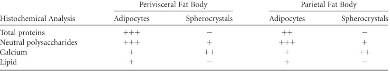
~Fontanetti et al., 2004!. In general, the fat body has a whitish appearance, with adipocytes displayed in rows.R. padbergi’s adipocytes are big with well-defined limits and a round nucleus located in different positions. Its cytoplasm is heterogeneous, holding several round structures—some of which have a concentric display—named spherocrystals
~see arrows in Figs. 1A, 1B!. Associated with adipocytes, smaller cells, namely oenocytes, can be found.
The histochemical analysis showed considerable quan-tities of protein ~Fig. 2A! and neutral polysaccharides
~Fig. 2B!distributed within the cytoplasmic cells. Granule-shaped calcium distributed both within the cytoplasm~ ar-Table 2. Histochemical Analysis of the Perivisceral Fat Body Exposed to Sewage Sludge.
Perivisceral Fat Body
Adipocytes Spherocrystals
Histochemical Analysis 1% 10% 50% 1% 10% 50%
Total proteins
Acute exposure ⫹⫹⫹ ⫹⫹⫹ ⫹ ⫺ ⫺ ⫺
Intermediate exposure ⫹⫹⫹ ⫹ ⫹ ⫺ ⫺ ⫺
Subchronic exposure ⫹ * * ⫺ * *
Neutral polysaccharides
Acute exposure ⫹⫹ ⫹ ⫹ ⫹ ⫹ ⫹
Intermediate exposure ⫹ ⫹ ⫹ ⫹ ⫹ ⫹
Subchronic exposure ⫹ * * ⫹ * *
Calcium
Acute exposure ⫹ ⫹ ⫹ ⫹⫹ ⫹⫹ ⫹⫹
Intermediate exposure ⫹ ⫹ ⫹ ⫹⫹ ⫹⫹ ⫹⫹
Subchronic exposure ⫹ * * ⫹⫹⫹ * *
Lipid
Acute exposure ⫹ ⫹ ⫹ ⫺ ⫺ ⫺
Intermediate exposure ⫹ ⫹ ⫹ ⫺ ⫺ ⫺
Subchronic exposure ⫹ * * ⫺ * *
Note:⫹⫹⫹, Strongly positive reaction;⫹⫹, moderately positive reaction;⫹, weakly positive reaction;⫺, negative reaction; *, animals did not survive.
Table 3. Histological Alterations in the Perivisceral Fat Body of the DiplopodR. padbergi.
Bioassays
Alterations Control Group
Sewage Sludge 1%
Sewage Sludge 10%
Sewage Sludge 50%
Loss of cellular limit
Acute exposure ⫺ ⫺ ⫺ ⫹
Intermediate exposure ⫺ ⫺ ⫺ ⫺
Subchronic exposure ⫺ ⫹⫹⫹ * *
Cytoplasmic disorganization
Acute exposure ⫺ ⫺ ⫺ ⫹
Intermediate exposure ⫺ ⫺ ⫹ ⫹⫹
Subchronic exposure ⫺ ⫹⫹⫹ * *
Increase in the quantity of spherocrystals
Acute exposure ⫺ ⫺ ⫹⫹ ⫹⫹
Intermediate exposure ⫺ ⫺ ⫹⫹ ⫹⫹
Subchronic exposure ⫺ ⫹⫹ * *
Nuclei with altered morphology
Acute exposure ⫺ ⫺ ⫺ ⫺
Intermediate exposure ⫺ ⫺ ⫹ ⫹⫹
Subchronic exposure ⫺ ⫹⫹⫹ * *
rowhead in Fig. 2C! and the spherocrystals ~arrows in Fig. 2C!was detected. A small amount of lipid was observed in the adipocytes~Fig. 2D!.
Acute Exposure
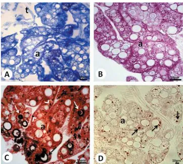
Specimens exposed to sludge at 1% or 10% concentrations presented intact fat body cells; an increase in the quantity of spherocrystals in the cells of animals exposed to the sludge at 10% concentrations was observed~Fig. 3A!. In the group exposed to the 50% concentration, some cells showed cyto-plasmic disorganization and loss of cellular limit~see arrow-head in Fig. 3B!, as well as an increase in the number of spherocrystals~Fig. 3B!.
The animals exposed to the 1% concentration did not show any alterations in the distribution pattern and the quantity of proteins, lipids, and calcium. There was a slight reduction in the quantity of neutral polysaccharides.
For the animals exposed to the 10% concentration, a noticeable reduction in the quantity of neutral polysaccha-rides, which were confined in some regions of the adipocyte cytoplasm, was observed~see arrows in Fig. 3C!. The tests done to detect proteins, calcium, and lipids showed no alterations.
It was observed that the animals exposed to sludge in the 50% concentration showed both protein~Fig. 3D!and neutral polysaccharide depletion; calcium and lipids suf-fered no alterations.
Intermediate Exposure
Histological analysis of the group exposed to the sludge in 1% concentration showed no alteration in relation to the control group. The animals exposed to the sludge in
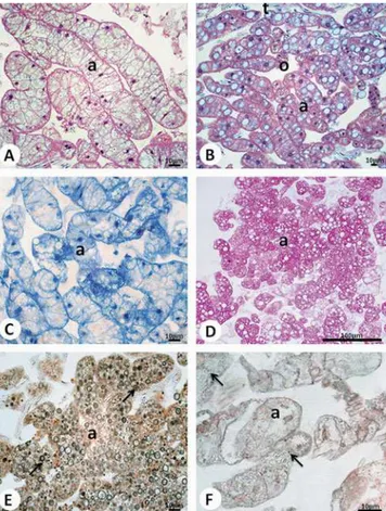
the 10% concentration showed an increase in the num-ber of spherocrystals, cells with cytoplasmic degradation
~Fig. 4A!, and nuclei with altered morphology ~see detail in Fig. 4A!. For animals exposed to the 50% sludge concen-tration, the same alterations described for the animal in the group exposed to the 10% sludge concentration were observed.
Upon applying the test to detect proteins, it was ob-served that the animals exposed to the 1% sludge concentra-tion did not show any alteraconcentra-tions in relaconcentra-tion to the control group, whereas for the animals exposed to the 10% and 50% concentrations, there was a decrease in these elements. A reduction in the quantity of neutral polysaccharides was observed for the animals exposed to all concentrations. There were no alterations concerning the amount of lipid and calcium.
Subchronic Exposure
The animals exposed in the bioassays with sludge at 10% and 50% concentration did not survive to the end of the experiment~90 days, as initially proposed!. Therefore, only animals exposed to sludge at 1% concentration were used to analyze subchronic exposure. Cells with a high cytoplasmic degradation rate were observed, with visible loss of cellular limit and deformed and pyknotic nuclei~Fig. 4B!. Most of the spherocrystals reacted positively to the technique used to detect calcium ~Fig. 4C!; a decrease in the quantity of total proteins and neutral polysaccharides was evidenced by both the bromophenol blue and the PAS techniques, respectively.
Figure 2. Histological sections of R. padbergi’s perivisceral fat body submitted to~A!bromophenol blue,~B!PAS,~C!von Kossa, and ~D! sudan black techniques. Control group. a⫽Adipocyte; arrowhead on panelC⫽calcium granules; arrows on panelC⫽ spherocrystals with calcium; arrows on panelD⫽lipid granules; t⫽tracheole.
Parietal Fat Body
Parietal fat body cells of the animals exposed to the three sewage sludge concentrations for 7, 15, or 90 days showed no histological or histochemical alterations in relation to the control group, being in accordance with the pattern described for the species~Fontanetti et al., 2004! ~Fig. 5!.
D
ISCUSSIONDepending on the origin, sewage sludge has quite a peculiar composition because it reflects various particularities of the local population, such as eating habits, sanitation condi-tions, and industrialization level~CETESB, 1999!. Accord-ing to Tsutiya ~1999!, taking its origin into consideration,
the sewage sludge used in this study may be classified as industrial and therefore presents higher concentrations of toxic substances compared to domestic sewage sludge.
In the first analysis, sludge toxicity may be evidenced by the mortality rate presented in the bioassays. The animals exposed to the 10% or 50% sludge concentrations did not survive until the end of the experiment so that it was not possible to conduct subchronic-related exposure analysis
~90 days!, also indicating that the animals were turning over the substrate.
Land invertebrates have been used as bioindicators of environmental pollution caused by metals such as cad-mium, copper, lead, and zinc, due to their constant contact with soil contaminants. The accumulation of metals in worms or invertebrates that feed on those metals has been commonly used as a parameter for assessing environmental risks~Roberts & Johnson, 1978; Ireland, 1979; Beyer et al., 1985; Heikens et al., 2001!.
Due to their habits, diplopods can be influenced by the disposal of metals in soil. Some species of millipedes can suffer a remarkable reduction in the number of their repre-Figure 4. Histological sections of R. padbergi’s perivisceral fat
body stained with~A,B!H-E and submitted to the~C!von Kossa technique. Fifteen-day exposure:~A!group exposed to 10% sludge concentration. 90-day exposure: ~B, C! group exposed to 1% sludge concentration. a⫽Adipocyte; arrowhead⫽loss of cellular limit; arrows⫽spherocrystals with calcium; detail on panelA⫽ nuclei with altered morphology; n⫽nucleus; o⫽oenocyte.
sentatives within very contaminated areas ~Hopkin et al., 1985!, whereas others have efficient organism detoxification strategies~Köhler et al., 1995!.
The epithelium of a millipedes’ digestive tract is made up of a layer of cells in direct contact with the external environment, thus guaranteeing a first line of defense against an array of toxic substances ~Köhler et al., 1995!. When invading agents manage to cross the integument and the digestive tract, they expose themselves to a variety of cellu-lar and humoral mechanisms acting on the host’s behalf. In some species of diplopods, however, the intestinal epithe-lium may not work efficiently as a barrier to passage of chemical compounds from the lumen into other body tis-sues~Hopkin et al., 1985!.
Due to a failure in this barrier,R. padbergi’s fat body was affected by toxic substances present in the sludge. However, alterations do not take place equally within the tissue. The parietal fat body, which is firmly stuck to the integument in a peripheral layer ~Fontanetti et al., 2004!, was not affected by the toxic substances. Therefore, neither morphologic nor histochemical alterations in this type of fat body were observed.
Alterations resulting from exposure to sludge were found in the perivisceral fat body, a tissue that fills the body cavity and involves different organs, such as the digestive tract and the gonads~Fontanetti et al., 2004!.
Acute exposure revealed that either 1% or 10% concen-trations were not enough to cause damage to cells because they were found intact. However, the sludge present in the substrate for these concentrations was responsible for the increase in the amount of mineralized structures. According to Hubert~1975!, these mineralized structures in Diplopoda happen in the shape of spherocrystals and granules. Hopkin and Read ~1992! state that fat body cells incorporate a variety of granules that contain metals, serving as perma-nent places for storing unnecessary substances. One of the detoxification mechanisms described for invertebrates is the precipitation of metals as intracellular granules of different types ~Hopkin, 1989!. Thus, chemical compounds that crossed the digestive tract were stored and inactivated in the fat body cells to occur subsequent release.
The two-week exposure ~intermediate exposure! also presented animals with an increase in the quantity of sphero-crystals in the cytoplasm. From this period of exposure, the reduction in the quantity of neutral polysaccharides and total proteins became more visible. Nath et al.~1997! and Nath ~2000! carried out studies involving silk worms ex-posed to organophosphate insecticides, in which they ob-served alterations in protein and carbohydrate reserves as a consequence of the stress.
In this study, the sludge may have acted as a stressor for adipocytes, changing their metabolism, and the carbohy-drate and protein reserves were used by the cell to deal with possible toxic effects of sludge substances.
Cytoplasmic degradation and loss of cellular limit were observed in the three exposure periods. In subchronic expo-sure, however, these alterations were much more visible,
followed by a large quantity of deformed and pyknotic nuclei.
According to Meyer and Da Silva~1999!, cells submit-ted to stress may either survive or die. As a result of stress, they may suffer necrosis, a kind of death in which there is cell volume increase, chromatin aggregation, cytoplasmic disorganization, integrity loss of plasma membrane, and consequent cell breakage~Grivicich et al., 2007!.
Comparative analysis of cell alterations indicates that the increase in sludge concentration is responsible for accel-erating tissue responses. Cytoplasmic disorganization was observed only for the 50% concentration in acute exposure, whereas the same alteration was largely visible for the 1% concentration in subchronic exposure. In a first phase, the animals exposed for shorter periods of time and in lower concentrations seem to be able to deal with toxic substances, storing material in spherocrystals and relocating certain com-ponents such as neutral polysaccharides and proteins to be used in other functions. As the exposure periods and/or concentrations are increased, this kind of response seems to reach its limit. From then on, a high degree of cell damage is observed, including deformed nuclei and loss of cellular limit. This defense mechanism was found in other studies involving diplopods of the same species; however, in those studies, the midgut was exposed. Godoy and Fontanetti
~2010!exposed the organ to crude sludge for 7 or 15 days. In the 15-day exposure, the accumulation of substances seems to have reached its limit; an intense epithelial renewal and an increase in the quantity of hemocytes were observed. Nogarol and Fontanetti ~2010! used substrates containing 1%, 10%, or 50% sewage sludge concentrations to obtain acute, intermediate, or subchronic responses. It was also observed that exposures to higher sludge concentrations may accelerate some responses, such as cytoplasmic granule increase, found as an acute response for animals exposed to 50% concentrations, and as a subchronic response for ani-mals exposed to 1% concentrations.
Therefore, low levels of toxic substances may be toler-ated by the animal studied for a short period of time. Nevertheless, as the period increases, the organism’s de-fenses become unable to protect it and morphological alter-ations resulting from contamination appear. On the other hand, high levels of toxic substances, associated with a long exposure period, exceed the maximum limits the organism can withstand, thus leading to its death.
C
ONCLUSIONThe data found in this study suggest one should be cautious when applying sewage sludge to the soil because soil-associated fauna may be negatively affected.
A
CKNOWLEDGMENTSsewage sludge samples; Gerson Mello de Souza for technical support; biologists Larissa Rosa Nagarol and Vlamir Boz-zato de Oliveira for their help during the experiments.
R
EFER ENCESAndreoli, C.V.~2001!.Resíduos sólidos do saneamento: Processa-mento, reciclagem e disposição final. Rio de Janeiro, Brazil: ABES.
Arab, A., Zacarin, G.G., Fontanetti, C.S., Camargo-Mathias, M.I., Santos, M.G.& Cabrera, A.C.~2003!. Composition of the defensive secretion of the neotropical millipedeRhinocricus padbergi Verhoeff, 1938 ~Diplopoda, Spirobolida, Rhinocri-cidae!.Entomotropica18, 79–82.
Beyer, W.N., Pattee, O.H., Sileo, L., Hoffman, D.J.&Mulhern, B.M.~1985!. Metal contamination in wildlife living near two zinc smelters.Environ Pollut Ser38, 63–86.
Calligaris, I.B., Boccardo, L., Sanches, M.R.& Fontanetti, C.S.~2005!. Morphometric analysis of a population of diplo-pods of the genusRhinocricusKarsch, 1881.Folia Biologica51, 40–46.
Camargo, O.A., Pires, A.M.M.& Bettiol, W.~2008!. Lodo na agricultura.Ciência Hoje42~248!, 68–70.
CETESB ~1999!. Aplicação de lodos de sistemas de tratamento biológico em áreas agrícolas—Critérios para projeto e operação. Manual Técnico—P4230. Companhia de Tecnologia de Sanea-mento Ambiental.
Conama~2006!. Resolução 375/2006. Conselho Nacional do Meio Ambiente. Available at http://www.mma.gov.br/port/conama/ legiabre.cfm?codleg⫽506~accessed June 6, 2008!.
Fantazzini, E.R., Fontanetti, C.S.&Camargo-Mathias, M.I.
~1998!. Anatomy of digestive tube, histology and histochemis-try of the foregut and salivary glands ofRhinocricus padbergi ~Diplopoda, Rhinocricidae!.Arthropoda Selecta7, 257–264.
Fantazzini, E., Fontanetti, C.S. & Camargo-Mathias, M.I.
~2002!. Midgut of the millipedeRhinodricus padbergiVerhoeff, 1938~Diplopoda: Spirobolida!: Histology and histochemistry. Arthropoda Selecta11, 135–142.
Fontanetti, C.S.&Camargo-Mathias, M.I.~2004!. Presence of calcium in oocytes of the diplopodRhinocricus padbergi Ver-hoeff~Spirobolida, Rhinocricidae!.Acta Histochemica37, 301– 306.
Fontanetti, C.S., Camargo-Mathias, M.I.& Tiritan, B.M.S.
~2004!. The fat body in Rhinocricus padbergi ~Diplopoda, Spirobolida!.Iheringia Sér Zool94~4!, 351–355.
Fontanetti, C.S., Tiritan, B.&Camargo-Mathias, M.I.~2006!. Mineralized bodies in the fat body of Rhinocricus padbergi ~Diplopoda!.Braz J Morphol Sci23~3–4!, 487–493.
Godoy, J.A.P.& Fontanetti, C.S.~2010!. Diplopods as bioindi-cators of soils: Analysis of midgut of individuals maintained in substract containing sewage sludge. Water Air Soil Pollut 210~1–4!, 389–398.
Gomes, S.B.V., Nascimento, C.W.A.&Biondi, C.M.~2005!. Produ-tividade e composição mineral de plantas de milho em solo adubado com lodo de esgoto.Revista Brasileira de Engenharia Agrícola e Ambiental11~5!, 459–465.
Grivicich, I., Regner, A.&Rocha, A.B.~2007!. Morte celular por apoptose.Revista Brasileira de Cancerologia53~3!, 335–343.
Heikens, A., Peijnenburg, W.J.G.M. & Hendriks, A.J. ~2001!. Bioaccumulation of heavy metals in terrestrial invertebrates. Environ Pollut113, 385–393.
Hopkin, S.P.~1989!.Ecophysiology of Metals in Terrestrial Inverte-brates. Barking, UK: Elsevier Applied Science.
Hopkin, S.P.&Read, H.J.~1992!.The Biology of Millipedes. New York: Oxford University Press.
Hopkin, S.P., Watson, K., Martin, M.H.&Mould, M.L.~1985!. The assimilation of heavy metals byLithobius variegatusand Glomeris marginata~Chilopoda; Diplopoda!.Bijdr Dierk55~1!, 88–94.
Hubert, M.~1975!. Sur la nature des accumulations minerales et puriquez chezCylindroiulus teutonicusPocock~londinensisCLK, Diplopoda, Iuloidea!.CR Acad Sc Paris281D, 151–154.
Ireland, M.P. ~1979!. Metal accumulation by the earthworms Lumbricus rubellus,Dendrbaena venetaandEiseniella tetraedra living in heavy metal polluted sites.Environ Pollut19, 201–206.
Junqueira, L.C.&Junqueira, L.M.M.S.~1983!.Técnicas Básicas de Citologia e Histologia. São Paulo, Brazil: Livraria Editora Santos.
Kammenga, J.E., Dallinger, R., Donker, M.H., Köhler, H.R., Simonsen, V., Triebskorn, R.&Weeks, J.M.~2000!. Biomak-ers in terrestrial invertebrates for ecotoxicological soil risk assessment.Rev Environ Contam Toxicol164, 93–147.
Köhler, H.R., Körtje, K.H. & Alberti, G. ~1995!. Content, absorption quantities and intracellular storage sites of heavy metals in Diplopoda~Arthropoda!.BioMetals8, 37–46.
Köhler, H.R.&Triebskorn, R.~1998!. Assessment of the cyto-toxic impact of heavy metals on soil invertebrates using a protocol integrate qualitative and quantitative components. Biomakers3~2!, 109–127.
Meyer, T.N.&Da Silva, A.L.~1999!. Resposta celular ao estresse. Rev Ass Med Brasil45~2!, 181–188.
Miyoshi, A.R., Gabriel, V.A., Fantazzini, E.R.& Fontanetti, C.S. ~2005!. Microspines in the pylorus of Pseudonannolene tricolorandRhinocricus padbergi~Arthropoda, Diplopoda!. Ihe-ringia95, 183–187.
Nath, B.S.~2000!. Changes in carbohydrate metabolism in hemo-lymph and fat body of the silkworm, Bombyx moriL. exposed to organophosphorus insecticides. Pestic Biochem Phys 68, 1504–1515.
Nath, B.S., Suresh, A., Varma, B.M. & Kumar, R.P.S. ~1997!. Changes in protein metabolism in hemolymph and fat body of the silkworm,Bombyx mori~Lepdoptera: Bombycidae! in re-sponse to organophosphorus insecticides toxicity.Toxicol Envi-ron Safety36, 169–173.
Nogarol, L.R.&Fontanetti, C.S.~2010!. Acute and subchronic exposure of diplopods to substrate containing sewage mud: Tissular responses of the midgut.Micron41, 239–246.
Pearse, A.G.E. ~1985!. Histochemistry: Theoretical and Applied. Edinburgh, UK: Churchill Livingstone.
Roberts, R.D.&Johnson, M.S.~1978!. Dispersal of heavy metals from abandoned mine workings and their transference through terrestrial food chains.Environ Pollut16, 293–310.