Research Article
Thyroid Hormone Status Interferes with Estrogen Target Gene
Expression in Breast Cancer Samples in Menopausal Women
Sandro José Conde,
1,2Renata de Azevedo Melo Luvizotto,
2Maria Teresa de Síbio,
2and Célia Regina Nogueira
21Department of Biological Science, S˜ao Paulo Federal Institute (IFSP), 18136-540 S˜ao Roque, SP, Brazil
2Department of Internal Medicine, Division of Endocrinology and Metabolism, UNESP, 18618-000 Botucatu, SP, Brazil
Correspondence should be addressed to Sandro Jos´e Conde; [email protected]
Received 11 November 2013; Accepted 11 January 2014; Published 20 February 2014
Academic Editors: J. Pachucki, V. Pezzi, and D. F. Skafar
Copyright © 2014 Sandro Jos´e Conde et al. This is an open access article distributed under the Creative Commons Attribution License, which permits unrestricted use, distribution, and reproduction in any medium, provided the original work is properly cited.
We investigated thyroid hormone levels in menopausal BrC patients and verified the action of triiodothyronine on genes regulated by estrogen and by triiodothyronine itself in BrC tissues. We selected 15 postmenopausal BrC patients and a control group of 18 postmenopausal women without BrC. We measured serum TPO-AB, TSH, FT4, and estradiol, before and after surgery, and used immunohistochemistry to examine estrogen and progesterone receptors. BrC primary tissue cultures received the following treatments: ethanol, triiodothyronine, triiodothyronine plus 4-hydroxytamoxifen, 4-hydroxytamoxifen, estrogen, or estrogen plus 4-hydroxytamoxifen. Genes regulated by estrogen (TGFA, TGFB1, and PGR) and by triiodothyronine (TNFRSF9, BMP-6, and
THRA)in vitrowere evaluated. TSH levels in BrC patients did not differ from those of the control group (1.34±0.60 versus 2.41
±1.10𝜇U/mL), but FT4 levels of BrC patients were statistically higher than controls (1.78±0.20 versus 0.95±0.16 ng/dL). TGFA
was upregulated and downregulated after estrogen and triiodothyronine treatment, respectively. Triiodothyronine increased PGR expression; however 4-hydroxytamoxifen did not block triiodothyronine action on PGR expression. 4-Hydroxytamoxifen, alone or associated with triiodothyronine, modulated gene expression of TNFRSF9, BMP-6, and THRA, similar to triiodothyronine treatment. Thus, our work highlights the importance of thyroid hormone status evaluation and its ability to interfere with estrogen target gene expression in BrC samples in menopausal women.
1. Introduction
For many years, associations between thyroid disorders and breast cancer (BrC) have raised questions regarding the involvement of thyroid hormone (TH) (either associated with estrogen receptor or not) on the development and progression of breast cancer, and significant research efforts have focused on this area [1–10]. Recently a study first showed that TH levels in postmenopausal women are positively related to BrC risk in a dose-response manner [9].
Prognostic and predictive factors are indispensable tools in neoplastic disease treatment [11], and estrogen recep-tor (ER) concentration is an important parameter in BrC prognosis [12]; ER status is an important consideration for BrC antiestrogen treatment [13]. Therefore, the pres-ence and concentration of ER provide crucial information
regarding tumors that respond to hormonal intervention [14]. Positive ER detection in BrC tissues is an indication of a tumor with hormonal dependence and indicates the benefit of endocrine therapy to this type of BrC [15]. Addi-tionally, identification of positive or negative ER tumors can direct therapeutic strategies and clinical prognosis [16–18].
Given the known effect of TH on BrC, there is little infor-mation about how this hormone binds with the receptors of breast tumor cells. Previous research demonstrated TH receptors in the nuclei of MCF-7 cells [19], while other works confirmed the presence of TH receptors in BrC tissue, but without correlation with other hormonal receptors (ER or progesterone receptor) or tumor progression [20]. Shao et al. [21] showed that triiodothyronine (T3) potentiates estrogen action on ER-positive BrC cell lines.
We previously performedin vitrostudies in MCF-7 and MDA-MB-231 cell lines to show that a supraphysiological concentration of T3 induces cell proliferation and expression of genes previously stimulated by estradiol (E2) independent of ER, with inhibition of T3 induction by tamoxifen (TAM) [7].
It is recognized that E2 and the hormonal status of a patient are important in BrC cell proliferation and treatment [15], and with respect to T3, while epidemiological studies have produced contradictory data regarding its effect on BrC [1, 22–27], laboratory studies have demonstrated its ability to induce BrC proliferation in a ER-dependent manner, possibly through crosstalk between the TH and E2 pathways [3,7,28].
We investigated thyroid hormones levels in menopausal BrC patients because of their lack of estrogen and verified the action of T3 on genes regulated by E2 (TGFA, TGFB1,
and PGR) [7] and T3 (TNFRSF9, BMP6,and THRA) [29] in primary BrC tissues exhibiting the early stages of tumor progression.
2. Methods
2.1. Patients. The study was approved by the Cancer Hospital, Antˆonio Prudente Foundation, S˜ao Paulo, Brazil, and Ethics Committee, and all patients signed an informed consent form. Patients recruited to this study were newly diagnosed with breast cancer and underwent surgery at the Cancer Hospital, Antˆonio Prudente Foundation, S˜ao Paulo, Brazil. All cases were classified as tumor node metastasis stage I or II. Ages ranged from 48 to 55 years, and all patients were menopausal (amenorrhea for at least 1 year).
Patients were excluded for the following reasons: radio-or chemotherapy administration befradio-ore surgery, hradio-ormone replacement therapy, any kind of previously diagnosed thy-roid disease, chronic kidney failure, or recent elevation in serum creatinine values over those normally expected for that particular age. Other exclusion factors were abnormal hepatic function with aspartate aminotransferase, alanine aminotransferase, bilirubin, or alkaline phosphatase concen-trations higher than twice the normal upper limit; use of
𝛽-blocking agents, aspirin, heparin, phenytoin, steroids, or dopamine in the month before or during the study; use of iodine-containing contrast agents in the six months before and during the study.
A control group consisted of 18 women aged 47 to 57 years whose recent mammograms indicated the absence of breast cancer. These mammograms were performed in the same week when anamnesis and blood samples were collected.
2.2. Immunohistochemistry. The presence of ER and proges-terone receptors (PR) in tumors was determined by immuno-histochemical staining using a monoclonal antibody to ER𝛼 (Upstate Biotechnology Inc., Lake Placid, NY, USA) and a monoclonal anti-PR antibody 636 (M3569, DakoCytoma-tion). Biotinylated secondary antibodies (anti-mouse IgG or anti-rabbit IgG) were obtained from Vector Laboratories (Burlingame, CA, USA). Endogenous peroxidase in tissue
sections was blocked by incubation with a solution of 1% hydrogen peroxide for 30 min, and antigen retrieval was performed by microwaving sections in 0.01 M citrate buffer (pH 6.0) for 20 min at 800 W. Antibodies were diluted individually in PBS containing 3% BSA. ER𝛼antibody was used at a dilution of 1 : 500 and PR antibody was used at 1 : 100. Prior to addition of secondary antibody, tissue sections were rinsed in PBS containing 0.05% Tween 20. The reactions were developed with an avidin-biotin-peroxidase complex. Tumors known to be positive for the studied marker were considered to be positive controls. Tumors were considered positive with a moderate intensity of staining and the proportion of this intensity at more than 10% of cells [30].
2.3. Serum Dosage. Serum aliquots were analyzed for thy-roid peroxidase antibody (TPOab), thythy-roid-stimulating hor-mone (TSH), free thyroxine (FT4), and E2 using commer-cially available kits (DPC, Los Angeles, CA, USA). The normal ranges were <35.0 UI/mL for anti-TPO negative, 0.4–4.0𝜇U/mL for TSH, 0.8–2.0 ng/dL for FT4, and 0.0– 30.0 pg/mL for E2 (after menopause).
2.4. Chemicals. E2, T3, and TAM were purchased from Sigma. Each was dissolved in ethanol to give a stock solution and used diluted for use in culture at the following concen-trations: E2 at a physiological concentration of 10−8M, T3 at a supraphysiological concentration of 10−8M, and TAM at a pharmacological concentration of 10−6M [7].
2.5. Primary Culture. Fresh human breast carcinoma tissue remaining after pathological and prognostic analysis was obtained from collaborating surgeons and pathologists; it was trimmed free of fat and placed in phosphate-buffered saline. A Krumdieck tissue slicer (Alabama Research Cor-poration) was used to obtain 0.3 mm slices. Slices were divided into six 35 mm dishes and placed on siliconized lens paper floating in 2 mL organ culture medium. Each treatment was performed in a single dish containing 2 to 3 slices. Primary tissue was cultured in phenol-red-free RPMI supplemented with 10 U/mL penicillin, 100𝜇g/mL streptomycin, 500𝜇g/mL BSA, and 5𝜇g/mL insulin (Life Technologies Corporation, Carlsbad, CA, USA). Dishes received the following treatments: dish 1: ethanol; dish 2: T3 (10−8M); dish 3: T3 (10−8M) plus TAM (10−6M); dish 4: TAM (10−6M); dish 5: E2 (10−8M); dish 6: E2 (10−8M) plus TAM (10−6M). Cultures were maintained at 37∘C in a humidified atmosphere of 95% air/5% CO2. Medium changes were performed after 24 h and harvesting was performed after 48 h [31].
2.7. Measurement of Gene Expression by Quantitative Real-Time PCR. The real-time RT-PCR method with an Assay-on-Demand Gene Expression Product (Life Technologies, P/N 4331182) consisted of unlabeled PCR primers and a TaqMan MGB probe (FAM dye-labeled) optimized to work with the TaqMan Universal PCR Master Mix (P/N 4304437) in an ABI Prism 7700 system (PerkinElmer Life Sciences, Boston, MA, USA) and was employed to quantitatively measure transforming growth factor alpha (TGFA; Hs00608187 m1), transforming growth factor beta 1 (TGFB1; Hs00998133 m1), progesterone receptor (PGR; Hs01556702 m1), tumor necrosis factor receptor superfam-ily member 9 (TNFRSF9; Hs00155512 m1), bone morpho-genetic protein 6 (BMP6; Hs01099594 m1), thyroid hor-mone receptors 𝛼/𝛽 (THRA; Hs00268470 m1, and THRB; Hs00230861 m1), and glyceraldehyde-3-phosphate dehydro-genase (GAPDH; Hs02758991 g1) mRNA expression (Applied Biosystems). All assays were performed in triplicate. The mRNA contents were normalized to GAPDH mRNA levels, and differences in expression were determined by the CT me-thod described in the ABI user’s manual (Life Technologies).
2.8. Statistical Analysis. Serum dosages were compared by nonparametric analysis of variance for the two-factor model (𝑃 < 0.05, Mann-Whitney test).
Gene expression comparisons were performed by the analysis of variance technique for an experiment with com-pletely randomized blocks, complemented by the Tukey multiple comparison test for pairs of measurements, or the equivalent nonparametric procedure when data were not normally distributed. The level of significance was set at 95% (𝑃 < 0.05) for all presented data.
3. Results
3.1. Hormonal Status and Immunohistochemistry. Fresh tu-mor samples were used for ER and PR immunohistochem-istry. Two samples were negative for ER, and six were nega-tive for PR. ER-neganega-tive samples were excluded from gene expression analysis.
Serum determinations were performed prior to surgery. After surgery, new serum determinations were performed to confirm the previously obtained data. Three patients (num-bers 01, 05, and 14) presented with clinical hyperthyroidism (low TSH with high FT4), while one patient (number 06) was positive for TPOab, and one patient (number 11) showed subclinical hypothyroidism (high TSH with normal FT4) (Table 1). There were no differences in E2 levels between controls and BrC patients. BrC patients presented with no significant differences in E2 and TSH levels compared with control patients. Mean serum values for thyroid hormone were statistically higher in BrC than control patients. TPOab was not observed in controls. FT4 levels were significantly higher in BrC patients than in controls (Table 2).
3.2. Primary Culture. All the tissues included in this study were positive for estrogen receptor and thyroid hormone receptor expression.
1 2 3 4 5 6
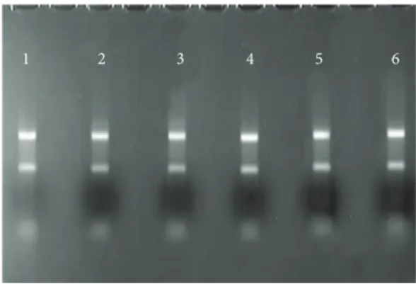
Figure 1: Total RNA from primary culture of breast tumor for different postsurgery treatment periods on a 1% agarose gel (1 : 8 h; 2 : 16 h; 3 : 24 h; 4 : 32 h; 5 : 40 h; 6 : 48 h).
To ascertain whether extracted mRNA quality was af-fected by treatment time, a 1% agarose gel test was used to evaluate intervals of 8, 16, 24, 32, 40, and 48 h (Figure 1). Of note, normal breast tissue from patients undergoing mammoplasty was negative for mRNA expression of the studied genes.
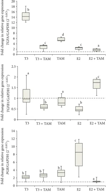
3.3. Gene Expression and Evidence of Thyroid Hormone Influ-ence. Figure 2shows box plots that represent the expression of each gene following stimulation by E2 (TGFA, TGFB1,and
PGR) after administration of the previously described treat-ments;Figure 3details the respective data for T3 (TNFRSF9, BMP-6,andTHRA). Patients 01, 05, and 14 (with amended thyroid hormone status) and patients 03 and10 (negative ER) were excluded from these data.
4. Discussion
4.1. Hormonal Status and Immunohistochemistry. There is much evidence suggesting a relationship between thyroid disease and BrC risk but it is controversial. Numerous studies have been conducted, and some have found no significant relationship between thyroid disease [32] or treatment of thyroid disease [33] and BrC risk, while others correlated BrC with hyperthyroidism [1,10,34,35], hypothyroidism [24,
36, 37], nontoxic goiter [8,38,39], or thyroid autoimmune diseases [8,37,38,40–42].
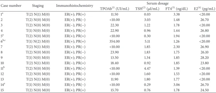
Table 1: Tumor staging, immunohistochemistry, and serum dosage of breast cancer patients.
Case number Staging Immunohistochemistry Serum dosage
TPOAb(1)(UI/mL) TSH(2)(𝜇I/mL) FT4(3)(ng/dL) E2(4)(pg/mL)
1a T(2) N(1) M(0) ER(+); PR(+) 11.50 0.03 3.38 <20.00
2 T(2) N(0) M(0) ER(+); PR(−) <10.00 3.03 1.48 26.70
3 T(2) N(0) M(0) ER(−); PR(−) 22.30 1.22 1.78 <20.00
4 T(1) N(0) M(0) ER(+); PR(+) 22.90 0.96 1.44 26.80
5a T(2) N(0) M(0) ER(+); PR(+) <10.00 0.30 1.94 <20.00
6b,c T(1) N(2) M(0) ER(+); PR(+) 354.00 5.11 1.26 <20.00
7 T(2) N(1) M(0) ER(+); PR(−) <10.00 1.85 2.30 26.90
8 T(2) N(0) M(0) ER(+); PR(−) 23.90 1.83 1.75 26.10
9 T(1) N(0) M(0) ER(+); PR(+) 13.50 1.34 1.85 28.20
10 T(2) N(1) M(0) ER(−); PR(+) 18.40 0.92 1.85 23.80
11b T(2) N(0) M(0) ER(+); PR(+) <10.00 4.47 1.39 <20.00
12 T(2) N(0) M(0) ER(+); PR(−) <10.00 1.60 1.53 <20.00
13 T(2) N(1) M(0) ER(+); PR(+) 11.90 1.80 1.77 <20.00
14a T(1) N(0) M(0) ER(+); PR(−) <10.00 0.29 3.06 26.70
15 T(2) N(0) M(0) ER(+); PR(+) 15.70 0.76 1.78 24.50
(1)Thyroid peroxidase antibody (TPOab):<35.00 UI/mL = negative.
(2)Thyroid-stimulating hormone (TSH): normality between 0.4 and 4.0 mUI/mL.
(3)Free thyroxine (FT4): normality between 0.8 and 1.9 ng/dL.
(4)Estradiol (E2): normality on postmenopause between 0 and 30 pg/mL.
a
Hyperthyroidism.
bSubclinical hypothyroidism.
cTPOab positive.
Table 2: Comparison of hormonal dosages between breast cancer and normal control patients.
Breast cancer (𝑁 = 15) Control (𝑁 = 18)
TSH (𝜇I/mL) 1.34±0.60 2.41±1.10
FT4 (ng/dL) 1.78±0.20∗ 0.95±0.16
E2 (pg/mL) 23.80±3.35 21.80±3.29
Data are reported as median±total semirange. TSH: thyroid-stimulating
hormone; FT4: free thyroxine; E2: estradiol.∗𝑃 < 0.05compared with
control group (Mann-Whitney test).
In our study, we identified a higher prevalence of thyroid disease (33.3%), with hyperthyroidism being the most fre-quent condition (20.0%), confirming previous reports [1,27].
4.2. Primary Culture. One of the major concerns for re-searchers working with breast cancer models is that these models may exhibit behavior that is dissimilar to that of breast cancer tissuein vivo. Burdall and colleagues [44] evaluated the use of breast cancer cell cultures and highlighted the advantages of these models in that they exhibit limitless self-replication, are easily replaced with frozen stock, and demon-strate a relatively high degree of homogeneity. However, cell culture models are prone to genotypic and phenotypic drift, which excludes this model when considering and comparing individual characteristics of patients. A recent alternative approach has been aimed at minimizing the differences between in vitro culture models and living breast cancer tissue.
In contrast, culture of breast tumor slices does not present the effects of genetic drift, which may occur in cell lines during the course of passaging, and this method maintains some in vivo characteristics. Furthermore, breast tumor slices have been primarily useful for evaluating responses to hormonal and pharmacological treatments, showing gene expression results that are close toin vivoresponses.
These data complement our understanding of how study models for breast cancer may present variations in results. It was previously observed by our group that TGFA expression in primary organ culture was not reproducible to the results seen in cell lines (5). Cell lines have advantages such as high genomic homogeneity that lead to little variation in the results when compared with primary organ cultures. However, it is now acknowledged that information regarding genomic variation is insignificant in comparison to the variability introduced during technical steps such as culture preparation and gene expression. In primary organ culture, variation in the data produced is higher than the natural variation introduced by the techniques used. These variations may reflect a heterogeneity that develops in different tumor samples because of the wide range of factors that lead to genomic instability.
0 2 4 6 8 10 12 14 16 18 20 F o ld c h an ge in r ela ti ve g ene exp re ssio n F o ld c h an ge in r ela ti ve g ene exp re ssio n F o ld c h an ge in r ela ti ve g ene exp re ssio n 0 0.5 1 1.5 2 2.5 0 2 4 6 8 10 12 14 b c d c a a c b b b a b a b c T3
T3 +TAM TAM E2 E2+TAM
T3
T3 +TAM TAM E2 E2+TAM
T3
T3 +TAM TAM E2 E2+TAM
TGF A /GAPD H ( 2 −Δ Δ Ct ) TGFB 1 /GAPD H ( 2 −Δ Δ Ct ) PGR /GAPD H ( 2 −Δ Δ Ct )
Figure 2: Gene expression of known estrogen-stimulated genes in primary cultures of breast tumors. Samples were treated as follows: ethanol vehicle, triiodothyronine (T3), T3 plus tamoxifen (TAM), TAM, estradiol (E2), and E2 plus TAM. After 48 h of
treatment,TGFA,TGFB1, andPGRwere quantified by real-time
RT-PCR. Glyceraldehyde-3-phosphate dehydrogenase (GAPDH) was
used to normalize gene expression. Relative mRNA expression was calculated using the expression level of the treated ethanol sample as the standard set to the dotted line and represented by letter “a.”
Different letters indicate𝑃 < 0.05.
estrogen receptor dependence [7]. When hormonal treat-ment was associated with TAM, variations in gene expression did not show statistical differences compared with control cells. However, although E2 and T3 treatment of primary cultures showed the same gene expression profile, TAM asso-ciation did not block T3 treatment but rather increased TGFA and decreased TGFB1 gene expression during T3 and TAM association. Here, we choose PGR as a marker of estrogen action, as reported elsewhere [45]. As expected, E2 treatment
0 1 2 3 4 5 6 7 8 0 5 10 15 20 25 0 0.5 1 1.5 2 2.5 b a d a d b c c a a b ab
a a ab
T3
T3 +TAM TAM E2 E2+TAM
T3
T3 +TAM TAM E2 E2+TAM
T3
T3 +TAM TAM E2 E2+TAM
F o ld c h an ge in r ela ti ve g ene exp re ssio n F o ld c h an ge in r ela ti ve g ene exp re ssio n F o ld c h an ge in r ela ti ve g ene exp re ssio n THR A /GAPD H ( 2 −Δ Δ Ct ) TNFRS F 9 /GAPD H ( 2 −Δ Δ Ct ) BMP 6 /GAPD H ( 2 −Δ Δ Ct )
Figure 3: Gene expression of known thyroid hormone-stimulated genes in primary cultures of breast tumors. Samples were treated as follows: ethanol vehicle, triiodothyronine (T3), T3 plus tamoxifen (TAM), TAM, estradiol (E2), and E2 plus TAM. After 48 h of
treat-ment,THRA,TNFRSF9, andBMP6were quantified by real-time
RT-PCR. Glyceraldehyde-3-phosphate dehydrogenase (GAPDH) was used to normalize gene expression. Relative mRNA expression was calculated using the expression level in the treated ethanol sample as the standard set to the dotted line and represented by letter “a.”
Different letters indicate𝑃 < 0.05.
here we provided evidence for the upregulation of PGR in primary culture treated with T3 plus TAM.
To confirm the action of T3 treatment, we verified the expression of previously described genes exhibiting upreg-ulation (TNFRSF9 and THRA) or downregupreg-ulation (4-1BB) under TH treatment [29]. Treatment of primary cultures with T3 and E2 reproduced equivalent results obtained in cell lines [7]. However, associations of these hormones with TAM did not reproduce these results. Acting as antagonist on ER, TAM associated with E2 maintained expression of TGFA and PGR to the control levels, but when T3 plus TAM association was applied, TGFA and TGFB1 expression was at the same levels as those following E2 treatment. This association may coincide with estrogenic response on gene expression and highlight the effect of TAM usage in hyperthyroid patients, especially considering the proliferative and apoptotic effects of TGFA and TGFB1, respectively, on the initial tumor progression stages. This finding was reinforced by observations of upregulated PGR expression under T3 treatment (although levels were less than those produced following E2 stimulation), which did not show changes with TAM association.
To assess whether E2 could influence the expression of T3 target genes, we examined the effect on TNFRSF9, BMP-6, and THRA expression and observed no differences in com-parison to the control. E2 and TAM treatment upregulated THRA expression; however, this effect may be due only to TAM because it exerted the same upregulation effects as E2 plus TAM.
Note that TAM alone or associated with T3 modulated gene expression of TNFRSF9, BMP-6, and THRA related to the control, similar to T3 treatment, showing that the TAM can interfere with gene expression modulated by T3.
Our results showed that thyroid dysfunctions correlate with ER positivity (Table 1). There are many possibilities of crosstalk between T3, E2, and TAM. Some previous research considered TH and ER binding, as demonstrated in BrC cell lines [7]. On the other hand, TH can alter ER-dependent gene transcription, with ER-thyroid hormone receptor dimer formation that results in flexible regulation of the consensus ERE [28], or interaction between thyroid hormone receptors and ERE [3]. Most recently, studies have emphasized the nongenomic actions of T3 and E2, commencing at the receptors at the plasma membrane or in the cytoplasm and activating intracellular signaling pathways such as PI3 K or MAPK. The E2 antagonist fulvestrant does not activate these signaling pathways by this mechanism, but other selective estrogen receptor modulators (SERM), such as TAM, do it in a similar manner to E2, showing that TAM confers an antagonistic action on nuclear ER but an agonistic action on membrane ER [47, 48]. AKT and MAPK phosphorylation can elevate agonistic TAM and other SERM activity [48].
5. Conclusions
Our work identified a high incidence of hyperthyroidism in menopausal women with BrC, who exhibited a higher degree of TH than the control group. Primary culture of tumor
samples was utilized to evaluate gene expression modified by T3 or E2 treatment and produced similar but not identical results to those observed in breast cancer cell lines. T3 had a significant effect on genes classically regulated by E2, but the combination of T3 with TAM did not reverse gene expression levels to those observed in the untreated control group, in contrast to E2 plus TAM, which resulted in maintained gene expression when E2 treatment was applied. Taking into account the known ability of TH to mimic the effects of E2, particularly in the presence of TAM, our results reinforce previous reports that the thyroid hormone status of BrC patients can influence E2-controlled mechanisms, even under TAM intervention and/or the absence of circulating E2 in postmenopausal women. Thus, our study highlights the importance of evaluation of thyroid hormone status when considering the prognosis and treatment options for individual patients.
Abbreviations
BrC: Breast cancer TH: Thyroid hormone ER: Estrogen receptor T3: Triiodothyronine E2: Estradiol
TAM: 4-Hydroxytamoxifen PR: Progesterone receptor TPOab: Thyroid peroxidase antibody TSH: Thyroid-stimulating hormone FT4: Free thyroxine
TGFA: Transforming growth factor alpha TGFB1: Transforming growth factor beta 1 PGR: Progesterone receptor
TNFRSF9: Tumor necrosis factor receptor superfamily 9 BMP-6: Bone morphogenetic protein 6
THRA: Thyroid hormone receptor alpha.
Conflict of Interests
The authors declare that there is no conflict of interests regarding the publication of this paper.
Acknowledgments
This study was supported by FAPESP Grant 05/55459-1 and 05/55459-13/05/55459-105/55459-105/55459-105/55459-105/55459-1-8 and supported by CAPES/PNPD Grant 00000.059158/2010.
References
[1] P. P. Saraiva, N. B. Figueiredo, C. R. Padovani, M. M. Brentani, and C. R. Nogueira, “Profile of thyroid hormones in breast
cancer patients,” Brazilian Journal of Medical and Biological
Research, vol. 38, no. 5, pp. 761–765, 2005.
[2] I. Conde, R. Paniagua, J. Zamora et al., “Influence of thyroid
hormone receptors on breast cancer cell proliferation,”Annals
[3] S. Dinda, A. Sanchez, and V. Moudgil, “Estrogen-like effects of thyroid hormone on the regulation of tumor suppressor
pro-teins, p53 and retinoblastoma, in breast cancer cells,”Oncogene,
vol. 21, no. 5, pp. 761–768, 2002.
[4] S. J. Conde, R. D. A. M. Luvizotto, M. T. de S´ıbio, and C. R. Nogueira, “Human breast tumor slices as an alternative approach to cell lines to individualize research for each patient,”
European Journal of Cancer Prevention, vol. 21, no. 4, pp. 333–
335, 2012.
[5] S. J. Conde, R. A. M. Luvizotto, M. T. Sibio, M. L. H. Katayama, M. M. Brentani, and C. R. Nogueira, “Tamoxifen inhibits
transforming growth factor-𝛼gene expression in human breast
carcinoma samples treated with triiodothyronine,”Journal of
Endocrinological Investigation, vol. 31, no. 12, pp. 1047–1051,
2008.
[6] S. H. Cestari, N. B. Figueiredo, S. J. Conde et al., “Influence of estradiol and triiodothyronine on breast cancer cell lines proliferation and expression of estrogen and thyroid hormone
receptors,”Arquivos Brasileiros de Endocrinologia e Metabologia,
vol. 53, no. 7, pp. 859–864, 2009.
[7] C. R. Nogueira and M. M. Brentani, “Triiodothyronine mimics
the effects of estrogen in breast cancer cell lines,”Journal of
Steroid Biochemistry and Molecular Biology, vol. 59, no. 3-4, pp.
271–279, 1996.
[8] O. Turken, Y. NarIn, S. DemIrbas et al., “Breast cancer in
association with thyroid disorders,”Breast Cancer Research, vol.
5, no. 5, pp. R110–R113, 2003.
[9] A. Tosovic, A.-G. Bondeson, L. Bondeson, U.-B. Ericsson, J. Malm, and J. Manjer, “Prospectively measured triiodothyronine levels are positively associated with breast cancer risk in
postmenopausal women,”Breast Cancer Research, vol. 12, no. 3,
article R33, 2010.
[10] M. Lemaire and L. Baugnet-Mahieu, “Thyroid function in
women with breast cancer,”European Journal of Cancer and
Clinical Oncology, vol. 22, no. 3, pp. 301–307, 1986.
[11] M. West, C. Blanchette, H. Dressman et al., “Predicting the clinical status of human breast cancer by using gene expression
profiles,”Proceedings of the National Academy of Sciences of the
United States of America, vol. 98, no. 20, pp. 11462–11467, 2001.
[12] M. Lacroix, G. Querton, P. Hennebert, D. Larsimont, and G. Leclercq, “Estrogen receptor analysis in primary breast tumors by ligand-binding assay, immunocytochemical assay,
and northern blot: a comparison,”Breast Cancer Research and
Treatment, vol. 67, no. 3, pp. 263–271, 2001.
[13] S. Sommer and S. A. W. Fuqua, “Estrogen receptor and breast
cancer,”Seminars in Cancer Biology, vol. 11, no. 5, pp. 339–352,
2001.
[14] S. A. W. Fuqua, G. C. Chamness, and W. L. McGuire, “Estrogen
receptor mutations in breast cancer,”Journal of Cellular
Bio-chemistry, vol. 51, no. 2, pp. 135–139, 1993.
[15] E. V. Jensen, G. Cheng, C. Palmieri et al., “Estrogen receptors and proliferation markers in primary and recurrent breast
cancer,”Proceedings of the National Academy of Sciences of the
United States of America, vol. 98, no. 26, pp. 15197–15202, 2001.
[16] D. P. Edwards, G. C. Chamness, and W. L. McGuire, “Estrogen
and progesterone receptor in breast cancer,” Biochimica et
Biophysica Acta, vol. 560, no. 4, pp. 457–486, 1979.
[17] L. Bernstein and R. K. Ross, “Endogenous hormones and breast
cancer risk,”Epidemiologic Reviews, vol. 15, no. 1, pp. 48–65,
1993.
[18] L. A. Habel and J. L. Stanford, “Hormone receptors and breast
cancer,”Epidemiologic Reviews, vol. 15, no. 1, pp. 209–219, 1993.
[19] R. E. Burke and W. L. McGuire, “Nuclear thyroid hormone
receptors in a human breast cancer cell line,”Cancer Research,
vol. 38, no. 11 I, pp. 3769–3773, 1978.
[20] M.-A. Cerbon, M.-F. Pichon, and E. Milgrom, “Thyroid
hor-mone receptors in human breast cancer,”Cancer Research, vol.
41, no. 10, pp. 4167–4173, 1981.
[21] Z.-M. Shao, M. S. Sheikh, A. K. Rishi et al., “Thyroid hormone enhancement of estradiol stimulation of breast carcinoma
proliferation,”Experimental Cell Research, vol. 218, no. 1, pp. 1–8,
1995.
[22] J. G. Spencer, “The influence of the thyroid in malignant
disease,”British Journal of Cancer, vol. 8, no. 3, pp. 393–411, 1954.
[23] O. Muhlbock and L. M. Boot, “Induction of mammary cancer in
mice without the mammary tumor agent by,”Cancer Research,
vol. 19, no. 4, pp. 402–412, 1959.
[24] D. P. Rose and T. E. Davis, “Plasma thyroid-stimulating
hor-mone and thyroxine concentrations in breast cancer,”Cancer,
vol. 41, no. 2, pp. 666–670, 1978.
[25] B. S. Thomas, R. D. Bulbrook, and M. J. Russell, “Thyroid
function in early breast cancer,”European Journal of Cancer and
Clinical Oncology, vol. 19, no. 9, pp. 1213–1219, 1983.
[26] H. Vorherr, “Thyroid function in benign and malignant breast
disease,”European Journal of Cancer and Clinical Oncology, vol.
23, no. 3, pp. 255–257, 1987.
[27] O. Takatani, T. Okumoto, H. Kosano, M. Nishida, H. Hiraide, and S. Tamakuma, “Relationship between the levels of serum thyroid hormones or estrogen status and the risk of breast
cancer genesis in Japanese women,”Cancer Research, vol. 49, no.
11, pp. 3109–3112, 1989.
[28] N. Vasudevan, N. Koibuchi, W. W. Chin, and D. W. Pfaff,
“Differential crosstalk between estrogen receptor (ER)𝛼and
ER𝛽 and the thyroid hormone receptor isoforms results in
flexible regulation of the consensus ERE,” Molecular Brain
Research, vol. 95, no. 1-2, pp. 9–17, 2001.
[29] T. Yamada-Okabe, Y. Satoh, and H. Yamada-Okabe, “Thyroid hormone induces the expression of 4-1BB and activation of caspases in a thyroid hormone receptor-dependent manner,”
European Journal of Biochemistry, vol. 270, no. 14, pp. 3064–
3073, 2003.
[30] S. R. Lakhani, M. J. van de Vijver, J. Jacquemier et al., “The pathology of familial breast cancer: predictive value of immunohistochemical markers estrogen receptor, progesterone receptor, HER-2, and p53 in patients with mutations in BRCA1
and BRCA2,”Journal of Clinical Oncology, vol. 20, no. 9, pp.
2310–2318, 2002.
[31] R. Mira-y-Lopez and L. Ossowski, “Preservation of steroid hor-mone receptors in organ cultures of human breast carcinomas,”
Cancer Research, vol. 50, no. 1, pp. 78–83, 1990.
[32] A. Kalache, M. P. Vessey, and K. McPherson, “Thyroid disease
and breast cancer: findings in a large case-control study,”British
Journal of Surgery, vol. 69, no. 7, pp. 434–435, 1982.
[33] H. A. Weiss, L. A. Brinton, N. A. Potischman et al., “Breast cancer risk in young women and history of selected medical
conditions,”International Journal of Epidemiology, vol. 28, no.
5, pp. 816–823, 1999.
[34] A. R. Moossa, D. A. Price Evans, and A. C. Brewer, “Thyroid status and breast cancer: reappraisal of an old relationship,”
Annals of the Royal College of Surgeons of England, vol. 53, no. 3,
pp. 178–188, 1973.
[35] M. B. Goldman, “Thyroid diseases and breast cancer,”
[36] I. Mittra and J. L. Hayward, “Hypothalamic-pituitary-thyroid
axis in breast cancer,”The Lancet, vol. 1, no. 7863, pp. 885–889,
1974.
[37] D. A. Adamopoulos, S. Vassilaros, and N. Kapolla, “Thyroid disease in patients with benign and malignant mastopathy,”
Cancer, vol. 57, no. 1, pp. 125–128, 1986.
[38] C. Giani, P. Fierabracci, R. Bonacci et al., “Relationship between breast cancer and thyroid disease: relevance of autoimmune
thyroid disorders in breast malignancy,” Journal of Clinical
Endocrinology and Metabolism, vol. 81, no. 3, pp. 990–994, 1996.
[39] P. P. A. Smyth, D. F. Smith, E. W. M. McDermott, M. J. Murray, J. G. Geraghty, and N. J. O’Higgins, “A direct relationship between
thyroid enlargement and breast cancer,” Journal of Clinical
Endocrinology and Metabolism, vol. 81, no. 3, pp. 937–941, 1996.
[40] K. Itoh and N. Maruchi, “Breast cancer in patients with
Hashimoto’s thyroiditis,”The Lancet, vol. 2, no. 7945, pp. 1119–
1121, 1975.
[41] B. Rasmusson, U. Feldt-Rasmussen, and L. Hegedus, “Thyroid
function in patients with breast cancer,”European Journal of
Cancer and Clinical Oncology, vol. 23, no. 5, pp. 553–556, 1987.
[42] P. P. A. Smyth, S. G. Shering, M. T. Kilbane et al., “Serum thyroid peroxidase autoantibodies, thyroid volume, and
out-come in breast carcinoma,”Journal of Clinical Endocrinology
and Metabolism, vol. 83, no. 8, pp. 2711–2716, 1998.
[43] A. J. Hedley, S. J. Jones, and D. J. Spiegelhalter, “Breast cancer in
thyroid disease: fact or fallacy?”The Lancet, vol. 1, no. 8212, pp.
131–133, 1981.
[44] S. E. Burdall, A. M. Hanby, M. R. J. Lansdown, and V. Speirs,
“Breast cancer cell lines: friend or foe?”Breast Cancer Research,
vol. 5, no. 2, pp. 89–95, 2003.
[45] J. Rokicki, P. M. Das, J. M. Giltnane et al., “The ER𝛼coactivator,
HER4/4ICD, regulates progesterone receptor expression in
normal and malignant breast epithelium,”Molecular Cancer,
vol. 9, article 150, 2010.
[46] C. K. Osborne and R. Schiff, “Estrogen-receptor biology:
continuing progress and therapeutic implications,”Journal of
Clinical Oncology, vol. 23, no. 8, pp. 1616–1622, 2005.
[47] E. R. Levin, “Cellular functions of plasma membrane estrogen
receptors,”Steroids, vol. 67, no. 6, pp. 471–475, 2002.
[48] J. Shou, S. Massarweh, C. K. Osborne et al., “Mechanisms of tamoxifen resistance: increased estrogen receptor-HER2/neu
cross-talk in ER/HER2-positive breast cancer,”Journal of the
Submit your manuscripts at
http://www.hindawi.com
Stem Cells
International
Hindawi Publishing Corporation
http://www.hindawi.com Volume 2014
Hindawi Publishing Corporation
http://www.hindawi.com Volume 2014
INFLAMMATION
Hindawi Publishing Corporation
http://www.hindawi.com Volume 2014
Behavioural
Neurology
Endocrinology
International Journal ofHindawi Publishing Corporation
http://www.hindawi.com Volume 2014 Hindawi Publishing Corporation
http://www.hindawi.com Volume 2014
Disease Markers
Hindawi Publishing Corporation
http://www.hindawi.com Volume 2014 BioMed
Research International
Oncology
Journal of Hindawi Publishing Corporationhttp://www.hindawi.com Volume 2014
Hindawi Publishing Corporation
http://www.hindawi.com Volume 2014 Oxidative Medicine and Cellular Longevity Hindawi Publishing Corporation
http://www.hindawi.com Volume 2014
PPAR Research
The Scientific
World Journal
Hindawi Publishing Corporationhttp://www.hindawi.com Volume 2014
Immunology Research Hindawi Publishing Corporation
http://www.hindawi.com Volume 2014
Journal of
Obesity
Journal ofHindawi Publishing Corporation
http://www.hindawi.com Volume 2014
Hindawi Publishing Corporation
http://www.hindawi.com Volume 2014
Computational and Mathematical Methods in Medicine
Ophthalmology
Journal of Hindawi Publishing Corporationhttp://www.hindawi.com Volume 2014
Diabetes Research
Journal ofHindawi Publishing Corporation
http://www.hindawi.com Volume 2014
Hindawi Publishing Corporation
http://www.hindawi.com Volume 2014
Research and Treatment
AIDS
Hindawi Publishing Corporation
http://www.hindawi.com Volume 2014
Gastroenterology Research and Practice
Hindawi Publishing Corporation
http://www.hindawi.com Volume 2014
Parkinson’s
Disease
Evidence-Based Complementary and Alternative Medicine
Volume 2014 Hindawi Publishing Corporation