Associated with AIDS Progression
Zhen Li1., Wei Li2., Ning Li3
, Yanmei Jiao2, Dexi Chen2, Lianxian Cui1, Yu Hu1, Hao Wu2*, Wei He1*
1Department of Immunology, School of Basic Medicine, Peking Union Medical College; National Key Laboratory of Medical Molecular Biology, Institute of Basic Medical Sciences, Chinese Academy of Medical Sciences, Beijing, China,2Center for Infectious Diseases, Beijing You’an Hospital, Capital Medical University, Beijing, China,3Beijing You’an Hospital, Capital Medical University, Beijing, China
Abstract
Background:Early diagnosis is vital to HIV control.cdT cells play critical roles in viral infections, but their activation in acute HIV infected patients and follow up to 18 months has not been described.
Methods:Changes incdT cells, including subsets, function and activation, in treated and untreated acutely HIV-infected patients (n = 79) were compared by cytotoxicity assay and flow cytometry with healthy controls (n = 21) at month 0, 6, 12 and 18.
Results: In acutely HIV-infected patients, Vd1 cell proportion was elevated (P= 0.027) with Vd2 population reduced (P= 0.002). Effector and central memorycd T cell factions were decreased (P= 0.006 and P= 0.001, respectively), while proportion of terminalcdT cells increased (P= 0.002).cdT cell cytotoxicity was compromised over time. Fraction of IL-17-producing cells increased (P= 0.008), and IFN-c-producing cells were unaffected (P= 0.115). Elevation of a microbial translocation marker, sCD14, was associated withcd T cell activation (P= 0.001), which increased in a time-dependent manner, correlating with CD4/CD8 T cell activation set-points and CD4 counts. Antiretroviral therapy did not affect these changes.
Conclusions:cdT cell subpopulation and functions change significantly in acute HIV infection and over time. EarlycdT cell activation was associated with CD4/CD8 T cell activation set-points, which predict AIDS progression. Therefore,cdT cell activation represents a potential surrogate marker of AIDS progression.
Citation:Li Z, Li W, Li N, Jiao Y, Chen D, et al. (2014)cdT Cells Are Involved in Acute HIV Infection and Associated with AIDS Progression. PLoS ONE 9(9): e106064. doi:10.1371/journal.pone.0106064
Editor:R. Keith Reeves, Beth Israel Deaconess Medical Center, Harvard Medical School, United States of America
ReceivedMarch 12, 2014;AcceptedJuly 31, 2014;PublishedSeptember 4, 2014
Copyright:ß2014 Li et al. This is an open-access article distributed under the terms of the Creative Commons Attribution License, which permits unrestricted use, distribution, and reproduction in any medium, provided the original author and source are credited.
Data Availability:The authors confirm that all data underlying the findings are fully available without restriction. All relevant data are within the paper and its Supporting Information files.
Funding:This work was supported by Twelve-five Project for Control and Prevention of Major Infectious Diseases [2012ZX10001-006], National Natural Science Foundation of China [81371332], and Beijing Key Laboratory [BZ0089]. The funders had no role in study design, data collection and analysis, decision to publish, or preparation of the manuscript.
Competing Interests:The authors have declared that no competing interests exist.
* Email: [email protected] (HW); [email protected] (WH)
.These authors contributed equally to this work.
Introduction
HumancdT cells are distinct from abT cells, and generally lack CD4 or CD8 antigen expression [1]. These cells represent approximately 3–4% of peripheral T cells in the blood, and are composed predominantly of two subsets, depending on expression of the Vd1 or Vd2 gene [2–5].Vd1 T cells respond to antigens from bacterial pathogens [6]. Vd2 T cells are believed to be activated by several chemical compounds, some of which are produced by activated host cells in response to pathogenic infections [7]. Vd2 T cells can be further divided into four subtypes: naı¨ve (Tnaı¨ve), central memory (TCM), effector memory (TEM) and terminally differentiated (TEMRA), based on differ-ential surface expression of CD45RA and CD27 [8,9]. Specific Tnaı¨ve and TCM cells are located in the lymph nodes and possess distinct functions [10–12]. TEM and TEMRA are located
preferentially at inflammatory sites and perform effector functions [10].
Functionally, cd T cells produce interleukin 17 (IL-17), interferon gamma (IFN-c), and other secreted protein factors after activation [13,14]. IL-17 is thought to be important for maintaining intestinal mucosal integrity and controlling microbial translocation [15]. IFN-clevels are elevated in many proinflam-matory events, and microbial translocation [16]. Furthermore,cd T cells are involved in the recruitment of neutrophils in bacterial infections [17]. Reports indicate that, the cytotoxic activity ofcdT lymphocytes against human neuroblastoma cells, Burkitt’s lym-phoma cells and various cancer cell lines (human colon carcinoma, erytholeukemia, human neuroepithelioma and renal adenocarci-noma) is augmented upon activation [18,19].
the mucosal barrier is significantly elevated [20,21], which may result in translocation of microbial products into the systemic circulation, leading to immune stimulation. A number of studies have described the use of soluble CD14 (sCD14) as a marker of microbial translocation, and even as a predictive marker of disease progression in HIV infection [22–24].
ThecdT cell responses to HIV infection are complicated and not well-elucidated [25,26]. Previous reports have indicated increased levels of Vd1 cells and decreased levels and function of Vd2 cells in chronically HIV-infected patients [26,27]. Further-more, changes in the Vd2 T cell population correlated positively with CD4 T cell counts and negatively with viral loads [26].
The therapeutic efficacy of antiretroviral therapy (ART) on Vd2 T cell recovery in HIV patients is controversial and inconclusive. A report in 2004 indicated partial recovery of the Vd2 T cell population following long-term HIV-suppressive therapy [28]. However, a subsequent study demonstrated that ART failed to restore the Vd2 repertoire in HIV-infected men [29].
In this study, we aimed to characterize the changes incdT cells, including subsets, function and activation status, in acutely HIV-infected patients who were followed from acute infection up to 18 months, and to define their possible correlation with other serum parameters, microbial translocation and AIDS progression.
Methods
Study subjects
The protocol for this study was approved by the Beijing You’an Hospital Ethics Committee and the Guidelines for Human Experimentation (PR. China) were followed throughout. Upon admission, all patients provided written informed consent for their information and clinical samples (blood and plasma) to be stored and used for research.
Seventy-nine homosexual men with acute HIV infection were enrolled in a prospective study (co-inclusion in the Beijing PRIMO cohort) conducted in Beijing You’an Hospital, Beijing, PR. China. Acute HIV infection was defined by a negative enzyme-linked immunosorbent assay (ELISA), and at least one of the following criteria: less than three bands on HIV Western blot (HIV BLOT2.2, 11039, Diagnostic Biotechnology Pte, Singapore), a positive p24 test or detectable plasma HIV-RNA. The estimated date of infection was calculated as follows: 2 weeks before onset of symptoms or 4 weeks before the first positive Western blot. All 79 enrolled HIV-infected patients were diagnosed early during the acute stage of HIV infection (median of estimated time post-infection: 41 days) and transmitted by homosexual activity. At baseline (day 0 of enrollment), all patients were treatment-naı¨ve. During the follow-up times, some of the patients (11/79) started combination antiretroviral treatment (ART), based on CD4 cell counts (e.g.,350/mL according to WHO recommendations) with
or without clinical symptoms, and the consensus of both physicians and patients. Patients who were treated during the study received a combination of AZT, 3TC, and EFV or LPV/r. The courses of their treatment were not influenced by the authors for the purpose of this study. Most of them (68/79) were still untreated [CD4 counts:.350/mL (n = 48),,350/mL (n = 20)]. Twenty-one male homosexual healthy subjects were included as controls (HC). The ages of all groups were matched.
Blood samples were collected from patients at baseline (day 0 of enrollment), month 6, month 12 and month 18. Plasma samples were also collected from all healthy volunteers. All tests were performed at baseline. In addition, CD38/HLA-DR antigen expression and the cytotoxicity activity of cd T cells were determined at baseline, month 6, month 12 and month 18.
Cell culture
Peripheral blood mononuclear cells (PBMCs) were isolated by Ficoll-Hypaque (TBD, Tianjin, China) centrifugation. The expansion of cd T cells was performed as previously described [30]. Briefly, 24-well plates were coated with 500mL purified anti-cdTCR antibody (IMMU510, 1 mg/mL; Immunotech, Beckman Coulter, Fullerton, CA, USA) at 37uC for 2 h. PBMCs were then added to the antibody-coated wells and cultured in RPMI 1640 medium supplemented with 10% FCS and 200 IU/mL recombi-nant human IL-2 (Beijing Read United Cross Pharmaceutical, Beijing, China). The culture medium was replaced every other day. The trypan blue exclusion method was employed to assess cell viability.
Antibodies and flow cytometry
Antibodies (Abs) used in this study were as follows: mouse phycoerythin (PE)-conjugated anti-human CD38 (HIT2) Ab, mouse allophycocyanin (APC)-conjugated anti-human HLA-DR (L243) Ab, mouse phycoerythin-cyanin7(PE-cy7)-conjugated anti-human CD3 (HIT3a) Ab, mouse peridinin-chlorophyll-protein complex cyanine 5.5 (PerCP-cy5.5) conjugated anti-human CD27 (O323) Ab, mouse APC-conjugated anti-human CD45RA (HI100) Ab were purchased from Biolegend (San Diego, CA, USA). Mouse Fluorescein isothiocyanate (FITC)-conjugated anti-human pan TCRcd Ab, mouse PE-conjugated anti-human pan TCRcd (IMMU510) Ab and mouse FITC-conjugated anti-human pan Vd2TCR (IMMU389) Ab were purchased from Immunotech (Beckman Coulter, Fullerton, CA, USA). Mouse FITC-conjugated anti-human pan Vd1TCR (TS8.2) Ab was purchased from Pierce (USA). Mouse PE-conjugated anti-human IL-17A (SCPL1362) Ab and mouse PE-conjugated anti-human IFN-c (B27) Ab were purchased from BD Pharmingen (San Diego, CA, USA). The isotype control antibodies were purchased from the corresponding company, respectively.
Cells were stained with appropriate target Abs and isotype Abs using conventional surface- and/or intracellular staining methods. When both surface and intracellular staining was required, cells were first fixed and permeabilized using BD Cytofix/Cytoperm Fixation and Permeabilization Solution (BD Pharmingen), fol-lowed by staining for intracellular proteins. Cells were then washed extensively with PBS to remove excess Ab, stained for extracellular targets, and fixed with 2% formaldehyde. Fluores-cence was evaluated with a FACSCalibur flow cytometer, and data were analyzed using FlowJo software (Tree Star, Ashland, OR, USA).
Cytotoxicity
The CytoTox 96 Non-Radioactive Cytotoxicity Assay (Pro-mega, Madison, WI, USA), based on the colorimetric detection of released lactate dehydrogenase (LDH), was used according to the manufacturer’s instructions. Tumor cells used as target cells were seeded into a 96-round well plate (56104cells/well). Expandedcd T cells (effector cells) were added directly to individual wells at different effector/target (E/T) ratios. The plate was incubated at 37uC for 4 h before supernatants were collected and LDH activity was determined. Controls for spontaneous LDH release from effector and target cells, the target maximum release, as well as the culture medium background were assayed simultaneously. The cytotoxicity was calculated as follows: %Cytotoxicity = (Exper-imental-Effector spontaneous-Target spontaneous)/(Target maxi-mum-Target spontaneous)6100%.
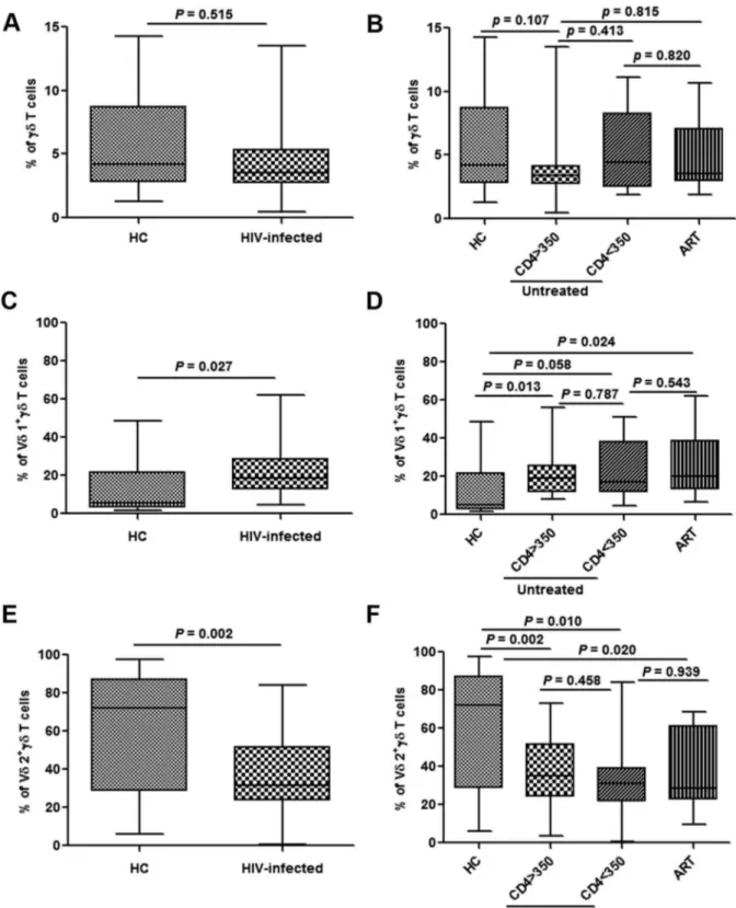
Figure 1. Changes in different subpopulations ofcdT cells in acutely HIV-infected patients.Frequencies of totalcdT cells, Vd1cdT and Vd2cdT cells were analyzed in healthy controls (HC) and acutely HIV-infected subjects at baseline (A, C, E). The patients were then subdivided based on the administration of antiretroviral therapy (ART) and CD4 levels (,or.than 350/mL). The frequencies were re-analyzed and compared with HC
(B, D, F).
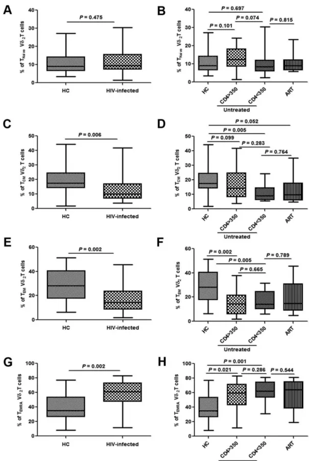
Figure 2. Changes in subpopulations of Vd2cdT cells in acutely HIV-infected patients.Frequencies of Tnaı¨ve, TCM, TEM and TEMRA cells were analyzed in healthy controls (HC) and acutely HIV-infected subjects at baseline (A, C, E, G). The patients were then subdivided based on the administration of antiretroviral therapy (ART) and CD4 levels (,or.than 350/mL). The frequencies were re-analyzed and compared with HC (B, D, F, H).
doi:10.1371/journal.pone.0106064.g002
Quantification of plasma lipopolysaccharide (LPS), LPS-binding protein (LBP) and soluble CD14 (sCD14)
Commercially available ELISA kits were used in this study following manufacturers’ instructions for measuring plasma concentrations of LBP (Cell Sciences, MA, USA) and sCD14 (soluble CD14) (R&D Systems, MN, USA). Plasma LPS levels were quantified using a Limulus amebocyte lysate (LAL) assay (Hycult Biotech, UDEN, Netherlands).
Statistical analysis
Non-parametric tests were used to avoid the impact of potential outlier values in a small study. Comparisons between groups were performed using the Mann-Whitney test. The Wilcox on matched pairs test was used to estimate the changes in the different variables throughout the follow-up. The Spearman’s non-parametric correlation was used to estimate the association of two continuous variables of interest. P-values below 0.05 were considered statistically significant. Data of patients on ART were compared only with data of untreated patients with CD4 counts lower than 350/mL.
Results
Changes incdT cell subpopulations in acutely HIV-infected patients
Compared with healthy subjects, there was no marked difference in the fraction of cd T cells in acutely HIV-infected patients (Fig. 1A). Nor were there any differences between patients with different CD4 counts, or those who did or did not receive ART (Fig. 1B). To characterize the changes incdT cells subpopulations in acutely HIV-infected patients, we first analyzed changes in the Vd1 and Vd2 subtypes. The proportion of Vd1 cells among cd T cells was elevated (P= 0.027), while the Vd2
population was significantly reduced (P= 0.002) (Fig. 1C and 1E). However, there were no significant differences in both the proportions of Vd1 and Vd2 cells between patients with different CD4 counts (bothP.0.05). Furthermore, Initiation of ART failed to bring about Vd2 subtype recover, and had no effect on the Vd1 population (Fig. 1D and 1F).
Changes in the levels of Vd2cd T subgroups in acutely HIV-infected patients were investigated by analysis of the expression of surface CD27 and CD45RA antigens [8,9]. There was no difference in the proportion of naı¨ve Vd2cdT cells (CD27+/ CD45RA+) observed (P= 0.475), on which CD4 levels and ART showed no impact (Fig. 2A and 2B). The fractions of the TEM (effector memory Vd2cdT cells, CD272/CD45RA2) and TCM (central memory Vd2cdT cells, CD27+/CD45RA2) populations were significantly decreased in acutely HIV-infected patients (P= 0.002 and P= 0.006, respectively), while the proportion of TEMRA (terminal Vd2 cd T cells, CD272/CD45RA+) was increased (P= 0.002) (Fig. 2C, 2E, and 2G). CD4 levels and the initiation of ART showed no correlation with these changes (Fig. 2D, 2F, and 2H).
CompromisedcdT cell functions in acutely HIV-infected patients
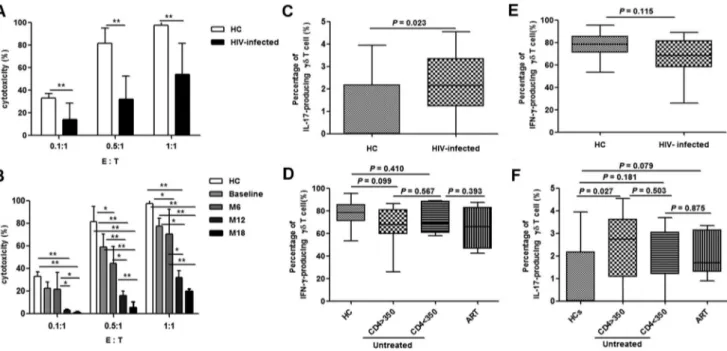
The cytotoxic activity ofcdT cells in the acute stage of HIV infection was analyzed at baseline, month 6, 12 and 18. Compared with data from healthy controls, cd T cell cytotoxicity was gradually compromised in acutely HIV-infected patients over time (Fig. 3A and 3B). Furthermore, the fraction of IL-17-producing cdT cells was elevated (P= 0.023), while the fraction of IFN-c-producing cd T cells was unchanged (P= 0.115) (Fig. 3C and 3E). These changes were not affected by CD4 counts or the initiation of ART (Fig. 3D and 3F).
Figure 3. Altered functions ofcdT cells in acute HIV infection.Cytotoxicity (%) was assessed at different effector (E,cdT cells) to target (T, Daudi cells) ratios (0.1:1, 0.5m1 and 1:1) at baseline (A). Subsequently, longitudinal follow-up analysis of cytotoxicity was conducted at different time-points [baseline, month 6 (M6), month 12 (M12) and month 18 (M18)] (B). Frequencies of IFN-c- and IL-17-producingcdT cells were analyzed in healthy controls (HC) and acutely HIV-infected subjects at baseline (C, E). The patients were then subdivided based on the administration of antiretroviral therapy (ART) and CD4 levels (,or.than 350/mL). The frequencies were re-analyzed and compared with HC (D, F). *:P,0.05, **:P, 0.01.
Activation ofcdT cells
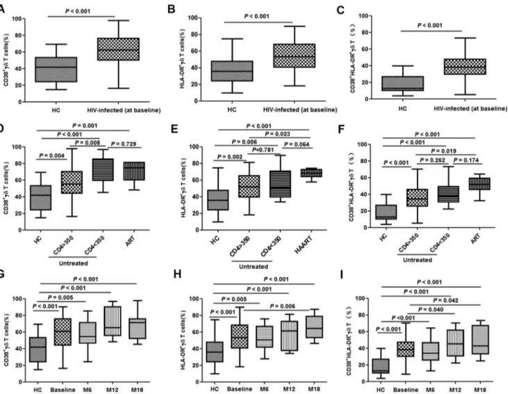
CD38 and HLA-DR are used as surrogate markers ofcdT cell activation [31,32]. Compared with healthy controls, the propor-tions of CD38+ or HLA-DR+ cd T cells (single-positive and double-positive) were significantly elevated in acutely HIV-infected patients (all P,0.001) (Fig. 4A–C). For patients with a CD4 count of less than 350/mL, the fraction of CD38+cdT cells was higher than for those with a CD4 count of greater than 350/
mL(P= 0.008) (Fig. 4D); however, this CD4-dependent change was not evident in HLA-DR+or double-positive cells (P= 0.781 and 0.262, respectively) (Fig. 4E and 4F). No significant differences were observed among patients on ART compared with untreated patients with a CD4 count of less than 350/mL (Fig. 4D–F).
To further characterize the activation ofcdT cells, CD38 and HLA-DR expression was determined at different time-points (baseline and months 6, 12, and 18). Steady increases in the fractions of all three groups ofcdT cells (CD38+, HLA-DR+and CD38+/HLA-DR+) were observed over time, except for CD38+
cells, which showed a marginal reduction at month 6 (Fig. 4G– I).No correlation was detected betweencd T cell activation and baseline viral load (data not shown).
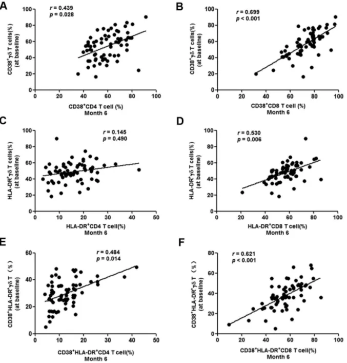
Correlations between earlycdT cell activation and CD4/CD8 T cell activation set-point
Activation of CD4/CD8 cells was determined on the basis of CD38 and HLA-DR surface marker expression at month 6 after baseline, when the T cell activation set-point of most patients should have been established [33]. Correlation analysis was performed by comparing CD4/CD8 activation at month 6 and cdT cell activation at baseline. CD4 and CD8 T cell activation set-points were both positively correlated tocdT cell activation (Fig. 5A–B and 5E–F). If HLA-DR alone was used, the CD8 T cell set-point was correlated (P= 0.006), but not the CD4 T cell activation set-point (P= 0.490) (Fig. 5C and 5D). These data demonstrated that early activation of cd T cells was positively correlated withCD4 and CD8 T cell activation set-points.
Figure 4. Activation ofcdT cells in acute HIV infection.Activation ofcdT cells was determined by analysis of CD38 and HLA-DR expression. Frequencies of CD38+, HLA-DR+and CD38+/HLA-DR+cdT cells were tested in healthy controls (HC) and acutely HIV-infected subjects at baseline (A– C). The patients were then subdivided based on the administration of antiretroviral therapy (ART) and CD4 levels (,or .than 350/mL). The
frequencies were re-analyzed and compared with HC (D–F). Longitudinal follow-up ofcdT cell activation in patients with acute HIV infection was conducted at baseline, month 6 (M6), month 12 (M12) and month 18 (M18). Frequencies of CD38+, HLA-DR+and CD38+/HLA-DR+cdT cells were tested in healthy controls (HC) and acutely HIV-infected subjects at baseline (G–I).
doi:10.1371/journal.pone.0106064.g004
Microbial translocation versuscdT cells
Microbial translocation in acute HIV infection was determined by analysis of the serum levels of LPS (lipopolysaccharides), LBP (LPS-binding protein) and sCD14 (soluble CD14). Only sCD14 levels were found to be elevated in acutely HIV-infected patients compared with those of healthy subjects (P= 0.001) (Fig. 6A–C). The levels of LPS and LBP did not differ between ART-treated and untreated patients, or between patients with different CD4 count levels (Fig. 6D–E). However, the sCD14 level in ART-treated patients was significant elevated compared with unART-treated patients with CD4.350 cells/ml (P= 0.003). Correlation analysis also showed no association between sCD14 levels and the totalcd
T cell population (P= 0.062) (data not shown); however, further investigation demonstrated positive correlations between sCD14 levels and differentcd T subgroups. Statistical analysis indicated that sCD14 levels were positively correlated withCD38+ /HLA-DR+(P= 0.002) and HLA-DR+(P= 0.011), but not with CD38+
(P= 0.105) (Fig. 6G). Fractions of Tnaı¨ve, TCM and TEM were not correlated with sCD14 levels (allP.0.05) (Fig. 6J). Propor-tions of Vd1, Vd2 and IL-17 levels were not associated with sCD14 levels (allP.0.05) (Fig. 6H, I, K).The fraction of IFN-c-producingcdT cells was negatively correlated with sCD14 levels (P= 0.046) (Fig. 6L). Furthermore, sCD14 was associated with LPS levels (P= 0.034) (data not shown).
Discussion
There is an increasing body of evidence indicating thatcdT cells play critical roles in HIV infections [25]. In this study conducted in a male homosexual population, we show that the proportion of Vd1 cells amongcdT cells was elevated in acutely HIV-infected patients, while the Vd2 population was significantly reduced. Among theVd2cdT cells, the fractions of TCM and TEM cells were significantly decreased, while the proportion of TEMRA was increased. In term ofcdT cell functions, cytotoxicity was compromised significantly in acutely HIV-infected patients over time. The fraction of IL-17-producing cells was elevated, but
not that of the IFN-c-producing cells. Activation of cd T cells increased over time and correlated withCD4 counts and CD4/ CD8 T cell activation set-points. In acute HIV infection, levels of the microbial translocation marker, sCD14, were significantly elevated, regardless ofCD4 counts. However, sCD14 levels exhibited a positive association withCD38/DR and HLA-DR expression and a negative association with the fraction of IFN-c-producingcdT cells. Initiation of ART had no impact on any of these changes.
It has been shown that the Vd2 population is depleted in chronically HIV-infected patients [25]. Our data indicated that this is an early event, occurring before the remarkable reduction in
Figure 6. Correlation between microbial translocation andcdT cells.Levels of LPS, LBP and sCD14 were analyzed in healthy controls (HC) and acutely HIV-infected subjects at baseline (A–C). The patients were then subdivided based on the administration of antiretroviral therapy (ART) and CD4 levels (,or.than 350/mL). The levels of LPS, LBP and sCD14 were re-analyzed and compared with HC (D–F). Correlation analysis between
the proportion of activatedcdT cells determined by CD38+, HLA-DR+or CD38+/HLA-DR and sCD14 levels at baseline was performed by Spearman’s rank correlation, where coefficients ‘r’ and correspondingP-values are indicated on panel G. Similar correlation analysis was performed between sCD14 levels and different subsets ofcdT cells [Vd1 (H) and Vd2 (I)], subgroups of Vd2 cells (Tnaı¨ve, TCM and TEM) (J), IL-17-producingcdT cells (K) or IFN-c-producingcdT cells (L).
doi:10.1371/journal.pone.0106064.g006
the totalcdT cell count, since baseline data showed no significant change in the fraction of cd T cells among the total T cell population (Fig. 1A, 1C and 1E). Our results also demonstrated that there were no significant changes in the proportion of Tnaı¨ve cells in the Vd2 cell population. The fractions of TEM and TCM cells were decreased, while the TEMRA fraction was elevated, which indicated increased aging of Vd2 cells in acute HIV infection (Fig. 2).
ActivatedcdT cells are the subtype of this population of cells that function in response to HIV infections, and therefore, were the primary target cell population of this study. Our data showed a gradual increase incdT cell activation, along with compromised cdT cell cytotoxicity over time, in addition to an elevation in the fraction of IL-17-producing cd T cells in acute HIV infection (Fig. 3, 4, 5).These observations indicated that early initiation of ART failed to bring about recovery of thecd T cell populations and their functions, which is consistent with a previous report [29], although additional studies are required to confirm this. More importantly, we found a positive correlation between baselinecdT cell activation and CD4/CD8 T cell activation set-points (Fig. 5), which were used to predict subsequent CD4 T cell loss, disease progression and outcome [32,34]. It can therefore be speculated that activation ofcdcells represents a surrogate marker for disease progression and outcome.
Our results indicated potential associations betweencd T cell activation and levels of sCD14, a monocyte activation marker, which is also commonly used as an indicator of microbial translocation (Fig. 6). The level of sCD14 is used to predict mortality and disease progression in chronic HIV patients [24,33], in addition to other disorders, such as different cancers and hemodialysis [35,36]. Microbial translocation results in the release of bacterial products, such as LPS and LBP, which in turn activate
monocytes in various clinical situations [37,38]. LPS and LBP levels have been shown to be increased in chronic HIV infection, but not in the acute stage. Furthermore, it has been reported that microbial translocation is not observed in primary HIV infection, although sCD14 levels are elevated [33,39]. Therefore, even though sCD14 levels were found to be elevated in acute HIV infection in our study, an association ofcdT cell activation with microbial translocation remains to be established.
No differences were observed between patients on early ART and untreated patients in any aspect investigated in this study. Although there are reports indicating that ART does not bring about recovery of the cd T cell depletion, this conclusion is controversial. Our study was conducted in only 11 patients on ART, and two treatment plans including two different protease inhibitors; therefore, a larger study with a more stringent design is required to confirm this conclusion.
In conclusion, in acute HIV infections, a significant shift in the proportions and functions of the variouscdT cell subgroups was observed. Elevation of the microbial translocation marker, sCD14, was associated withcdT cell activation. More importantly, early cd T cell activation was associated with CD4 and CD8 T cell activation set-points, which predict AIDS progression. Therefore, cd T cell activation represents a potential surrogate marker of AIDS progression. This conclusion requires confirmation in larger prospective cohort studies.
Author Contributions
Conceived and designed the experiments: WL WH HW. Performed the experiments: ZL YJ LC YH. Analyzed the data: WL NL ZL YJ HW LC. Contributed reagents/materials/analysis tools: NL DC. Contributed to the writing of the manuscript: WL WH.
References
1. Constant P, Davodeau F, Peyrat MA, Poquet Y, Puzo G, et al. (1994) Stimulation of human gamma delta T cells by nonpeptidic mycobacterial ligands. Science 264: 267–270.
2. Dik WA, Pike-Overzet K, Weerkamp F, de Ridder D, de Haas EF, et al. (2005) New insights on human T cell development by quantitative T cell receptor gene rearrangement studies and gene expression profiling. J Exp Med 201: 1715– 1723.
3. Hayday AC (2000) [gamma][delta] cells: a right time and a right place for a conserved third way of protection. Annu Rev Immunol 18: 975–1026. 4. Xi X, Guo Y, Chen H, Xu C, Zhang H, et al. (2009) Antigen specificity of
gammadelta T cells depends primarily on the flanking sequences of CDR3delta. J Biol Chem 284: 27449–27455.
5. Bukowski JF, Morita CT, Brenner MB (1999) Human gamma delta T cells recognize alkylamines derived from microbes, edible plants, and tea: implications for innate immunity. Immunity 11: 57–65.
6. Vincent MS, Roessner K, Sellati T, Huston CD, Sigal LH, et al. (1998) Lyme arthritis synovial gamma delta T cells respond to Borrelia burgdorferi lipoproteins and lipidated hexapeptides. J Immunol 161: 5762–5771. 7. Sanders JM, Ghosh S, Chan JM, Meints G, Wang H, et al. (2004) Quantitative
structure-activity relationships for gammadelta T cell activation by bisphospho-nates. J Med Chem 47: 375–384.
8. Battistini L, Caccamo N, Borsellino G, Meraviglia S, Angelini DF, et al. (2005) Homing and memory patterns of human gammadelta T cells in physiopath-ological situations. Microbes Infect 7: 510–517.
9. Qin G, Liu Y, Zheng J, Xiang Z, Ng IH, et al. (2012) Phenotypic and functional characterization of human gammadelta T-cell subsets in response to influenza A viruses. J Infect Dis 205: 1646–1653.
10. Dieli F, Poccia F, Lipp M, Sireci G, Caccamo N, et al. (2003) Differentiation of effector/memory Vdelta2 T cells and migratory routes in lymph nodes or inflammatory sites. J Exp Med 198: 391–397.
11. Eberl M, Engel R, Beck E, Jomaa H (2002) Differentiation of human gamma-delta T cells towards distinct memory phenotypes. Cell Immunol 218: 1–6. 12. Ribot JC, deBarros A, Pang DJ, Neves JF, Peperzak V, et al. (2009) CD27 is a
thymic determinant of the balance between interferon-gamma- and interleukin 17-producing gammadelta T cell subsets. Nat Immunol 10: 427–436. 13. Fenoglio D, Poggi A, Catellani S, Battaglia F, Ferrera A, et al. (2009) Vdelta1 T
lymphocytes producing IFN-gamma and IL-17 are expanded in HIV-1-infected patients and respond to Candida albicans. Blood 113: 6611–6618.
14. Battistini L, Borsellino G, Sawicki G, Poccia F, Salvetti M, et al. (1997) Phenotypic and cytokine analysis of human peripheral blood gamma delta T cells expressing NK cell receptors. J Immunol 159: 3723–3730.
15. Brenchley JM, Douek DC (2012) Microbial translocation across the GI tract. Annu Rev Immunol 30: 149–173.
16. Santos-Oliveira JR, Regis EG, Giacoia-Gripp CB, Valverde JG, Alexandrino-de-Oliveira P, et al. (2013) Microbial translocation induces an intense proinflammatory response in patients with visceral leishmaniasis and HIV type 1 coinfection. J Infect Dis 208: 57–66.
17. Shibata K, Yamada H, Hara H, Kishihara K, Yoshikai Y (2007) Resident Vdelta1+gammadelta T cells control early infiltration of neutrophils after Escherichia coli infection via IL-17 production. J Immunol 178: 4466–4472. 18. Schilbach K, Geiselhart A, Handgretinger R (2001) Induction of proliferation
and augmented cytotoxicity of gammadelta T lymphocytes by bisphosphonate clodronate. Blood 97: 2917–2918.
19. Thedrez A, Harly C, Morice A, Salot S, Bonneville M, et al. (2009) IL-21-mediated potentiation of antitumor cytolytic and proinflammatory responses of human V gamma 9V delta 2 T cells for adoptive immunotherapy. J Immunol 182: 3423–3431.
20. Kotler DP, Gaetz HP, Lange M, Klein EB, Holt PR (1984) Enteropathy associated with the acquired immunodeficiency syndrome. Ann Intern Med 101: 421–428.
21. Epple HJ, Schneider T, Troeger H, Kunkel D, Allers K, et al. (2009) Impairment of the intestinal barrier is evident in untreated but absent in suppressively treated HIV-infected patients. Gut 58: 220–227.
22. Romero-Sanchez M, Gonzalez-Serna A, Pacheco YM, Ferrando-Martinez S, Machmach K, et al. (2012) Different biological significance of sCD14 and LPS in HIV-infection: importance of the immunovirology stage and association with HIV-disease progression markers. J Infect 65: 431–438.
23. Wittkop L, Bitard J, Lazaro E, Neau D, Bonnet F, et al. (2013) Effect of cytomegalovirus-induced immune response, self antigen-induced immune response, and microbial translocation on chronic immune activation in successfully treated HIV type 1-infected patients: the ANRS CO3 Aquitaine Cohort. J Infect Dis 207: 622–627.
25. Li H, Chaudhry S, Poonia B, Shao Y, Pauza CD (2013) Depletion and dysfunction of Vgamma2Vdelta2 T cells in HIV disease: mechanisms, impacts and therapeutic implications. Cell Mol Immunol 10: 42–49.
26. Li H, Peng H, Ma P, Ruan Y, Su B, et al. (2008) Association between Vgamma2Vdelta2 T cells and disease progression after infection with closely related strains of HIV in China. Clin Infect Dis 46: 1466–1472.
27. Zheng NN, McElrath MJ, Sow PS, Mesher A, Hawes SE, et al. (2011) Association between peripheral gammadelta T-cell profile and disease progression in individuals infected with HIV-1 or HIV-2 in West Africa. J Acquir Immune Defic Syndr 57: 92–100.
28. Bordon J, Evans PS, Propp N, Davis CE Jr, Redfield RR, et al. (2004) Association between longer duration of HIV-suppressive therapy and partial recovery of the V gamma 2 T cell receptor repertoire. J Infect Dis 189: 1482– 1486.
29. Hebbeler AM, Propp N, Cairo C, Li H, Cummings JS, et al. (2008) Failure to restore the Vgamma2-Jgamma1.2 repertoire in HIV-infected men receiving highly active antiretroviral therapy (HAART). Clin Immunol 128: 349–357. 30. Zhou J, Kang N, Cui L, Ba D, He W (2012) Anti-gammadelta TCR
antibody-expanded gammadelta T cells: a better choice for the adoptive immunotherapy of lymphoid malignancies. Cell Mol Immunol 9: 34–44.
31. Sodora DL, Silvestri G (2008) Immune activation and AIDS pathogenesis. AIDS 22: 439–446.
32. Deeks SG, Kitchen CM, Liu L, Guo H, Gascon R, et al. (2004) Immune activation set point during early HIV infection predicts subsequent CD4+T-cell changes independent of viral load. Blood 104: 942–947.
33. Chevalier MF, Petitjean G, Dunyach-Remy C, Didier C, Girard PM, et al. (2013) The Th17/Treg ratio, IL-1RA and sCD14 levels in primary HIV infection predict the T-cell activation set point in the absence of systemic microbial translocation. PLoS Pathog 9: e1003453.
34. Karim R, Mack WJ, Stiller T, Operskalski E, Frederick T, et al. (2013) Association of HIV clinical disease progression with profiles of early immune activation: results from a cluster analysis approach. AIDS 27: 1473–1481. 35. Alunno A, Bartoloni E, Bistoni O, Nocentini G, Ronchetti S, et al. (2012)
Balance between regulatory T and Th17 cells in systemic lupus erythematosus: the old and the new. Clin Dev Immunol 2012: 823085.
36. Braga WM, Atanackovic D, Colleoni GW (2012) The role of regulatory T cells and TH17 cells in multiple myeloma. Clin Dev Immunol 2012: 293479. 37. Adib-Conquy M, Cavaillon JM (2007) Stress molecules in sepsis and systemic
inflammatory response syndrome. FEBS Lett 581: 3723–3733.
38. Munoz C, Carlet J, Fitting C, Misset B, Bleriot JP, et al. (1991) Dysregulation of in vitro cytokine production by monocytes during sepsis. J Clin Invest 88: 1747– 1754.
39. Brenchley JM, Price DA, Schacker TW, Asher TE, Silvestri G, et al. (2006) Microbial translocation is a cause of systemic immune activation in chronic HIV infection. Nat Med 12: 1365–1371.