ISSN 0001-3765 www.scielo.br/aabc
Inhibition of caspase-8 activity reduces IFN-
γ
expression by T cells from
Leishmania major
infection
WÂNIA F. PEREIRA, LANDI V.C. GUILLERMO, FLÁVIA L. RIBEIRO-GOMES and MARCELA F. LOPES
Instituto de Biofísica Carlos Chagas Filho, Centro de Ciências da Saúde, Universidade Federal do Rio de Janeiro, Av. Carlos Chagas Filho, Ilha do Fundão, 21941-902 Rio de Janeiro, RJ, Brasil
Manuscript received on July 11, 2007; accepted for publication on August 15, 2007; presented byROGERIOMENEGHINI
ABSTRACT
Following infection withLeishmania major, T cell activation and apoptosis can be detected in draining lymph nodes of C57BL/6-infected mice. We investigated the mechanisms involved in apoptosis and cytokine expression following T cell activation. After two weeks of infection, apoptotic T cells were not detected in draining lymph nodes but activation with anti-CD3 induced apoptosis in both CD4 and CD8 T cells. Treatment with anti-Fas Ligand, 8 or caspase-9 inhibitors did not block activation-induced T-cell death. We also investigated whether the blockade of caspase-8 activity would affect the expression of type-1 or type-2 cytokines. At early stages of infection, both CD4 and CD8 T cells expressed IFN-γupon activation. Treatment with the caspase-8 inhibitor zIETD-fmk (benzyl-oxycarbonyl-Ile-Glu(OMe)-Thr-Asp(OMe)-fluoromethyl ketone) reduced the proportion of CD8 T cells and IFN-γexpression in both CD4 and CD8 T cells. We conclude that a non apoptotic role of caspase-8 activity may be required for T cell-mediated type-1 responses duringL. majorinfection.
Key words:apoptosis, caspase-8, CD8 T cells, IFN-γ, Leishmaniasis.
INTRODUCTION
Immunity toLeishmaniainfection correlates with
pro-tective type 1 response in resistant hosts. CD4 Th1 cells
have been considered as a major source of IFN-γ, with
NK cells and CD8 T cells playing a minor role in pri-mary infection (McMahon-Pratt and Alexander 2004).
Susceptibility toLeishmania majorinfection has been
as-sociated with type 2 cytokine responses; whereas IL-10 contributes to parasite persistence (Belkaid et al. 2002a), the role of IL-4 produced by CD4 Th2 cells remains con-troversial (Mohrs et al. 1999, Noben-Trauth et al. 1999, Noben-Trauth et al. 2003).
Apoptosis of both CD4 and CD8 T cells has been ob-served within cutaneous lesions in human leishmaniasis (Bertho et al. 2000), as well as in lymph nodes from mice
Correspondence to: Marcela de Freitas Lopes E-mail: marcelal @ biof.ufrj.br
infected withL. major(Desbarats et al. 2000).
Apopto-sis contributes to T cell anergy toLeishmania
amazonen-sisantigens (Pinheiro et al. 2004), but the mechanisms
involved in T cell death in cutaneous leishmaniasis are not known.
T cell apoptosis can be mediated by intrinsic path-way involving growth factor deprivation and caspase-9 activation (Alves et al. 2007). By contrast, activation-induced cell death (AICD) occurs upon induction of Fas Ligand (FasL) expression, interaction with death re-ceptor Fas and recruitment of caspase-8 (Lenardo et al. 1999, Krammer 2000). Caspase-8 is also involved in T cell signaling for proliferation, IL-2 production and de-velopment of CD8 T cell memory (Alam et al. 1999, Kennedy et al. 1999, Chun et al. 2002, Wu et al. 2004). We investigated the role of caspase-8 in T-cell apoptosis and control of cytokine responses in draining
Activation-in-from infected mice. Nonetheless, inhibitors of FasL, caspase-8 or caspase-9 did not affect T cell apoptosis,
whereas caspase-8 inhibition reduced IFN-γ expression
upon stimulation of T cells. These results suggest that caspase-8 activity may be required in the development of
protective immune responses toLeishmaniainfection.
MATERIALS AND METHODS
MICE ANDL. majorINFECTION
Female C57BL/6 mice were obtained from the Federal
University of Rio de Janeiro. L. major LV39 isolated
from BALB/c mice were cultured in Schneider’s medium
at 28◦C during 4 days and used at the stationary phase
of the culture. Mice, aging 7-9 wk, were infected in the
left hind footpad with sc injection of 3×10⁶metacyclic
promastigotes/30µL. Mice were killed during the acute
phase at 2, 6, and 13 weeks post infection. All exper-iments and animal handling were conducted according to approved institutional protocols.
CELLSUSPENSIONS ANDCULTURES
Cells were obtained from draining (popliteal) lymph nodes from infected mice or from a pool of inguinal and popliteal lymph nodes from normal mice. Cells were re-suspended in DMEM (Invitrogen Life
Technolo-gies), supplemented with 2 mM glutamine, 5×10⁻⁵M
2-ME, 10µg/mL gentamicin, 1 mM sodium pyruvate,
0.1 mM MEM nonessential amino acids, and 10 mM HEPES plus 10% FBS (Invitrogen Life Technologies).
Cells (1 ×10⁶/0.5mL) were cultured in duplicate in
medium only or stimulated with 10µg/mL plate-bound
anti-CD3 (mAb 2C11; BD Pharmingen) in 48-well
ves-sels. Cultures were set at 37◦C and 7% CO
2 in a
hu-mid atmosphere for 24 h. In some experiments, cells
were activated with anti-CD3 and incubated with 10µg/
mL anti-FasL (clone MFL3) or IgG control mAb (BD
Pharmingen), or with 40µM of caspase-8 inhibitor
zI-ETD (zIzI-ETD-fmk: benzyl-oxycarbonyl-Ile-Glu(OMe)-Thr-Asp(OMe)-fluoromethyl ketone), or caspase-9 in-hibitor zLEHD (zLEHD-fmk: benzyl-oxycarbonyl-Leu-Glu(OMe)-His-Asp(OMe)-fmk) from Enzyme System Products, or 0.4% DMSO as stock diluent control. Apop-tosis was evaluated in T cell subsets by flow cytometry as described below.
Fresh or cultured cells were washed in sorting buffer (containing 2% FBS) and incubated with anti-CD16/ CD32 for Fc blocking, followed by staining with allo-phycocyanin-labeled anti-CD8 or anti-CD4 for 30 min
at 4◦C. All mAbs used in flow cytometry are from BD
Pharmingen. For apoptosis detection, cells were washed to remove excess of surface staining reagents, and then stained with FITC-annexin V (apoptosis detection kit; R&D Systems) for 20 min at room temperature in an-nexin buffer or with 7-AAD, which was added just prior to flow cytometry. Cells were also stained with FITC-labeled anti-CD44 as a marker for T cell activation. Cells were washed and acquired on a FACSCalibur system, by using Cell quest software (BD Biosciences). For anal-ysis, FlowJo software was used (TreeStar).
INTRACELLULARCYTOKINES
Lymph node cells(1×10⁶/0.5 mL)obtained fromL.
major-infected mice (2 wk upon infection) were cultured
in 48 well plates with plate bound anti-CD3(10µg/mL)
in the presence or absence of IL-4 (1 ng/mL), zIETD
(40µM)or 0.4% DMSO. Upon 72 h, PMA (10 ng/mL)
and ionomycin(0.5µg/mL)from Sigma were added to
cultures. Some cultures were left without further
stim-ulation. After 1 h, brefeldin A (10µg/mL, Sigma) was
added to all cells. Cells were collected upon 3 h, counted, stained, and analyzed by flow cytometry. For surface markers, cells were treated with allophycocyanin-labeled anti-CD8 or anti-CD4, washed, fixed and stained with
PE-anti-IL-10 and FITC-anti-IFNγ.
DATAANALYSIS
Results are expressed as average and SEM in figures and
the number(n)of animals per group was indicated in
fig-ure legend. Forin vitroexperiments, data are expressed
as average of 2-3 determinations per treatment, in each of at least 3 repeat experiments, and significant differences
detected in Student’s t test were shown forp <0.05(∗).
RESULTS
TCELLAPOPTOSISDURINGL.majorINFECTION
We infected B6 mice in the footpads withL. majorand
ex-0 2 6 13 0 20 40 60 80 Normal Infected CD4 T cells:
weeks post infection
C D 4 4 h ice ll s (%)
0 2 6 13
0 20 40 60 80 Normal Infected CD8 T cells:
weeks post infection
C D 4 4 h ice ll s (%)
0 2 6 13
0 5 10 15 20 Normal Infected CD4 T cells:
weeks post infection
7 A A D +ce ll s ( % )
0 2 6 13
0 5 10 15 20 Normal Infected CD8 T cells:
weeks post infection
7 A A D +ce ll s ( % )
A
D
C
B
Fig. 1 – T cell activation and apoptosis duringL. majorinfection. Lymph node cells from normal andL. major-infected mice were analysed by flow cytometry. Activation of CD4 (A) and CD8 (B) T cells was determined by high expression of CD44. Apoptosis was assed upon staining with 7-AAD in CD4 (C) and CD8 (D) T cells. Open bars represents T-cell activation and apoptosis in a pool of (inguinal and popliteal) lymph nodes from normal mice, black bars represent draining (popliteal) lymph nodes fromL. major-infected mice. Results are expressed as average and SEM (n=2-3 mice/group).
pression of CD44 and staining with 7-AAD were as-sessed as markers of activation and apoptosis in T cells, respectively. We observed increased activation and
apoptosis 6 wk uponL. majorinfection in both CD4 and
CD8 T cells (Fig. 1). After 13 wk of infection, how-ever, activation returned to baseline levels (Fig. 1A and B), whereas apoptosis remained elevated (Fig. 1C and D). We investigated the mechanisms of T cell apoptosis
in vitro. Activation with anti-CD3 induced apoptosis in
both CD4 and CD8 T cells from draining lymph nodes
as early as 2 wk (Fig. 2A and B) upon infection withL.
major. AICD was more evident in CD4 than in CD8 T
cells upon 2 (Fig. 2A and B) or 6 (Fig. 2C and D) wk of infection. To investigate the role of Fas-death
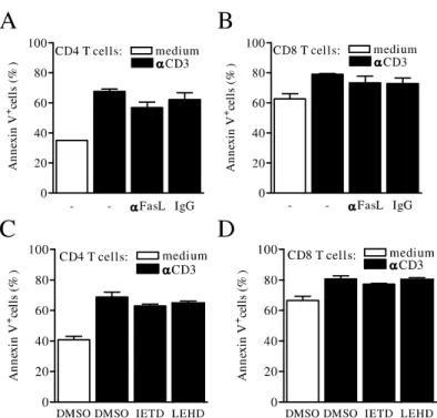
path-way, we treated T cell cultures with anti-FasL or control IgG antibody and assessed apoptosis by annexin V stain-ing upon 24 h. Treatment with anti-FasL did not inhibit apoptosis induced upon T cell activation (Fig. 3A and B). To address whether caspase-8 or caspase-9 were in-volved in apoptosis, T cells were activated in the presence of caspase-8 (zIETD) or caspase-9 (zLEHD) inhibitor. Neither zIETD nor zLEHD inhibited AICD in T cells
fromL. majorinfection (Fig. 3C and D).
CASPASE-8ANDCYTOKINEEXPRESSION BYT CELLS
cyto-Fig. 2 – Activation-induced cell death upon infection withL. major. Lymph node cells from normal andL. major-infected mice were cultured in the presence of medium only (open bars) or stimulated with anti-CD3 (black bars). After 24 h, cells were stained with anti-CD4 (A and C), anti-CD8 (B and D), 7-AAD and evaluated by flow cytometry. Panels show percentages of apoptotic (7-AAD+) CD4 and CD8 T cells upon 2 (A and B) or 6 (C and D) weeks of infection. Results are expressed as average and SEM. Significant differences between cells treated with medium and anti-CD3 were indicated for p<0.05(∗).
metry cytokine expression in T cell subsets, and whether
caspase-8 controls type-1/type-2 cytokines uponL.
ma-jorinfection. First, T cells from draining lymph nodes
(2 wk post infection) were activated with anti-CD3 in the presence of caspase-8 inhibitor zIETD or DMSO only during 72 h. Then, T cells were re-stimulated with ionomycin and PMA, in the presence of brefeldin A, to prevent cytokine secretion. The presence of zIETD did not affect the number of cells recovered from T cell cul-tures (not shown). The proportions of CD4 and CD8 T cells were similar (30-40%) in cultures (Fig. 4A and B). Caspase-8 inhibition negatively affected CD8, but not CD4, T cells (Fig. 4A and B) and the proportion of cells
expressing IFN-γ, but not IL-10 (Fig. 4C and D). Both
CD4 and CD8 T cells expressed IFN-γ(Fig. 5A and B),
whereas expression of IL-4 was not detected even upon
culture with exogenous IL-4 (not shown). The inhibition
of caspase-8 decreased the expression of IFN-γ in both
CD4 and CD8 T cells (Fig. 5A and B). These results sug-gest that caspase-8 plays a role in the control of cytokine
responses toL. majorinfection.
DISCUSSION
Apoptosis of CD4 T cells has been suggested as a
mech-anism to reduce Th1 responses inLeishmania donovani
infection (Das et al. 1999). In addition, Fas and/or FasL expression has been detected in T cells from lymph nodes (Desbarats et al. 2000) or lesions (Eidsmo et al. 2005)
duringL. majorinfection, and mice with defective Fas/
FasL expression show increased Th1 responses uponL.
Fig. 3 – T-cell death pathways duringL. majorinfection. Lymph node cells from infected (2 wk) mice were cultured with medium only (open bars) or stimulated with anti-CD3 (black bars) in the presence of anti-FasL or control IgG (A and B) or in the presence of zIETD, zLEHD or stock diluent DMSO (C and D). After 24 h, cultured cells were stained with anti-CD4, anti-CD8, and annexin V. Panels show percentages of apoptotic (annexin V+) CD4 (A and C) and CD8 (B and D) T cells. Results are expressed as average and SEM.
et al. 1998) infections. We observed increased apop-tosis in both CD4 and CD8 T cells from draining lymph
nodes only at later stages ofL. majorinfection. T cell
apoptosis can also be induced by stimulation in vitro
(AICD) as early as 2 wk and after 6 wk, at the peak of
lymphocyte activationin vivo. Nonetheless, AICD was
not blocked by inhibitors of FasL, 8 or caspase-9, although these reagents have been successfully used at the same doses in previous unrelated experiments (Silva et al. 2005, Guillermo et al. 2007). Therefore, AICD
in lymph nodes fromL. majorinfection may be
medi-ated by other mechanisms, such as granzyme-B-induced apoptosis (Devadas et al. 2006).
By contrast, caspase-8 inhibition by zIETD peptide negatively affected CD8 T cells and the expression of
IFN-γin T cells fromL. major-infected mice. Similarly,
CD8 T cells have defective survival and IFN-γ
expres-sion in mice bearing a T cell-restricted transgene for the viral-FLICE/caspase-8 inhibitory protein (vFLIP) (Wu
et al. 2004). These results suggest a paradoxical role of caspase-8 preventing, rather than inducing T cell death. Although caspase-8 inhibition could affect CD8 T-cell
responses duringL. majorinfection, the role of CD8 T
cells as an important source of IFN-γin the immunity to
primaryL. majorinfection is disputed (Huber et al. 1998,
Belkaid et al. 2002b). However, it has been suggested
that CD8 T cells can favor IFN-γ expression by
protec-tive CD4 Th1 cells duringL. majorinfection. (Herath et
al. 2003).
Based on evidence suggesting a non apoptotic role of caspase-8 in T cell signaling for cytokine expression (Su et al. 2005), we studied how caspase-8 inhibition would affect the development of type-1 vs type-2
re-sponses followingL. majorinfection. Caspase-8
inhibi-tion decreased the expression of IFN-γby both CD4 and
CD8 T cells. It is possible that these effects are due to de-fective signaling for cytokine expression upon caspase-8
- DMSO IETD 0
10 20 30 40 50
Lymph node:
Ion + PMA medium
C
D
4
+ce
ll
s (%)
- DMSO IETD
0 10 20 30 40
50 Lymph node: medium
Ion + PMA
*
C
D
8
+ ce
ll
s (%)
- DMSO IETD
0 1 2 3 4 5 6
Lymph node: medium
Ion+PMA
IL
-1
0
+ce
ll
s (%)
- DMSO IETD
0 10 20 30 40
Lymph node: medium
Ion+PMA
*
IF
N
+ ce
ll
s (
%
)
D
C
Fig. 4 – Expression of type 1 and type 2 cytokines inL. majorinfection. Lymph node cells from infected (2 wk) mice were stimulated with anti-CD3 only or in the presence of zIETD or stock diluent DMSO for 72 h. Cells were re-stimulated with PMA and ionomycin for 4 h, in the presence of brefeldin A. Cells were stained with anti-CD4 (A), anti-CD8 (B) or permeabilized and stained with anti-IL-10 (C) and anti-IFN-γ (D). Results are expressed as average and SEM. Significant differences between cells treated with zIETD and DMSO were indicated forp<0.05(∗).
and IL-2 secretion (Silva et al. 2005, Su et al. 2005).
In agreement with this idea, defective NF-κB activation
affects negatively both proliferation and the production
of IFN-γby T cells (Corn et al. 2003). By contrast,
IL-10 expression in T cells fromL. majorinfection was not
affected by inhibition of caspase-8 even in the presence of IL-4 (not shown). Therefore, non apoptotic activity of caspase-8 is not necessary for the expression of type-2 cytokines, but may be required for the induction of
protective type-1 response inL. majorinfection.
ACKNOWLEDGMENTS
We thank George A. DosReis (IBCCF-UFRJ) for helpful suggestions. This investigation received financial sup-port from the UNICEF/UNDP/World Bank/WHO Spe-cial Program for Research and Training in Tropical Dis-eases (TDR) (grant A60281), Conselho Nacional de De-senvolvimento Científico e Tecnológico (CNPq),
Fun-dação Carlos Chagas Filho de Amparo à Pesquisa do Estado do Rio de Janeiro (FAPERJ), and Institute for In-vestigation in Immunology (iii), Millennium Institutes, Brazilian Ministry of Science and Technology. W.F.P. receives an MSc fellowship from Coordenação de Aper-feiçoamento de Pessoal de Nível Superior (CAPES). M.F.L. is a research fellow at CNPq, Brazil.
RESUMO
A
DMSO IETDB
36.7% 18.3%
34.1% 25.2%
IFN-γ
CD
8
CD
4
Fig. 5 – Inhibition of caspase-8 reduces IFN-γexpression by T cells fromL. majorinfected mice. Lymph node cells from infected (2 wk) mice were stimulated with anti-CD3 in the presence of zIETD (right panels) or stock diluent DMSO (left panels), re-stimulated as described above and stained with anti-CD4 (A), anti-CD8 (B) and anti-IFN-γ(A and B). Panels show percentages of IFN-γ+CD4 T cells (A), and IFN-γ+CD8 T cells (B) as assessed by flow cytometry.
bloqueou a morte induzida por ativação das células T. Investi-gamos também se a inibição da atividade da caspase-8 poderia afetar a expressão de citocinas tipo-1 ou tipo-2. Nos estágios iniciais da infecção, células T CD4 e CD8 de animais infectados comL. majorexpressaram IFN-γapós ativação. O tratamento com o inibidor de caspase-8 zIETD (benzoil-oxicarbonil-Ile-Glu(OMe)-Thr-Asp(OMe)-fluorometilcetona) durante a esti-mulação de células T reduziu a proporção de células T CD8 e a expressão de IFN-γpor células T CD4 e CD8. Concluimos que a atividade não apoptótica de caspase-8 pode ser neces-sária para o estabelecimento da imunidade mediada por células T durante a infecção porL. major.
Palavras-chave: apoptose, caspase-8, células T CD8, IFN-gama, Leishmanioses.
REFERENCES
ALAM A, COHENLY, AOUADS ANDSEKALY RP. 1999. Early activation of caspases during T lymphocyte
stimula-tion results in selective substrate cleavage in nonapoptotic cells. J Exp Med 190: 1879–1890.
ALEXANDERCE, KAYEPM ANDENGWERDA CR. 2001. CD95 is required for the early control of parasite burden in the liver ofLeishmania donovani-infected mice. Eur J Immunol 31: 1199–1210.
ALVESNL,VANLIERRAANDELDERINGE. 2007. With-drawal symptoms on display: Bcl-2 members under in-vestigation. Trends Immunol 28: 26–32.
BELKAIDY, PICCIRILLO CA, MENDEZS, SHEVACH EM ANDSACKSDL. 2002a. CD4+CD25+ regulatory T cells control Leishmania major persistence and immunity. Nature 420: 502–507.
BELKAID Y, VON STEBUT E, MENDEZ S, LIRA R, CALERE, BERTHOLET S, UDEY MCANDSACKS D. 2002b. CD8+ T cells are required for primary immunity in C57BL/6 mice following low-dose, intradermal challenge withLeishmania major. J Immunol 168: 3992–4000. BERTHOAL, SANTIAGO MA, DA-CRUZ AMANDCOU
with localized cutaneous leishmaniasis. Braz J Med Biol Res 33: 317–325.
CHUNHJET AL. 2002. Pleiotropic defects in lymphocyte activation caused by caspase-8 mutations lead to human immunodeficiency. Nature 419: 395–399.
CORN RA ET AL. 2003. T cell-intrinsic requirement for NF-κB induction in postdifferentiation IFN-γproduction and clonal expansion in a Th1 response. J Immunol 171: 1816–1824.
DAS G, VOHRA H, RAOK, SAHAB ANDMISHRA GC. 1999. Leishmania donovani infection of a susceptible host results in CD4+ T-cell apoptosis and decreased Th1 cytokine production. Scand J Immunol 49: 307–310. DESBARATSJ, STONEJE, LINL, ZAKERIZF, DAVISGS,
PFEIFFER LM, TITUS RG AND NEWELL MK. 2000. Rapid early onset lymphocyte cell death in mice resis-tant, but not susceptible to Leishmania majorinfection. Apoptosis 5: 189–196.
DEVADASS, DASJ, LIUC, ZHANGL, ROBERTSAI, PAN Z, MOOREPA, DASGANDSHIY. 2006. Granzyme B is critical for T cell receptor-induced cell death of type 2 helper T cells. Immunity 25: 237–247.
EIDSMOL, NYLENS, KHAMESIPOURA, HEDBLADMA, CHIODIF ANDAKUFFO H. 2005. The contribution of the Fas/FasL apoptotic pathway in ulcer formation during Leishmania major-induced cutaneous Leishmaniasis. Am J Pathol 166: 1099–1108.
GUILLERMO LV, SILVA EM, RIBEIRO-GOMES FL, DE MEISJ, PEREIRAWF, YAGITAH, DOSREISGAAND LOPES MF. 2007. The Fas death pathway controls co-ordinated expansions of type 1 CD8 and type 2 CD4 T cells inTrypanosoma cruziinfection. J Leukoc Biol 81: 942–951.
HERATHS, KROPFPANDMULLERI. 2003. Cross-talk be-tween CD8(+) and CD4(+) T cells in experimental cu-taneous leishmaniasis: CD8(+) T cells are required for optimal IFN-γ production by CD4(+) T cells. Parasite Immunol 25: 559–567.
HUANGFP, XUD, ESFANDIARIEO, SANDSW, WEIXQ ANDLIEWFY. 1998. Mice defective in Fas are highly susceptible toLeishmania majorinfection despite elevated IL-12 synthesis, strong Th1 responses, and enhanced nitric oxide production. J Immunol 160: 4143–4147.
HUBERM, TIMMS E, MAK TW, ROLLINGHOFF MAND LOHOFF M. 1998. Effective and long-lasting immunity against the parasite Leishmania majorin CD8-deficient mice. Infect Immun 66: 3968–3970.
1999. Caspase activation is required for T cell prolifera-tion. J Exp Med 190: 1891–1896.
KRAMMERPH. 2000. CD95’s deadly mission in the immune system. Nature 407: 789–795.
LENARDOM, CHANKM, HORNUNGF, MCFARLANDH, SIEGELR, WANG J ANDZHENG L. 1999. Mature T lymphocyte apoptosis–immune regulation in a dynamic and unpredictable antigenic environment. Annu Rev Im-munol 17: 221–253.
MCMAHON-PRATTDANDALEXANDERJ. 2004. Does the Leishmania majorparadigm of pathogenesis and protec-tion hold for New World cutaneous leishmaniases or the visceral disease? Immunol Rev 201: 206–224.
MOHRSM, LEDERMANNB, KOHLERG, DORFMULLERA, GESSNER AANDBROMBACHER F. 1999. Differences between IL-4- and IL-4 receptor alpha-deficient mice in chronic leishmaniasis reveal a protective role for IL-13 receptor signaling. J Immunol 162: 7302–7308. NOBEN-TRAUTHN, PAULWEANDSACKSDL. 1999.
IL-4- and IL-4 receptor-deficient BALB/c mice reveal differ-ences in susceptibility toLeishmania majorparasite sub-strains. J Immunol 162: 6132–6140.
NOBEN-TRAUTHN, LIRAR, NAGASEH, PAULWEAND SACKSDL. 2003. The relative contribution of IL-4 re-ceptor signaling and IL-10 to susceptibility toLeishmania major. J Immunol 170: 5152–5158.
PINHEIRORO, PINTOEF, BENEDITOAB, LOPESUGAND ROSSI-BERGMANNB. 2004. The T-cell anergy induced byLeishmania amazonensisantigens is related with de-fective antigen presentation and apoptosis. An Acad Bras Cienc 76: 519–527.
SEHRA S, PATEL D, KUSAM S, WANGZY, CHANG CH ANDDENTAL. 2005. A role for caspases in controlling IL-4 expression in T cells. J Immunol 174: 3440–3446. SILVAEMET AL. 2005. Caspase-8 activity prevents type 2
cytokine responses and is required for protective T cell-mediated immunity againstTrypanosoma cruziinfection. J Immunol 174: 6314–6321.
SUH, BIDEREN, ZHENGL, CUBREA, SAKAIK, DALEJ, SALMENAL, HAKEMR, STRAUSSANDLENARDOM. 2005. Requirement for caspase-8 in NF-κB activation by antigen receptor. Science 307: 1465–1468.