O R I G I N A L A R T I C L E UDC: 577.1::617.7 DOI: 10.2298/VSP1203231D
Transforming growth factor
β
1, matrix metalloproteinase-2 and its
tissue inhibitor in patients with pseudoexfoliation glaucoma/syndrome
Transformišu
ć
i faktor rasta
β
1, matriks metaloproteinaza-2 i njen tkivni
inhibitor kod bolesnika sa pseudoeksfolijativnim sindromom/glaukomom
Jasmina Djordjević-Jocić*, Gordana Zlatanović*, Dragan Veselinović*, Predrag Jovanović*, Vidosava Djordjević*, Lilika Zvezdanović†, Gordana Stanković-Babić*,
Milena Vujanović*, Sonja Cekić*, Matthias Zenkel‡, Ursula Schlotzer-Schrehardt‡
*Clinic of Ophthalmology, Clinical Center Niš, Niš Serbia;
†
Centre for Medical Biochemistry, Clinical Center Niš, Niš, Serbia; ‡Department of Ophthalmology, University of Erlangen-Nuernberg, Germany
Abstract
Background/Aim. Transforming growth factor- (TGF-), oxidative stress and imbalance between matrix metalloproteinases (MMPs) and their tissue inhibitors (TIMPs) may play an important role in pathogenesis of pseudoexfoliation syndrome/glaucoma (PEX Sy/Gl). The aim of the study was to measure concentrations of TGF-1, MMP-2, TIMP-2 in the aqueous humor in the exam-ined group, as well as to compare the biochemical findings with the following clinical parameters: degree of chamber angle pigmantation, presence of pseudoexfoliation and the value of intraocular pressure (IOP). Methods. Aqueous samples from 30 patients with cataract, 30 patients with PEX Sy, 36 patients with PEX Gl, and 42 patients with primary open-angle glaucoma (POAG) were collected during phacoemulsification cataract surgery. TGF MMP-2, TIMP-2 Fluotokine Multi Analyze Profiling kits and Luminex technology were used to simultaneously measure TGF MMP-2 and TIMP-2. Results. TGF-β1, MMP-2, TIMP-2 were detected in human aqueous from all the groups with the highest level in the group
with PEX Gl. Statistically, a significant correlation be-tween the levels of TGF 1, MMP-2, TIMP-2 in the aque-ous humor of the patients with PEX Gl and the IOP value was confirmed (p < 0.05). In this group, the positive cor-relations between the TGF 1 concentration in the aque-ous humor and the presence of pseudoexfoliation (p < 0.01), on the one hand, and between the TIMP-2 level and the presence of pseudoexfoliation (p < 0.05), on the other, were reported. A statistically significant positive correlation of TGF-1 and MMP-2, and the degree of chamber angle pigmentation in the PEX Gl group was confirmed (p < 0.05). In the POAG group, TIMP-2 values were in a negative correlation with the degree of pigmen-tation (p < 0.05), and the IOP value (p < 0.05). Conclu-sion. TGF 1 and MMP-2 affect the degree of chamber angle pigmentation and the degree of pseudoexfoliation in patients with pseudoexfoliative glaucoma.
Key words:
transforming growth factor beta 1; matrix metaloproteinase 2; tissue inhibitor of metaloproteinase-2; exfoliation syndrome.
Apstrakt
Uvod/Cilj. Transformišući faktor rasta ß1 (TGF-ß1), oksi-dativni stres i disbalans između matriks metaloproteinaza (MMPs) i njihovih tkivnih inhibitora (TIMPs) igraju važnu ulogu u patogenezi pseudoeksfolijativnog sindro-ma/glaukoma PEX Sy/Gl. Cilj ove studije bio je da se utvr-di koncentracija TGF-ß1, MMP-2, TIMPs-2 u očnoj voutvr-dici izabranih grupa bolesnika, kao i da se uporede biohemijski nalazi sa odgovarajućim kliničkim parametrima: stepen pig-mentacije komornog ugla, prisustvo pseudoekfolijacija i vrednost intraokularnog pritiska (IOP). Metode. Tokom operacije fakoemulzifikacije katarakte prikupljena je očna
prisu-stva pseudoeksfolijacija sa druge strane (p < 0,05). Potvrđe-na je i statistički zPotvrđe-načajPotvrđe-na pozitivPotvrđe-na korelacija nivoa TGF-ß1, MMP-2 i stepena pigmentacije komornog ugla (p < 0,05) u grupi bolesnika sa PEX Gl. U grupi bolesnika sa POAG vrednost TIMP-2 je u negativnoj korelaciji sa stepenom pi-gmentacije komornog ugla i vrednostima IOP (p < 0,05). Zaključak. TGF-ß1 i MMP-2 utiču na stepen pigmentacije
komornog ugla i prisustva pseudoeksfolijacija kod bolesnika sa PEX glaukomom.
Ključne reči:
faktor rasta, transformišući, beta 1; matriks metaloproteinaza 2; tkivni inhibitor matriks metaloproteinaze-2; eksfolijativni sindrom.
Introduction
Pseudoexfoliation syndrome (PEX Sy) is an elastosis-like systemic disease characterized by the production and progressive accumulation of extracellular fibrillar material, known as pseudoexfoliative material, on the tissues of the anterior segment of the eye and different visceral organs 1–3. In many countries PEX Sy is common in population over the age of 60, and in many cases it leads to the appearance of pseudoexfoliation glaucoma (PEX Gl), one of the most fre-quent causes of poor visual acuity and blindness 4, 5. The real pathogenesis of PEX Sy is still not known enough. Recent studies have shown that PEX Sy is a microfibrilopathy and that transforming growth factor (TGF-), oxidative stress and an imbalance between matrix metalloproteinases (MMPs) and their tissue inhibitors (TIMPs) play a role in its appearance. The aim of the study was to determine concen-trations of TGF-1, MMP-2, TIMP-2 in the aqueous humor in patients with PEX Gl, PEX Sy, primary open-angle glau-coma (POAG) and cataract (Cat), and also, to compare the biochemical findings with the following clinical parameters: degree of angle pigmantation, presence of pseudoexfoliation and the value of intraocular pressure (IOP).
Methods
Four groups of patients were included in this prospec-tive study: the group I – 42 patients with PEX Gl, group II – 30 patients with PEX Sy, group III – 36 patients with POAG, and the group IV – 30 patients with Cat.
The ophthalmological examination was conducted at the Clinic of Ophthalmology, Clinical Center Niš (Niš, Serbia).
Biochemical analyses were done in the Center of Medi-cal Biochemistry of CliniMedi-cal Center Niš, and the Ophthol-mology, Department of the University of Erlangen-Nürnberg (Germany).
Tranforming growth factor 1 was determined by the ELISA method using a commercially available kit (Quan-tikine; R&D System, UK).
The MMP-2 vlaues in the aqueous humor of patients were determined with the multiplex method for quantitative measurement using a commercially available test (Quan-tikine; R&D System, UK). The values were read by the Lu-minex analyser.
For this study the aqueous humor of the patients was used. Aqueous humor was extracted by paracentesis through the limbus of the cornea during the trabeculectomy or routine phacoemulsification paying special attention not to touch the endothelium, iris or lens as well as extracting the aqueous
humor without any traces of blood. After its aspiration and securing in sterile test tubes, aqueous humor samples (80– 100 l) were immediately frozen in liquid nitrogen and then stored and kept at the temperature of -80°C.
All the patients gave their written informed consent. All the patients underwent pupillary dilation a day before the surgery so that the presence of pseudoexfiliation could be confirmed. The classification of the presence of pseudoexfo-liation was done from I0 to III0. Thus, I0 marked that PEX material was visible only on the anterior side of the lens after the pupillary dilation; II0 – PEX material was occasionally visible on the pupillary edge, and III0 – pseudoexfoliation was costantly visible along the whole circumference of the pupillary edge and on the anterior side of the lens, with or without iridodonesis and phacodonesis. The width and pig-mentation of the chamber angle were classified according to the Scheie classification.
A one-way analysis of variance (One –Way ANOVA) and a Post Hoc (Tukey HSD) analysis were used to check the difference in the average age between the examined groups. To check the hypotheses that there are differences in the presence of certain attributes between the groups, Fisher’s Exact Probability Test was used. To evaluate the differences in IOP values between the patients with different kinds of glaucoma, the Mann-Whitney U-Wilcoxon Rank Sum W-test was used. This test was also used to compare the values of the concentration of certain enzymes in the aqueous humor of the patients. The level of significance was adapted by the Bonferoni inequality and for the time being it is 0.01. The correlation between enzymes and clinical parameters was conducted with the help of Pearson's linear correlation coef-ficient. For the statistical analysis of the data, SPSS Win-dows (Ver. 8.0) was used.
Results
The average age of the patients with PEX Gl was 68.9 ± 7.6 years; of the patients with PEX Sy 66.8 ± 4.6 years; of the patients with POAG 62.8 ± 8.8 years; and, finally, of the pa-tients with cataract 64.8 ± 3.7 years. Statistically, the papa-tients with PEX Gl were significantly older in relation to the patients with POAG (p < 0.001). The average value of IOP was 43.1 ± 13 mm Hg in the group with PEX Gl, while in the group with POAG the average value of IOP was 34.8 ± 11.4 mm Hg and this difference proves to be statistically significant (p < 0.001).
Fig. 1 – The presence of pseudoexfoliation (PEX) in the patients with PEX glaucoma and PEX syndrome
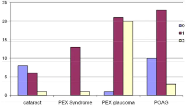
Figure 2 shows a degree of chamber angle pigmentation (according to Scheie) in the patients with POAG and those with pseudoexfiloation syndrome.
Fig. 2 – Degree of chamber angle pigmentation (according to Scheie) in the patients with primary open-angle glaucoma
(POAG) and pseudoexfiloation (PEX) syndrome
The greatest number of POAG patients had Io chamber angle pigmentation according to Scheie (96.7%). In the PEX Gl group, the gonioscopic findings mostly showed IIo ili IIIo chamber angle pigmentation (86.7%). The observed difference in the chamber angle pigmantation degree between these two groups was statistically highly significant (p < 0.0001).
The values of TGF-β1, MMP-2 i TIMP-2 in the aqueous humor of the patients in all the groups is shown in Table 1. The average level of TGF-β1 in the aqueous humor of the patients with PEX Gl was 147.29 76.54 pg/mL, while in the group with senile cataract it was 38.85 28.47 pg/mL (p < 0.0001).
The mean values of this protease were 29031.5 16725.8 pg/mL in the PEX Gl group, 24250.12 42741.74 pg/mL in the PEX Sy group, 19346.07 10871.13 pg/mL in the POAG group, and 15195.40 11225.02 pg/mL in the Cataract group. Significant differences were found between MMP-2 in PEX Gl and both POAG and cataract groups (p < 0.001, and p < 0.0001, respectively). The difference in the values of MMP-2 in the aqueous humor of the patients with PEX Sy in comparison with the senile Cataract and POAG groups, respectively, was signifi-cant (p < 0.05).
The levels of TIMP-2 in aqueous humor ranged from 19.947 pg/mL in the patients with cataract to 43.521 pg/mL in the patients with PEX Gl. A significant increase in aque-ous humor TIMP-2 levels was mesaured in both PEX Gl and POAG groups compared with PEX Sy and Cataract groups (p < 0.001 and p < 0.01, respectively).
Table 2 shows a correlation of the levels of TGF-1, MMP-2, TIMP-2 in the aqueous humor of patients with the degree of pigmentation, the presence of pseudoexfoliation and IOP value. Statistically, a significant correlation between
Table 1 Concentration of transforming growth factor β1 (TGF-β1), matrix metaloproteinase-2 (MMP-2) and its tissue inhibitor
(TIMP-2) in aqueous humor of the patients
Group TGF- 1 (pg/mL) MMP-2 (pg/mL) TIMP-2 (pg/mL)
PEX Gl 147.29 76.54**, *** 29031.5 16725.8 *, **, *** 43521 19737 **, *** POAG 73.96 48.50 ## 19346.07 10871.13 26103 14989 PEX Sy 108.26 30.85***, # 24250.12 42741.74 # 39103 4961 Senile cataract 38.85 28.47 15195.40 11225.02 19947.7 5181
PEX Gl – pseudoexfoliation glaucoma; PEX Sy – pseudoexfoliation syndrome; POAG – primary open-angle glaucoma.
*p < 0.05 in relation to PEX syndrome
**p < 0.001 in relation to POAG ***p < 0.0001 in relation to senile cataract
#
p < 0.05 in relation to POAG
##
p < 0.01 in relation to senile cataract
Table 2 Correlation of transforming growth factor β1 (TGF-β1), matrix metaloproteinase-2 (MMP-2) and its tissue inhibitor (TIMP-2) in aqueous humor of the patients with the degree of pigmentation, presence of pseudoexfoliation (PEX) and
intraocular pressure (IOP) values
PEXGl POAG
Biochemical
pa-rameters Degree
of PEX IOP
Pigmentation of the chamber angle
Pigmentation of the chamber angle
IOP
MMP-2 c = 0.33
p < 0.05
c = 0.36
p < 0.05
TIMP-2 c = 0.46
p < 0.05
c = 0.43
p < 0.05
c = -0.48
p < 0.01
c = -0.36
p < 0.05
TGF-1 c = 0.47
p < 0.01
c = 0.48
p < 0.01
c = 0.312
p < 0.05
the levels of TGF-β1, MMP-2, TIMP-2 in the aqueous hu-mor of the patients with PEX Gl and the IOP value was con-firmed (p < 0.05). In this group, positive correlations were established between the level of TGF 1 and TIMP-2 in aqueous humor and the presence of pseudoexfoliation (c = 0.47; p < 0.01 and c = 0.46, p < 0.05, respectively). In the PEX Gy group a statistically significant positive correla-tion of TGF-1 and MMP-2, and the degree of angle pig-mentation was confirmed (c = 0.312; p < 0.05). In the POAG group, the TIMP-2 values were in a negative correlation with the degree of angle pigmentation (c = -0.48, p < 0.05), and the IOP value (c = 0.36; p < 0.05).
Discussion
The pathological accumulation of abnormal fibrillar extracellular material, which is a characteristic of PEX Sy in numerous extra- and intraocular tissues, can result in a great number of clinical complications and the development of PEX Gl 1–4. PEX syndrome is one of the main causes of PEX Gl. Clinical changes in the eye are often asymmetrical and can be manifested as trabeculopathy, iridopathy, zonulopa-thy, endotheliopazonulopa-thy, pigment dispersion and increased trab-ecular pigmentation, high values of IOP, as well as great daily fluctuations of IOP accompanied by the rapid deterio-ration of the optic nerve head and a progressive visual field loss 3, 6–10.
However, a precise etyology of this systemic disease of extracellular matrix still remains unknown, though it is con-sidered TGF 1 causes an imbalance between MMPs and their TIMPs, leading to a progressive accumulation of exfo-liation material in the trabecular tissue, which further results in elevated IOP 11–13. The normal balance requires a balanced interaction of MMPs and TIMPs, and the normal relation of enzymes to the inhibitor is 1 : 1. Any changes in the balance can result in the excessive accumulation or degradation of extracellular matrix (ECM) 6, 7, 14, 15.
In this study the values of total TGF-β1 in aqueous humor of the patients with PEX Gl and PEX Sy, respec-tively, were significantly higher in relation to the values of this enzyme in aqueous humor of the patients with POAG and cataract, respectively (p < 0.0001), while the activity of total TGF-β1 in the aqueous humor of the patients with PEX Gl was different from and higher than the activity of this enzyme in the aqueous humor of patients with PEX Sy, but the difference was not statistically significant – it can be said that it was at the verge of statistical significance (p < 0.01).
Statistically, the values of total TGF-β1 in aqueous hu-mor of the patients with POAG were considerably higher in relation to the values of aqueous humor inpatients with senile cataract (p < 0.0001). The values of TGF-β1 in the aqueous humor of the patients with POAG statistically were consid-erably lower than the values in the aqueous humor of the pa-tients with PEX Sy (p < 0.05).
The patients of different groups with different ophthal-mological diseases had different – statistically significant – values of MMP-2 in aqueous humor (p < 0.0001).
MMP-2 was detectable in aqueous humor of all the pa-tients. The values of MMP-2 in the aqueous humor of the patients with PEX Gl were statistically significantly higher in comparison with the values in the patients of senile cataract (p < 0.0001), while that difference in comparison with the values of this proen-zyme in aqueous humor of the patients with POAG was also sig-nificant (p < 0.001).
The MMP-2 values in aqueous humor of the patients with PEX Gl were not significantly higher in comparison with the val-ues of this proenzyme in aqueous humor of the patients with PEX Sy (p > 0.05), while the difference in the values of this proenzyme in the aqueous humor of the patients with PEX Sy in comparison with the senile cataract and POAG groups, respectively, was (p < 0.05). The TIMP-2 values ranged from 19,947 pg/mL to 43,521 pg/mL. A significant increase in TIMP-2 in aqueous humor of the patients both with PEX Gl and PEX Sy was no-ticed in comparison with the values measured in the patients both with POAG and cataract (p < 0.001).
In their study, Schlotzer-Schrehard et al. 16 examined whether they could detect latent and active 1 i TGF-2 in aqueous humor using the ELISA method. Both latent and active ones could be detected in aqueous humor and se-rum of the patients with the PEX eye. The level of total and active TGF-1 respectively was statistically significantly higher in patients both with PEX Sy and PEX Gl in compari-son with the group with cataract and open-angle glaucoma respectively, while the difference was not noticed between the groups with PEX changes. These authors also found that the TGF-1 values in serum statistically were not signifi-cantly different among the examined groups.
Koliakos et al. 13 also found increased levels of TGF-1 in the patients with PEX Sy ranging from 6.1 to 54.6 (me-dian value 17.06 ± 11.02) pg/mL, which statistically was a significant rise in comparison with the group with cataract.
Slotzer-Schrehard et al. 14 measured a total and active quantity of MMPs and TIMPs in patients with PEX Gl, PEX Sy and POAG, using the Western Blot, elektrophoresis and the ELISA method. MMP-2 (both as proenzyme and in its complex form) was found in considerable quantity ranging from 18.6 to 232.4 ng/mL. Despite this high varibility, the total quantity of MMP-2 statistically was considerably ele-vated in aqueous humor of the patients with PEX Sy with or without glaucoma, respectively, in comparison with aqueous humor of the patients with POAG and cataract, respectively. Free, unbound MMP-2 made 22–24% of the total quantity in aqueous humor and was signifigantly increased in the pa-tients with PEX Sy. The concentrations of TIMPs in aqueous humor were six to seven times as high as the concentrations of MMPs, and they had the predominant role in the activa-tion of MMPs. MMP-2 and TIMP-2 should be balanced and any imbalance can affect the biological activity of the cell. It was noticed that the concentrations of MMPs and TIMPs in aqueous humor were considerably higher in patients with pseudoexfoliation with or withour glaucoma, respectively, in comparison with the patients with primary open-angle glau-coma, especially MMP-2, -3, TIMP-1, -2 17-25.
from patients who had PEX Gl and used the prostaglandin, latanoprost (0.005% eye drops) monotherapy in comparison with the patients who had PEX Gl and used beta blockers in the form of timolol maleate, (0.5% eye drops). These authors believed that TGF-1 increased the TIMP-2 and MMP-2 ex-pressions in the eyes with pseudoexfoliation, and that latano-prost interrupted the positive feedback mechanism of TGF-1 accumulation. However, the reduction mechanism of TGF-1, MMP-2 i TIMP-2 by latanoprost required furthur clarification.
Patients with PEX Gl had a statistically significantly higher degree of pseudoexfoliative changes in comparison with patients with PEX Sy (p < 0.0001). The greatest number of POAG patients had Io chamber angle pigmentation ac-cording to Scheie (96.7%). In the PEX Gl group, the goneo-scopic findings mostly showed IIo or IIIo chamber angle pigmentation (86.7%). The observed difference in chamber angle pigmantation degree between these two groups was statistically highly significant (p < 0.0001).
In the PEX Gl group, it was confirmed that TGF 1 was positively correlated with MMP-2 (c = 0.51; p < 0.01), and a positive correlation between MMP-2 and its tissue inhibitor TIMP-2 was also noticed (c = 0.54; p < 0.01).
Statistically, a significant correlation between the levels of TGF 1, MMP-2, TIMP-2 in aqueous humor of the pa-tients with PEX Gl and the IOP value was confirmed (p < 0.05). In this group, positive correlations between TGF
1 concentration in aqueous humor and the presence of pseudoexfoliation (c = 0.47; p < 0.01), on the one hand, and between TIMP-2 level and the presence of pseudoexfoliation (c = 0.46, p < 0.05), on the other, were reported.
In this study the relation of TGF 1 and MMP-2 levels, and the degree of chamber angle pigmentation was estab-lished, but that connection was not statistically significant in the PEX Sy group (p > 0.05), while a statistically significant positive correlation of TGF 1 and MMP-2, and the degree of chamber angle pigmentation in the PEX Gl group was confirmed (c = 0.312; p < 0.05). In the POAG group, the TIMP-2 values were in a negative correlation with the degree of pigmentation (c = -0.48, p < 0.05), and the IOP value (c = 0.36; p < 0.05).
At the same time, the MMP-2 values, on one hand, and the presence of exfoliation PEX, on the other, did not show a statistically significant connection in the PEX Gl patients and PEX Sy patients.
Conclusion
Concentrations of TGF 1 and MMP-2 in the aqueous humor of patients with pseudoexfoliative changes are con-siderably elevated, and TGF 1 and MMP-2 affect the de-gree of chamber angle pigmentation and the dede-gree of pseu-doexfoliation presence in patients with pseudoexfoliative glaucoma.
R E F E R E N C E S
1. Ritch R, Schlötzer-Schrehardt U, Konstas AG. Why is glaucoma as-sociated with exfoliation syndrome? Prog Retin Eye Res 2003; 22(3): 253−75.
2. Ritch R. Exfoliation syndrome-the most common identifiable cause of open-angle glaucoma. J Glaucoma 1994; 3(2): 176−7.
3. Ritch R, Schlötzer-Schrehardt U. Exfoliation syndrome. Surv Ophthalmol 2001; 45(4): 265−315.
4. Schlötzer-Schrehardt U, Küchle M, Jünemann A, Naumann GO. Relevance of the pseudoexfoliation syndrome for the glauco-mas. Ophthalmologe 2002; 99(9): 683−90. (German)
5. Topouzis F, Harris A, Wilson MR, Koskosas A, Founti P, Yu F, et al. Increased likelihood of glaucoma at the same screening in-traocular pressure in subjects with pseudoexfoliation: the Thessaloniki Eye Study. Am J Ophthalmol 2009; 148(4): 606−13.e1.
6. Lütjen-Drecoll E. Functional morphology of the trabecular meshwork in primate eyes. Prog Retin Eye Res 1999; 18(1): 91−119.
7. Grierson I, Pfeiffer N, Cracknell KP, Appleton P. Histology and fine structure of the iris and outflow system following latanoprost therapy. Surv Ophthalmol 2002; 47 Suppl 1: S176−84. 8. Gabelt BT, Kaufman PL. Changes in aqueous humor dynamics
with age and glaucoma. Prog Retin Eye Res 2005; 24(5): 612−37.
9. Alexander JP, Samples JR, Acott TS. Growth factor and cytokine modulation of trabecular meshwork matrix metalloproteinase and TIMP expression. Curr Eye Res 1998; 17(3): 276−85. 10.Chintala SK, Wang N, Diskin S, Mattox C, Kagemann L, Fini ME,
et al. Matrix metalloproteinase gelatinase B (MMP-9) is associ-ated with leaking glaucoma filtering blebs. Exp Eye Res 2005; 81(4): 429−36.
11.Yüksel N, Karabaş VL, Arslan A, Demirci A, Cağlar Y. Ocular hemodynamics in pseudoexfoliation syndrome and pseudoex-foliation glaucoma. Ophthalmology 2001; 108(6): 1043−9. 12.Esaki K, Ito K, Matsunaga K, Sugimoto K, Sasoh M, Uji Y.
Ante-rior chamber structural change in postural variation in pseudo-exfoliation syndrome. Nihon Ganka Gakkai Zasshi 2001; 105(8): 524−9. (Japanese)
13.Koliakos GG, Schlötzer-Schrehardt U, Konstas AG, Bufidis T, Geor-giadis N, Dimitriadou A. Transforming and insulin-like growth factors in the aqueous humour of patients with exfoliation syndrome. Graefes Arch Clin Exp Ophthalmol 2001; 239(7): 482−7.
14.Schlötzer-Schrehardt U, Lommatzsch J, Küchle M, Konstas AG, Naumann GO. Matrix metalloproteinases and their inhibitors in aqueous humor of patients with pseudoexfoliation syn-drome/glaucoma and primary open-angle glaucoma. Invest Ophthalmol Vis Sci 2003; 44(3): 1117−25.
15.Schlötzer-Schrehardt U, Naumann GO. Ocular and systemic pseu-doexfoliation syndrome. Am J Ophthalmol 2006; 141(5): 921−37.
16.Schlötzer-Schrehardt U, Zenkel M, Küchle M, Sakai LY, Naumann GO. Role of transforming growth factor-beta1 and its latent form binding protein in pseudoexfoliation syndrome. Exp Eye Res 2001; 73(6): 765−80.
17.Visse R, Nagase H. Matrix metalloproteinases and tissue in-hibitors of metalloproteinases: structure, function, and bio-chemistry. Circ Res 2003; 92(8): 827−39.
19.el-Shabrawi Y, Eckhardt M, Berghold A, Faulborn J, Auboeck L, Mangge H, et al. Synthesis pattern of matrix metalloproteinases (MMPs) and inhibitors (TIMPs) in human explant organ cul-tures after treatment with latanoprost and dexamethasone. Eye (Lond) 2000; 14( Pt 3A): 375−83.
20.Pang IH, Hellberg PE, Fleenor DL, Jacobson N, Clark AF. Expres-sion of matrix metalloproteinases and their inhibitors in hu-man trabecular meshwork cells. Invest Ophthalmol Vis Sci 2003; 44(8): 3485−93.
21.Gartaganis SP, Georgakopoulos CD, Mela EK, Exarchou A, Ziouti N, Assouti M, et al. Matrix metalloproteinases and their in-hibitors in exfoliation syndrome. Ophthalmic Res 2002; 34(3): 165−71.
22.Määttä M, Tervahartiala T, Vesti E, Airaksinen J, Sorsa T. Levels and activation of matrix metalloproteinases in aqueous humor are elevated in uveitis-related secondary glaucoma. J Glaucoma 2006; 15(3): 229−37.
23.Weinstein WL, Dietrich UM, Sapienza JS, Carmichael KP, Moore PA, Krunkosky TM. Identification of ocular matrix metallo-proteinases present within the aqueous humor and
iridocor-neal drainage angle tissue of normal and glaucomatous canine eyes. Vet Ophthalmol 2007; 10 Suppl 1: 108−16.
24.Rönkkö S, Rekonen P, Kaarniranta K, Puustjärvi T, Teräsvirta M, Uusitalo H. Matrix metalloproteinases and their inhibitors in the chamber angle of normal eyes and patients with primary open-angle glaucoma and exfoliation glaucoma. Graefes Arch Clin Exp Ophthalmol 2007; 245(5): 697−704.
25.Ho SL, Dogar GF, Wang J, Crean J, Wu QD, Oliver N, et al. Ele-vated aqueous humour tissue inhibitor of matrix metallopro-teinase-1 and connective tissue growth factor in pseudoexfo-liation syndrome. Br J Ophthalmol 2005; 89(2): 169−73. 26.Konstas AG, Koliakos GG, Karabatsas CH, Liakos P,
Schlötzer-Schrehardt U, Georgiadis N, et al. Latanoprost therapy reduces the levels of TGF beta 1 and gelatinases in the aqueous hu-mour of patients with exfoliative glaucoma. Exp Eye Res 2006; 82(2): 319−22.