Study of crystallite size and strain as a function of morphological evolution
in zinc oxide powder obtained from hydroxycarbonate precursor
Fernando Aparecido Sigoli, Carlos de Oliveira Paiva-Santos, Miguel Jafelicci, Jr., and Marian Rosaly Davolosa)
Instituto de Quimica, Universidade Estadual Paulista-UNESP, P.O. Box 355, 14801-970, Araraquara, SP, Brazil
(Received 12 February 2000; accepted 3 April 2001)
In this work, zinc oxide samples were obtained from hydroxycarbonate by thermal decomposition at 300 °C. Zinc hydroxycarbonate samples were produced by homogeneous precipitation over different periods of time. The method used to obtain zinc oxide produces different morphologies as a function of the precursor precipitation time. Among the obtained particle shapes were porous spherical aggregates, spherulitic needle aggregates, and single acicular particles. This work investigated spherulitic needle-aggregate formation and the correlation among morphology, domain size, and microstrain. Transmission electron microscopy data revealed that the acicular particles that form the spherulitic needle aggregates consist of nanometer crystallites. Apparent crystallite size and microstrain in the directions perpendicular to (hOO), (hOl), (hkO), and (00/) planes were invariable as a function of precursor precipitation time. From the results, it was possible to conclude that the precursor precipitation period directly influenced the morphology of the zinc oxide but did not influence average crystallite size and microstrain for ZnO samples. Therefore, using this route, it was possible to prepare zinc oxide with different morphologies without microstructural alterations. © 2001 International Centre for Diffraction Data. [DOI: 10.1154/1.127606]
Key words: zinc oxide, crystallite size, strain, morphology
I. INTRODUCTION
Precursors with low thermal stability, e.g., oxalates, hy-droxynitrates, and hydroxycarbonates, can be transformed to oxides by endothermic thermal decomposition at about 300 °C, generally as finely divided solid particles (Auffredic
et al, 1995). The importance of endothermic decomposition in the formation of solids with high surface area is recog-nized in several fields of material science, e.g., sintering, catalysts, pigments, and reactivity of solids (Langford et al,
1993). It is commonly accepted that morphology and micro-structure of the particles in these solids depend on the nature of the precursor and also on the experimental parameters chosen for carrying out the solid state decomposition of pre-cursors (Auffredic et al, 1995). In recent years a large num-ber of methods have been developed to prepare particles of different compositions, but with uniform shapes and narrow size distributions in the micron and submicron range (Matjevic, 1985). The most convenient method for prepara-tion of such products is based on the control of chemical processes taking place in solutions (Haruta et al, 1986).
Zinc oxide is one of the most promising materials among AnBVI wide-band semiconductors (Georgobiami et al,
1993). The utilization of zinc oxide as a semiconductor is limited by the difficulties in obtaining samples with the proper electrical and optical properties (Georgobiami et al,
1993). Energy absorption in this semiconductor occurs by exciting electrons to the empty conduction band leaving holes in the completely filled valence band. Recombination usually occurs close to or in electronic defects of the crystal lattice (Blasse and Grabmaier, 1994). Such defects normally depend on morphology, structure, particle size, composition,
''Electronic mail: [email protected]
and crystallinity (Kouldelka and Horak, 1994), which play an important role in luminescent devices and pigment tech-nologies (Vanheusden et al, 1997). ZnO is industrially pro-duced in large quantities by the oxidation of zinc vapor, re-sulting in various morphologies, and also by thermal decomposition of a topotactic precursor such as zinc oxalate or zinc hydroxynitrate (Collins and Taylor, 1992). The zinc hydroxycarbonate precursor allows zinc oxide particles to be prepared with controlled size and shape. It is prepared by aging aqueous zinc salt solutions in the presence of urea at a controlled temperature (Castellano and Matijevic, 1989). Spherulitic needle aggregates of zinc oxide were obtained from a chloride medium and large porous spherical aggre-gates from a sulfate medium using this method (Zampronio
etal, 1995).
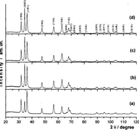
Figure 1. X-ray diffractogram of zinc oxide samples obtained at different precursor precipitation times: (a) ZnO-30, (b) ZnO-180, (c) ZnO-250, (d) ZnO-440.
20 30 100 110 120
2 8/ degree
In a recent work, we have obtained zinc oxide samples containing porous spherical, spherulitic needle aggregates or needle particles, depending on the precipitation time, from a zinc chloride medium (Sigoli et al., 1997). The present work investigated spherulitic needle-aggregate formation and the correlation among morphology, domain size, and microstrain in the ZnO obtained from endothermic decomposition of zinc hydroxide carbonate.
II. EXPERIMENT A. Sample preparation
Zinc oxide samples, ZnO-30, ZnO-180, ZnO-250, and ZnO-440, were obtained from thermal decomposition carried
out in an oven (EDG FIVE-4) at 300 °C for 240 min from zinc hydroxycarbonate, Zn5(CO3)2(OH)6. The latter was pre-cipitated from homogeneous precipitation at different times: 30, 180, 250, and 440 min, respectively, as described by Sigoli et al. (1997).
B. Powder characterization
Zinc oxide samples were characterized by a transmission electron microscopy (ZEISS EM 902), specific surface using the BET method (SCIENTIFIC INSTRUMENTS CG 2000) and X-ray diffraction (SIEMENS D5000) using a Cu radia-tion source and a graphite monochromator. The X-ray dif-fraction patterns for ZnO samples were obtained over the
1,6-
1,4-
1,2-O)
1,0-CM
0,8-S
0,6-U.
0,40 , 2
-0,0
A A
A A
A A
AA A
-ee—o-Figure 2. FWHM values as function of 2ft (O) BaF2,
internal reference factor (IRF), (A) ZnO-30 obtained by thermal decomposition of precursor precipitated at 30
IRF
0 10 20 30 40 50 60 70 80 90 100 110 120 130 140
angular range 4 to 120° 26, with a step size of 0.02° 26, and counting time of 10 sstep~'.
C. Microstructural analysis
Zinc oxide X-ray diffraction data (Figure 1) were used to investigate the mean crystallite size and the microstrain. In-strumental broadening corrections were performed using a standard of BaF2 annealed for 36 h at 900 °C. The standard X-ray diffraction XRD pattern was obtained using the same conditions as for the ZnO samples. The full width at half-maximum (FWHM) for peaks of all ZnO samples and the standard was obtained by using the program PROFIT (Toraya, 1990). The FWHM of the BaF2 standard together with one of the zinc oxide samples is plotted in Figure 2, showing the good quality of the standard used. For microstrain and ap-parent crystallite size investigations the integral breadth (f3f = area/maximum intensity), was obtained considering that the line broadening for the samples, h(x), and the instrumen-tal broadening, g(x), are expressed by the Voigt function. The use of this function can be justified by ((p
= 2FWHM/yS) whose values must be between the Lorentz-ian (tp = 0.634) and Gaussian ( 9 = 0.939) limits (Langford
et al., 1993). By assuming the line profiles as a Voigt func-tion, the integral breadth, (/?/), and instrumental corrections can be obtained by the method proposed by Langford et al.
(1988). The apparent crystallite sizes in the directions per-pendicular to the (100), (002), (101), (102), (110), (103)
TABLE I. Experimental (SBET) and calculated (SXRD) surface areas and
agglomeration degree (UF) for ZnO samples.
ZnO-30 ZnO-180 ZnO-250 ZnO-440
<//„> (nm)
23 (1.2) 20 (1.0) 18(1.1) 18 (0.9)
(D,) (nm) S
14.14 (0.7) 14.14 (0.8) 14.14 (0.7) 14.14 (0.7)
' X R D O H V
65.20 67.50 69.45 69.45
) SB E T(m2g-')
31.55 28.28 30.87 30.62
UF
0.48 0.41 0.44 0.44
planes and the lattice strain were obtained from the single-line method (Langford et al, 1988) and are listed in Table I. In this method it is normally assumed that {3Lf is due only to
size effects and (3Gf is attributed to strain. Since this
ap-proach is applied to data for each line separately, information on crystallite shape and strain can be obtained, but the results will clearly be influenced by this assumption (Langford, 1992). Using the single-line method the crystallite size was obtained by Eq. (1), the strain, 77, was calculated by expres-sion (2), and the rms strain, e, was obtained from Eq. (3):
(1)
(2)
(3)
The specific surface areas obtained from the BET method, 5B E T, and those calculated from the crystallite
TABLE II. Calculated apparent size (e,lW), rj, and rms strain from single-line method. hkl 100 002 101 102 110 103 e(A) 168 229 154 92 122 98 ZnO-30 V (XlO2)
2.4 1.6 2.3 1.7 1.7 1.6 Strain (XlO3)
4.8 3.3 4.7 3.3 3.4 3.3 6 (A) 140 198 133 99 122 57 ZnO-180 V (xlO2)
1.9 1.6 2.1 1.8 1.7 1.6 Strain (XlO3)
3.8 3.2 4.3 3.5 3.5 3.3 e(A) 141 177 123 83 119 90 ZnO-250 V (XlO2)
2.1 1.8 2.1 1.7 1.8 1.8 Strain (XlO3)
4.2 3.6 4.3 3.4 3.6 3.6 e(A) 155 183 127 86 121 84 ZnO-440 V (XlO2)
2.3 1.8 2.2 1.8 1.8 1.7 Strain (XlO3)
4.5 3.6 4.4 3.6 3.6 3.4
shape, SXRD, were compared in order to evaluate the degree
of agglomeration among crystallites. For the comparison the agglomeration factor, \IF, is defined as (4). The surface area obtained from XRD, SXRD >
w a s
calculated by means of Eq. 3 (5) where p is the density of ZnO, taken as 5.673 gcm~3, and
SXRD is expressed in m2g~'. The volume-weighted mean height (Hv) is equal to the apparent size for cylindrical
crys-tallites from the (001) reflection and the equivalent diameter
(Dv) is given by Eq. (6),
1/F=SBET/SXRD,
SXRD=(4(HV) + 2(DV))X W3/(Hv)(Dv)p,
(4)
(5)
(6)
The calculated values SXRD> ues SBET a r e listed in Table I.
, and the obtained
val-III. RESULTS AND DISCUSSIONS
The method used to obtain zinc oxide produces different particle shapes as a function of the precursor precipitation time. Among the shapes are porous spherical aggregates, ob-tained within the period of time from first turbidity until 75 min of precipitation. At longer periods of precipitation they
are transformed to spherulitic needle aggregates and single acicular particles. SEM micrographs were published else-where (Sigoli et al., 1997). This morphological evolution is in agreement with thermodynamic principles and conse-quently with the natural occurrence of the precursor (Ghose, 1964). Transmission electron microscopy (Figure 3) revealed that the spherulitic aggregates are formed by acicular par-ticles that consist of small crystallites [Figure 3(a)]. Although there are many terms for particle size and shape (Matyi
et al., 1987), by considering these crystallites as cylindrical or spherical particles, the definitions of the Feret's diameter, maximum caliper diameter, equivalent circle, and equivalent sphere can be used to infer their diameter or size. From this consideration, the crystallites are approximately 15 nm across, appearing roughly isotropic [Figures 3(b) and 3(c)]. It is interesting to note the remarkable linear organization of these subparticles at the surface of a pseudomorph. This re-sult is an indication of a crystallographic orientation relation-ship between the oxide and its precursor crystals. This rela-tionship is a consequence of a topotactic transformation process. These results are similar to those observed by Bolis
etal. (1989) and Auffredic et al. (1995). The diameters of the crystallites seen in Figures 3(b) and 3(c) are close to the apparent crystallite sizes calculated in the direction perpen-dicular to the (100) plane (Table II).
The morphology was also evaluated by X-ray
diffracto-o X #*~ CO. 28,0- 26,3-24,5 22,8- 21,0- 19,3- 17,5- 15,8- 14,0- 12,3- 10,5- 8,8- 7,0- 5,3- 3,5- 1,8-0,0
i i s
2 i s g 2
VNAVV V V—vwv^7 V V 2 3
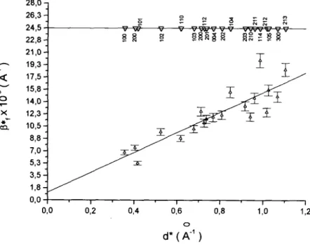
Figure 4. Indexed Williamson-Hall plot (/?* vs d*) of ZnO-30 obtained by thermal decomposition of precur-sor precipitated at 30 min.
6 <
O
X * *~ ca.
O 1 ,%J 29 8 2 8 , 0 2 6 , 3 2 4 , 5 2 2 , 8 2 1 , 0 1 9 , 3 - 17,5- 15,8- 14,0- 12,3-
10,58 , 10,58 7 , 0 5 , 3 3 , 5 - 1,80 , 1,80
-a
V
1
- T - ^
? 8 8 5 § g S 8 ? J S §
31
1 ' 1 ' 1 '
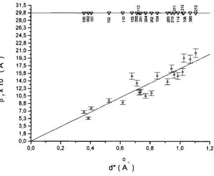
Figure 5. Indexed Williamson-Hall plot (pf vs d*) of ZnO-180 obtained by thermal decomposition of precur-sor precipitated at 180 min.
0,0 0,2 0,4 0,6 0,8 1,0 1,2
0
metric analysis. Zinc oxide samples obtained by thermal de-composition are crystalline and their X-ray diffractograms are characteristic of zincite (Figure 1). The intensity of the reflection (002) is substantially increased as a function of precursor precipitation time. However, from data in Table II crystallite size does not increase for the (002) indicating that the reflection intensity increase is related to preferred orien-tation in the samples. Moreover, the intensity increase of the reflection (002) is correlated to the number of isolated acicu-lar particles or spherulitic needle aggregates in the samples. These shapes depend on precursor precipitation time as veri-fied from electron scanning microscopy by Sigoli et al.
(1997). The apparent crystallite size (ehkl) in the direction
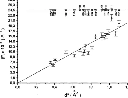
perpendicular to the (002) plane is larger than for (100) (Table II), and consequently the crystallite shape may be regarded as almost cylindrical. These data are in agreement with the transmission electron microscopy results. From the Williamson-Hall plots (Langford et al, 1993) (Figures 4-7)
it is immediately apparent that there is a scatter in the values of /3f suggesting the utility of the single-line method to dis-tinguish size and strain. There is a positive slope, and y3* lines for (hOO) and (00/) reflections have nonzero and slightly different slopes (plots not shown), indicative of the presence of microstrain. In this case, according to the rms strain calculated from the single-line method (Table II), all ZnO samples have a similar degree of microstrain, indepen-dent of the precursor precipitation time (Table II). For zinc oxide samples the agglomeration factor (1/F) was calculated from the data shown in Table I. The constant results indicate that the agglomeration degree between crystallites does not depend on precursor precipitation time.
In recent work, (Sigoli et al., 1999) it was observed that the zinc oxide excitation spectra present different bands as a function of particle shapes. Excitation spectra showed three bands at 377, 405, 430 nm for porous spherical aggregates
0 <
28,0-] 26,3-24,5 22,8-
21,0-
19,3- 17,5- 15,8-
14,0-12,3
-
10,58 , 10,58 7 , 0 5 , 3 3 , 5
- 1,8-0,0
0,0
v w v V"
182 S 1 v v—vwv
1
IS^
I
Figure 6. Indexed Williamson-Hall plot (/3* vs d*) of ZnO-250 obtained by thermal decomposition of precur-sor precipitated at 250 min.
0,2 0,4 0,6
d*(A"1)
0,8 1,0 1,2
X
§ S g
Figure 7. Indexed Williamson-Hall plot (Pf vs d*) of ZnO-440 obtained by thermal decomposition of precur-sor precipitated at 440 min.
d*(A"1)
and only a well-defined band at 377 nm for spherulitic-shaped particles and needle particles. These different bands were correlated with: (i) the morphological changes, (ii) the urea decomposition during precursor precipitation process, (iii) the thermal decomposition of the precursor, and (iv) with lattice defects in the zinc oxide structure. The present results allow us to infer that the different excitation bands verified before (Sigoli et al., 1999) can be attributed to elec-tronic defects originating from either particle shape or from oxygenated groups present on ZnO surface instead of lattice defects, which are invariable as a function of precursor pre-cipitation time.
IV. CONCLUSIONS
From the results it is possible to conclude that the pre-cursor precipitation period directly influences the zinc oxide morphology but influences neither the average crystallite size nor the microstrain for ZnO samples obtained by this route. It is shown that the spherulitic aggregates are formed by needlelike particles, which are built of anisotropic crystal-lites. All the zinc oxide samples presented microstrain that is qualitatively shown by Williamson-Hall plots. The presence of microstrain in the samples is likely a consequence of the low temperature and short period of time used for precursor thermal decomposition. The results presented in this work also allow us to infer that the different excitation bands veri-fied by Sigoli et al. (1999), can be attributed to electronic defects originating from the particle shape or from oxygen-ated groups present on the ZnO surface instead of lattice defects.
Therefore, using this route, it is possible to prepare zinc oxide with different morphologies without microstructural alterations. Such preparation may be very important in af-fecting several properties of ZnO that may be useful in the manufacturing of optical devices.
ACKNOWLEDGMENTS
Financial support by CNPq and FAPESP is gratefully acknowledged. F.A.S. thanks CNPq for a scholarship. The
authors would like to thank Professor Maria do Carmo Gon-calves from UNICAMP for her assistance in TEM analysis.
Auffredic, J. P., Boultif, A., Langford, J. I., and Louer, D. (1995). "Early stages of crystallite growth of ZnO obtained from an oxalate precursor," J. Am. Ceram. Soc. 78, 323-328.
Blasse, G., and Grabmaier, B. C. (1994). Luminescent Materials (Springer, Berlin), pp. 60-64.
Bolis, V., Fubini, B., Giamello, E., and Reller, A. (1989). "Effect of form on the surface reactivity of differently prepared zinc-oxides," J. Chem. Soc., Faraday Trans. 1 85, 855.
Castellano, M. A., and Matijevic, E. (1989). "Uniform colloidal zinc com-pounds of various morphologies," Chem. Mater. 1, 78-82.
Collins, I. R., and Taylor, E. S. (1992). "Nonaqueous thermal-decomposition route to colloidal inorganic oxides," J. Mater. Chem. 2, 1277-1281.
Georgobiani, A. N., Butkhuzi, T. V., Zadauly, E., Kekelidze, N. P., and Khulordava, T. G. (1993). "Optical-properties of zinc oxide dielectric layers," Inorg. Mater. (Transl. of Neorg. Mater.) 29, 1249-1252. Ghose, S. (1964). "The crystal structure of hydrozincite, Zn5(OH)6
(CO3)2,"ActaCrystall0gr. 17, 1051-1057.
Haruta, M., and Delmon, B. (1986). "Preparation of homodisperse solids," J. Chim. Phys. Phys.-Chim. Biol. 83, 859-868.
Koudelka, L., and Horak, J. (1994). "Morphology of polycrystalline ZnO and its physical-properties," J. Mater. Sci. 29, 1497-1500.
Langford, J. I. (1992). "The use of the Voigt function in determining micro-structural properties from diffraction data by means of pattern decompo-sition," in Accuracy in Powder Diffraction II, edited by E. Prince and J. K. Stalik (NIST Special Publication No. 846, Gaithersburg), pp.
110-126.
Langford, J. I., Boultif, A., Auffredic, J. P., and Louer, D. (1993). "The use of pattern decomposition to study the combined X-ray diffraction effects of crystallite size and stacking faults in ex-oxalate zinc oxide," J. Appl. Crystallogr. 26, 22-33.
Langford, J. I., Delhez, R., Keijser, Th. H., and Mittemeijer, E. J. (1988).
"Profile analysis for microcrystalline properties by the Fourrier and other methods," Aust. J. Phys. 41, 173-187.
Louer, D., Auffredic, J. P., Langford, J. I., Ciosmak, D., and Niepce, J. C.
(1983). "A precise determination of the shape, size and distribution of
crystallites in zinc oxide by X-ray line-broadening analysis," J. Appl. Crystallogr. 16, 183-191.
Matijevic, E. (1985). "Production of monodispersed colloidal particles," Annu. Rev. Mater. Sci. 15, 483-516.
Sigoli, F. A., Davolos, M. R., and Jafelicci, Jr., M. (1997). "Morphological evolution of zinc oxide originating from zinc hydroxide carbonate," J. Alloys Compd. 262-263. 292-295.
Sigoli, F. A., Davolos, M. R., and Jafelicci, Jr., M., (1999). "Morphological control and luminescence properties of zinc oxide," Adv. Sci. Technol. (Faenza, Italy) 27, 45-52.
Toraya, H. (1990). "Operating manuals for computer programs: PROFIT and
Vanheusden, K., Seager, C. H., Warren, W. L., Tallant, D. R., Caruso, J., Hampden-Smith, M. J., and Kodas, T. J. (1997). "Green photolumines-cence efficiency and free-carrier density in ZnO phosphor powders pre-pared by spray pyrolysis," J. Lumin. 75, 11-16.
Zampronio, C. G., Davolos, M. R., Stucchi, E. B., and Jaffelicci, Jr., M.
(1995). "Spherical particles of pure and manganese doped zinc oxide