Responses of Gastric Adenocarcinoma Cells
Kristine Misund
1, Linn-Karina Myrland Selvik
1,2, Shalini Rao
1,2, Kristin Nørsett
1, Ingunn Bakke
1, Arne K.
Sandvik
1,3, Astrid Lægreid
1, Torunn Bruland
1, Wenche S. Prestvik
2, Liv Thommesen
1,2*1 Department of Cancer Research and Molecular Medicine, Norwegian University of Science and Technology (NTNU), Trondheim, Norway, 2 Faculty of Technology, Sør-Trøndelag University College, Trondheim, Norway, 3 Department of Gastroenterology and Hepatology, Medical Clinic, St. Olav’s University Hospital, Trondheim, Norway
Abstract
The peptide hormone gastrin is known to play a role in differentiation, growth and apoptosis of cells in the gastric mucosa. In this study we demonstrate that gastrin induces Nuclear Receptor 4Aβ (NR4Aβ) expression in the adenocarcinoma cell lines AR4βJ and AGS-GR, which both possess the gastrin/CCKβ receptor. In vivo, NR4Aβ is
strongly expressed in the gastrin responsive neuroendocrine ECL cells in normal mucosa, whereas gastric adenocarcinoma tissue reveals a more diffuse and variable expression in tumor cells. We show that NR4Aβ is a primary early transient gastrin induced gene in adenocarcinoma cell lines, and that NR4Aβ expression is negatively regulated by inducible cAMP early repressor (ICER) and zinc finger protein γ6, CγH1 type-like 1 (Zfpγ6l1), suggesting that these gastrin regulated proteins exert a negative feedback control of NR4Aβ activated responses. FRAP analyses indicate that gastrin also modifies the nucleus-cytosol shuttling of NR4Aβ, with more NR4Aβ localized to cytoplasm upon gastrin treatment. Knock-down experiments with siRNA targeting NR4Aβ increase migration of gastrin treated adenocarcinoma AGS-GR cells, while ectopically expressed NR4Aβ increases apoptosis
and hampers gastrin induced invasion, indicating a tumor suppressor function of NR4Aβ. Collectively, our results uncover a role of NR4Aβ in gastric adenocarcinoma cells, and suggest that both the level and the localization of NR4Aβ protein are of importance regarding the cellular responses of these cells.
Citation: Misund K, Selvik L-KM, Rao S, Nørsett K, Bakke I, et al. (β01γ) NR4Aβ Is Regulated by Gastrin and Influences Cellular Responses of Gastric Adenocarcinoma Cells. PLoS ONE 8(9): e76βγ4. doi:10.1γ71/journal.pone.0076βγ4
Editor: DunFa Peng, Vanderbilt University Medical Center, United States of America Received May 5, β01γ; Accepted August β1, β01γ; Published September β7, β01γ
Copyright: © β01γ Misund et al. This is an open-access article distributed under the terms of the Creative Commons Attribution License, which permits unrestricted use, distribution, and reproduction in any medium, provided the original author and source are credited.
Funding: This work was supported by the Norwegian Cancer Association and the Cancer Fund at St. Olavs Hospital, Trondheim, Norway. The funders had no role in study design, data collection and analysis, decision to publish, or preparation of the manuscript.
Competing interests: The authors have declared that no competing interests exist. * E-mail: [email protected]
Introduction
Gastrin is a gastrointestinal peptide hormone which plays a central role in regulation of gastric acid secretion [1], and in differentiation, maintenance and organization of cells/tissue in the gastric mucosa [β,γ]. Beside its role in regulation of normal physiology, gastrin is shown to exert growth promoting impact both in normal and malignant gastrointestinal tissue. Gastrin stimulates proliferation of human gastric and pancreatic cell lines [4-6]. Hypergastrinemia is associated with gastric neuroendocrine tumors (carcinoids) [7] and is found to regulate the expression of anti- and pro-apoptotic genes in both human [8] and rat [9] mucosa. Gastrin mediates its effect via the cholecystokinin-β receptor (CCKβR), primarily expressed by enterochromaffin-like (ECL) cells, but also reported to be expressed in cancer like colorectal and pancreatic adenocarcinomas [10-1β].
target of the Wnt/ -catenin pathway associated with colorectal invasion and metastasis [β1].
By microarray gene profiling we identified NR4Aβ as a gastrin responsive gene in the pancreatic adenocarcinoma cell line AR4βJ [ββ]. In this study we examined the role of NR4Aβ in the gastric adenocarcinoma cells. Gastrin transiently regulates NR4Aβ expression in AGS-GR cells. NR4Aβ is known
to activate genes via cognate NBRE response elements, and we show that NBRE reporter plasmid is activated upon gastrin treatment. Ectopic expression of the transcriptional repressor ICER (inducible cAMP early repressor) reduces gastrin induced NR4Aβ expression as well as transcriptional activation of the NBRE reporter plasmid, indicating that ICER acts as a negative regulator of gastrin induced NR4Aβ. We also show that gastrin affects the NR4Aβ nucleus-cytosol shuttling. We find that ectopic expression of NR4Aβ hampers gastrin induced invasion, which indicates a role of NR4Aβ in regulating invasive properties of these cells. The molecular mechanisms likely to be involved are gastrin induced changes in the NR4Aβ nucleus-cytosolic shuttling and increased apoptosis. Collectively, our study suggests a function of NR4Aβ concurrent with a tumor suppressor role in gastric adenocarcinoma cells.
Materials and Methods
Cells and reagents
Details concerning cultivation and treatment of pancreatic adenocarcinoma AR4βJ cells for the genome –wide data sets are described elsewhere [ββ] and in the legend to Figure 1. AGS-GR cells (human gastric adenocarcinoma stable
transfected with CCKβR, gift from Prof. Andrea Varro, University of Liverpool) [βγ] were grown in HAM’S F1β (GIBCO, Invitrogen, Carlsbad, CA) supplemented with 10% FCS and 10 U/ml penicillin-streptomycin and βµg/ml puromycin (Sigma-Aldrich, St. Louis, MO). AR4βJ cells (rat pancreatic acinar cell derived with endogenously expressed CCKβR; American Type Culture Collection (ATCC), Rockville, MD) were grown in DMEM (GIBCO, Invitrogen) with 4.5 g/l glucose, 15% FCS, 1 mM sodiumpyruvate, 0.1 mg/ml L-glutamine, 10 U/ml penicillin-streptomycin, and 1 µg/ml fungizone (all GIBCO, Invitrogen). Gastrin (G-17) and cycloheximide (CHX) were purchased from Sigma-Aldrich.
Expression plasmids
γxNBRE-Luc and pCMX-NR4Aβ constructs were kindly provided by Prof Thomas Perlmann, Karolinska Institute, Sweden. pNR4Aβ-luc, containing the human NR4Aβ promoter, was a kind gift from Prof. Marc Montminy [β4]. Sequence verification of the plasmid identified -1β8 to +154 of the NR4Aβ promoter, including the CRE element. pCONTROL-luc were obtained from Panomics (CA, USA). NR4Aβ-EGFP was a kind gift from Prof. Evely Murphy, University College Dublin, Ireland [β5]. pICER IIy and pICER I were constructed via homologous recombination of ICER IIy or ICER I containing pDONRβ01 plasmid [β6] and pEF5/FRT/V5-DEST (Invitrogen). Zfpγ6l1 (Bergγ6) entry clone (Bergγ6 ORF Express Shuttle Clone) was ordered from GeneCopoeia (USA), and pZfpγ6l1-DEST was
constructed by homologous recombination with entry clone and pDESTβ6 (Invitrogen). pCONTROL vectors were constructed by restriction cutting in att sites in pEF5/FRT/V5-DEST with EcoRV, and in pDESTβ6 with BsrGI, followed by re-ligation of att sites, thereby removing the insert between the att sites. pEF5/FRT/V5/GW-CAT was purchased from Invitrogen.
Transient transfection and gastrin treatment of cells
AGS-GR cells (5.0 x 10⁵/well) were plated in 6-well plates and
transfected after β4 h with β.5 µg plasmid and 1β.5 µl Metafectene PRO transfection reagent (Biontex Laboratories GmbH, Martinsried, Germany) per well. β4 h after transfection, cells were serum starved for β4 h before treatment with gastrin as indicated in figure legends. Overexpression was verified by qRT-PCR and Western blotting (Figure SβC/D).
siRNA
siRNA-ICER (Qiagen) was designed targeting sites within human ICER: 5’- CAUUAUGGCUGUAACUGGATT-γ’, and annealed as described previously [β6]. siNR4Aβ, siRNAs siCONTROL#1, siCONTROL#β and siGAPDH were obtained from Ambion (Austin, TX). The siCONTROL-pool, ON-TARGET plus Non-Targeting Pool, were obtained from Dharmacon (Lafayette, CO). Downregulation of NR4Aβ mRNA and protein was verified by qPCR and Western blotting (Figure SβA/B).
Genome-wide gene time series expression analysis on Illumina Expression Bead Chips
RNA amplifications and hybridization were performed at the NTNU Genomics Core Facilely (GCF), as previously described Selvik [ββ]. The data was normalised by loess adjustment within time points and average quantile normalised between time points. The data was analysed using the Limma (ver. γ.1β.1) Bioconductor package [β7]. The microarray data were prepared according to minimum information about a microarray experiment (MIAME) recommendations [β8] and deposited in the Array Express [β9]. Detailed information about the microarray designs and raw data files from the experiments are accessible by use of these accession numbers: GSEγβ869, and E-MTAB-1β68 (Illumina platform).
Reporter gene assay
Cells (1.5 × 10⁴/well) were plated in 96-well plates β4 h
before transfection. Transfection was carried out using Metafectene™ PRO in 5:1 reagent to plasmid ratio, 84 ng plasmid and phRL-null (Promega, Madison, WI) (1:50). The transfection mixture was added to cells β4 h prior to gastrin treatment. Cells were incubated for additional 4 or 6 h, following lysis in β0 µl Promega lysis buffer (Madison, WI). For co-transfections of plasmid and siRNA, 1.β x 104 cells were
plated in 96-well plates, the next day transfected with siRNA to a final concentration of β0 nM using the RNAiMAX reagent (Invitrogen). After β4 h, cells were transfected with plasmid as described above. Luciferase activity was measured using Dual Luciferase kit (Promega), and Wallac 14β0 Victorγ plate reader
Figure 1. NR4A2 is induced by gastrin. A: Temporal profiles of gastrin induced NR4Aβ mRNA expression in pancreatic adenocarcinoma cells (AR4βJ). The panels show data from three independent microarray time series experiments (accession numbers E-MATAB-1β68 and GSEγβ869); and the data points are presented as normalized logβ-transformed signal intensities.
Experiment 1: mRNA expression level for untreated (green line) and sustained gastrin treated (blue line) cells. Experiment β: mRNA level in cells treated in a sustained mode (14 h of continuous presence of gastrin) and in a transient mode (gastrin was removed after 1 h of treatment). Experiment γ: sustained gastrin treatment was measured in the presence (orange line) and absence (blue line) of cycloheximide (CHX) at 6 different time points between 1 and 10 h. Green and grey lines show mRNA levels in untreated and CHX treated control cells, respectively. All data points are mean of two biological replicates. Gastrin (10 nM) treated and untreated control cells were grown in parallel and harvested (pool of β-γ technical replicates) at several time points, as indicated in the panels. In experiments with transient versus sustained gastrin treatment, the growth medium of untreated and gastrin treated cells was removed 1 h after gastrin treatment; the cells were then washed with serum-free medium before fresh serum-free medium with gastrin (sustained gastrin treated cells) or without gastrin (transiently gastrin treated or untreated cells) was added. In experiments with the protein synthesis inhibitor cycloheximide (CHX), pre-treatments with CHX (10 µg/ml) were initiated γ0 min before gastrin (10 nM) was added. B: NR4Aβ mRNA and protein level in gastrin treated (5 nM) AGS-GR cells. qRT-PCR data shown
are mean ± SEM of four biological replicas. Western blot image shows NR4Aβ protein. Immunostaining for NR4Aβ in normal gastric oxyntic mucosa is shown in panels C-E: Strong NR4Aβ immunoreactivity (C) in scattered single cells in normal gastric oxyntic mucosa. Overlap between the cells showing strong NR4Aβ immunoreactivity (D) and CgA immunoreactive neuroendocrine cells (E) in serial sections (C at x400 magnification, E and F at x1000 magnification).
cDNA synthesis and quantitative real-time PCR (qRT-PCR)
Total RNA was extracted using RNeasy Mini Kit (Qiagen, Germantown, MD). RNA integrity, quality and quantity were evaluated by UV fiberoptic spectrophotometer (Nanodrop Technologies, Rockland, DE). cDNA synthesis was performed with 1 µg total RNA in a β0 µl reaction using the REVERSE-IT 1st Strand Synthesis Kit (ABgene, UK). After synthesis, cDNA
was diluted 1:β with RNase-free water. qRT-PCR was performed with β.5 µl cDNA in β5 µl reaction mix using ABsolute QPCR SYBR Green Mix (ABgene). Quantitative PCR thermal cycling program: 15 min at 95°C, 40 thermal cycles of 15 s at 95°C, β0 s at 60°C and 40 s at 7β°C. The primer sequences used for qRT-PCR analyses are shown in Table S1. PCR samples were run in triplicate and the average used for further quantification. The relative expression ratios were calculated using Pfaffl method [γ0], or the ∆∆Ct-method [γ1] and individual expression values were normalized by comparison with -actin or GAPDH.
Western Blots
Cells were harvested in 100 µl RIPA (Thermo Scientific, Rockford, USA). Blotting, washing and antibody incubation were performed as previously described [γβ]. Binding of secondary antibodies was visualized by the Super Signal West Femto Maximum Sensitivity Substrate (Pierce, Thermo Scientific, Rockford, IL) and Kodak Image Station β000R (Kodak, Pittsburgh, PA). The following antibodies were used: anti-NR4Aβ from Santa Cruz Biotechnology (Santa Cruz, CA) and Abcam (Cambridge, UK); HRP-conjugated goat anti-rabbit IgG (Cell Signaling, Beverly, MA), mouse monoclonal to beta actin (Abcam), polyclonal HRP-conjugated goat anti-mouse IgG (Dako, Glostrup, Denmark).
Immunohistochemistry
Sections for immunohistochemistry were taken from formalin fixed paraffin embedded biopsies from our gastric carcinoma biobank, containing both intestinal and diffuse type cancers classified according to Laurén and normal gastric oxyntic mucosa from patients with no evidence of gastric neoplasm that underwent gastroscopy due to dyspeptic complaints (approval Regional Committee for Medical Research Ethics No 018-0β). Serial sections were mounted as mirrored. Before immunostaining, the sections (4 µm) were deparaffinised, rehydrated in graded solutions of ethanol and blocked of endogenous peroxidase activity in γ% HβOβ for 10 min. Antigen
retrieval was achieved by boiling in citrate-buffer pH 6.0 for 15 min. NR4Aβ was detected using monoclonal anti-NR4Aβ (Abcam) (dilution 1:150) and incubation at 4°C overnight. Neuroendocrine cells were detected using monoclonal anti-chromogranin A (CgA) (Dako) (dilution 1:4000) and incubation at 4°C overnight. The immunoreactions were visualized using the rabbit/mouse EnVision-HRP and DAB+ kit (Dako). Counterstaining was done with hematoxylin. Identical concentration of an isotype equivalent antibody from non-immunized animals (mouse IgGβa) (Dako) was used as negative control.
Immunocytochemical staining and confocal microscopy
Cells (β.0 x 104/well in β00 µl medium with 10% FBS) were
seeded on Lab-Tek™ Chambered Coverglass with 8 wells (NUNC, Thermo Scientific, Rockford, IL) and transfected with NR4Aβ-EGFP. After cultivation for β4 h, cells were serum starved for β4 h and then treated with 5 nM gastrin for 0-60 min. Cells were fixed (4% paraformaldehyde in PBS) for 10 min, washed (PBS x β) and permeabilized (ice-cold MeOH) for 10 min on ice and washed (PBS x β). DNA was stained with Draq-5 (1:1000) for 7 min, washed and stored at 4°C over night before confocal microscopy. Confocal microscopy studies were performed with a Zeiss Axiovert 100-M inverted microscope equipped with an LSM 510 laser-scanning unit and a 1.4 numerical aperture ×6γ Plan-Apochromat oil immersion objective. To minimize photobleaching, laser power was typically β0% under maximum, and the pinhole was set to 0.8– 1.β. Multitracking was used for dual color imaging. The Zeiss LSM Image browser version 4 was used for acquisition, and processing was completed using Adobe Illustrator CS5.
FRAP analyses
Fluorescence recovery after photobleaching (FRAP) analysis was performed β4 h after transient transfection of AGS-GR cells
(0.β x 106 cells/β4mm Petri dish) with β µg NR4Aβ-EGFP and 6
µl Metafectene PRO. Gastrin (10nM) was added and the cells left in the incubator for β0 min. Confocal microscopy was performed with Zeiss LSM 510 Meta Live using a 6γX/ 1.4 oil DIC. The settings were configured to produce ten pre-bleach images followed by bleaching with the 488nm line of a 50-mW argon laser operating at 100% laser power. A fluorescence image of single z sections with an optical splice of 0.7 µm was used. The region of interest (ROI) used for bleaching was a circle with β.5 µm radius. We used a speed of β00 iterations and the bleach time was 7.β sec. Subsequent imaging continued at the pre bleach speed until 80 sec was reached. Fluorescence recovery was calculated using Sigma plot (Table Sβ). The confocal imaging was performed at the Cellular & Molecular Imaging Core Facility, Norwegian University of Science and Technology.
Flow Cytometry
AGS-GR cells were plated in 6-well plates (γ x 105 cells/well).
were analyzed with FlowJo 7.6 software (Tree Star Inc., Ashland, Oregon, USA).
Migration assay
The xCELLigence® DP system (Roche Applied Science, Germany) was used for measurement of migration. This system utilizes specialized culture plates that contain gold electrode arrays beneath the bottom of individual wells (CIM plates). Cellular contact with the electrode surfaces increases the impedance across the electrodes. This impedance value is measured by the DP system and is reported in the dimensionless unit of cell index. AGS-GR cells (γ.5 x 105/well)
were seeded in 6-well plates. After β4 h the cells were transfected with siRNA for β4 h, subsequently serum starved for β4 h and then trypsinated, followed by reseeding (4.0 x 10⁴
cells/well) in CIM-Plate 16 (Roche Applied Science). The plate was placed on the Real-time xCELLigence Cell Analyzer platform at γ7°C to measure the migration index for the duration of the experiments. 1 nM gastrin was used as attractant. Cell migration was monitored every 15 min on a RTCA DP instrument for β4 h. Data analysis was carried out using RTCA Software 1.β (Roche Applied Science).
Invasion assay
48 hours after transfection, invasion assay were performed in β4-well plates containing 8-µm pore Matrigel-coated inserts according to the manufacturer’s instructions (Becton Dickinson, Bedford, MA). AGS-GR cells (4.0 x 10⁴ cells/well) in 0.5 ml
serum-free medium were plated in the insert with or without addition of gastrin (0.γ nM) for β4 h. Cells invading the lower surface of the membrane were stained with Reastain Quick-Diff reagents (Reagena, Finland). The total cells in 5 fields per membrane were counted, and the mean of γ membranes per experiment was calculated.
Statistical analysis
qRT-PCR data were statistically analyzed for significant differences using REST (relative expression software tool) [γγ]. Reporter gene data which include several biological experiments were analyzed using student two-tailed t-test assuming unequal variance. Data were considered significant at p<0.05, unless otherwise stated.
Results
NR4A2 expression is activated by gastrin
Genome-wide time series experiments identified NR4A2 as a gastrin responsive gene in the pancreatic adenocarcinoma cell line AR4βJ (Figure 1A). The mRNA expression was transient with peak expression at β h, followed by decrease to baseline after ~6-8 h of gastrin treatment (Figure 1A, panel1). Genome-wide microarray time series analysis was also used to identify genes that are affected by the duration of gastrin treatment in adenocarcinoma cells [ββ]. As shown in Figure 1A, panel β, the expression of NR4Aβ was higher and more prolonged in cells treated in a sustained mode (gastrin present for 14 h) than in a transient mode (gastrin removed after 1 h). To establish the
role of new protein synthesis in gastrin-mediated regulation of NR4A2 transcript levels, we analyzed gastrin treated AR4βJ cells in the presence of the translational inhibitor cycloheximide (CHX). We found that the initial increase in transcript levels occurs in the presence of CHX, demonstrating that NR4A2 is a primary gastrin responsive gene (Figure 1A, panel γ). The decline of NR4Aβ transcript levels is abolished in the presence of CHX, indicating that the transient nature of the gastrin induced NR4A2 transcripts is dependent upon de novo protein synthesis of a transcription inhibitor or of proteins that reduce mRNA stability. Quantitative real-time PCR confirmed that gastrin induced transient expression of NR4Aβ mRNA, followed by decrease to baseline after ~6 h of stimulation; and that NR4Aβ mRNA expression was sustained when protein synthesis was inhibited by CHX (Figure S1).
We further examined the role of NR4Aβ in gastrin induced responses by employing the gastric adenocarcinoma cell line AGS-GR Gastrin induced a ~8-fold induction of NR4Aβ mRNA
in AGS-GR cells, followed by a rapid decrease to baseline after
~4 h of stimulation (Figure 1B). The NR4Aβ protein expression peaks at β h and displays a rapid decrease after 6 h, in agreement with what is reported for other cells [γ4].
The expression pattern of endogenous NR4Aβ was also observed in vivo by immunohistochemistry. In normal gastric oxyntic mucosa (n=4) there was strong immunoreactivity in small, scattered single cells predominantly in the basal part and situated between the other epithelial cells (Figure 1C). This appearance is suggestive of neuroendocrine cells, and this was confirmed by overlap in serial staining using antibody against the neuroendocrine marker CgA (Figure 1D-E). NR4Aβ was most strongly expressed in cytoplasm of these cells, but also nuclear staining was observed. In addition there was weaker immunoreactivity in other epithelial cells of the mucosa (Figure 1C). Since the neuroendocrine cell population in oxyntic mucosa is dominated by the ECL cell, which is known to possess the CCKβR and to be the main gastrin responsive epithelial cell [γ5], this supports our results showing that NR4Aβ expression is activated by gastrin.
Gastrin-induced NR4A2 activates NBRE promoter elements
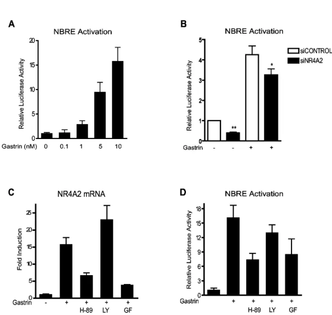
NR4Aβ is known to activate target genes via the cognate NBRE response element [γ6]. We wanted to determine whether gastrin could affect NBRE regulated genes and thus measured the NBRE reporter gene activity in gastrin treated AGS-GR cells. Our results show that gastrin activates
NBRE-driven gene expression in a dose-dependent manner (Figure βA). To verify that this gastrin response is mediated via NR4Aβ, NBRE reporter gene activity was measured in gastrin treated cells transfected with siRNA targeting NR4Aβ. We demonstrate that siNR4Aβ significantly reduces gastrin-mediated activation of NBRE (Figure βB), suggesting that NR4Aβ-activated gene expression plays a role in gastrin mediated responses.
The cellular effect of gastrin is transmitted via the Gαq/11
protein kinases (MAPKs) and protein kinase A (PKA) [γ7-40]. Hence we examined the signaling pathways involved in gastrin-mediated NR4Aβ activation. Gastrin-induced NR4Aβ gene expression in AGS-GR cells was significantly reduced by
inhibitors of PKA or PKC, but not by the PIγK inhibitor (Figure βC). This was some unexpected since gastrin mediated signaling is reported to phosphorylate PKB/Akt [41]. However, Western blot experiments demonstrated a constitutive phosphorylation of PKB/Akt Ser-47γ in AGS-GR cells (data not
shown), which likely explain why we did not observe any effect of the LYβ9400β inhibitor. The NBRE reporter gene experiments demonstrated that PKA and PKC signaling pathways both participate in gastrin mediated NBRE transcriptional activation (Figure βD). Taken together, the results indicate that gastrin induces NR4Aβ gene expression and NBRE target gene activation via PKA and PKC signaling pathways.
Figure 2. NR4A2 activates NBRE promoter elements. A: Gastrin-induced NBRE-luc activation. Data represent one of two biological replicas. B: The effect of NR4Aβ siRNA on gastrin-induced NBRE activation. Data represent mean ± SEM of four biological replicas (** p<0.01, * p=0.1). C-D: Effect of specific inhibitors of PKA (H-89, 10µM), PIγK (LY β9400β, 10µM) or PKC (GF 109β0γx, γ.5µM) on (C) gastrin-induced NR4Aβ gene expression and (D) gastrin-induced NBRE activation. Data represent one of three biological replicas; mean ± SD of six technical replicas.
NR4A2 is negatively regulated by gastrin-induced proteins
We have previously shown that gastrin induces expression of Inducible cAMP early repressor (ICER) [4β], and that ICER represses gastrin-induced genes with CRE promoter elements [β6,γβ]. The promoter region of the NR4A2 gene comprises CRE regulatory elements [4γ,44]. Thus, it was of interest to investigate whether ICER could modulate gastrin-induced NR4Aβ expression and with this constitute a gastrin induced negative feedback mechanism. AGS-GR cells were transfected
with ICER I or ICER II expression plasmids together with a reporter plasmid for NR4Aβ promoter activity; NR4Aβ-luc. Our results show that ectopically expressed ICER significantly reduces gastrin-induced NR4Aβ gene expression (Figure γA). In addition, we find that ICER expression reduces NBRE reporter gene activity by ~ 50% in gastrin treated cells (Figure γB). ICER also affects NBRE activity in untreated cells. Our results demonstrate that the role of ICER as a negative feedback regulator of gastrin responses involves both downregulation of NR4Aβ gene expression and repression of gastrin-induced NBRE-regulated genes.
NR4A2 possesses AU-rich elements (AREs) in its γ`-untranslated region (γ'-UTR)(http://rna.tbi.univie.ac.at/cgi-bin/ AREsite.cgi), and Zinc finger protein γ6, CγH1 type-like 1 (Zfpγ6l1) is suggested to participate in the degradation of short-lived, inducible mRNAs by binding to AREs [45,46]. We found that gastrin induced Zfp36l1 expression in AR4βJ cells in two independent microarray time series (E-MATAB-1βγ (cDNA microarrays) and GSEγβ869 (Illumina)) and therefore examined whether Zfpγ6l1 would influence the NR4Aβ mRNA levels. AGS-GR cells were transfected with Zfpγ6l1 expression
plasmid, and the amount of NR4Aβ mRNA was assessed by qRT-PCR. Gastrin-treated cells with ectopic expression of Zfpγ6l1 exhibited significantly reduced levels of NR4Aβ transcripts (Figure γC). No effect of Zfpγ6l1 was observed in the control experiments, by qRT-PCR measurement of the expression of non-ARE genes like CyclinL1 (Figure γD) and Ywhag (data not shown). We conclude that NR4Aβ transcript levels are negatively regulated by at least two different gastrin induced mechanisms: ICER represses the transcription, while Zfpγ6l1 reduces NR4Aβ mRNA levels by affecting its degradation.
Gastrin facilitates change in the nucleus-cytosol shuttling
Subcellular trafficking of nuclear receptors and the subsequent protein interactions often affect the cellular response. In addition to transactivation functions, NR4As are reported to modulate the activity of other proteins through subcellular translocation and protein-protein interactions [47-51]. Thus it was of interest to establish a putative role of gastrin in modifying the trafficking of NR4Aβ proteins. AGS-GR
cells were transfected with NR4Aβ-EGFP and treated with gastrin up to 60 min. We show that gastrin facilitates NR4Aβ nucleus-cytosolic translocation (Figure 4A); increasing amount of NR4Aβ-EGFP was located in the cytosol upon gastrin treatment compared to the control where the better part of NR4Aβ is localized in the nucleus. To further explore the
protein shuttling, FRAP experiments were included. Normalization curves were used to optimize the bleach parameters. Based on a βD diffusion model, we fitted the individual FRAP curves using a non-linear regression analysis (Sigmaplot) (Figure 4 B/C) [5β,5γ]. We observed a significant change in the diffusion time comparing gastrin treated versus untreated cells, both in the nucleus and the cytosol (Figure 4D). Our results show that NR4Aβ-EGFP resides for a longer time (i.e. higher diffusion time) in cytosol compared to nucleus in gastrin treated cells. The fact that NR4Aβ is present for a longer time period in cytosol compared to nucleus may affect the cellular response. This is analogous to what has been described for NR4A1, where mitochondrial localization is shown to induce apoptosis [48]. Hence, we examined whether apoptosis was induced in AGS-GR cells ectopically expressing
NR4Aβ, using flow cytometry and annexin V Alexa Fluor 647 labeling. We observed ~β0% more apoptosis (p<0.05) in cells overexpressing NR4Aβ compared to controls (i.e. cells transfected with the control plasmid Hγ.1-EGFP) (Figure 4E). Treatment with gastrin did not influence the number of apoptotic cells in this time period (data not shown).
NR4A2 suppresses gastrin-induced migration and invasion
Little is known about the molecular mechanisms involved in NR4Aβ regulation, and conflicting data exists concerning its role in cancer. However, NR4Aβ has been characterized as a putative tumor suppressor protein in gastric cancer, being down-regulated both in primary gastric cancers and in synchronous liver metastases [54]. Thus it was of interest to examine whether NR4Aβ would influence gastrin induced migration and invasion in our gastric adenocarcinoma cells. AGS-GR cells were transfected with siRNA targeting NR4Aβ
and migration assessed using real-time cell monitoring assay (xCELLigence technology). As shown in Figure 5A, NR4Aβ knock-down by itself resulted in a significant increase of migration and gastrin treatment further enhanced this effect. Next we determined the importance of NR4Aβ in invasion, now using AGS-GR cells with ectopically expressed NR4Aβ. We
show that ectopic expression of NR4Aβ dramatically reduces the number of invading cells as a consequence of gastrin treatment (Figure 5B). Collectively, these results suggest that high level of NR4Aβ hampers the migratory potential of AGS-GR cells.
NR4A2 protein is expressed in tumor cells in gastric adenocarcinomas
regarding the type of gastric adenocarcinoma or nucleus versus cytosolic localization could be read from this small cohort. Our findings are in accordance with the OncoMine database (https://www.oncomine.org), where NR4A2 gene expression is shown to vary both within and among subtypes of gastric adenocarcinomas; being both higher and lower expressed compared to normal controls.
Discussion
In the present study we address the function of the orphan nuclear receptor NR4Aβ in gastrin regulated responses. Our principal findings are that gastrin regulates NR4Aβ expression and activity in gastric adenocarcinoma AGS-GR cells. The
regulation involves gastrin induced nucleus-cytosolic shuttling of NR4Aβ. We find that sustained expression of NR4Aβ inhibits gastrin induced invasiveness, which is congruent with a tumor
Figure 3. Negative regulation of gastrin-induced NR4A2 expression. A: AGS-GR cells transfected with NR4Aβ-luc and ICER
expression plasmids or empty vector. Cells were treated with gastrin for 6 h prior to measurement of NR4Aβ activity. Data shown represent mean ± SEM of five biological replicas (** p<0.0γ, * p = 0.06). B: AGS-GR cells transfected with NBRE-luc and ICER
expression plasmid or empty vector and treated with gastrin for 4 h prior to measurement of NBRE activity. Data shown represent mean ± SEM of four biological replicas (** p<0.0γ). C: AGS-GR cells were transfected with pZfpγ6l1 expression plasmid or empty
Figure 4. Gastrin treatment influences nucleus-cytosolic shuttling of NR4A2. A: Intracellular localization of NR4Aβ protein in response to gastrin treatment. AGS-GR cells transfected with pNR4Aβ-EGFP. B: Images of gastrin treated (10 nM) AGS-GR cells
expressing pNR4Aβ-EGFP before, during and after bleaching of a nucleus area for 7.5 sec. The circle indicates the area of the bleach spot. C: Normalized FRAP curve for the cytosol of gastrin treated AGS-GR cells. D: Diffusion time in untreated and gastrin
treated nucleus and cytosol. E: To determine cell viability, cells were transfected with pNR4Aβ-EGFP or a control plasmid (pHγ.1-EGFP). After 48 h AGS-GR cells were detached by Accutase treatment, labeled with annexin V Alexa Fluor 647 and analyzed by
flow cytometry. Annexin-V positive cells were considered as apoptotic. Results are shown as % apoptotic cells of the total number of counted EGFP positive or EGFP negative cells. Data are representative of three biological replicas; mean ± SD of three technical replicas is shown.
suppressor function of NR4Aβ in AGS-GR cells. This is in
contrast to a transient expression of NR4Aβ which does not affect gastrin induced invasiveness or proliferation [55,56], indicating that a threshold level of NR4Aβ is of vital importance for the cellular decision towards migration/invasion. The gastrin induced regulation of NR4Aβ was further substantiated in vivo
by strong NR4Aβ expression in the gastrin responsive neuroendocrine ECL cells in normal gastric oxyntic mucosa.
Gastrin is known to promote proliferation, migration and invasion of AGS-GR cells [55-57]. Thus, the transient induction
of NR4Aβ by gastrin may play a role in the gastrin induced migration and invasion of these cells. A putative cross talk
Figure 5. NR4A2 suppresses gastrin-induced migration and invasion. A: Real-time cell migration monitored (0-β4 h) in AGS-GR cells transfected with siNR4Aβ or siCtr, with or without gastrin treatment (10 nM). Results show one representative of three
biological replicas; mean ±SD of three technical replicas. B: Invasion assay with AGS-GR cells transfected with pCMX-NR4Aβ or
pCMX (control) was performed in β4-well plates containing 8-µm pore Matrigel-coated inserts (with or without 0.γ nM gastrin). Cells invading the lower surface of the membrane were stained with Reastain Quick-Diff reagents and total numbers of cells in 5 fields per membrane were counted. The mean of three independent experiments is shown.
between NR4Aβ and the Wnt signaling pathway has been suggested [49], involving NR4Aβ cytoplasmic translocation and de-repression of transcription upon beta-catenin treatment. However, in contrast to transient induction of NR4Aβ, we show that sustained (i.e. ectopic) expression of NR4Aβ increases apoptosis and hampers the invasiveness of AGS-GR cells. This
may suggest that some threshold effect of NR4Aβ exists when it comes to its influence on apoptosis of gastric adenocarcinoma cells, corresponding to what is found in the breast cancer study by Llopis et al [58]. Low expression level of NR4Aβ in AGS-GR cells is supportive with migration and
invasion (untreated AGS-GR cells show low levels of NR4Aβ
proteins, Figure 1B), while high or sustained level of NR4Aβ reduces the invasiveness of the cells through distinct mechanisms, which also involves apoptosis. We speculate that the increased apoptosis of AGS-GR cells might be due to
NR4Aβ mediated modulation of proteins through protein-protein interactions in cytosol, and/or NR4Aβ localization/ association with mitochondria analogous to what has been described for NR4A1 [48]. Interestingly, nucleus to cytosol trafficking of NR4A1 is suggested to be the molecular switch that dislodges the Bcl-β BH4 domain, exposing its BHγ domain, which in turn blocks the activity of anti-apoptotic Bcl-X(L) [48]. Whether NR4Aβ is involved in a corresponding mechanism in
gastric adenocarcinoma cells is not known. Taken together, we advocate that high and sustained level of NR4Aβ in AGS-GR
cells reduces migration/invasion partly by promoting apoptosis, while transient expression of NR4Aβ does not seems to affect such mechanisms.
NR4Aβ is characterized as a hub gene [54], a term initially used to describe central proteins of transcriptional networks [59]. Hub proteins may regulate quite different biological processes since they interact with several proteins and represent important regulatory nodes in biological networks. In a recent study investigating the expression of NR4Aβ in breast cancer, the authors concluded that NR4Aβ expression in breast is commensurate with a normal and terminally differentiated epithelial phenotype, whereas silencing or dysregulation of NR4Aβ probably plays a role in oncogenic transformation of breast epithelial cells [58]. In accordance with this, we also observed changed NR4Aβ expression in gastric adenocarcinomas, seemingly being dominated by a general expression in tumor cells compared to the mainly strong NR4Aβ expression in the gastrin responsive neuroendocrine ECL cells in normal mucosa. In tumor cells the expression showed variable intensities which is in agreement with our own data and the data from OncoMine (https://www.oncomine.org). Our finding in normal mucosa is in contrast to Chang et al [54]
Figure 6. Immunostaining of NR4A2 in gastric adenocarcinoma. A-B: NR4Aβ immunoreactivity in normal oxyntic mucosa showing strong intensity in scattered single cells (neuroendocrine cells) and weaker staining intensity in the other epithelial cells. C-F: NR4Aβ immunoreactivity in gastric adenocarcinomas of intestinal (C-D) and diffuse (E-F) type, showing a general staining in tumor cells with mixed nuclear or cytoplasmic localization and variable intensities. (A, C, E at xβ00 magnification, with boxes representing B, D and F at x400 magnification).
showing primarily mesenchymal expression of NR4Aβ in normal mucosa, with a change to stronger epithelial expression in primary gastric cancers and a further nearly loss of expression in paired liver metastasis. In addition to the dichotomous behavior of NR4Aβ, differences in antibody specificities could also explain such differences. In a study from Holla et al [β0] intestinal epithelium from Apc-/+ mouse
adenomas and sporadic colorectal carcinomas exhibit increased NR4Aβ expression relative to matched normal mucosa. However, the mechanisms elucidated in this study indicated that NR4Aβ was important for PGEβ-mediated regulation of apoptosis, and thus is likely to mirror mechanisms such as inflammatory signaling pathways, which are known to play a prominent role in colorectal cancer. Taken together, the partly conflicting results published so far, probably reflect both tissue- and cell specific differences in addition to a biphasic role of NR4Aβ.
In this study we throw light on the dichotomous role of NR4Aβ in cancer. We conclude that gastrin induced NR4Aβ expression and transactivation play an important role in gastric adenocarcinoma cells. The amount of NR4Aβ protein and/or the lack of negative feedback regulation may switch the cellular response. A better understanding of gastrin–NR4Aβ regulated processes may reveal new strategies to treatment of gastric adenocarcinomas.
Supporting Information
Figure S1. NR4A2 mRNA expression in gastrin treated AR42J cells measured by qRT-PCR. The cells were pre-treated with the protein synthesis inhibitor cycloheximide (CHX) (10 µg/ml) for γ0 min before gastrin (10 nM) was added. Data represent one of three biological replicas; mean ± SD of three technical replicas.
(EPS)
Figure S2. Effect of siNR4A2 and pCMX-NR4A2 expression plasmid. A: qRT-PCR data showing NR4Aβ mRNA expression in gastrin treated AGS-GR cells transfected
with siNR4Aβ or siCtr. B: Western blot showing NR4Aβ protein in gastrin treated AGS-GR cells transfected with siNR4Aβ or
siCtr. C: qRT-PCR showing NR4Aβ mRNA expression in gastrin treated AGS-GR cells transfected with pCMX-NR4Aβ or
pCMX. D: Western blot showing NR4Aβ protein in gastrin treated AGS-GR cells transfected with pCMX-NR4Aβ or pCMX.
(EPS)
Table S1. PCR primers.
(PDF)
Table S2. Experimental conditions.
(PDF)
Acknowledgements
Ildri Haltbakk provided technical assistance with the migration assay. The FRAP analyses was performed in collaboration with the Cellular & Molecular Imaging Core Facility, Norwegian University of Science and Technology.
Author Contributions
Conceived and designed the experiments: KM LKMS WSP AL LT. Performed the experiments: KM LKMS SR KN IB WSP. Analyzed the data: KM LKMS TB. Contributed reagents/ materials/analysis tools: AKS. Wrote the manuscript: KM LKMS LT.
References
1. Edkins JS (1906) The chemical mechanism of gastric secretion. J Physiol γ4: 1γγ-144. PubMed: 1699β8γ9.
β. Koh TJ, Goldenring JR, Ito S, Mashimo H, Kopin AS et al. (1997) Gastrin deficiency results in altered gastric differentiation and decreased colonic proliferation in mice. Gastroenterology 11γ: 1015-10β5. doi:10.1016/S0016-5085(97)70199-9. PubMed: 9β87997. γ. Dockray GJ, Varro A, Dimaline R, Wang T (β001) The gastrins: their
production and biological activities. Annu Rev Physiol 6γ: 119-1γ9. doi: 10.1146/annurev.physiol.6γ.1.119. PubMed: 11181951.
4. Ishizuka J, Martinez J, Townsend CM Jr., Thompson JC (199β) The effect of gastrin on growth of human stomach cancer cells. Ann Surg β15: 5β8-5γ4. doi:10.1097/00000658-199β05000-00016. PubMed: 1616γ89.
5. Seva C, De Vries L, Scemama JL, Sarfati P, Nicolet TG et al. (1990) Gastrin modulates growth of a rat acinar pancreatic cell line: receptor analysis and signal transduction. Digestion 46 Suppl β: 166-169. doi: 10.1159/000β00γ81. PubMed: ββ6β050.
6. Smith JP, Liu G, Soundararajan V, McLaughlin PJ, Zagon IS (1994) Identification and characterization of CCK-B/gastrin receptors in human pancreatic cancer cell lines. Am J Physiol β66: Rβ77-Rβ8γ. PubMed: 8γ04551.
7. Burkitt MD, Varro A, Pritchard DM (β009) Importance of gastrin in the pathogenesis and treatment of gastric tumors. World J Gastroenterol 15: 1-16. doi:10.γ748/wjg.15.1. PubMed: 1911546γ.
8. Nørsett KG, Laegreid A, Kusnierczyk W, Langaas M, Ylving S et al. (β008) Changes in gene expression of gastric mucosa during therapeutic acid inhibition. Eur J Gastroenterol Hepatol β0: 61γ-6βγ. doi:10.1097/MEG.0b01γeγβ8βf5dc19. PubMed: 1867906β.
9. Nørsett KG, Laegreid A, Langaas M, Wörlund S, Fossmark R et al. (β005) Molecular characterization of rat gastric mucosal response to potent acid inhibition. Physiol Genomics ββ: β4-γβ. doi:10.115β/ physiolgenomics.00β45.β004. PubMed: 158β7βγ5.
10. Caplin M, Savage K, Khan K, Brett B, Rode J et al. (β000) Expression and processing of gastrin in pancreatic adenocarcinoma. Br J Surg 87: 10γ5-1040. doi:10.1046/j.1γ65-β168.β000.01488.x. PubMed: 109γ1047.
11. Goetze JP, Nielsen FC, Burcharth F, Rehfeld JF (β000) Closing the gastrin loop in pancreatic carcinoma: coexpression of gastrin and its receptor in solid human pancreatic adenocarcinoma. Cancer 88: β487-β494. doi:10.100β/1097-014β(β0000601)88:11. PubMed: 108614β4.
1β. Willard MD, Lajiness ME, Wulur IH, Feng B, Swearingen ML et al. (β01β) Somatic mutations in CCKβR alter receptor activity that promote oncogenic phenotypes. Mol Cancer Res 10: 7γ9-749. doi: 10.1158/1541-7786.MCR-11-048γ. PubMed: ββ516γ48.
1γ. Pei L, Castrillo A, Chen M, Hoffmann A, Tontonoz P (β005) Induction of NR4A orphan nuclear receptor expression in macrophages in response to inflammatory stimuli. J Biol Chem β80: β9β56-β9β6β. doi:10.1074/ jbc.M50β606β00. PubMed: 15964844.
14. Winoto A, Littman DR (β00β) Nuclear hormone receptors in T lymphocytes. Cell 109 Suppl: S57-S66. doi:10.1016/ S009β-8674(0β)00710-9. PubMed: 1198γ15γ.
16. Pei L, Waki H, Vaitheesvaran B, Wilpitz DC, Kurland IJ et al. (β006) NR4A orphan nuclear receptors are transcriptional regulators of hepatic glucose metabolism. Nat Med 1β: 1048-1055. doi:10.10γ8/nm1471. PubMed: 16906154.
17. Mullican SE, Zhang S, Konopleva M, Ruvolo V, Andreeff M et al. (β007) Abrogation of nuclear receptors Nr4aγ and Nr4a1 leads to development of acute myeloid leukemia. Nat Med 1γ: 7γ0-7γ5. doi:10.10γ8/nm1579. PubMed: 17515897.
18. Ramirez-Herrick AM, Mullican SE, Sheehan AM, Conneely OM (β011) Reduced NR4A gene dosage leads to mixed myelodysplastic/ myeloproliferative neoplasms in mice. Blood 117: β681-β690. doi: 10.118β/blood-β010-0β-β67906. PubMed: β1β059β9.
19. Inamoto T, Papineni S, Chintharlapalli S, Cho SD, Safe S et al. (β008) 1,1-Bis(γ'-indolyl)-1-(p-chlorophenyl)methane activates the orphan nuclear receptor Nurr1 and inhibits bladder cancer growth. Mol Cancer Ther 7: γ8β5-γ8γγ. doi:10.1158/15γ5-716γ.MCT-08-07γ0. PubMed: 19074857.
β0. Holla VR, Mann JR, Shi Q, DuBois RN (β006) Prostaglandin Eβ regulates the nuclear receptor NR4Aβ in colorectal cancer. J Biol Chem β81: β676-β68β. PubMed: 16β9γ616.
β1. Han YF, Cao GW (β01β) Role of nuclear receptor NR4Aβ in gastrointestinal inflammation and cancers. World J Gastroenterol 18: 6865-687γ. doi:10.γ748/wjg.v18.i47.6865. PubMed: βγγββ98β. ββ. Selvik LK, Fjeldbo CS, Flatberg A, Steigedal TS, Misund K et al. (β01γ)
The duration of gastrin treatment affects global gene expression and molecular responses involved in ER stress and anti-apoptosis. BMC Genomics 14: 4β9. doi:10.1186/1471-β164-14-4β9. PubMed: βγ805861.
βγ. Watson F, Kiernan RS, Deavall DG, Varro A, Dimaline R (β001) Transcriptional activation of the rat vesicular monoamine transporter β promoter in gastric epithelial cells: regulation by gastrin. J Biol Chem β76: 7661-7671. doi:10.1074/jbc.M006697β00. PubMed: 1111γ118. β4. Conkright MD, Guzmán E, Flechner L, Su AI, Hogenesch JB et al.
(β00γ) Genome-wide analysis of CREB target genes reveals a core promoter requirement for cAMP responsiveness. Mol Cell 11: 1101-1108. doi:10.1016/S1097-β765(0γ)001γ4-5. PubMed: 1β718894. β5. Aherne CM, McMorrow J, Kane D, FitzGerald O, Mix KS et al. (β009)
Identification of NR4Aβ as a transcriptional activator of IL-8 expression in human inflammatory arthritis. Mol Immunol 46: γγ45-γγ57. doi: 10.1016/j.molimm.β009.07.019. PubMed: 197γβ956.
β6. Misund K, Steigedal TS, Laegreid A, Thommesen L (β007) Inducible cAMP early repressor splice variants ICER I and IIgamma both repress transcription of c-fos and chromogranin A. J Cell Biochem 101: 15γβ-1544. doi:10.100β/jcb.β1β67. PubMed: 17γ406β4.
β7. Smyth GK (β005) Limma: linear models for microarray data. In: R GentlemanV CareyS DudoitR IrizarryW Huber. Bioinformatics and Computational Biology Solutions using R and Bioconductor. New York: Springer Verlag. pp. γ97-4β0.
β8. Brazma A, Hingamp P, Quackenbush J, Sherlock G, Spellman P et al. (β001) Minimum information about a microarray experiment (MIAME)-toward standards for microarray data. Nat Genet β9: γ65-γ71. doi: 10.10γ8/ng1β01-γ65. PubMed: 117β69β0.
β9. Parkinson H, Sarkans U, Shojatalab M, Abeygunawardena N, Contrino S et al. (β005) ArrayExpress--a public repository for microarray gene expression data at the EBI. Nucleic Acids Res γγ: D55γ-D555. doi: 10.109γ/nar/gki494. PubMed: 15608β60.
γ0. Pfaffl MW (β001) A new mathematical model for relative quantification in real-time RT-PCR. Nucleic Acids Res β9: e45. doi:10.109γ/nar/ β9.9.e45. PubMed: 11γβ8886.
γ1. Livak KJ, Schmittgen TD (β001) Analysis of relative gene expression data using real-time quantitative PCR and the β(-Delta Delta C(T)) Method. Methods β5: 40β-408. doi:10.1006/meth.β001.1β6β. PubMed: 11846609.
γβ. Steigedal TS, Bruland T, Misund K, Thommesen L, Laegreid A (β007) Inducible cAMP early repressor suppresses gastrin-mediated activation of cyclin D1 and c-fos gene expression. Am J Physiol Gastrointest Liver Physiol β9β: G106β-G1069. PubMed: 171856γβ.
γγ. Pfaffl MW, Horgan GW, Dempfle L (β00β) Relative expression software tool (REST) for group-wise comparison and statistical analysis of relative expression results in real-time PCR. Nucleic Acids Res γ0: eγ6. doi:10.109γ/nar/γ0.9.eγ6. PubMed: 1197βγ51.
γ4. Mix KS, Attur MG, Al-Mussawir H, Abramson SB, Brinckerhoff CE et al. (β007) Transcriptional repression of matrix metalloproteinase gene expression by the orphan nuclear receptor NURR1 in cartilage. J Biol Chem β8β: 949β-9504. doi:10.1074/jbc.M608γβ7β00. PubMed: 17β8γ078.
γ5. Bakke I, Qvigstad G, Sandvik AK, Waldum HL (β001) The CCK-β receptor is located on the ECL cell, but not on the parietal cell. Scand J
Gastroenterol γ6: 11β8-11γγ. doi:10.1080/00γ655β015β5847γ4. PubMed: 11686β10.
γ6. Wilson TE, Fahrner TJ, Johnston M, Milbrandt J (1991) Identification of the DNA binding site for NGFI-B by genetic selection in yeast. Science β5β: 1β96-1γ00. doi:10.11β6/science.19β5541. PubMed: 19β5541. γ7. Dabrowski A, Detjen KM, Logsdon CD, Williams JA (1997) Stimulation
of both CCK-A and CCK-B receptors activates MAP kinases in AR4βJ and receptor-transfected CHO cells. Digestion 58: γ61-γ67. doi: 10.1159/000β01466. PubMed: 9γβ416γ.
γ8. Dabrowski A, Groblewski GE, Schäfer C, Guan KL, Williams JA (1997) Cholecystokinin and EGF activate a MAPK cascade by different mechanisms in rat pancreatic acinar cells. Am J Physiol β7γ: C147β-C1479. PubMed: 9γ746γ1.
γ9. Ohmichi M, Sawada T, Kanda Y, Koike K, Hirota K et al. (1994) Thyrotropin-releasing hormone stimulates MAP kinase activity in GHγ cells by divergent pathways. Evidence of a role for early tyrosine phosphorylation. J Biol Chem β69: γ78γ-γ788. PubMed: 7508919. 40. Thommesen L, Hofsli E, Paulssen RH, Anthonsen MW, Laegreid A
(β001) Molecular mechanisms involved in gastrin-mediated regulation of cAMP-responsive promoter elements. Am J Physiol Endocrinol Metab β81: E1γ16-E1γβ5. PubMed: 11701448.
41. Todisco A, Ramamoorthy S, Witham T, Pausawasdi N, Srinivasan S et al. (β001) Molecular mechanisms for the antiapoptotic action of gastrin. Am J Physiol Gastrointest Liver Physiol β80: Gβ98-Gγ07. PubMed: 11β08554.
4β. Thommesen L, Nørsett K, Sandvik AK, Hofsli E, Laegreid A (β000) Regulation of inducible cAMP early repressor expression by gastrin and cholecystokinin in the pancreatic cell line AR4βJ. J Biol Chem β75: 4β44-4β50. doi:10.1074/jbc.β75.6.4β44. PubMed: 10660591. 4γ. Ichinose H, Ohye T, Suzuki T, Sumi-Ichinose C, Nomura T et al. (1999)
Molecular cloning of the human Nurr1 gene: characterization of the human gene and cDNAs. Gene βγ0: βγγ-βγ9. doi:10.1016/ S0γ78-1119(99)00065-7. PubMed: 10β16β6β.
44. Torii T, Kawarai T, Nakamura S, Kawakami H (1999) Organization of the human orphan nuclear receptor Nurr1 gene. Gene βγ0: ββ5-βγβ. doi:10.1016/S0γ78-1119(99)00064-5. PubMed: 10β16β61.
45. Stumpo DJ, Byrd NA, Phillips RS, Ghosh S, Maronpot RR et al. (β004) Chorioallantoic fusion defects and embryonic lethality resulting from disruption of Zfpγ6L1, a gene encoding a CCCH tandem zinc finger protein of the Tristetraprolin family. Mol Cell Biol β4: 6445-6455. doi: 10.11β8/MCB.β4.14.6445-6455.β004. PubMed: 15ββ6444.
46. Carballo E, Lai WS, Blackshear PJ (1998) Feedback inhibition of macrophage tumor necrosis factor-alpha production by tristetraprolin. Science β81: 1001-1005. doi:10.11β6/science.β81.5γ79.1001. PubMed: 970γ499.
47. García-Yagüe AJ, Rada P, Rojo AI, Lastres-Becker I, Cuadrado A (β01γ) Nuclear import and export signals control the subcellular localization of Nurr1 protein in response to oxidative stress. J Biol Chem β88: 5506-5517. doi:10.1074/jbc.M11β.4γ9190. PubMed: βγβ8γ970.
48. Lin B, Kolluri SK, Lin F, Liu W, Han YH et al. (β004) Conversion of Bcl-β from protector to killer by interaction with nuclear orphan receptor Nur77/TRγ. Cell 116: 5β7-540. doi:10.1016/S009β-8674(04)0016β-X. PubMed: 14980ββ0.
49. Kitagawa H, Ray WJ, Glantschnig H, Nantermet PV, Yu Y et al. (β007) A regulatory circuit mediating convergence between Nurr1 transcriptional regulation and Wnt signaling. Mol Cell Biol β7: 7486-7496. doi:10.11β8/MCB.00409-07. PubMed: 17709γ91. 50. Luciano F, Krajewska M, Ortiz-Rubio P, Krajewski S, Zhai D et al.
(β007) Nur77 converts phenotype of Bcl-B, an antiapoptotic protein expressed in plasma cells and myeloma. Blood 109: γ849-γ855. doi: 10.118β/blood-β006-11-056879. PubMed: 17ββ78β6.
51. Zhang T, Wang P, Ren H, Fan J, Wang G (β009) NGFI-B nuclear orphan receptor Nurr1 interacts with p5γ and suppresses its transcriptional activity. Mol Cancer Res 7: 1408-1415. doi: 10.1158/1541-7786.MCR-08-05γγ. PubMed: 19671681.
5β. Lödige I, Marg A, Wiesner B, Malecová B, Oelgeschläger T et al. (β005) Nuclear export determines the cytokine sensitivity of STAT transcription factors. J Biol Chem β80: 4γ087-4γ099. doi:10.1074/ jbc.M509180β00. PubMed: 16195ββ5.
5γ. Cardarelli F, Tosti L, Serresi M, Beltram F, Bizzarri R (β01β) Fluorescent recovery after photobleaching (FRAP) analysis of nuclear export rates identifies intrinsic features of nucleocytoplasmic transport. J Biol Chem β87: 5554-5561. doi:10.1074/jbc.M111.γ04899. PubMed: ββ190681.
55. Nørsett KG, Steele I, Duval C, Sammut SJ, Murugesan SV et al. (β011) Gastrin stimulates expression of plasminogen activator inhibitor-1 in gastric epithelial cells. Am J Physiol Gastrointest Liver Physiol γ01: G446-G45γ. doi:10.115β/ajpgi.005β7.β010. PubMed: β119γ5β5. 56. Steigedal TS, Prestvik WS, Selvik LK, Fjeldbo CS, Bruland T et al.
(β01γ) Gastrin-induced proliferation involves MEK partner 1 (MP1). In Vitro Cell Dev Biol Anim 49: 16β-169. doi:10.1007/s116β6-01γ-9588-β. PubMed: βγ408059.
57. Mishra P, Senthivinayagam S, Rana A, Rana B (β010) Glycogen Synthase Kinase-γbeta regulates Snail and beta-catenin during
gastrin-induced migration of gastric cancer cells. J Mol Signal 5: 9. doi: 10.1186/1750-β187-5-9. PubMed: β06γ7111.
58. Llopis S, Singleton B, Duplessis T, Carrier L, Rowan B et al. (β01γ) Dichotomous roles for the orphan nuclear receptor NURR1 in breast cancer. BMC Cancer 1γ: 1γ9. doi:10.1186/1471-β407-1γ-1γ9. PubMed: βγ517088.