Effect of Fatty Acids on Human Bone Marrow
Mesenchymal Stem Cell Energy Metabolism
and Survival
Natasha Fillmore, Alda Huqi, Jagdip S. Jaswal, Jun Mori, Roxane Paulin, Alois Haromy, Arzu Onay-Besikci, Lavinia Ionescu, Bernard Thébaud, Evangelos Michelakis, Gary D. Lopaschuk*
Cardiovascular Research Centre, Mazankowski Alberta Heart Institute, University of Alberta, Edmonton, Alberta, Canada
Abstract
Successful stem cell therapy requires the optimal proliferation, engraftment, and differentia-tion of stem cells into the desired cell lineage of tissues. However, stem cell therapy clinical trials to date have had limited success, suggesting that a better understanding of stem cell biology is needed. This includes a better understanding of stem cell energy metabolism be-cause of the importance of energy metabolism in stem cell proliferation and differentiation. We report here the first direct evidence that human bone marrow mesenchymal stem cell (BMMSC) energy metabolism is highly glycolytic with low rates of mitochondrial oxidative metabolism. The contribution of glycolysis to ATP production is greater than 97% in undiffer-entiated BMMSCs, while glucose and fatty acid oxidation combined only contribute 3% of ATP production. We also assessed the effect of physiological levels of fatty acids on human BMMSC survival and energy metabolism. We found that the saturated fatty acid palmitate induces BMMSC apoptosis and decreases proliferation, an effect prevented by the unsatu-rated fatty acid oleate. Interestingly, chronic exposure of human BMMSCs to physiological levels of palmitate (for 24 hr) reduces palmitate oxidation rates. This decrease in palmitate oxidation is prevented by chronic exposure of the BMMSCs to oleate. These results suggest that reducing saturated fatty acid oxidation can decrease human BMMSC proliferation and cause cell death. These results also suggest that saturated fatty acids may be involved in the long-term impairment of BMMSC survivalin vivo.
Introduction
The potential for stem cell therapy to regenerate injured tissue has recently generated consider-able interest. Two major problems facing stem cell heart therapy include low stem cell survival
in vivoand negligible stem cell-to-target cell differentiationin vivo[1–6]. The development of
strategies to solve these problems should be facilitated by a better understanding of stem cell bi-ology. One aspect of this biology that we believe will be particularly important to better
a11111
OPEN ACCESS
Citation:Fillmore N, Huqi A, Jaswal JS, Mori J, Paulin R, Haromy A, et al. (2015) Effect of Fatty Acids on Human Bone Marrow Mesenchymal Stem Cell Energy Metabolism and Survival. PLoS ONE 10(3): e0120257. doi:10.1371/journal.pone.0120257
Academic Editor:Xing-Ming Shi, Georgia Regents University, UNITED STATES
Received:October 16, 2014
Accepted:January 24, 2015
Published:March 13, 2015
Copyright:© 2015 Fillmore et al. This is an open access article distributed under the terms of the
Creative Commons Attribution License, which permits unrestricted use, distribution, and reproduction in any medium, provided the original author and source are credited.
Data Availability Statement:All relevant data are within the paper.
Funding:Supported by a grant from the Canadian Institutes of Health Research 123472. http://www.cihr-irsc.gc.ca/e/193.html. GDL is an Alberta Heritage Foundation for Medical Research Scientist. NF holds an Alberta Innovates Health Solutions studentship.
http://www.aihealthsolutions.ca/. The funders had no role in study design, data collection and analysis, decision to publish, or preparation of the manuscript.
understand is the regulation of energy metabolism because of its potential importance in differ-entiation and cell proliferation, important characteristics of stem cells [7–12].
The concept that energy metabolism is involved in mediating cell proliferation was first in-troduced by Otto Warburg. His finding, referred to as the Warburg effect, was that highly pro-liferative cancer cells have high rates of glycolysis even under aerobic conditions [13,14]. The survival and proliferation of these highly glycolytic cells correlate with high glycolysis rates [15]. Increasing the coupling of glycolysis to glucose oxidation by treating cancer cells with dichloroacetate, a drug that increases pyruvate dehydrogenase (PDH) activity by inhibiting py-ruvate dehydrogenase kinase (PDK), not only increases glucose oxidation but also decreases glycolysis, decreases proliferation, and increases apoptosis [9]. Genetically decreasing PDK ex-pression also increases overall oxidative metabolism and decreases the proliferation of cancer cells [9,16]. While not identical, embryonic stem cells (ESCs) and embryonal carcinoma cells have similar levels of metabolites, especially those involved in glycolysis [17]. Therefore, cancer cell metabolism may provide a clue to the metabolism of stem cells. While there is relatively lit-tle evidence, the data do indicate that high glycolysis and low oxidative metabolism is impor-tant in stem cell survival and proliferation [18–21].
Glycolysis is believed to be important in proliferation because it provides the cell with sub-strates needed to maintain high rates of macromolecular synthesis. For example, lipogenesis re-quires NADPH, which is produced by the pentose phosphate cycle that temporarily shunts substrates away from glycolysis. NADPH production and its use in lipogenesis appears to be essential for cancer cell proliferation [22,23]. In addition, a key transcription factor regulating glycolysis, hypoxia inducible factor 1α(HIF1α), enhances macromolecular synthesis by
in-creasing the protein expression of isocitrate dehydrogenase (IDH) 2 [24]. IDH2 helps convert
αketoglutarate back to citrate which can be transported out of the mitochondria and used
in lipogenesis.
The concept that high glycolysis and low oxidative metabolism is necessary for proliferation and survival of proliferating cells is not completely straightforward. For example, stimulation of fatty acid oxidation protects glioblastoma cells, which are normally dependent on Akt for anaerobic glycolysis and survival, from death induced by glucose deprivation [25]. It has also been shown that expression of carnitine palmitoyltransferase 1c, a protein involved in mito-chondrial fatty acid transport, or uncoupling protein 2 (UCP2) protects cancer cells from hyp-oxia and glycolysis inhibition by providing an alternative pathway for energy production [11,26]. This capacity for fatty acid oxidation to maintain cancer cell proliferation and survival is not true for all cancer cells and may be unique to cancer cells. These findings do suggest that oxidative metabolism, and specifically fatty acid oxidation, does not always hinder proliferative cell survival.
Despite the potential importance of glycolysis and fatty acid oxidation on stem cell viability and proliferation, very little is known about the control of energy metabolism in stem cells. In-deed, very little is known about the viability of stem cells exposed to the concentrations of fatty acids normally seenin vivo. We therefore characterized bone marrow mesenchymal stem cell
(BMMSC) energy metabolism and investigated the effect of fatty acids on BMMSC metabolism and survival. We report here the first direct energy metabolic rate profile of BMMSCs, confirm-ing that BMMSCs are highly glycolytic. We also examined what effect physiological levels of fatty acids present in the circulation have on BMMSC glucose and fatty acid metabolism and survival. We demonstratein vitrothat fatty acids induce BMMSC death which suggests that
fatty acids may be involved in the low survival observed in stem cellsin vivoand that an
Materials and Methods
Cell culture
Human BMMSCs were used in this study. Standard cell culture procedures were used. Human BMMSCs were treated with media containing low glucoseα-MEM, 16.5% fetal bovine serum
(FBS), 1% glutamine, and 1% streptomycin/penicillin. Cells were cultured at 37°C and 5% CO2.
During experiments assessing the chronic effect of fatty acids on BMMSCs this media was also supplemented with 4% fatty acid free bovine serum albumin (BSA) (Equitech-Bio Inc BAH66) or 4% BSA bound to the indicated type and concentration of fatty acid (Palmitate, Sigma P9767; Oleate, Fluka Analytical 60420; Stearate, Sigma S3381). For experiments assessing the acute effect of fatty acids, cells were exposed to the normal media described above until begin-ning measurement of metabolism. More information on this assay and the media used that was supplemented with BSA and fatty acids is provided below in the metabolic rates and fatty acid uptake section. Human BMMSCs from a single donor were purchased from the Texas A&M Health Science Center already characterized. Some of the measurements involved in this characterization included confirmation of ability to undergo adipogenesis and osteogenesis, ex-pression of CD105, CD73, and CD90, and absence of CD45, CD34, and CD14 exex-pression. Human BMMSCs were passaged at 70% confluency with 60 cells seeded per cm2.
MTT assay
A standard MTT assay protocol was used to assess cell viability. Briefly, 0.5 mg/ml MTT (Invi-trogen M6494) was added to aspirated wells for 2 hr at 37°C. Wells were then aspirated, rinsed with PBS and the product, formazan, was dissolved in DMSO. If necessary, 200μl were
trans-ferred to a 96 well plate. Absorbance was measured at 550 nm.
Caspase activity assay
Caspase activity was measured using a DEVD-AMC (Sigma A1086) kit. The standard proce-dure provided by Sigma was used.
Immunofluorescence
Standard immunofluorescence methods were used. Images were taken with the confocal mi-croscope Zeiss LSM 510 NLO. Terminal deoxynucleotidyl transferase dUTP nick end labeling (TUNEL) and Ki67 staining in fixed cells was performed as described previously [27]. Mito-chondrial membrane potential was measured in live cells with TMRM staining as described previously [9]. The average intensity of mitochondrial membrane potential was assessed using Zeiss LSM 510 software.
Western blots
Western blotting was performed using standard procedures. Briefly, samples were loaded into wells in Tris-HCl gels and run at 60V for 10 min initially and then switched to 120V. Protein in the gel was then transferred onto nitrocellulose at 90 V for 2 hr. Membranes were then blocked for 1 hr in 5% non-fat dry milk (NFDM) in TBST, probed overnight at 4°C with pri-mary antibody, rinsed 4 x 5 min in TBST, probed with appropriate secondary antibody for 1 hr at room temperature, and then rinsed in TBST 4 x 5 min. Primary antibodies included HIF1α
primary antibody included anti rabbit (Santa Cruz, sc-2054), anti mouse (Santa Cruz, sc-2055), or anti goat (Santa Cruz, sc-2056). Chemiluminescent detection was then performed using en-hanced chemiluminescence (ECL) and was detected using autoradiography film. Western blots were analyzed using Image J. Ponceau red staining was used to correct for any variation in pro-tein loading between samples. Values presented in graphs were normalized against the
BSA group.
Metabolic rates and fatty acid uptake
Glycolysis, glucose oxidation, palmitate oxidation, and oleate oxidation were measured in cells grown in T25 flasks. At the beginning of each of these assays, cell culture media was switched out for Krebs Henseleit buffer (118 mM NaCl, 4.7 mM KCl, 1.2 mM KH2PO4, 1.2 mM
MgSO47H2O, 2.5 mM CaCl22H2O, 25 mM NaHCO3) supplemented with at least 5 mM
glu-cose, 0.55 mM fatty acid free BSA (Equitech-Bio Inc BAH66) and the appropriate radioactive labeled fatty acid or glucose (as indicated below). The presence of 0.4 mM palmitate (Sigma P0500) and/or 0.4 mM oleate (Fluka Analytical 60420) in the Krebs Henseleit buffer is indicat-ed in the figure legends. Because fatty acids are normally bound to albumin in the blood, and the actual concentration of free fatty acid levels that the cell is exposed to depends on the fatty acid to albumin ratio [28], all metabolic measurements were performed with 0.55 mM albu-min, which is the concentration of albumin normally seen in the blood. Fatty acids were bound to albumin. Glycolysis was measured using cells incubated with [5–3H]glucose and the3H2O
released at the enolase step of glycolysis was measured. Glucose oxidation, palmitate oxidation, and oleate oxidation was measured using [U-14C]glucose, [1–14C]palmitate, or [1–14C]oleate, respectively.14CO2formed was captured over a period of 3 hr. For3H2O detection, following
addition of [5–3H]glucose flasks were kept at 37°C for 2 hr. Duplicate 200μl samples of the
media were then transferred from each flask into 1.5 ml capless centrifuge tubes placed inside a scintillation vial with 500μl of ddH2O at the bottom of the vial.3H2O standard and the
unme-tabolized buffer were also added in parallel. Capped scintillation vials were left at 50°C for 24 hr and then transferred to 4°C overnight. Care was taken to keep any H2O on the sides of
each tube inside the scintillation vials. In duplicate, 200μl of the3H2O standard and
unmetabo-lized buffer were also placed into empty scintillation vials in order to calculate transfer efficien-cy and specific activity. For14CO2detection the flask was attached to a CO2capture device and
placed in a dry incubator at 37°C for 3 hr.14CO2in the media was released by adding 1 ml of
9 M sulphuric acid to the flask with a needle through a rubber stopper in order to not compro-mise the closed system. CO2was captured using a hyamine hydroxide soaked filter paper at the
top of the CO2capture device. The CO2capture device has been previously described [29]. If
fatty acid uptake was measured instead of oxidation, cells were washed three times with PBS. Following homogenization, the supernatant was counted to determine the amount of palmitate uptake. All hyamine soaked filter papers from the flasks were placed in scintillation vials. Scin-tillation fluid (Fisher, SX23–5) was added to all scintillation vials and counted in a scintillation counter (Perkin Elmer, 2800TR). Rates of ATP production from energy metabolism were cal-culated based on 2 ATP produced/ molecule of glucose passing through glycolysis, 30 ATP for each molecule of glucose oxidized, and 105 ATP for each palmitate molecule oxidized.
Statistical analysis
experimental replicates is indicated in figure legends. Differences are considered statistically significant if p<0.05. Values are presented as mean ± SEM.
Results
Profile of human BMMSC energy metabolism
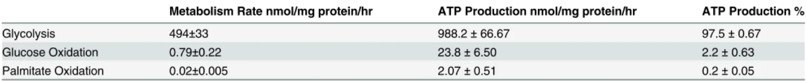
We report here the first direct measurements of energy metabolic rates in BMMSCs (Table 1). These studies were performed under experimental conditions in which cells were incubated with concentrations of glucose (5 mM), fatty acids (0.4 mM palmitate), and albumin
(0.55 mM). Glucose and albumin levels mimic the concentrations of these substrates normally present in the blood. The normal circulating level of fatty acids under non-fasting conditions is 0.4 mM while the concentration of the fatty acid palmitate is about 0.2 mM. Palmitate was used as the representative fatty acid in this assay. In human BMMSCs glycolysis rates (494±33 nmol/mg protein/hr) are extremely high compared to glucose oxidation (0.79±0.22 nmol/mg protein/hr) and palmitate oxidation rates (0.02±0.005 nmol/mg protein/hr). From these rates ATP production from each of these pathways was calculated: glycolysis (988.2±66.7 nmol ATP/mg protein/hr), glucose oxidation (23.8±6.5 nmol ATP/mg protein/hr), and palmitate ox-idation (2.07±0.51 nmol ATP/mg protein/hr) (Table 1). Based on these rates, 97.5% of ATP in the BMMSCs is derived from glycolysis, 2.2% of ATP from glucose oxidation, and 0.2% of ATP from palmitate oxidation. This confirms the previous assumptions that stem cells are primarily deriving their energy from glycolysis.
During each assay Krebs Henseleit buffer was supplemented with 5 mM glucose and 0.4 mM palmitate bound to 0.55 mM albumin. In addition, the Krebs Henseleit buffer was sup-plemented with either [U-14C]glucose, [1–14C]palmitate, or [5–3H]glucose in order to measure glucose oxidation, palmitate oxidation, or glycolysis, respectively. Bone marrow mesenchymal stem cells (BMMSCs) were exposed to normal cell culture media immediately up to the point it was switched to this Krebs Henseleit buffer at the start of each assay. Calculations to determine ATP production and % ATP production were made from the metabolic rate results. n = 5–8 Values are shown as the mean ± SEM.
Fatty acids affect BMMSC survival
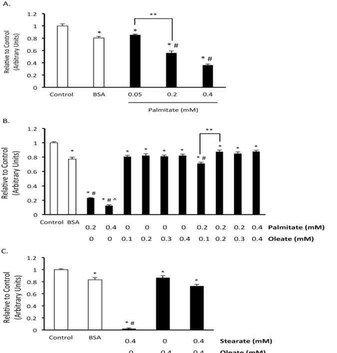
When stem cells are introduced into the target organ they become exposed to the blood. Some of the blood’s contents include glucose, fatty acids, and albumin. Surprisingly, a survey of cell culture media indicate that the level of fatty acids and albumin are much lower than what is present in the circulation [30]. We, therefore, were interested in what effect fatty acids might have on BMMSC viability. When we exposed BMMSCs to media supplemented with levels of palmitate (0.05–0.4 mM) and albumin (0.55 mM) normally present in the blood, we noticed that palmitate in a concentration and time dependent manner decreased BMMSC viability (Fig. 1). In contrast, exposure of the BMMSCs to only 0.55 mM albumin did not result in any major decrease in cell viability (Fig. 1). Importantly, not only pathological levels (0.4 mM), but
Table 1. Contribution of energy metabolism pathways to ATP production in human BMMSCs.
Metabolism Rate nmol/mg protein/hr ATP Production nmol/mg protein/hr ATP Production %
Glycolysis 494±33 988.2±66.67 97.5±0.67
Glucose Oxidation 0.79±0.22 23.8±6.50 2.2±0.63
Palmitate Oxidation 0.02±0.005 2.07±0.51 0.2±0.05
also normal circulating levels of palmitate (0.2 mM) decreased BMMSC viability (Fig. 1). Fur-ther, another saturated fatty acid, stearate, also decreased BMMSC viability (Fig. 1c). Because both unsaturated and saturated fatty acids are present in the circulation, we also examined what effect the unsaturated fatty acid oleate, the most abundant fatty acid in the circulation, has on BMMSC viability. When we treated BMMSCs with physiologically relevant levels of the unsaturated fatty acid oleate (bound to 0.55 mM albumin) we did not observe a change in BMMSC viability (Fig. 1b). Further, if palmitate treated BMMSCs were also exposed to oleate, BMMSC viability was protected (Fig. 1). Oleate also protected against stearate-induced BMMSC death. These results indicate that saturated fatty acids could be a contributing factor in the loss in viability observed when stem cells are introduced into the body for
therapeutic purposes.
Palmitate induces apoptosis and decreases proliferation
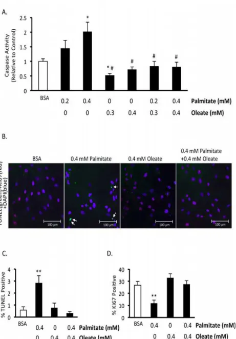
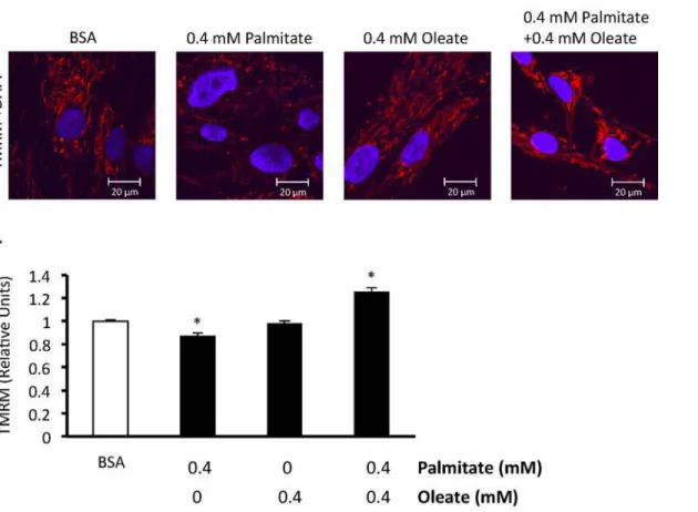
To confirm that palmitate induces BMMSC death and to assess whether the type of cell death involves apoptosis, we measured mitochondrial membrane potential, performed TUNEL stain-ing, and assessed caspase 3 activity. Chronic exposure of BMMSCs to palmitate (48 hr expo-sure) resulted in an increase in caspase 3 activity (Fig. 2A). Palmitate also increased TUNEL staining after 24 hr (Fig. 2B,C) and decreased mitochondrial membrane potential (Fig. 3A,B).
We also investigated the effect of palmitate on BMMSC proliferation. To do this, we as-sessed the nuclear expression of Ki67, a marker of proliferation. There is a lower percentage of Ki67 positive human BMMSC nuclei following palmitate exposure (Fig. 2D).
Oleate inhibits palmitate-induced human BMMSC apoptosis and
reduction in proliferation
To examine the relationship between saturated and unsaturated fatty acids on BMMSC viabili-ty we treated BMMSCs with varying ratios of palmitate and oleate up to a normal physiological range (0.2 mM palmitate and 0.3 mM oleate) during non-fasting conditions. When BMMSCs are exposed to equal or greater amounts of oleate BMMSC viability is preserved (Fig. 1B,Cand
Fig. 2A,C). At the lower ratio tested (0.1 mM oleate and 0.2 mM palmitate) oleate is only par-tially protective. In addition, oleate prevents the increase in caspase activity and TUNEL posi-tive nuclei induced by palmitate treatment (Fig. 2A,B). Oleate also protects against the drop in mitochondrial membrane potential induced by palmitate (Fig. 3A,B). Finally, oleate prevents the drop in Ki67 positive human BMMSCs following 24 hr of palmitate treatment (Fig. 2D). We also looked to see if cyclin D1/Rb signaling, which can regulate proliferation and has been shown to be affected by hematopoietic stem cell exposure to palmitate [31], is involved in this drop in proliferation. However, phosphorylation of S780 Rb and total cyclin D1 protein expres-sion were not significantly affected following 24 hr treatment with palmitate and/or oleate (data not shown).
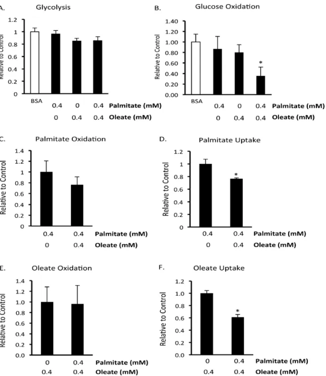
Acute effect of the fatty acids palmitate and oleate on BMMSC energy
metabolism
BMMSCs were only given these treatments while energy metabolism was being measured. Up until these assays began BMMSCs were only exposed to standard cell culture media. Palmitate or oleate alone did not affect glycolysis, glucose oxidation, or oleate oxidation rates (Fig. 4). However, combined treatment with palmitate and oleate did significantly reduce glucose Fig 1. Oleate prevents palmitate-induced human BMMSC death.A) Effect of palmitate on human bone marrow mesenchymal stem cell (BMMSC) viability was measured by the MTT assay after 48 hr treatment with indicated treatments. B) Effect of the ratio of palmitate and oleate on palmitate-induced BMMSC death after 72 hr treatment was measured by MTT assay. C) Effect of 72 hr treatment with stearate and/or oleate on the amount of viable cells was measured by the MTT assay. The BSA group was treated with media supplemented with 0.55 mM albumin. All fatty acid treated groups were also treated with media supplemented with 0.55 mM albumin in addition to the type and amount of fatty acid indicated in the figures. The Control group was treated with media identical to the BSA group minus supplementation with albumin. n = 7–12*Significantly different from Control group. # Significantly less than BSA group. ^ Significantly less than 0.2 mM Palmitate Group.**Groups are significantly different. Values are shown as the mean±SEM.
oxidation rates (Fig. 4B), a condition known to be associated with increased cell proliferation [9]. As expected, palmitate and oleate inhibited each other’s uptake (Fig. 4E,F), although nei-ther inhibited each onei-ther’s oxidation (Fig. 4C,E). This suggests that oleate resulted in a better coupling of palmitate uptake to palmitate oxidation, resulting in less palmitate entering other cellular pathways.
Fig 2. Oleate prevents palmitate-induced human BMMSC apoptosis and reduction in proliferation.A) Caspase activity after 48 hr treatment with indicated treatments. B) Images of terminal deoxynucleotidyl transferase dUTP nick end labeling (TUNEL) and Ki67 staining of bone marrow mesenchymal stem cells (BMMSCs) treated for 24 hr with indicated treatments. Image is 28X. C) % nuclei positive for TUNEL. D) % nuclei positive for Ki67. n = 5–7 The BSA group was treated with media supplemented with 0.55 mM albumin. All fatty acid treated groups were also treated with media supplemented with 0.55 mM albumin in addition to the type and amount of fatty acid indicated in the figures.*Significantly different from BSA group. # Significantly different from 0.2 mM Palmitate and 0.4 mM Palmitate groups.**Significantly different from all groups. Values are shown as the mean±SEM.
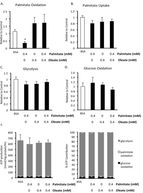
Chronic effects of the fatty acids palmitate and oleate on BMMSC energy
metabolism
Since chronic exposure of BMMSCs results in decreased cell viability, we also investigated what effect exposure of human BMMSCs to fatty acids had on energy metabolism (Fig. 5). Since 72 hr exposure of BMMSCs to palmitate resulted in a substantial decrease in cell viability (Fig. 1), cells were treated with palmitate for 24 hr prior to measurements of energy metabo-lism, a time period where no decrease in cell viability was observed. BMMSCs were treated for 24 hr with 5 mM glucose and either 0.55 mM BSA, 0.4 mM palmitate bound to 0.55 mM BSA, 0.4 mM oleate bound to 0.55 mM BSA, or 0.4 mM palmitate and 0.4 mM oleate bound to 0.55 mM BSA. Interestingly, chronic palmitate treatment decreases palmitate oxidation rates (Fig. 5A), without altering palmitate uptake rates (Fig. 5B). Chronic exposure to oleate prevents the decrease in palmitate oxidation without altering palmitate uptake rates (Fig. 5A,B), thereby improving the coupling between palmitate uptake and oxidation. Neither chronic palmitate and/or oleate treatment affects BMMSC glucose oxidation or glycolysis (Fig. 5C,D). Overall, glycolysis remains the major source of ATP production in BMMSCs which are chronically ex-posed to palmitate and/or oleate (Fig. 5E,F).
Fig 3. Effect of 24 hr exposure to palmitate and oleate on human BMMSC mitochondrial membrane potential.A) Images of tetramethylrhodamine methyl ester (TMRM) stained bone marrow mesenchymal stem cells (BMMSCs) treated for 24 hr with indicated treatments. B) Relative TMRM levels. BMMSCs were treated for 24 hr with 0.55 mM bovine serum albumin (BSA) alone or palmitate and/or oleate bound to 0.55 mM albumin. TMRM and Hoechst stain were added to the medium to measure mitochondrial membrane potential and stain nuclei, respectively, and images were taken. All fatty acid treated groups were also treated with media supplemented with 0.55 mM albumin in addition to the type and amount of fatty acid indicated in the figures.4separate observations. Values are shown as the mean±SEM.*Significantly different from the BSA group.
Fig 4. Effect of acute exposure to fatty acids on human BMMSC energy metabolism.A) Glycolysis, B) glucose oxidation, C) palmitate oxidation, D) palmitate uptake, E) oleate oxidation, and F) oleate uptake were measured in untreated human bone marrow mesenchymal stem cells (BMMSCs). n = 5–7 During each assay Krebs Henseleit buffer was supplemented with 5 mM glucose and, as indicated in each graph, either 0.55 mM albumin (BSA group) or 0.55 mM albumin bound to 0.4 mM palmitate and/or 0.4 mM oleate. In addition, the Krebs Henseleit buffer was supplemented with either [U-14C]glucose,
[1–14C]palmitate, [1–14C]oleate, or [5–3H]glucose for the measurement of glucose oxidation, palmitate oxidation and uptake, oleate oxidation and uptake, or glycolysis, respectively. Since these experiments assessed the acute effect of palmitate and oleate, BMMSCs were maintained in cell culture media used to culture these immediately up to the start of each assay when the media was switched to Krebs Henseleit buffer supplemented with fatty acids. The levels and type of fatty acid BMMSCs were exposed to is indicated on the x-axis of the figures.*Significantly different from all groups. Values are shown as the mean± SEM.
Fig 5. Effect of 24 hour exposure to fatty acids on human BMMSC energy metabolism.A) Palmitate oxidation, B) palmitate uptake, C) glycolysis, and D) glucose oxidation were measured in human BMMSCs that had been treated for 24 hr with either 0.55 mM albumin (BSA group) or 0.55 mM albumin and 0.4 mM palmitate and/or 0.4 mM oleate prior to these metabolism measurements being made. n = 5–8 The graphs indicate which groups were exposed to these different treatments for the 24 hr prior to the metabolism measurements. During each assay all groups were given Krebs buffer supplemented with 5 mM glucose and 0.4 mM palmitate bound to 0.55 mM albumin. In addition, the Krebs buffer was supplemented with either [U-14C]glucose, [1
–14C]palmitate, or [5–3H]glucose for the measurement of glucose oxidation, palmitate oxidation and uptake, or glycolysis, respectively. E) The contribution of metabolic pathways to ATP production were calculated from the metabolic rate results.*Significantly different from all groups. Values are shown as mean±SEM.
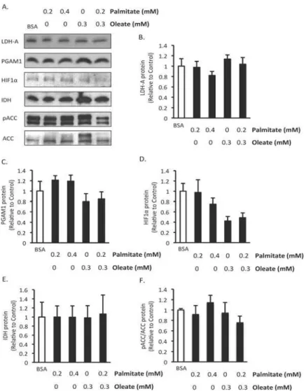
Chronic effects of palmitate and/or oleate on BMMSC expression of
proteins involved in glycolysis and oxidative metabolism
To further investigate the effect of fatty acids on BMMSC energy metabolism we assessed the effect of 24 hr treatment with palmitate and/or oleate on the expression of proteins involved in glycolysis and oxidative metabolism. We chose this length of treatment because it was long enough to potentially observe changes in protein expression but soon enough that there would still be cells present to make measurements in. No significant changes occurred in the protein expression of PGAM1 and LDH-A, two proteins involved in glycolysis (Fig. 6A,B). Interesting-ly, HIF1αprotein expression, a key transcription factor involved in regulating glycolysis, trends
towards being reduced in both groups treated with oleate but palmitate alone did not affect its expression (Fig. 6C). Isocitrate dehydrogenase, an enzyme involve in the TCA cycle, was not af-fected by any of the treatments (Fig. 6D). There were also no changes in ACC expression or phosphorylation of ACC (Fig. 6E), a key enzyme involved in the synthesis of malonyl CoA, which is a potent inhibitor of mitochondrial fatty acid uptake and oxidation. This suggests that regulation of ACC is not an explanation for the reduction in palmitate oxidation observed fol-lowing 24 hr treatment with palmitate.
Discussion
This is the first study to directly determine the energy metabolic rate profile of human BMMSCs. We confirmed the previous assumption that BMMSCs derive most of their ATP from glycolysis (>97%) (Table 1). This finding is in agreement with indirect measurements of energy metabolism including those showing elevated lactate levels and low oxygen consump-tion rates in several types of stem cells including mesenchymal, embryonic, and induced plu-ripotent stem cells [8,32–34]. In support of high rates of glycolysis being important for pluripotency, studies have shown that osteogenic differentiation of mesenchymal stem cells and ESC-to-cardiomyocyte differentiation are accompanied by a decline in lactate production [8,33]. We also examined the effect of various fatty acids on the energy substrate metabolism, survival, and proliferation of human BMMSCs. We show that physiologically relevant levels of saturated fatty acids induce BMMSC death and decrease BMMSC proliferation, effects which are prevented by the unsaturated fatty acid oleate. These experiments were designed to assess the effect of levels of fatty acids present in the circulation on BMMSCs. It will be interesting in the future to also assess the effect of the level of fatty acids present in the bone marrow on BMMSC survival. We also show that decreasing saturated fatty acid oxidation may induce BMMSC death. This has important implications on the therapeutic strategy of using BMMSCs for tissue regeneration, and suggests that strategies should be implemented that minimize cir-culating saturated fatty acid levels during the therapy.
the albumin concentration used was low) [38]. Regardless, the data highlight the need to care-fully consider both the fatty acid concentration and albumin concentration to which the BMMSC is exposed during any attempts at stem cell therapy.
Fatty acids can regulate flux through energy metabolic pathways, and may thereby regulate cell survival. The survival and proliferation of cells with high glycolytic rates tends to be posi-tively correlated with glycolysis [9,15]. In other cell types a process referred to as the glucose-fatty acid cycle, or the Randle cycle, has been observed, where increased glucose-fatty acid oxidation can inhibit glucose oxidation and glycolysis [39]. However, in the heart it has frequently been reported that elevating fatty acid oxidation results in uncoupling of glycolysis from glucose oxi-dation, due to a greater inhibition of glucose oxidation than glycolysis [40]. In agreement with this, inhibiting fatty acid oxidation via MCD inhibition results in pulmonary artery smooth Fig 6. Effect of 24 hr exposure to fatty acids on expression of proteins involved in energy metabolism. A) Representative western blots, B) Lactate dehydrogenase A (LDHA), C) Phosphoglycerate mutase 1 (PGAM1), D) Hypoxia inducible factor 1α(HIF1α), E) Isocitrate dehydrogenase (IDH), F) phospho Acetyl CoA carboxylase (ACC)/ACC protein expression. n = 5–6 Protein expression was measured in human bone marrow mesenchymal stem cells (BMMSCs) treated for 24 hr with indicated treatments. The BSA group was treated with media supplemented with 0.55 mM albumin. All fatty acid treated groups were also treated with media supplemented with 0.55 mM albumin in addition to the type and amount of fatty acid indicated in the figures. Values are shown as mean±SEM.
muscle cell apoptosis and decreased proliferation [10]. This is probably detrimental to these cells because decreasing palmitate oxidation likely results in an improved coupling of glycolysis to glucose oxidation. Therefore, this link between fatty acid oxidation and glucose metabolism could explain why fatty acid oxidation seems to regulate cell proliferation and survival.
An alternative explanation for the effects of fatty acids on cell survival is that fatty acid oxi-dation could be beneficial independent of its effects on glycolysis. It has been suggested that under conditions where glycolysis is reduced fatty acid oxidation can be used by cancer cells for energy production [25]. We therefore decided to determine whether fatty acids inhibit human BMMSC glucose metabolism and induce BMMSC death via modulation of glucose and fatty acid energy metabolism. Acute exposure to palmitate and/or oleate did not affect glycoly-sis or fatty acid oxidation rates (Fig. 4). However, combined acute exposure to palmitate and oleate did reduce glucose oxidation (Fig. 4B). These results indicate that the Randle Cycle exists at least to some extent in human BMMSCs. Following chronic treatment with palmitate and/or oleate we observed that only palmitate exposure reduced palmitate oxidation rates (Fig. 5). In-terestingly, combined treatment with oleate, which prevented palmitate-induced death, pre-vented this reduction in palmitate oxidation (Fig. 5). Neither palmitate or oleate affected the expression of proteins involved in oxidative metabolism or glycolysis that we measured (Fig. 6). These palmitate oxidation results agree with a previous report showing that 20 hr ex-posure of neonatal cardiac myocytes to palmitate induced apoptosis and decreased palmitate oxidation rates [41]. This suggests that palmitate induces BMMSC death via inhibition of pal-mitate oxidation and that oleate is protective because it prevents palpal-mitate oxidation from de-creasing. This is supported by a previous study in BMMSCs in which AICAR (an activator of AMPK and fatty acid oxidation) prevented palmitate-induced death [35]. However, we did not observe a change in phosphorylation of ACC, an indicator of AMPK activity and an important pathway by which AMPK increases fatty acid oxidation. This may not actually be that surpris-ing, since AMPK activation can decrease proliferation but as we show here oleate protects against the drop in proliferation induced by palmitate [42]. It is still a possibility, however, that a reduction in glycolysis may be involved in palmitate-induced BMMSC death, but changes in glycolysis that occur in response to 24 hr treatment with palmitate and/or oleate were masked by switching all groups to the same buffer during the measurement of glycolysis rates.
Oleate had a dramatic effect of preventing palmitate-induced BMMSC death. This may have occurred secondary to inhibiting palmitate uptake. Acutely, oleate and palmitate reduced each other’s uptake (Fig. 4). However, after 24 hr of exposure to palmitate and/or oleate, palmi-tate uptake was not different between groups (Fig. 5). It is still possible, however, that oleate did in fact reduce palmitate uptake at 24 hr but it was an acute effect and therefore was not measured (since during the assay the cells in all groups were exposed to 0.4 mM palmitate). Therefore, oleate may be at least partially protecting against palmitate-induced cell death by re-ducing intracellular palmitate levels by decreasing palmitate uptake.
Unfortunately, experimental conditions precluded us from measuring ceramide levels in these cells. However, there is evidence that palmitate at least does not always work through cer-amides to induce cell death [36,37,43].
Conclusion
We demonstrate that human BMMSC energy production is predominantly derived from gly-colysis, and we show that modulation of energy metabolism is important in the proliferation and survival of human BMMSCs. In particular, physiologically relevant levels of saturated fatty acids reduce BMMSC proliferation and induce BMMSC apoptosis, all effects that can be pre-vented by oleate. The decrease in saturated fatty acid oxidation induced by chronic exposure to palmitate may be involved in these deleterious effects of palmitate on BMMSCs. These observa-tions indicate that saturated fatty acids could be contributing to the lowin vivosurvival of
BMMSCs, and therefore to the disappointing results of stem cell therapy clinical trials includ-ing those focused on treatinclud-ing heart and pulmonary diseases [1–6,44–46].
Acknowledgments
Human BMMSCs were provided by the Texas A&M Health Science Center College of Medi-cine Institute for Regenerative MediMedi-cine at Scott & White through a grant from NCRR of the NIH, Grant # P40RR017447.
Author Contributions
Conceived and designed the experiments: NF A. Huqi JSJ GDL. Performed the experiments: NF A. Huqi JM RP A. Haromy AO. Analyzed the data: NF GDL. Contributed reagents/materi-als/analysis tools: LI BT EM. Wrote the paper: NF A. Huqi JSJ JM RP A. Haromy AO LI BT EM GDL.
References
1. Copland IB, Galipeau J. Death and inflammation following somatic cell transplantation. Semin Immuno-pathol. 2011; 33: 535–550. doi:10.1007/s00281-011-0274-8PMID:21533908
2. Behfar A, Yamada S, Crespo-Diaz R, Nesbitt JJ, Rowe LA, Perez-Terzic C, et al. Guided cardiopoiesis enhances therapeutic benefit of bone marrow human mesenchymal stem cells in chronic myocardial in-farction. J Am Coll Cardiol. 2010; 56: 721–734. doi:10.1016/j.jacc.2010.03.066PMID:20723802 3. Mangi AA, Noiseux N, Kong D, He H, Rezvani M, Ingwall JS, et al. Mesenchymal stem cells modified
with Akt prevent remodeling and restore performance of infarcted hearts. Nat Med. 2003; 9: 1195–1201. PMID:12910262
4. Lim SY, Kim YS, Ahn Y, Jeong MH, Hong MH, Joo SY, et al. The effects of mesenchymal stem cells transduced with Akt in a porcine myocardial infarction model. Cardiovasc Res. 2006; 70: 530–542. PMID:16563361
5. Li W, Ma N, Ong LL, Nesselmann C, Klopsch C, Ladilov Y, et al. Bcl-2 engineered MSCs inhibited apo-ptosis and improved heart function. Stem Cells. 2007; 25: 2118–2127. PMID:17478584
6. Toma C, Wagner WR, Bowry S, Schwartz A, Villanueva F. Fate of culture-expanded mesenchymal stem cells in the microvasculature: in vivo observations of cell kinetics. Circ Res. 2009; 104: 398–402. doi:10.1161/CIRCRESAHA.108.187724PMID:19096027
7. Chung S, Arrell DK, Faustino RS, Terzic A, Dzeja PP. Glycolytic network restructuring integral to the en-ergetics of embryonic stem cell cardiac differentiation. J Mol Cell Cardiol. 2010; 48: 725–734. doi:10.
1016/j.yjmcc.2009.12.014PMID:20045004
8. Chung S, Dzeja PP, Faustino RS, Perez-Terzic C, Behfar A, Terzic A. Mitochondrial oxidative metabo-lism is required for the cardiac differentiation of stem cells. Nat Clin Pract Cardiovasc Med. 2007;4 Suppl 1: : S60–67.
10. Sutendra G, Bonnet S, Rochefort G, Haromy A, Folmes KD, Lopaschuk GD, et al. Fatty acid oxidation and malonyl-CoA decarboxylase in the vascular remodeling of pulmonary hypertension. Sci Transl Med. 2010; 2: 44ra58. doi:10.1126/scitranslmed.3001327PMID:20702857
11. Pecqueur C, Bui T, Gelly C, Hauchard J, Barbot C, Bouillaud F, et al. Uncoupling protein-2 controls pro-liferation by promoting fatty acid oxidation and limiting glycolysis-derived pyruvate utilization. FASEB J. 2008; 22: 9–18. PMID:17855623
12. Wanet A, Remacle N, Najar M, Sokal E, Arnould T, Najimi M, et al. Mitochondrial remodeling in hepatic differentiation and dedifferentiation. Int J Biochem Cell Biol. 2014; 54: 174–185. doi:10.1016/j.biocel. 2014.07.015PMID:25084555
13. Warburg O. On respiratory impairment in cancer cells. Science. 1956; 124: 269–270. PMID:13351639
14. Warburg O, Posener K, Negelein E. On the metabolism of carcinoma cells. Biochemische Zeitschrift. 1924; 152: 309–344.
15. Vander Heiden MG, Plas DR, Rathmell JC, Fox CJ, Harris MH, Thompson CB. Growth Factors Can In-fluence Cell Growth and Survival through Effects on Glucose Metabolism. Molecular and Cellular Biolo-gy. 2001; 21: 5899–5912. PMID:11486029
16. Kaplon J, Zheng L, Meissl K, Chaneton B, Selivanov VA, Mackay G, et al. A key role for mitochondrial gatekeeper pyruvate dehydrogenase in oncogene-induced senescence. Nature. 2013; 498: 109–112. doi:10.1038/nature12154PMID:23685455
17. Abu Dawud R, Schreiber K, Schomburg D, Adjaye J. Human embryonic stem cells and embryonal car-cinoma cells have overlapping and distinct metabolic signatures. PLoS One. 2012; 7: e39896. doi:10. 1371/journal.pone.0039896PMID:22768158
18. Burgess RJ, Agathocleous M, Morrison SJ. Metabolic regulation of stem cell function. J Intern Med. 2014; 276: 12–24. doi:10.1111/joim.12247PMID:24697828
19. Guarnerio J, Coltella N, Ala U, Tonon G, Pandolfi PP, Bernardi R. Bone marrow endosteal mesenchy-mal progenitors depend on HIF factors for maintenance and regulation of hematopoiesis. Stem Cell Re-ports. 2014; 2: 794–809. doi:10.1016/j.stemcr.2014.04.002PMID:24936467
20. Mylotte LA, Duffy AM, Murphy M, O'Brien T, Samali A, Barry F, et al. Metabolic flexibility permits mesen-chymal stem cell survival in an ischemic environment. Stem Cells. 2008; 26: 1325–1336. doi:10.1634/ stemcells.2007-1072PMID:18308942
21. Kang JX, Wan JB, He C. Concise review: Regulation of stem cell proliferation and differentiation by es-sential fatty acids and their metabolites. Stem Cells. 2014; 32: 1092–1098. doi:10.1002/stem.1620
PMID:24356924
22. Calvisi DF, Wang C, Ho C, Ladu S, Lee SA, Mattu S, et al. Increased lipogenesis, induced by AKT-mTORC1-RPS6 signaling, promotes development of human hepatocellular carcinoma. Gastroenterol-ogy. 2011; 140: 1071–1083. doi:10.1053/j.gastro.2010.12.006PMID:21147110
23. Yamashita T, Honda M, Takatori H, Nishino R, Minato H, Takamura H, et al. Activation of lipogenic pathway correlates with cell proliferation and poor prognosis in hepatocellular carcinoma. J Hepatol. 2009; 50: 100–110. doi:10.1016/j.jhep.2008.07.036PMID:19008011
24. Wise DR, Ward PS, Shay JE, Cross JR, Gruber JJ, Sachdeva UM, et al. Hypoxia promotes isocitrate dehydrogenase-dependent carboxylation of alpha-ketoglutarate to citrate to support cell growth and viability. Proc Natl Acad Sci U S A. 2011; 108: 19611–19616. doi:10.1073/pnas.1117773108PMID:
22106302
25. Buzzai M, Bauer DE, Jones RG, Deberardinis RJ, Hatzivassiliou G, Elstrom RL, et al. The glucose de-pendence of Akt-transformed cells can be reversed by pharmacologic activation of fatty acid beta-oxi-dation. Oncogene. 2005; 24: 4165–4173. PMID:15806154
26. Zaugg K, Yao Y, Reilly PT, Kannan K, Kiarash R, Mason J, et al. Carnitine palmitoyltransferase 1C pro-motes cell survival and tumor growth under conditions of metabolic stress. Genes Dev. 2011; 25: 1041–1051. doi:10.1101/gad.1987211PMID:21576264
27. Paulin R, Meloche J, Courboulin A, Lambert C, Haromy A, Courchesne A, et al. Targeting cell motility in pulmonary arterial hypertension. Eur Respir J. 2014; 43: 531–544. doi:10.1183/09031936.00181312 PMID:23845719
28. Sorrentino D, Robinson RB, Kiang CL, Berk PD. At physiologic albumin/oleate concentrations oleate uptake by isolated hepatocytes, cardiac myocytes, and adipocytes is a saturable function of the un-bound oleate concentration. Uptake kinetics are consistent with the conventional theory. J Clin Invest. 1989; 84: 1325–1333. PMID:2794064
30. Boone CW, Mantel N, Caruso TD Jr., Kazam E, Stevenson RE. Quality control studies on fetal bovine serum used in tissue culture. In Vitro. 1971; 7: 174–189. PMID:4131329
31. Ito K, Carracedo A, Weiss D, Arai F, Ala U, Avigan DE, et al. A PML-PPAR-delta pathway for fatty acid oxidation regulates hematopoietic stem cell maintenance. Nat Med. 2012; 18: 1350–1358. PMID:
22902876
32. Varum S, Rodrigues AS, Moura MB, Momcilovic O, Easley CAt, Ramalho-Santos J, et al. Energy me-tabolism in human pluripotent stem cells and their differentiated counterparts. PLoS One. 2011; 6: e20914. doi:10.1371/journal.pone.0020914PMID:21698063
33. Chen CT, Shih YR, Kuo TK, Lee OK, Wei YH. Coordinated changes of mitochondrial biogenesis and antioxidant enzymes during osteogenic differentiation of human mesenchymal stem cells. Stem Cells. 2008; 26: 960–968. doi:10.1634/stemcells.2007-0509PMID:18218821
34. Pattappa G, Heywood HK, de Bruijn JD, Lee DA. The metabolism of human mesenchymal stem cells during proliferation and differentiation. J Cell Physiol. 2011; 226: 2562–2570. doi:10.1002/jcp.22605
PMID:21792913
35. Lu J, Wang Q, Huang L, Dong H, Lin L, Lin N, et al. Palmitate causes endoplasmic reticulum stress and apoptosis in human mesenchymal stem cells: prevention by AMPK activator. Endocrinology. 2012; 153: 5275–5284. doi:10.1210/en.2012-1418PMID:22968644
36. Listenberger LL, Ory DS, Schaffer JE. Palmitate-induced apoptosis can occur through a ceramide-inde-pendent pathway. J Biol Chem. 2001; 276: 14890–14895. PMID:11278654
37. Miller TA, LeBrasseur NK, Cote GM, Trucillo MP, Pimentel DR, Ido Y, et al. Oleate prevents palmitate-induced cytotoxic stress in cardiac myocytes. Biochem Biophys Res Commun. 2005; 336: 309–315. PMID:16126172
38. Smith AN, Muffley LA, Bell AN, Numhom S, Hocking AM. Unsaturated fatty acids induce mesenchymal stem cells to increase secretion of angiogenic mediators. J Cell Physiol. 2012; 227: 3225–3233. doi:
10.1002/jcp.24013PMID:22105830
39. Randle PJ. Regulatory interactions between lipids and carbohydrates: the glucose fatty acid cycle after 35 years. Diabetes Metab Rev. 1998; 14: 263–283. PMID:10095997
40. Lopaschuk GD, Ussher JR, Folmes CD, Jaswal JS, Stanley WC. Myocardial fatty acid metabolism in health and disease. Physiol Rev. 2010; 90: 207–258. doi:10.1152/physrev.00015.2009PMID:
20086077
41. Hickson-Bick DL, Buja LM, McMillin JB. Palmitate-mediated alterations in the fatty acid metabolism of rat neonatal cardiac myocytes. J Mol Cell Cardiol. 2000; 32: 511–519. PMID:10731449
42. de Meester C, Timmermans AD, Balteau M, Ginion A, Roelants V, Noppe G, et al. Role of AMP-activat-ed protein kinase in regulating hypoxic survival and proliferation of mesenchymal stem cells. Cardio-vasc Res. 2014; 101: 20–29. doi:10.1093/cvr/cvt227PMID:24104879
43. Kong JY, Rabkin SW. Mitochondrial effects with ceramide-induced cardiac apoptosis are different from those of palmitate. Archives of Biochemistry and Biophysics. 2003; 412: 196–206. PMID:12667483
44. Clifford DM, Fisher SA, Brunskill SJ, Doree C, Mathur A, Watt S, et al. Stem cell treatment for acute myocardial infarction. Cochrane Database Syst Rev. 2012; 2: CD006536. doi:10.1002/14651858. CD006536.pub3PMID:22336818
45. Tzouvelekis A, Laurent G, Bouros D. Stem cell therapy in chronic obstructive pulmonary disease. Seek-ing the Prometheus effect. Curr Drug Targets. 2013; 14: 246–252. PMID:23256721