Phosphorus-32, a Clinically Available Drug,
Inhibits Cancer Growth by Inducing DNA
Double-Strand Breakage
Yulan Cheng1, Ana P. Kiess2, Joseph M. Herman2, Martin G. Pomper3, Stephen J. Meltzer1*, John M. Abraham1
1Department of Medicine, The Johns Hopkins University School of Medicine, Baltimore, Maryland, United States of America,2Department of Radiation Oncology and Molecular Radiation, The Johns Hopkins University School of Medicine, Baltimore, Maryland,3Department of Radiology and Radiological Science, The Johns Hopkins University School of Medicine, Baltimore, Maryland, United States of America
*smeltzer@jhmi.edu
Abstract
Radioisotopes that emit electrons (beta particles), such as radioiodine, can effectively kill target cells, including cancer cells. Aqueous32P[PO
4] is a pure beta-emitter that has been
used for several decades to treat non-malignant human myeloproliferative diseases.32P
[PO4] was directly compared to a more powerful pure beta-emitter, the clinically important 90
Y isotope.In vitro,32P[PO4] was more effective at killing cells than was the more powerful
isotope90Y (P0.001) and also caused substantially more double-stranded DNA breaks than did90Y.In vivo, a single low-dose intravenous dose of aqueous elemental32P
signifi-cantly inhibited tumor growth in the syngeneic murine cancer model (P0.001). This effect
is exerted by direct incorporation into nascent DNA chains, resulting in double-stranded breakage, a unique mechanism not duplicatable by other, more powerful electron-emitting radioisotopes.32P[PO4] should be considered for human clinical trials as a potential novel
anti-cancer drug.
Introduction
Beta particles (electrons) emitted by radioisotopes are known to efficiently kill cancer cells. This finding has already been clinically exploited by using131I to treat thyroid cancer [1], a strategy still employed successfully in more than 50% of such patients in the United States, with over a 90% cure rate. Similarly, beta particle-emitting radiolabeled antibodies directed against CD20, including131I-Bexxar (tositumomab) and90Y-Zevalin (ibritumomab tiuxetan), have been used against non-Hodgkin’s lymphoma [2,3]. Moreover,90Y-labeled somatostatin receptor ligand is utilized to treat neuroendocrine tumors [4]. Electrons emitted by32P have an energy level intermediate between those of131I and the more powerful90Y, resulting in a path length of up to 5 mm in human tissues [5]. Electrons emitted from radioisotopes can strike thousands of cells. The resulting bystander effect amplifies the lethal potential of each beta par-ticle emitted in or near a tumor. However, as we shall show below, we have discovered that
a11111
OPEN ACCESS
Citation:Cheng Y, Kiess AP, Herman JM, Pomper MG, Meltzer SJ, Abraham JM (2015) Phosphorus-32, a Clinically Available Drug, Inhibits Cancer Growth by Inducing DNA Double-Strand Breakage. PLoS ONE 10(6): e0128152. doi:10.1371/journal.pone.0128152
Academic Editor:Abhijit De, ACTREC, Tata Memorial Centre, INDIA
Received:December 19, 2014
Accepted:April 22, 2015
Published:June 1, 2015
Copyright:© 2015 Cheng et al. This is an open access article distributed under the terms of the Creative Commons Attribution License, which permits unrestricted use, distribution, and reproduction in any medium, provided the original author and source are credited.
Data Availability Statement:All relevant data are within the paper.
Funding:This work was supported by the National Cancer Institute CA133012-01, National Institutes of Health Roadmap DK087454-01, National Cancer Institute CA146799, National Cancer Institute CA190040, and a Sidney Kimmel Cancer Center Pilot Project Grant. The funders had no role in study design, data collection and analysis, decision to publish, or preparation of the manuscript.
among all available beta-emitting isotopes,32P possesses a unique chemically and
radiologically-based double-strand breakage mechanism, which confers greater anti-tumor ef-ficacy than other beta-emitters of comparable power.
Human cancer-derived cell lines established in immunocompromised mice are a valuable tool for testing the effectiveness of candidate anti-cancer agents [6–9]. We previously found that a single, low-dose intravenous injection of [32P]ATP significantly inhibits tumor growth for several weeks in murine xenograft models [10,11]. Because ATP is a small naturally-occurring molecule, its radiolabeled form poses some advantages over larger synthetic com-pounds as a potential anti-cancer therapeutic, including lower immunogenicity, greater tumor penetration, and superior pharmacokinetics [12]. Inorganic [32P]PO4, a simple aqueous ion,
has been used for decades as a therapeutic agent for polycythemia vera and essential thrombo-cythemia [13]. This ion was also previously used for palliation of bone pain due to metastases, where it was thought to be incorporated into the extracellular matrix [14]. However, aqueous
32P use has never been established as a primary anti-cancer strategy
per se.
The clinical application of32P was first attempted in the 1930’s [15–18]. Since that time,32P usage has generally been restricted to a colloidal suspension form, wherein32P forms a compo-nent of a complex, insoluble particle [19–22]. This form of32P is typically injected directly into the tumor, with the colloidal suspension preventing the radioisotope from leaving the intended target and disseminating throughout the body. The administration of aqueous32P as a primary anti-cancer agent has not been studied, aside from its palliative use for relief of pain due to bone metastases.
Recent experimental findings have led to the development and use of the alpha- and beta-emitter223Ra to selectively target bone metastases in patients with castration-resistant prostate cancer [23,24]. Originally developed by a Norwegian company Algeta, Alpharadin was ap-proved for use in the United States in 2013, and is now marketed by Bayer under the name Xofigo [25]. Thus,223Ra is the latest simple radioactive element to become an effective anti-cancer drug.
We now report that a single, low-dose intravenous injection of aqueous32P results in rapid, significant growth inhibition of pre-established tumor growth in an immunocompetent (syn-geneic) murine model. We also show that32P is more efficient than equivalent doses of higher-energy extracellular electrons, such as those emitted by90Y, a beta-emitting radioisotope in common use today. We provide evidence that this higher efficiency results from the direct in-corporation of32P into nascent DNA, causing double-strand DNA breakage via a combined chemical-radiological mechanism that cannot duplicated by other beta-emitting radioisotopes, such as131I and90Y. This finding has immediate ramifications for the expanded treatment of primary human cancers.
Methods
Measurement of
in vitro
cell killing by
32P and
90Y
Two thousand cells of the murine BALB/c CRL2836 cell line or the human HeLa S3 cell line were grown in complete medium in a 96-well plate and were exposed at Day 0 to either 0, 1, 2.5, or 5μCi of90Y radioisotope or the [32P]PO4radioisotope in complete medium. After a
Assessment of double-strand DNA breaks
Ten thousand HeLa S3 cells were seeded onto Lab-TekII chamber slides (Thermo Fisher Scien-tific Inc., Waltham, MA). At Day 0, cells were treated with 0 or 3μCi of32P or90Y in complete
medium. At Day 1, all wells were gently washed and fresh non-radioactive medium was added. At Day 1, Day 2, or Day 3, cells were fixed with 10% formalin at room temperature for 10 min, washed with PBS for two min, and permeabilized with 0.2% Triton X-100 with 10% FBS in PBS for 15 min. After a rinse with PBS, primary mouse anti-human H2AX antibody at 1:1000 dilution (Millipore, Billerica, MA) was incubated for 1 h at ambient temperature, then washed twice with PBS for 5 min. A dilution of 1:400 goat anti-mouse IgG with Alexa Fluor (Life Tech-nologies, Grand Island, NY) was incubated for 1 h at ambient temperature and washed twice with PBS for 5 min. Cells were stained with Hoechst solution (1:1000).
Assessement of
32P incorporated into DNA
One hundred and fifty thousand mouse CRL2836 or human HeLa S3 cells were seeded onto a six-well cell culture plate and grown for 24 h (defined as Day 0). Cells were then either incubat-ed overnight with32P[PO4], grown for 2 d in non-radioactive medium and the DNA extracted
(3 Days); or grown for 24 h, incubated with32P[PO4] for 24 h, grown for 24 h in
non-radioac-tive medium and the DNA extracted (2 Days); or grown for 48 h, incubated with32P[PO4] for
24 hours, washed with complete medium and the nucleic acid extracted (1 Day). DNA was ex-tracted using the DNAeasy Blood and Tissue Kit (Qiagen, Valencia, CA) and aliquots were in-cubated with or without four units of DNase I (New England Biolabs, Ipswich, MA) for two h at 37°C before the samples were run on a 5% polyacrylamide gel, exposed to film overnight at 4°C and developed.
Assay for apoptosis in cell lines incubated with
32P.
One hundred thousand mouse CRL2836 cells or HeLa S3 cells were seeded into each well of 6-well culture plates and grown for 24 h. Cells were then incubated with 0, 2.5, 5, 10 or 20 uCi
32P[PO
4] in two ml complete medium for 24 h, and non-radioactive medium added for an
ad-ditional 24 h. Protein was extracted from each well using RIBA buffer (Cell Signaling Technol-ogy, Boston, MA), the protein was quantified using a BCA protein assay kit (Pierce, Rockford, IL),and identical quantities were run on a 10 to 20% polyacrylamide gel, and blotted to nitro-cellulose. A western blot using a primary antibody to cleaved caspase-3 protein (Cell Signaling Technology, Boston, MA) was used to assay for apoptosis. A second western blot with identical protein quantities was probed with antibody against beta-actin (Cell Signaling Technology, Boston, MA).
Establishment of mouse tumors
Syngeneic BALB/c mouse tumors were established by injecting 2 X 10⁶BALB/c tumor CRL2836 cells (American Type Cell Culture, Manassas, VA) in a volume of 0.2 mL (50% Matrigel, 50% 1 X PBS) subcutaneously in the left rear and right rear flank. All mice were fe-male, 10 weeks of age, and purchased from Charles River Laboratories (Wilmington, MA).
32
P-Mediated tumor growth inhibition
After ten days, during which time well-vascularized tumors became well-established, an injec-tion of 5μCi of the monophosphate form of32P (Perkin-Elmer, Cat. # NEX06000, Waltham,
per week with a digital caliper and the volume was determined using the formula: volume = ½(width)2X (length). Mice were maintained at the Johns Hopkins University Facility in accor-dance with Laboratory Animal Resources Commission standards under the supervision and approval of the Johns Hopkins University Institutional Animal Care and Use Committee (IACUC).
Statistical analysis
The data from the WST-1 cell proliferation were presented as means ± standard deviation, and the significance was determined using the unpaired Student’sttest. Tumor volumes of the un-treated and un-treated mice were displayed as means ± SE; no outliers were excluded for any rea-son. Significance was determined using the unpaired Student’sttest.
Results
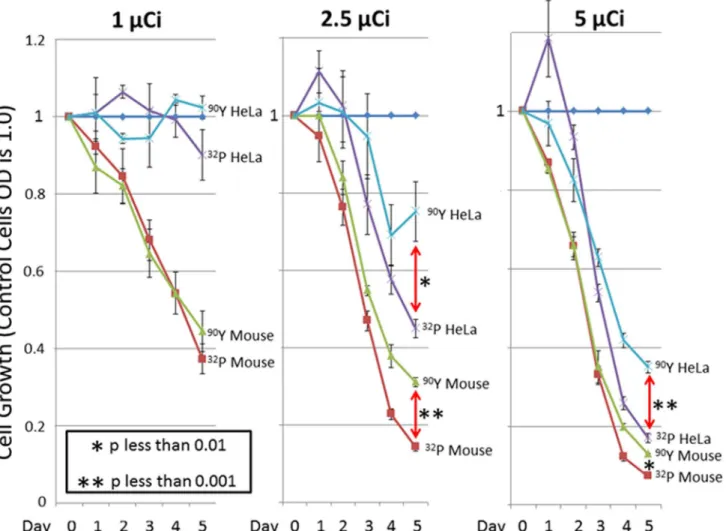
Cells exposed to [32P]PO4were compared with those exposed to identical counts per minute of
the more powerful beta-particle emitter,90Y, after which WST-1 cell proliferation assays were performed (Fig 1). Different cell lines were expected to demonstrate varying levels of suscepti-bility to radioisotopes. The 1μCi dose showed that HeLa cells were less susceptible to
beta-emitting isotopes than were BALB/c mouse CRL2836 cells, which originated as an osteosarco-ma and were isolated after it had metastasized to lung. Both the 2.5μCi and 5μCi doses
dem-onstrated similar results in both cell lines. Although90Y would had been expected to be more lethal than32P (based on its higher-energy electrons),32P killed cells more efficiently than did
90Y. In comparisons of the 2.5
μCi and 5μCi doses, [32P]PO4produced survival rates at Day 5
that were barely half of those produced by90Y.
The H2AX assays were used to compare double-strand DNA breakage in cells incubated with32Pvs.90Y (Fig 2) [26,27]. This assay accurately detects breakage in both strands of DNA at the same genomic locus. Nuclear staining of HeLa S3 cells demonstrated substantial, time-dependent double-strand DNA breakage in cells exposed to32P, while those exposed to identi-cal levels of90Y-based radiation had much less or no detectable DNA double-strand breakage. Digestion with DNase I showed that administered32P had been directly incorporated into cel-lular DNA (Fig 3A). More than half of the32P retained by the cells that were incubated with
32P[PO
4]for 24 h and then grown in non-radioactive medium for 48 h before the DNA was
ex-tracted had been permanently incorporated into cellular DNA. To determine whether cell death involves apoptosis as well as necrosis, mouse CRL2836 cells or HeLa S3 cells were incu-bated with varying amounts of32P for 24 hours, replaced with non-radioactive medium for an additional 24 hours, and the cells analyzed by western blot for the presence of cleaved caspase-3 indicating apoptosis (Fig 3B). The CRL2836 cells clearly demonstrated that apoptosis was in-volved in cellular death, while the HeLa S3 cells showed no detectable cleaved caspase-3 (data not shown). Antibody directed against beta-actin was used to verify equal loading of protein amounts in the gel wells. HeLa S3 cells express E6 from HPV18 and are rendered p53 null which severely inhibits apoptosis functions [28,29].
Previously, we demonstrated significant inhibition of HeLa S3 cell xenograft growths in nude mice by a single low-dose intravenous (IV) injection of [32P]ATP. Here, we chose immu-nocomponent syngeneic BALB/c mice to more closely recapitulate human malignancy. A sin-gle IV injection of aqueous [32P]PO4significantly inhibited established syngenic tumor growth
in BALB/c mice (Fig 4). There were no apparent detrimental effects of [32P]PO4comparing the
Discussion
This study documents our discovery that a single intravenous dose of the32P radioisotope sig-nificantly inhibits the growth of pre-established tumors in a murine syngeneic model, while si-multaneously establishing the mechanism underlying this anti-cancer effect. Specifically, we show that aqueous32P is incorporated into nascent DNA, where isotopic decay shears both strands, causing double-strand breakage as proven by phosphorylation of the histone H2-AX. Ourin vitroexperiments also demonstrate that the pure beta-emitter32P is superior to the more powerful pure beta-emitter90Y in tumor cytotoxicity, and finally, that apoptosis contrib-utes to this cytotoxicity.
Fig 5depicts a proposed mechanism for32P-induced cell killing. In this schematic,32P is in-corporated directly into one strand of replicating DNA. Radioactive decay of32P to32S causes chemical breakage of that same DNA strand. Next, the electron released by this decay event needs to travel only 2 nm to reach the contralateral strand of the double helix, severing it and thus causing a double-strand break at this genomic locus. This mechanism stands in stark con-trast to non-incorporated beta-emitting radioisotopes, where only a small fraction of emitted Fig 1. Inhibition of cell growth by [32P] PO4or [90Y].The WST-1 proliferation assay was done to determine the level of cell killing by32P or by90Y. BALB/c tumor CRL2836 cells or HeLa S3 cells were exposed to 0 Ci, 1μCi, 2.5μCi, or 5μCi in complete medium. After 24 hours, the medium was changed and cells were grown in non-radioactive complete medium. WST-1 cell proliferation assays were done at Days 1, 2, 3, 4, and 5 in triplicate. The mean is shown plus/ minus the standard deviation. The student’s two-sided t-test determined thePvalue shown.
electrons travel in the precise orientation necessary to strike one strand plus its opposite strand and cause a double-strand DNA break [30,31]. With32P, the extreme proximity of the contra-lateral target strand to the decay-produced electron makes this double-strand breakage much more likely to occur [32].
Aqueous [32P]PO4offers many potential advantages over other anti-cancer therapeutic
agents. Firstly, it allows for rapid systemic distribution and incorporation into primary tumors and32P is preferentially absorbed by rapidly proliferating cells, such as cancer cells. In addition, [32P]PO4improves on the previous direct injection of particulate colloidal32P into primary
tu-mors, since aqueous [32P]PO4allows for a simple intravenous injection [33–35].
Secondly,32P is already an FDA-approved drug, with a known low toxicity profile [36]. Pre-vious clinical studies of32P-orthophosphate [PO4] in aqueous solution for polycythemia vera
and essential thrombocythemia have established tolerable dose levels, particularly with respect to myelosuppression [36]. In this context, it is noteworthy that no obvious toxic side effects Fig 2. Determination of double-strand DNA breaks in cells caused by exposure to [32P]PO
4or90Y.HeLa S3 cells or mouse BALB/c CRL2836 cells were grown in multiple sections of chamber slides and exposed to 0μCi or 3μCi of [32P]PO4or90Y in complete medium at Day 0. After 24 hours, the medium
was changed and cells were grown in non-radioactive complete medium. At Day 1, 2, or 3 the presence of double-strand DNA breaks in the cells was determined by staining for phosphorylated H2-AX histones which indicate double-strand DNA damage.
occurred in any of the model systems we have studied to date. The other concern with its use in these benign hematologic disorders has been an increased incidence of subsequent acute mye-loid leukemia (AML) [37,38], but in patients with advanced solid tumors this is less of a con-cern, since subsequent AML occurs in only 10%ten yearsafter32P treatment [39]. Moreover, this new indication and method of use for an existing drug saves considerable time and ex-pense, relative to the investment required for new anticancer agents.
Thirdly, in contrast to other beta-emitting isotopes such as131I and90Y,32P is incorporated directly into nascent DNA [40,41]. Our data suggest that this incorporation dramatically in-creases the cell-killing efficiency of32P, since the decay of incorporated32P to sulfur chemically breaks the first strand of the DNA and the released electron needs to travel only 2 nm to reach its contralateral DNA strand. Thus, this process efficiently causes double-strand DNA break-age, which is required to overcome innate DNA repair pathways and achieve cell death. In con-trast to32P, other electron-emitting isotopes (such as131I and90Y) emit electrons from
distances of 1,000 to 5,000 nm away from DNA, some 500- to 2,500-fold farther than the dis-tance of an incorporated32P atom from its sister DNA strand [42–44].
Fig 3. Characterization of32P uptake by the cell.A.32
P is directly incorporated into cellular DNA. Mouse CRL2836 or human HeLa S3 cell lines were incubated overnight with32P[PO
4] and then grown for 48 h in non-radioactive medium (lanes 1 through 4), or grown for 24 h in non-radioactive medium,
grown for 24 h with32P[PO
4], and then grown for 24 h in non-radioactive medium (lanes 5 through 8), or grown for 48 h in non-radioactive medium, then
grown for 24 h with32P[PO
4] (lanes 9 through 12). The extracted nucleic acids were incubated with DNase I, the digestion products were run on a 5%
polyacrylamide gel and exposed to film. B. Apoptosis induced by32P in mouse CRL2836 cells. Mouse CRL2836 cells were incubated with 0, 2.5, 5, 10 or 20μCi32P[PO4] for 24 h, and non-radioactive medium added for an additional 24 h. Protein was extracted from each well and analyzed for apoptosis by
western blots using antibody to cleaved caspase-3 protein (Lanes 1 through 5). Antibody against beta-actin was used to verify identical amounts of protein were loaded (lanes 6 through 10).
It is intriguing to note that early researchers performing Sanger sequencing with [32P]dATP in the 1980’s noted that sequencing products required electrophoresis within two days after the sequencing reaction, otherwise bands seemed to disperse and were difficult to interpret [45]. This same principle may operate with32P as an anti-cancer agent. The decay of32P to sulfur chemically shears the strand of DNA into which it is incorporated. We hypothesize that this event, coupled with the extremely close proximity of the incorporated radioisotope to its sister DNA strand, results in a dramatic increase in cell-killing efficiencyvs. other beta-particle emit-ters such as131I and90Y, which are not incorporated into nascent DNA. The resulting implica-tions for the potential clinical treatment of primary human tumors are obvious and far-reaching.
Fig 4. Inhibition of BALB/c syngeneic tumor growth by [32P]PO
4.Syngeneic BALB/c CRL2836 tumors were established in the rear flanks of BALB/c mice at Day 0. After ten days, during which the tumors became well vascularized, mice received an injection of 5μCi of [32P]PO4intravenously via the tail vein. The
tumor volumes are shown as the mean of six tumors plus/minus the standard error of the mean. The student’s two-sided t-test determined thePvalue shown. Inset: Representative picture at 35 days post CRL2836 cell injection, showing two control mice on the left, and one mouse that received one 5 uCi [32P]PO
4
dose (right).
Acknowledgments
We wish to thank Mr. Gilbert Green for expert technical assistance in injecting the mice.
Author Contributions
Conceived and designed the experiments: YC APK JMH MGP SJM JMA. Performed the exper-iments: YC APK JMA. Analyzed the data: YC APK JMH MGP SJM JMA. Contributed re-agents/materials/analysis tools: YC APK MGP SJM JMA. Wrote the paper: YC APK JMH MGP SJM JMA.
References
1. Goldsmith SJ. (2011) To ablate or not to ablate: issues and evidence involved in 131I ablation of residu-al thyroid tissue in patients with differentiated thyroid carcinoma. Semin Nucl Med. 41: 96–104. doi:10. 1053/j.semnuclmed.2010.11.002PMID:21272683
2. Wiseman GA, Leigh B, Erwin WD, Lamonica D, Kornmehl E, Spies SM, et al. (2002) Radiation dosime-try results for Zevalin radioimmunotherapy of rituximab-refractory non-Hodgkin lymphoma. 94: 1349–
1357. PMID:11877765
3. Horning SJ, Younes A, Jain V, Kroll S, Lucas J, Podoloff D, et al. (2005) Efficacy and safety of tositumo-mab and iodine-131 tositumotositumo-mab (Bexxar) in B-cell lymphoma, progressive after rituxitositumo-mab. J Clin Oncol. 23: 712–719. PMID:15613695
Fig 5. Model of double-strand DNA break after incorporation of [32P]PO
4.The radioisotope is incorporated into the ribose-phosphate backbone of DNA in dividing cells. The process of decaying to sulfur (32S) breaks the backbone bond of the initial strand at a 67% rate and releases a high energy beta particle
(electron) that must only travel two nm across the helix to the opposite target strand. Although an emitted electron that travels in the perfect orientation from this32P decay to sever the opposite strand will only occur at a low percentage of the time, it is still much higher and more efficient than those electrons which are generated by other beta-producing radioisotopes on the cell surface or in the cytosol that must travel distances that are usually one thousand times or more longer in length.
4. Nisa L, Savelli G, Giubbini R. (2011) Yttrium-90 DOTATOC therapy in GEP-NET and other SST2 ex-pressing tumors: a selected review. Ann. Nucl. Med. 25: 75–85. doi:10.1007/s12149-010-0444-0
PMID:21107762
5. Lechner A, Blaickner M, Gianolini S, Poljanc K, Aiginger H, Georg D. (2008) Targeted radionuclide ther-apy: theoretical study of the relationship between tumour control probability and tumour radius for a 32P/33P radionuclide cocktail. Phys. Med. Biol. 53:1961–1974. doi:10.1088/0031-9155/53/7/011
PMID:18354241
6. Richmond A, Su Y. (2008) Mouse xenograft models vs GEM models for human cancer therapeutics. Dis. Model Mech 1: 78–82. doi:10.1242/dmm.000976PMID:19048064
7. Morton CL, Houghton PJ. (2007) Establishment of human tumor xenografts in immunodeficient mice. Nat. Protoc. 2: 247–250. PMID:17406581
8. Wang T, Weigt SS, Belperio JA, Lynch JP. (2011) Immunosuppressive and cytotoxic therapy: pharma-cology, toxicities, and monitoring. Semin. Respir. Crit Care Med. 32: 346–370. doi: 10.1055/s-0031-1279831PMID:21674420
9. Mellor HR, Snelling S, Hall MD, Modok S, Jaffar M, Hambley TW, et al. (2005) The influence of tumour microenvironmental factors on the efficacy of cisplatin and novel platinum(IV) complexes. Biochem. Pharmacol. 70: 1137–1146. PMID:16139250
10. Cheng Y, Yang J, Agarwal R, Green GM, Mease RC, Pomper MG,et al. (2011) Strong inhibition of xenografted tumor growth by low-level doses of [(32)P]ATP. Oncotarget 2: 461–466. PMID:21646686 11. Cheng Y, Senthamizhchelvan S, Agarwal R, Green GM, Mease RC, Sgouros G, et al. (2012) [32P]ATP
inhibits the growth of xenografted tumors in nude mice. Cell Cycle 11: 1878–1882. doi:10.4161/cc. 19955PMID:22544324
12. Patrick MR, Chester KA, Pietersz GA. (1998) In vitro characterization of a recombinant 32P-phosphory-lated anti-(carcinoembryonic antigen) single-chain antibody. Cancer Immunol. Immunother. 46: 229–237. PMID:9671146
13. Barbui T, Finazzi G. (1998) Treatment of polycythemia vera. Haematologica 83: 143–149. PMID:
9549926
14. Silberstein EB. (2005) Teletherapy and radiopharmaceutical therapy of painful bone metastases. Semin. Nucl. Med. 35:152–158. PMID:15765378
15. Varia MA, Stehman FB, Bundy BN, Benda JA, Clarke-Pearson DL, Alvarez RD et al. (2003) Intraperito-neal radioactive phosphorus (32P) versus observation after negative second-look laparotomy for stage III ovarian carcinoma: a randomized trial of the Gynecologic Oncology Group. J Clin. Oncol. 21: 2849–
2855. PMID:12885800
16. Gao W, Liu L, Liu ZY, Li XD, Li C. (2009) Interstitial injection of (32)P-chromic phosphate during lung cancer resection to treat occult lymphatic metastasis. Nucl. Med. Commun. 30: 420–426. doi:10.1097/ MNM.0b013e328310af19PMID:19398935
17. Powell JL, Burrell MO, Kirchner AB. (1987) Intraperitoneal radioactive chromic phosphate P 32 in the treatment of ovarian cancer. South. Med. J. 80: 1513–1517. PMID:3423895
18. Grady ED. (1984) Adjuvant therapy of Dukes' C colon cancer by intarterial P-32 colloid for internal ra-diation therapy of the liver. Am. Surg. 50: 488–492. PMID:6476612
19. Zubillaga MB, Boccio JR, Nicolini JO, Ughetti R, Lanari E, Caro RA. (1996) Use of colloids of chromic [32P] phosphate in treatment of solid tumors. Nucl. Med. Biol. 23: 907–910. PMID:8971858
20. Soper JT, Wilkinson RH, Bandy LC, Clarke-Pearson DL, Creasman WT. (1985) Intraperitoneal chromic phosphate P 32 suspension therapy of malignant peritoneal cytology in endometrial carcinoma. Am. J. Obstet. Gynecol. 153: 191–196. PMID:4037013
21. Heath R, Rosenman J, Varia M, Walton L. (1988) Peritoneal fluid cytology in endometrial cancer: its sig-nificance and the role of chromic phosphate (32P) therapy. Int. J. Radiat. Oncol. Biol. Phys. 15: 815–822. PMID:3182321
22. Gao W, Liu L, Liu ZY, Wang Y, Jiang B, Liu XN. (2010) Intratumoral injection of 32P-chromic phosphate in the treatment of implanted pancreatic carcinoma. Cancer Biother. Radiopharm. 25: 215–224. doi:
10.1089/cbr.2008.0596PMID:20423235
23. Larsen RH, Saxtorph H, Skydsgaard M, Borrebaek J, Jonasdottir TJ, Bruland OS, et al. (2006) Radio-toxicity of the alpha-emitting bone-seeker 223Ra injected intravenously into mice: histology, clinical chemistry and hematology. In Vivo 20: 325–331. PMID:16724665
24. Pandit-Taskar N, Larson SM, Carrasquillo JA. (2014) Bone-seeking radiopharmaceuticals for treatment of osseous metastases, Part 1:αtherapy with 223Ra-dichloride. J Nucl Med. 55: 268–274. doi:10. 2967/jnumed.112.112482PMID:24343987
bone metastases: results from a phase 3, double-blind, randomised trial. Lancet Oncol. 15: 738–746. doi:10.1016/S1470-2045(14)70183-4PMID:24836273
26. Fernandez-Capetillo O, Chen HT, Celeste A, Ward I, Romanienko PJ, Morales JC, et al. (2002) DNA damage-induced G2-M checkpoint activation by histone H2AX and 53BP1. Nat. Cell Biol. 4: 993–997. PMID:12447390
27. Ward IM, Minn K, Jorda Kg, Chen J. (2003) Accumulation of checkpoint protein 53BP1 at DNA breaks involves its binding to phosphorylated histone H2AX. J. Biol. Chem. 278: 19579–19582. PMID:
12697768
28. Cotugno R, Basile A, Romano E, Gallotta D, Belisario MA. (2014) BAG3 down-modulation sensitizes HPV18(+) HeLa cells to PEITC-induced apoptosis and restores p53. Cancer Lett. 354:263–271. doi:
10.1016/j.canlet.2014.08.022PMID:25175321
29. Yi JW, Jang M, Kim SJ, Kim SS, Rhee JE. (2013) Degradation of p53 by natural variants of the E6 pro-tein of human papillomavirus type 16. Oncol Rep. 29:1617–1622. doi:10.3892/or.2013.2281PMID:
23404471
30. Fallahi P, Mazzi V, Vita R, Ferrari SM, Materazzi G, Gallieri D, et al. (2015) New Therapies for Dediffer-entiated Papillary Thyroid Cancer. Int J Mol Sci. 16:6153–6182 doi:10.3390/ijms16036153PMID:
25789503
31. Knoop K, Schwenk N, Schmohl K, Müller A, Zach C, Cyran C, et al. (2015) Mesenchymal stem cell-mediated, tumor stroma-targeted radioiodine therapy of metastatic colon cancer using the sodium io-dide symporter as theranostic gene. J Nucl Med. 56:600–606. doi:10.2967/jnumed.114.146662PMID:
25745085
32. Mayson SE, Yoo DC, Gopalakrishnan G. (2015) The evolving use of radioiodine therapy in differentiat-ed thyroid cancer. Oncology. 88:247–256. doi:10.1159/000369496PMID:25503797
33. Lee I, Wallner PE. (1997) Evaluation of cellular uptake, tumor retention, radiation response, and tumor pathophysiology in experimental solid tumors after an intratumoral infusion of colloidal 32P. Cancer. 80:2611–2617. PMID:9406715
34. Nguyen H, Ghanem G, Morandini R, Verbist A, Larsimont D, Fallais C, et al. (1997) Tumor type and vascularity: important variables in infusional brachytherapy with colloidal 32P. Int J Radiat Oncol Biol Phys. 39:481–7. PMID:9308954
35. Order SE, Siegel JA, Lustig RA, Principato R, Zeiger LS, Johnson E, et al. (1994) A new method for de-livering radioactive cytotoxic agents in solid cancers. Int J Radiat Oncol Biol Phys. 30:715–720. PMID:
7928505
36. Tennvall J, Brans B. (2007) EANM procedure guideline for 32P phosphate treatment of myeloprolifera-tive diseases. Eur. J. Nucl. Med. Mol. Imaging 34: 1324–1329. PMID:17396258
37. Najean Y, Rain JD. (1997) Treatment of polycythemia vera: use of 32P alone or in combination with maintenance therapy using hydroxyurea in 461 patients greater than 65 years of age. Blood 89: 2319–2327. PMID:9116275
38. Brandt L, Anderson H. (1995) Survival and risk of leukaemia in polycythemia vera and essential throm-bocythemia treated with oral radiophosphorus. Eur. J. Haematol. 54: 21–26. PMID:7859872 39. Perkins A. (2005) In vivo molecular targeted radiotherapy. Biomed. Imaging Interv. J. 1: e9. doi:10.
2349/biij.1.2.e9PMID:21625282
40. Kim DH, Jung JH, Son SH, Kim CY, Hong CM, Jeong SY, et al. (2014) Difference of clinical and radio-logical characteristics according to radioiodine avidity in pulmonary metastases of differentiated thyroid cancer. Nucl Med Mol Imaging. 48:55–62. doi:10.1007/s13139-013-0239-zPMID:24900139 41. Fröhlich E, Wahl R. (2014) The current role of targeted therapies to induce radioiodine uptake in thyroid
cancer. Cancer Treat Rev. 40:665–674. doi:10.1016/j.ctrv.2014.01.002PMID:24485648
42. Saxena A, Meteling B, Kapoor J, Golani S, Danta M, Morris DL, et al. (2014) Yttrium-90 radioemboliza-tion is a safe and effective treatment for unresectable hepatocellular carcinoma: a single centre experi-ence of 45 consecutive patients. Int J Surg. 12:1403–1408. doi:10.1016/j.ijsu.2014.07.269PMID:
25091398
43. Kao YH. (2014) General theory of predictive dosimetry for yttrium-90 radioembolization to sites other than the liver. Cardiovasc Intervent Radiol. 37:1114–1117. PMID:24232040
44. Lam MG, Abdelmaksoud MH, Chang DT, Eclov NC, Chung MP, Chung AC, et al. (2013) Safety of 90Y radioembolization in patients who have undergone previous external beam radiation therapy.Int J Radiat Oncol Biol Phys. 87:323–329. doi:10.1016/j.ijrobp.2013.05.041PMID:23849697
![Fig 2. Determination of double-strand DNA breaks in cells caused by exposure to [ 32 P]PO 4 or 90 Y](https://thumb-eu.123doks.com/thumbv2/123dok_br/18243696.341486/6.918.53.764.108.695/fig-determination-double-strand-breaks-cells-caused-exposure.webp)

![Fig 4. Inhibition of BALB/c syngeneic tumor growth by [ 32 P]PO 4 . Syngeneic BALB/c CRL2836 tumors were established in the rear flanks of BALB/c mice at Day 0](https://thumb-eu.123doks.com/thumbv2/123dok_br/18243696.341486/8.918.61.769.114.717/inhibition-syngeneic-growth-syngeneic-balb-tumors-established-flanks.webp)
![Fig 5. Model of double-strand DNA break after incorporation of [ 32 P]PO 4 . The radioisotope is incorporated into the ribose-phosphate backbone of DNA in dividing cells](https://thumb-eu.123doks.com/thumbv2/123dok_br/18243696.341486/9.918.62.772.111.558/double-strand-incorporation-radioisotope-incorporated-phosphate-backbone-dividing.webp)
