Innate Immunity Drives the Initiation of a
Murine Model of Primary Biliary Cirrhosis
Chao-Hsuan Chang1, Ying-Chun Chen1, Weici Zhang2, Patrick S. C. Leung2, M. Eric Gershwin2, Ya-Hui Chuang1*
1Department of Clinical Laboratory Sciences and Medical Biotechnology, College of Medicine, National Taiwan University, Taipei, Taiwan,2Rheumatology, Allergy and Clinical Immunology, University of California at Davis, Davis, CA, 95616, United States of America
Abstract
Invariant natural killer T (iNKT) cells play complex roles in bridging innate and adaptive im-munity by engaging with glycolipid antigens presented by CD1d. Our earlier work suggested that iNKT cells were involved in the initiation of the original loss of tolerance in primary biliary cirrhosis (PBC). To address this issue in more detail and, in particular, to focus on whether iNKT cells activated by a Th2-biasing agonist (2s,3s,4r)-1-O-(α-D-galactopyranosyl)-N -tet-racosanoyl-2-amino-1,3,4-nonanetriol (OCH), can influence the development of PBC in a xenobiotic-induced PBC murine model. Groups of mice were treated with either OCH or, as a control,α-galactosylceramide (α-GalCer) and thence serially followed for cytokine
produc-tion, markers of T cell activaproduc-tion, liver histopathology and anti-mitochondrial antibody re-sponses. Further, additional groups of CD1d deleted mice were similarly studied. Our data indicate that administration of OCH has a dramatic influence with exacerbation of portal in-flammation and hepatic fibrosis similar to mice treated withα-GalCer. Further, iNKT cell
defi-cient CD1d knockout mice have decreased inflammatory portal cell infiltrates and reduced anti-mitochondrial antibody responses. We submit that activation of iNKT cells can occur via overlapping and/or promiscuous pathways and highlight the critical role of innate immu-nity in the natural history of autoimmune cholangitis. These data have implications for hu-mans with PBC and emphasize that therapeutic strategies must focus not only on suppressing adaptive responses, but also innate immunity.
Introduction
Invariant natural killer T (iNKT) cells, expressing semi-invariant Vα14-Jα28 chain
preferen-tially pair with Vβ2, Vβ7, or Vβ8.2 chain, hypersecrete Th1 and Th2 cytokines and chemokines
upon stimulation with an appropriate ligand, such asα-galactosylceramide (α-GalCer) [1,2].
Activation of iNKT cellsin vivoleads to subsequent activation of other cells, such as dendritic
cells, NK cells, T cells and B cells. Through the release of specific types of cytokines, iNKT cells regulate a cascade of immune reactions that alter the balance of subsequent Th1 and Th2
re-sponses [3].α-GalCer is a well-defined potent and specific ligand for iNKT cell activation in
OPEN ACCESS
Citation:Chang C-H, Chen Y-C, Zhang W, Leung PSC, Gershwin ME, Chuang Y-H (2015) Innate Immunity Drives the Initiation of a Murine Model of Primary Biliary Cirrhosis. PLoS ONE 10(3): e0121320. doi:10.1371/journal.pone.0121320
Academic Editor:William Ridgway, University of Cincinnati College of Medicine, UNITED STATES
Received:December 16, 2014
Accepted:January 30, 2015
Published:March 25, 2015
Copyright:© 2015 Chang et al. This is an open access article distributed under the terms of the
Creative Commons Attribution License, which permits unrestricted use, distribution, and reproduction in any medium, provided the original author and source are credited.
Data Availability Statement:All data are found within the paper.
Funding:This work was supported by a grant from the National Health Research Institutes, Taiwan (NHRI-EX103-10027SC) and a National Institutes of Health grant, DK067003. The funders had no role in study design, data collection and analysis, decision to publish, or preparation of the manuscript.
both humans and mice. Upon ligation of their invariant T cell receptors withα-GalCer pre-sented by CD1d of antigen presenting cells, iNKT cells rapidly produce large amount of
cyto-kines, including IFN-γand IL-4 [4,5,6].
Moreover, modification of the length of the lipid chain ofα-GalCer results in the generation
of glycolipids with predominant Th1 or Th2 cytokine skewing profiles [7]. (2s,3s,4r)-1-O
-(α-D-galactopyranosyl)-N-tetracosanoyl-2-amino-1,3,4-nonanetriol (OCH), a synthetic analog
ofα-GalCer with a truncated sphingosine chain, stimulates iNKT cells to produce
predomi-nantly Th2 cytokines [8]. Studies using models of experimental autoimmune diseases,
includ-ing arthritis, diabetes, and experimental autoimmune encephalomyelitis (EAE), have indicated that activation of iNKT cells by OCH ameliorates or prevents these Th1-mediated diseases,
which was attributed to its induction of IL-4 and Th2 skewing [9,10,11,12].
Primary biliary cirrhosis (PBC) is a progressive autoimmune liver disease with female predi-lection characterized by immune-mediated destruction of intrahepatic small bile ducts, leading
to decreased bile secretion, fibrosis, and eventual liver failure [13,14,15]. Although the role of
cytokines in the pathogenesis of PBC remains unresolved [16,17], PBC is considered a Th1
and/or Th17 dominant autoimmune responses similar to other organ-specific autoimmune
diseases [13,14,15] and activation of natural killer T cells accelerates PBC in murine models
[18,19,20]. Further, iNKT cell activation byα-GalCer leads to a profound exacerbation of
por-tal inflammation, granuloma formation, bile duct damage, and hepatic fibrosis in a chemical
xenobiotic murine model of PBC [18,19]. Despite major advances in our understanding of the
immunobiology of PBC in both humans and murine models, there remain major voids in our
dissection of initiating and exacerbating factors [21,22,23,24,25,26,27].
In this study, we investigated the effect of administration of OCH on the natural history of xenobiotic induced autoimmune cholangitis. We report herein that mice administered with OCH have exacerbated portal inflammation and hepatic fibrosis similar to mice treated with
α-GalCer. iNKT cell deficient CD1d knockout mice immunized with 2-OA-BSA have reduced
anti-mitochondrial antibody (AMA) production and cell infiltrates in the liver compared to that of immunized wild type mice. Thus, activation of iNKT cells is critical for the pathogenesis of autoimmune cholangitis and highlights the important roles of innate immunity in this mu-rine model of PBC.
Materials and Methods
Experimental mice
Female C57BL/6 mice aged 8–10 weeks were obtained from the National Laboratory Animal
Center, Taiwan. CD1d-/-mice on a B6 background were kindly provided by Dr. Chyung-Ru
Wang (Northwestern University Feinberg School of Medicine, Chicago, IL, USA). All mice were maintained in the Animal Center of the College of Medicine, National Taiwan University and all experiments performed following approval of The Institutional Animal Care and Use Committee (IACUC) of National Taiwan University College of Medicine and College of Public Health.
Experimental protocol
Mice were intraperitoneally immunized with 2-OA-BSA (150μg) in the presence of complete
phenotypes. For the glycolipid study, mice were injected intravenously using the retro-orbital
route with 2μg ofα-GalCer (KRN7000, Funakoshi, Tokyo, Japan), (2s,3s,4r)-1-O-(α-D
-galac-topyranosyl)-N-tetracosanoyl-2-amino-1,3,4-nonanetriol (OCH) (Enzo Life Sciences,
Farm-ingdale, NY, USA) or PBS simultaneously with first and second 2-OA-BSA immunizations.
Determination of anti-PDC-E2 antibodies
Serum and supernatant titers of IgM and IgG anti-PDC-E2 autoantibodies were measured by
ELISA using recombinant murine PDC-E2. Briefly, purified recombinant PDC-E2 at 1μg/ml
in carbonate buffer (pH 9.6) was coated onto ELISA plates at 4°C overnight. After blocking with 1% casein for 1 hour, supernatants or diluted sera were added for 2 hours at room temper-ature. The ELISA plates were washed with PBS-tween 20 followed by the addition of horserad-ish peroxidase (HRP)-conjugated goat anti-mouse IgG (1:5000, Zymed Laboratories, Carlsbad, CA, USA) and IgM (1:5000, Invitrogen, Camarillo, CA, USA). The plates were incubated for another 1 hr and immunoreactivity detected by measuring optical density (O.D.) at 450 nm after exposure for 20 minutes to tetramethylbenzidine (TMB) substrate (R&D systems, Minne-apolis, MN, USA).
Mononuclear cell preparation
Livers were perfused with PBS containing 0.2% BSA (PBS/0.2% BSA), passed through a 100μm
nylon mesh, and re-suspended in PBS/0.2% BSA. The parenchymal cells were removed as pel-lets after centrifugation at 50 g for 5 minute and the non-parenchymal cells were then isolated using Histopaque-1077 (Sigma-Aldrich). After centrifugation, collected cells were washed with PBS/0.2% BSA and viability of cells was confirmed by trypan blue dye exclusion.
Flow cytometry
Subsets of liver mononuclear cells were measured by flow cytometry. Before staining with a previously defined optimal dilution of monoclonal antibodies (Abs), the cells were
pre-incubat-ed with anti-CD16/32 (clone 93) to block non-specific FcRγbinding. The following Abs were
used in this study: anti-CD69, anti-CD44, anti-CD3, anti-CD4, anti-CD8a, anti-CD19, (Biole-gend, San Diego, CA, USA), anti-NK1.1 (eBioscience, San Diego, CA, USA) and PE-conjugated mouse CD1d tetramers loaded with PBS57 (PBS57 loaded CD1d tetramer, originally produced by the NIH tetramer facility, and supplied through Dr. David Serreze). Stained cells were as-sessed on a FACSCalibur (BD Biosciences, San Diego, CA, USA) using FlowJo software (Tree Star, Inc., Ashland, OR, USA). Optimal concentrations of the mAbs were used throughout and all assays included positive and negative controls.
Histopathology
Portions of liver were excised and immediately fixed with 10% buffered formalin solution for
2 days at room temperature. Paraffin-embedded tissue sections were then cut into 4-μm slices
for routine hematoxylin and eosin (H-E) and Masson’s Trichrome staining. Liver
inflamma-tion was evaluated using light microscopy using our previously defined scoring system
[17,28,29].
Cytokine expression by liver mononuclear cells
Liver mononuclear cells (2x10⁶/ml) were cultured with 1μg/ml anti-CD3 Ab and 1μg/ml
anti-CD28 Ab (Biolegend) for 48 hours. ELISA (Duoset, R&D, Minneapolis, MN, USA) assay
Statistical analysis
Data are presented as the mean ±standard error of the mean (SEM). All graphing and statistical analyses were performed using the Prism graphing program (GraphPad Software, San Diego,
CA, USA). p-values were calculated using a two-tailed unpaired Mann–Whitney U test.
Signifi-cance levels were set at a p-value of 0.05.
Results
Th2-biased cytokine profile was induced by OCH
As expected and as controls when these studies were initiated, intravenousα-GalCer induced
high levels of IL-4 and IFN-γwithin 18 hours of injection; neither IFN-γnor IL-4 were detected
in control injected mice (Fig. 1). In OCH injected mice, both IFN-γand IL-4 were significantly
induced and elevated IFN-γproduction (391.3 ±165.7 pg/ml) was found at 2 hours. However,
significantly lower IFN-γwas found at 18 hours by OCH compared toα-GalCer (794.1±428.4
pg/ml vs. 2735.0 ±1042.0 pg/ml, respectively) (Fig. 1A). A predominant production of IL-4 was
detected at 2 hours of OCH administration and it became undetectable at 18 hours (Fig. 1B).
Fig 1. Lower serum levels of IFN-γafter OCH injection.C57BL/6 mice were intravenously injected withα -GalCer, OCH, or PBS. Serum samples were collected at 2 and 18 hours afterα-GalCer, OCH, or PBS injection. IFN-γ(A) and IL-4 (B) were measured by ELISA. n = 10 mice per group.***, p<0.001.
IL-17 was undetectable in PBS,α-GalCer or OCH injected mice (data not shown). Thus, these
data indicate thatα-GalCer initiated a rapid IL-4 production and a prolonged high level of
IFN-γ, whereas OCH induced a predominant IL-4 production and a weak IFN-γlevel.
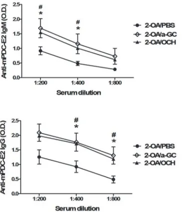
OCH promoted antigen-specific B cell response in 2-OA-BSA
immunized mice
Sinceα-GalCer and OCH initiate different cytokine profiles, we sought to address whether
these two glycolipids induced different antigen-specific B cell responses. As shown inFig. 2,
serum IgM and IgG antibodies to PDC-E2 were increased in 2-OA-BSA/ OCH (2-OA/OCH) immunized mice compared to 2-OA-BSA/PBS (2-OA/PBS) immunized mice. There were no significant differences in the titers of anti-PDCE2 antibodies between 2-OA/a-GC group and
2-OA/OCH group (Fig. 2).
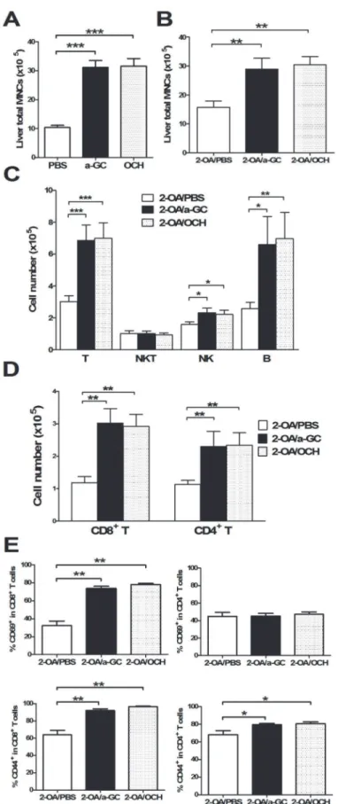
Increased mononuclear inflammatory infiltrate in OCH injected
2-OA-BSA immunized mice
Increased numbers of liver mononuclear cells were noted as early as 3 days after administration
with OCH orα-GalCer compared to PBS controls (Fig. 3A), indicating that both glycolipids
drive the recruitment of mononuclear cells into the liver. At 12 weeks post immunization, 2-OA-BSA/ OCH immunized mice had significantly higher liver total mononuclear cell
infil-trates, increased numbers of T, B and NK cells, and increased CD4+and CD8+T cells
Fig 2. Increased serum AMAs in mice injected with 2-OA-BSA/OCH.Wild type mice were immunized with 2-OA-BSA andα-GalCer (group name: 2-OA/a-GC), OCH (group name: 2-OA/OCH) or PBS (group name: 2-OA/PBS) at weeks 0, 2, 4, 6 and 8. At week 12, serum levels of autoantibodies to mPDC-E2 were measured by ELISA. n = 9–10 mice per group.*, p<0.05 in 2-OA/a-GC to 2-OA/PBS; #, p<0.05 in 2-OA/ OCH to 2-OA/PBS.
compared to 2-OA-BSA/PBS immunized mice (Fig. 3B, C, and D). The frequencies of CD44
expressing CD8+T cells and CD69 expressing CD8+T cells were significantly increased in
2-OA-BSA/OCH immunized mice compared to 2-OA-BSA/PBS immunized mice. Moreover,
the frequency of CD44 expressing CD4+T cells was significantly increased in 2-OA-BSA/OCH
immunized mice (Fig. 3E). There were no differences in mononuclear cells, cell subsets, and
ac-tivating T cells between 2-OA/α-GC group and 2-OA/OCH group (Fig. 3). Histologically,
there were significant increases in portal inflammation and fibrosis in the 2-OA/OCH group compared to 2-OA/PBS group mice and no differences between the 2-OA/a-GC group and
2-OA/OCH group (Fig. 4). Taken together, not onlyα-GalCer but, importantly, OCH
adminis-tration induced more inflammatory cells to liver, including activating CD4+and CD8+T, NK,
and B cells, which led to portal inflammation and liver fibrosis.
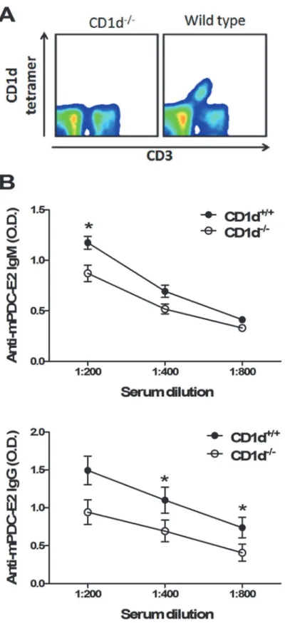
Decreased AMAs, cell infiltrates, and IFN-
γ
production of liver
mononuclear cells in 2-OA-BSA immunized CD1d
-/-mice
To further define the role of natural iNKT cells in the pathogenesis of autoimmune cholangitis, we immunized 2-OA-BSA to iNKT cell deficient CD1d knockout mice and monitored disease progress. At 12 weeks, serum IgM and IgG antibodies to PDC-E2 were significantly decreased
in 2-OA-BSA immunized CD1d-/-mice as compared to that of wild type mice (Fig. 5B). Liver
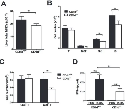
total mononuclear cell infiltrates were significantly lower in 2-OA-BSA immunized CD1d
-/-mice than wild type -/-mice (Fig. 6A). The total numbers of CD4+T cells, NK cells and B cells
were significantly decreased in 2-OA-BSA immunized CD1d-/-mice (Fig. 6B and C).
More-over, decreased expression of IFN-γin activated liver mononuclear cells of 2-OA-BSA
immu-nized CD1d-/-mice was noted compared to wild type mice (Fig. 6D).
Discussion
Cytokine secretion profile, timing of activation, and type of lipid antigens are important factors
determining the pathogenic or protective function of NKT subsets afterin vivostimulation and
activation [3,30,31]. In xenobiotic immunized mice, iNKT cell activation by a synthetic
glyco-plipid, such asα-GalCer, leads to the exacerbation of portal inflammation, granuloma
forma-tion, bile duct damage and in particular hepatic fibrosis [18,19]. Moreover,N.aromaticivorans
is a microorganism that expresses the conserved mammalian PDC-E2 autoepitopes and also activates NKT cells via cell wall glycosphingolipids and finally induces cholangitis following
ex-posure in wild-type mice [32]. These results suggest that activated iNKT cells exacerbate
PBC-like disease. Herein we demonstrate decreased AMAs, CD4+T, NK, and B cell infiltrates and
IFN-γproduction of liver mononuclear cells in 2-OA-BSA immunized iNKT cell deficient
CD1d-/-mice.
β-glucosylceramide is a natural plant glycospingolipid and inhibitsα-GalCer-mediated
acti-vation of NKT cells by binding to its receptor [33]. Administration ofβ-glucosylceramide
ame-liorates liver inflammation in TGF-βreceptor II dominant-negative (dnTGF-βRII) PBC mice
[34]. Of note, administration of either OCH orα-GalCer led to significantly elevated levels of
PDC-E2-specific IgM and IgG autoantibodies in 2-OA-BSA immunized mice compared to
3 days afterα-GalCer, OCH, or PBS injection. n = 10–13 mice per group.***, p<0.001. (B-E) Wild type mice were immunized with 2-OA-BSA andα-GalCer (group name: 2-OA/a-GC), OCH (group name:2-OA/OCH) or PBS (group name: 2-OA/PBS) at weeks 0, 2, 4, 6 and 8 and sacrificed at week 12. (B) Liver total
mononuclear cells (MNC) were measured. (C) The numbers of T (CD3+NK1.1-), NKT (CD3+NK1.1+), NK
(CD3-NK1.1+) and B (CD19+) cells were measured. (D) The numbers of CD4+and CD8+T cells were detected. (E) The expression of CD69 and CD44 in CD4+and CD8+T cells was measured by flowcytometry.
n = 9–10 mice per group.*, p<0.05;**, p<0.01;***, p<0.001.
controls, indicating that activated iNKT cells provide help for antibody production. In addi-tion, 2-OA-BSA immunized CD1d knockout controls have lower levels of AMA and reduced cellular infiltrates compared to controls, suggesting that iNKT cell activation occurs by an
en-dogenous ligand or via the use of complete Freund's adjuvant [35]. Our findings are consistent
with our previous studies that activation of iNKT cells by glycolipid antigens enhance autoanti-body production. In addition, the lack of iNKT cells will reduce autoantiautoanti-body production
[36,37]. Hence our thesis that iNKT cells regulate autoimmune responses at more than
one level.
Studies using models of experimental autoimmune diseases such as arthritis, diabetes, and experimental autoimmune encephalomyelitis (EAE) have indicated that activation of iNKT cells by OCH ameliorates or prevents these Th1-mediated diseases, attributed to induction of
IL-4 and Th2 skewing [9,10,11,12]. However, in this study, we found OCH exacerbates the
Fig 4. The increase of portal inflammation and fibrosis in mice injected with 2-OA-BSA/OCH.Mice were immunized with 2-OA-BSA andα-GalCer (group name: 2-OA/a-GC), OCH (group name: 2-OA/OCH) or PBS (group name: 2-OA/PBS) at weeks 0, 2, 4, 6 and 8 and sacrificed at week 12. (A) Representative stained liver sections of haematoxylin and eosin (H&E) and Masson’s trichrome stain. (B) Histopathological scores of individual livers on portal inflammation and fibrosis. 0 = no significant change, 1 = minimal, 2 = mild, 3 = moderate, and 4 = severe pathology. Individual symbols each represent a single mouse.
Fig 5. Decreased AMAs in 2-OA-BSA immunized CD1d-/-mice.(A) iNKT cell deficiency in CD1d
-/-mice was confirmed by staining with CD3 and CD1d tetramer. (B) CD1d-/-and wild type mice were immunized with
2-OA-BSA at 0, 2, 4, 6 and 8 and sacrificed at week 12. Serum levels of IgM and IgG to mPDC-E2 were measured by ELISA. n = 10 mice for each group.*, p<0.05.
manifestations of autoimmune cholangitis in 2-OA-BSA immunized mice to approximately
the same levels observed with administration ofα-GalCer. The pathogenesis of organ-specific
autoimmune diseases has been previously thought to be orchestrated by Th1 and/or Th17, not
Th2 cells [38]. PBC is considered a Th1 and/or Th17 dominant autoimmune responses. In the
serum of patients with PBC, the most significant increases were noted for IFN-γand IL-17,
al-though increased levels of IL-2, IL-4, IL-5, and IL-10 have also been reported
[16,39,40,41,42,43]. In addition, an increased in the frequency of IL-17+lymphocytic
infiltra-tion in liver has also been noted [40,42]. Our results suggest that activation of other immune
networks by activated NKT cells may be equally important for the pathogenesis of cholangitis. Thus, the importance of Th subsets and cytokines in disease progression requires further study
involving IFN-γ, IL-4 and IL-17 and/or blocking of cytokine signals by
cytokine-neutralizing antibodies.
In patients with PBC, there are increased number of liver NK cells [44]. We report herein
that NK cells are increased in bothα-GalCer and OCH injected 2-OA-BSA immunized mice
while decreased in CD1d-/-mice immunized with 2-OA-BSA. In a previous study,
administra-tion of polyI:C, a viral RNA mimetic and Toll-like receptor 3 agonist, to activate NK cells in Fig 6. Decreased cell infiltrates and IFN-γproduction of liver mononuclear cells in 2-OA-BSA immunized CD1d-/-mice.(A-C) CD1d-/-and wild type mice were immunized with 2-OA-BSA at 0, 2, 4, 6 and
8 and sacrificed at week 12. (A) Liver total mononuclear cells (MNCs) were measured. (B) The numbers of T (CD3+NK1.1-), NKT (CD3+NK1.1+), NK (CD3-NK1.1+) and B (CD19+) cells were measured. (C) The numbers
of CD4+and CD8+T cells were detected. n = 16
–20 per group.*, p<0.05. (D) CD1d-/-or wild type mice were immunized with 2-OA-BSA at weeks 0, 2, and 4 and sacrificed 3 days after last immunization. IFN-γ production of liver mononuclear cells stimulated with anti-CD3 and anti-CD28 Abs for 2 days was measured by ELISA. n = 8 mice for PBS treated group, n = 13–17 mice for 2-OA-BSA immunized group.*, p<0.05;**, p<0.01.
2-OA-BSA immunized mice, induces profound exacerbation of cholangitis [45]. In fact, long
term administration of polyI:C alone also induces a PBC-like disease [46]. Moreover, NK cells
isolated from PBC patients have greater ability to kill autologous biliary epithelial cells than
those from patients with other liver diseases [47], suggesting that NK cells may be involved in
bile duct damage of PBC. However, depletion of NK cells and NKT cells by intravenous injec-tion of NK1.1 Abs in 2-OA-BSA immunized PBC mice, demonstrate that there is a marked suppression of AMAs and cytokine production of T cells without change in portal
inflamma-tion [48]. Taken together, our data reinforce the concept that iNKT cells has a critical influence
in the pathogenesis of autoimmune cholangitis.
Acknowledgments
We thank Dr. Chyung-Ru Wang (Northwestern University Feinberg School of Medicine, Chi-cago, IL, USA) for providing CD1d knockout mice and Dr. D. Serreze (The Jackson Laborato-ry) for providing the CD1d-tetramer staining reagent.
Financial Support: This work was supported by grant from the National Health Research Institutes, Taiwan (NHRI-EX103-10027SC) and a National Institutes of Health grant, DK067003.
Author Contributions
Conceived and designed the experiments: C-HC Y-CC WZ PSCL MEG Y-HC. Performed the experiments: C-HC CC PSCL HC. Analyzed the data: C-HC CC WZ PSCL MEG Y-HC. Contributed reagents/materials/analysis tools: C-HC Y-CC WZ PSCL MEG Y-Y-HC. Wrote the paper: C-HC Y-CC WZ PSCL MEG Y-HC.
References
1. Pellicci DG, Patel O, Kjer-Nielsen L, Pang SS, Sullivan LC, Kyparissoudis K, et al. Differential recogni-tion of CD1d-alpha-galactosyl ceramide by the V beta 8.2 and V beta 7 semi-invariant NKT T cell recep-tors. Immunity 2009; 31: 47–59. doi:10.1016/j.immuni.2009.04.018PMID:19592275
2. Benlagha K, Weiss A, Beavis A, Teyton L, Bendelac A. In vivo identification of glycolipid antigen-specif-ic T cells using fluorescent CD1d tetramers. J Exp Med 2000; 191: 1895–1903. PMID:10839805
3. Brennan PJ, Brigl M, Brenner MB. Invariant natural killer T cells: an innate activation scheme linked to diverse effector functions. Nat Rev Immunol 2013; 13: 101–117. doi:10.1038/nri3369PMID:23334244
4. Godfrey DI, Kronenberg M. Going both ways: immune regulation via CD1d-dependent NKT cells. J Clin Invest 2004; 114: 1379–1388. PMID:15545985
5. Godfrey DI, MacDonald HR, Kronenberg M, Smyth MJ, Van Kaer L. NKT cells: what's in a name? Nat Rev Immunol 2004; 4: 231–237. PMID:15039760
6. Kronenberg M. Toward an understanding of NKT cell biology: progress and paradoxes. Annu Rev Immunol 2005; 23: 877–900. PMID:15771592
7. Cerundolo V, Silk JD, Masri SH, Salio M. Harnessing invariant NKT cells in vaccination strategies. Nat Rev Immunol 2009; 9: 28–38. doi:10.1038/nri2451PMID:19079136
8. Cullen R, Germanov E, Shimaoka T, Johnston B. Enhanced tumor metastasis in response to blockade of the chemokine receptor CXCR6 is overcome by NKT cell activation. J Immunol 2009; 183: 5807–
5815. doi:10.4049/jimmunol.0803520PMID:19812206
9. Chiba A, Oki S, Miyamoto K, Hashimoto H, Yamamura T, Miyake S. Suppression of collagen-induced arthritis by natural killer T cell activation with OCH, a sphingosine-truncated analog of alpha-galactosyl-ceramide. Arthritis Rheum 2004; 50: 305–313. PMID:14730629
10. Miyamoto K, Miyake S, Yamamura T. A synthetic glycolipid prevents autoimmune encephalomyelitis by inducing TH2 bias of natural killer T cells. Nature 2001; 413: 531–534. PMID:11586362
12. Walker KM, Rytelewski M, Mazzuca DM, Meilleur SA, Mannik LA, Yue D, et al. Preventing and curing citrulline-induced autoimmune arthritis in a humanized mouse model using a Th2-polarizing iNKT cell agonist. Immunol Cell Biol 2012; 90: 630–639. doi:10.1038/icb.2011.78PMID:21912419
13. Gershwin ME, Mackay IR. The causes of primary biliary cirrhosis: Convenient and inconvenient truths. Hepatology 2008; 47: 737–745. PMID:18098322
14. He XS, Ansari AA, Ridgway WM, Coppel RL, Gershwin ME. New insights to the immunopathology and autoimmune responses in primary biliary cirrhosis. Cell Immunol 2006; 239: 1–13. PMID:16765923
15. Chuang YH, Ridgway WM, Ueno Y, Gershwin ME. Animal models of primary biliary cirrhosis. Clin Liver Dis 2008; 12: 333–347; ix. doi:10.1016/j.cld.2008.02.011PMID:18456184
16. Nagano T, Yamamoto K, Matsumoto S, Okamoto R, Tagashira M, Ibuki N, et al. Cytokine profile in the liver of primary biliary cirrhosis. J Clin Immunol 1999; 19: 422–427. PMID:10634216
17. Kawata K, Tsuda M, Yang GX, Zhang W, Tanaka H, Tsuneyama K, et al. Identification of potential cyto-kine pathways for therapeutic intervention in murine primary biliary cirrhosis. PLoS One 2013; 8: e74225. doi:10.1371/journal.pone.0074225PMID:24040208
18. Wu SJ, Yang YH, Tsuneyama K, Leung PS, Illarionov P, Gershwin ME, et al. Innate immunity and pri-mary biliary cirrhosis: activated invariant natural killer T cells exacerbate murine autoimmune cholangi-tis and fibrosis. Hepatology 2011; 53: 915–925. doi:10.1002/hep.24113PMID:21374662
19. Chang CH, Chen YC, Yu YH, Tao MH, Leung PS, Ansari AA, et al. Innate immunity drives xenobiotic-induced murine autoimmune cholangitis. Clin Exp Immunol 2014; 177: 373–380. doi:10.1111/cei. 12298PMID:24547942
20. Chuang YH, Lian ZX, Yang GX, Shu SA, Moritoki Y, Ridgway WM, et al. Natural killer T cells exacer-bate liver injury in a transforming growth factor beta receptor II dominant-negative mouse model of pri-mary biliary cirrhosis. Hepatology 2008; 47: 571–580. PMID:18098320
21. Gershwin ME, Mackay IR, Sturgess A, Coppel RL. Identification and specificity of a cDNA encoding the 70 kd mitochondrial antigen recognized in primary biliary cirrhosis. J Immunol 1987; 138: 3525–3531. PMID:3571977
22. Huang W, Kachapati K, Adams D, Wu Y, Leung PS, Yang GX, et al. Murine autoimmune cholangitis re-quires two hits: cytotoxic KLRG1(+) CD8 effector cells and defective T regulatory cells. J Autoimmun 2014; 50: 123–134. doi:10.1016/j.jaut.2014.01.034PMID:24556277
23. Liaskou E, Hirschfield GM, Gershwin ME. Mechanisms of tissue injury in autoimmune liver diseases. Semin Immunopathol 2014; 36: 553–568. doi:10.1007/s00281-014-0439-3PMID:25082647
24. Lleo A, Zhang W, McDonald WH, Seeley EH, Leung PS, Coppel RL, et al. Shotgun proteomics: identifi-cation of unique protein profiles of apoptotic bodies from biliary epithelial cells. Hepatology 2014; 60: 1314–1323. doi:10.1002/hep.27230PMID:24841946
25. Yang CY, Ma X, Tsuneyama K, Huang S, Takahashi T, Chalasani NP, et al. IL-12/Th1 and IL-23/Th17 biliary microenvironment in primary biliary cirrhosis: implications for therapy. Hepatology 2014; 59: 1944–1953. doi:10.1002/hep.26979PMID:24375552
26. Yao Y, Yang W, Yang YQ, Ma HD, Lu FT, Li L, et al. Distinct from its canonical effects, deletion of IL-12p40 induces cholangitis and fibrosis in interleukin-2Ralpha(-/-) mice. J Autoimmun 2014; 51: 99–108. doi:10.1016/j.jaut.2014.02.009PMID:24651036
27. Zhang J, Zhang W, Leung PS, Bowlus CL, Dhaliwal S, Coppel RL, et al. Ongoing activation of autoanti-gen-specific B cells in primary biliary cirrhosis. Hepatology 2014; 60: 1708–1716. doi:10.1002/hep. 27313PMID:25043065
28. Dhirapong A, Yang GX, Nadler S, Zhang W, Tsuneyama K, Leung P, et al. Therapeutic effect of cyto-toxic T lymphocyte antigen 4/immunoglobulin on a murine model of primary biliary cirrhosis. Hepatology 2013; 57: 708–715. doi:10.1002/hep.26067PMID:22996325
29. Kawata K, Yang GX, Ando Y, Tanaka H, Zhang W, Kobayashi Y, et al. Clonality, activated antigen-spe-cific CD8(+) T cells, and development of autoimmune cholangitis in dnTGFbetaRII mice. Hepatology 2013; 58: 1094–1104. doi:10.1002/hep.26418PMID:23532950
30. Viale R, Ware R, Maricic I, Chaturvedi V, Kumar V. NKT Cell Subsets Can Exert Opposing Effects in Autoimmunity, Tumor Surveillance and Inflammation. Curr Immunol Rev 2012; 8: 287–296. PMID: 25288922
31. Ji Y, Sun S, Xia S, Yang L, Li X, Qi L. Short term high fat diet challenge promotes alternative macro-phage polarization in adipose tissue via natural killer T cells and interleukin-4. J Biol Chem 2012; 287: 24378–24386. doi:10.1074/jbc.M112.371807PMID:22645141
33. Ortaldo JR, Young HA, Winkler-Pickett RT, Bere EW Jr., Murphy WJ, Wiltrout RH. Dissociation of NKT stimulation, cytokine induction, and NK activation in vivo by the use of distinct TCR-binding ceramides. J Immunol 2004; 172: 943–953. PMID:14707067
34. Zhang W, Moritoki Y, Tsuneyama K, Yang GX, Ilan Y, Lian ZX, et al. Beta-glucosylceramide amelio-rates liver inflammation in murine autoimmune cholangitis. Clin Exp Immunol 2009; 157: 359–364. doi: 10.1111/j.1365-2249.2009.03971.xPMID:19664143
35. Lee IF, van den Elzen P, Tan R, Priatel JJ. NKT cells are required for complete Freund's adjuvant-medi-ated protection from autoimmune diabetes. J Immunol 2011; 187: 2898–2904. doi:10.4049/jimmunol. 1002551PMID:21844383
36. Zeng SG, Ghnewa YG, O'Reilly VP, Lyons VG, Atzberger A, Hogan AE, et al. Human invariant NKT cell subsets differentially promote differentiation, antibody production, and T cell stimulation by B cells in vitro. J Immunol 2013; 191: 1666–1676. doi:10.4049/jimmunol.1202223PMID:23851681
37. Detre C, Keszei M, Garrido-Mesa N, Kis-Toth K, Castro W, Agyemang AF, et al. SAP expression in in-variant NKT cells is required for cognate help to support B-cell responses. Blood 2012; 120: 122–129. doi:10.1182/blood-2011-11-395913PMID:22613797
38. Dardalhon V, Korn T, Kuchroo VK, Anderson AC. Role of Th1 and Th17 cells in organ-specific autoim-munity. J Autoimmun 2008; 31: 252–256. doi:10.1016/j.jaut.2008.04.017PMID:18502610
39. Harada K, Van de Water J, Leung PS, Coppel RL, Ansari A, Nakanuma Y, et al. In situ nucleic acid hy-bridization of cytokines in primary biliary cirrhosis: predominance of the Th1 subset. Hepatology 1997; 25: 791–796. PMID:9096578
40. Lan RY, Salunga TL, Tsuneyama K, Lian ZX, Yang GX, Hsu W, et al. Hepatic IL-17 responses in human and murine primary biliary cirrhosis. J Autoimmun 2009; 32: 43–51. doi:10.1016/j.jaut.2008.11. 001PMID:19101114
41. Rong G, Zhou Y, Xiong Y, Zhou L, Geng H, Jiang T, et al. Imbalance between T helper type 17 and T regulatory cells in patients with primary biliary cirrhosis: the serum cytokine profile and peripheral cell population. Clin Exp Immunol 2009; 156: 217–225. doi:10.1111/j.1365-2249.2009.03898.xPMID: 19302244
42. Harada K, Shimoda S, Sato Y, Isse K, Ikeda H, Nakanuma Y. Periductal interleukin-17 production in as-sociation with biliary innate immunity contributes to the pathogenesis of cholangiopathy in primary bili-ary cirrhosis. Clin Exp Immunol 2009; 157: 261–270. doi:10.1111/j.1365-2249.2009.03947.xPMID: 19604266
43. Krams SM, Cao S, Hayashi M, Villanueva JC, Martinez OM. Elevations in IFN-gamma, IL-5, and IL-10 in patients with the autoimmune disease primary biliary cirrhosis: association with autoantibodies and soluble CD30. Clin Immunol Immunopathol 1996; 80: 311–320. PMID:8811053
44. Chuang YH, Lian ZX, Tsuneyama K, Chiang BL, Ansari AA, Coppel RL, et al. Increased killing activity and decreased cytokine production in NK cells in patients with primary biliary cirrhosis. J Autoimmun 2006; 26: 232–240. PMID:16730427
45. Ambrosini YM, Yang GX, Zhang W, Tsuda M, Shu S, Tsuneyama K, et al. The multi-hit hypothesis of primary biliary cirrhosis: polyinosinic-polycytidylic acid (poly I:C) and murine autoimmune cholangitis. Clin Exp Immunol 2011; 166: 110–120. doi:10.1111/j.1365-2249.2011.04453.xPMID:21910728
46. Okada C, Akbar SM, Horiike N, Onji M. Early development of primary biliary cirrhosis in female C57BL/ 6 mice because of poly I:C administration. Liver Int 2005; 25: 595–603. PMID:15910497
47. Shimoda S, Harada K, Niiro H, Shirabe K, Taketomi A, Maehara Y, et al. Interaction between Toll-like receptors and natural killer cells in the destruction of bile ducts in primary biliary cirrhosis. Hepatology 2011; 53: 1270–1281. doi:10.1002/hep.24194PMID:21400555