NATAN DE JESUS PIMENTEL FILHO
EFFECT OF BACTERIOCINS ON THE ADHESION AND PROTEIN EXPRESSION PROFILING OF Staphylococcus aureus
Tese apresentada à Universidade Federal de Viçosa, como parte das exigências do Programa de Pós-Graduação em Microbiologia Agrícola, para obtenção do título de Doctor Scientiae.
VIÇOSA
Ficha catalográfica preparada pela Biblioteca Central da Universidade Federal de Viçosa - Câmpus Viçosa
T
Pimentel Filho, Natan de Jesus, 1983-P644e
2014
Effect of bacteriocins on the adhesion and protein
expression profiling of Staphylococcus aureus / Natan de Jesus Pimentel Filho. – Viçosa, MG, 2014.
xi, 60f. : il. (algumas color.) ; 29 cm.
Orientador: Maria Cristina Dantas Vanetti.
Tese (doutorado) - Universidade Federal de Viçosa. Inclui bibliografia.
1. Alimentos - Microbiologia. 2. Biofilme. 3. Proteômica. 4. Bacteriocinas. 5. Staphylococcus aureus. I. Universidade Federal de Viçosa. Departamento de Microbiologia. Programa de Pós-graduação em Microbiologia Agrícola. II. Título.
NATAN DE JESUS PIMENTEL FILHO
EFFECT OF BACTERIOCINS ON THE ADHESION AND PROTEIN EXPRESSION PROFILING OF Staphylococcus aureus
Tese apresentada à Universidade Federal de Viçosa, como parte das exigências do Programa de Pós-Graduação em Microbiologia Agrícola, para obtenção do título de Doctor Scientiae.
APROVADA: 25 de fevereiro de 2014.
Hilário Cuquetto Mantovani Denise Mara Soares Bazzolli (Coorientador)
Maria Cristina Baracat-Pereira Stephan Fuchs
Maria Cristina Dantas Vanetti (Orientadora)
Às mulheres da minha vida, Minha mãe Teresinha Minha irmã Caroline Minha noiva Larissa
AGRADECIMENTOS
Ao Deus, criador do universo, que me ama e cuida de mim, por me dar saúde, força e esperança.
À Universidade Federal de Viçosa e ao Departamento de Microbiologia, pela oportunidade de realização do curso e ao Conselho Nacional de Desenvolvimento Científico e Tecnológico, pela concessão da bolsa de estudos.
À Coordenação de Aperfeiçoamento de Pessoal de Nível Superior pelo apoio financeiro durante o período de estágio no exterior (programa doutorado sanduíche).
À minha orientadora, professora Maria Cristina Dantas Vanetti, o espelho de profissional que almejo ser, por depositar em mim confiança, pelos conselhos e por me assistir nestes seis anos de orientação (mestrado e doutorado). Serei eternamente grato por todos os ensinamentos e instruções para as pesquisas e para a vida.
À professora Katharina Riedel pela oportunidade e por me receber tão bem em seu laboratório.
Ao professor Hilário Cuquetto Mantovani, co-orientador, por me ensinar a fisiologia de micro-organismos, este complexo, porém apaixonante campo da ciência, e pelas relevantes discussões.
À professora Maria Cristina Baracat-Pereira, por me apresentar à proteômica, pelos conselhos, discussões, oportunidades e por participar da banca de defesa de tese.
Ao pesquisador Stephan Fuchs, por me ensinar a manipular as proteínas, pelas orientações e valiosas contribuições.
À professora Denise Mara Soares Bazzolli, pela capacidade de transmitir de maneira simples e entusiástica a genética de micro-organismos. Por todas as conversas, instruções e ajudas durante o curso e, por participar da banca de defesa de tese.
Aos meus pais, Natan e Teresinha, por me trazer a este mundo proporcionando-me toda educação, ensinaproporcionando-mentos e muito amor e, por não proporcionando-medirem esforços para ajudar a conquistar cada um dos meus objetivos.
Aos meus irmãos, Caroline, Giulio e David por todo carinho e companheirismo. Nossa amizade e união me deram muita força para chegar até aqui!
Aos meus sobrinhos, Henzo e Sara, por trazerem refrigério para minha alma por meio da inocência e da meiguice.
A todos os professores do Departamento de Microbiologia que contribuíram imensamente para a minha formação.
Aos funcionários do Departamento de Microbiologia, em especial Adriana, Nilcéa e Sr. Cesário, pela dedicação, eficiência e momentos de descontração.
Aos colegas dos laboratórios de Microbiologia de Alimentos, Microbiologia de Anaeróbios e Genética de Micro-organismos, pela ajuda e sempre boa, divertida e leve convivência.
Aos colegas do Laboratório de Microbiologia Molecular da Universidade de Greifswald, em especial à Maria Drewicke e Chi Bui, pela recepção calorosa, pelos cafezinhos, bate-papos e por me ajudar a vencer cada dia longe de casa.
Ao Guilherme Bicalho Nogueira, pela amizade e pela ajuda nos experimentos de RT-qPCR.
Aos queridos amigos do Grupo de Convivência, pelo companheirismo, orações e momentos de descontração.
À Patrícia Fernandes, Patrícia Bernardes e Roberta, pela ajuda nas medidas dos ângulos de contato.
A todos meus amigos de Viçosa e Mariana, por torcerem por mim.
BIOGRAFIA
NATAN DE JESUS PIMENTEL FILHO, filho de Natan de Jesus Pimentel e Teresinha Rizzo Pimentel, nasceu em Itabira, Minas Gerais no dia 06 de janeiro de 1983.
Em maio de 2002 iniciou o curso de Ciência e Tecnologia de Laticínios na Universidade Federal de Viçosa, tornando-se bacharel em outubro de 2006.
Em março de 2008, iniciou o curso de Mestrado em Microbiologia Agrícola na Universidade Federal de Viçosa, concentrando seus estudos na área de Microbiologia de Alimentos e Controle de Qualidade, submetendo-se à defesa de dissertação em 23 de fevereiro de 2010.
SUMÁRIO
RESUMO ... viii
ABSTRACT ... x
GENERAL INTRODUCTION ... 1
CHAPTER 1 ... 3
BOVICIN HC5 AND NISIN REDUCE Staphylococcus aureus ADHESION TO POLYSTYRENE BY CHANGES IN THE HYDROPHOBICITY PROFILE AND GIBBS FREE ENERGY OF ADHESION ... 3
ABSTRACT ... 3
1. INTRODUCTION ... 4
2. MATERIALS AND METHODS ... 6
2.1 Bacterial strains and culture conditions ... 6
2.2 Bacteriocins ... 7
2.3 Susceptibility to the bacteriocins and adhesion testing ... 8
2.4 Contact angle measurement ... 8
2.4.1 Surface ... 8
2.4.2 Microorganism ... 9
2.4.3 Determination of the total interfacial tension (γs tot) ... 9
2.4.4 Free energy of interaction (∆GswsTOT) ... 10
2.4.5 Determination of the total free energy of adhesion (∆Gadhesion) ... 11
2.5. Impact of bovicin HC5 and nisin on biofilm-related gene expression in S. aureus ... 11
2.5.1 Sample preparation and total RNA extraction ... 11
2.5.2 cDNA synthesis and RT-qPCR ... 12
3. RESULTS ... 13
γ.2 Contact angle and total free energy of interaction (∆GswsTOT) ... 14
3.2.1 Free energy adhesion (∆Gadhesion) ... 15
3.3 Effect of bovicin HC5 and nisin on the expression of biofilm-related genes in S. aureus ... 16
4. DISCUSSION ... 17
ACKNOWLEDGMENTS ... 20
REFERENCES ... 20
CHAPTER 2 ... 29
PROTEIN EXPRESSION PROFILING OF Staphylococcus aureus IN RESPONSE TO BOVICIN HC5 ... 29
1. INTRODUCTION ... 29
2. MATERIALS AND METHODS ... 31
2.1 Bacterial strains and culture conditions ... 31
2.2 Bacteriocins preparation... 31
2.3 L-[35S]methionine labeling and preparation of pulse-labeled protein extracts ... 31
2.4 Preparation of cytoplasmic proteins for preparative 2D gel electrophoresis ... 32
2.5 Analytical and preparative 2D PAGE ... 32
2.6 Protein quantitation ... 33
2.7 Protein identification ... 33
2.7.1 In gel digest ... 33
2.7.2 Mass spectrometry (MS) ... 34
2.7.3 MS data generation and database searching ... 34
3. RESULTS AND DISCUSSION ... 35
3.1 Growth behavior of stressed cells of S. aureus by bovicin HC5 ... 35
3.2 Effect of bovicin HC5 on global protein expression in S. aureus ... 35
4. CONCLUSIONS ... 53
REFERENCES ... 55
RESUMO
PIMENTEL FILHO, Natan de Jesus, D.Sc., Universidade Federal de Viçosa, fevereiro de 2014. Efeito de bacteriocinas sobre a adesão e o perfil de expressão de proteínas
de Staphylococcus aureus. Orientador: Maria Cristina Dantas Vanetti. Coorientador:
Hilário Cuquetto Mantovani.
Staphylococcus aureus é um patógeno humano oportunista responsável por uma série de
ABSTRACT
PIMENTEL FILHO, Natan de Jesus, D.Sc., Universidade Federal de Viçosa, February, 2014. Effect of bacteriocins on the adhesion and protein expression profiling of Staphylococcus aureus. Adviser: Maria Cristina Dantas Vanetti. Co-adviser: Hilário Cuquetto Mantovani.
Staphylococcus aureus is a human opportunistic pathogen responsible for a broad
GENERAL INTRODUCTION
Staphylococcus aureus is an opportunistic human pathogen that causes a range
of illnesses from mild skin infections to life-threatening diseases. Its pathogenicity results from a combination of toxin-mediated virulence, invasiveness and antibiotic resistance.
In the food industry, S. aureus causes great concern mainly due to the ability to adhere and form highly resistant biofilms on surfaces of food processing representing a high-risk source of cross-contamination, and once contaminating food, their growth can result in the production of enterotoxins, the causative agent of frequent food poisonings. Bacterial adhesion and, consequently biofilm formation, depends on the physical-chemical properties of the bacterial surface and food contact surfaces and also the characteristics of growth medium and other environmental conditions. These properties include hydrophobicity, electrical charge and roughness of biotic and abiotic surfaces.
Biofilms are difficult to eradicate due to their resistant characteristic. However, conventional cleaning and disinfection regimens may also contribute to inefficient biofilm control and to the dissemination of resistance. Thus, new control strategies are constantly emerging with main incidence in the use of enzymes, phages, interspecies interactions, and antimicrobial molecules from microbial origin.
can exert the antimicrobial effect through different mechanisms at which the cell envelope is generally the target.
Bacteriocins at subinhibitory concentrations interfere with the cell homeostasis causing a stress response leading the bacteria to regulate a number of mechanisms in an attempt to resist and survive to the action of these antimicrobial agents, especially in damage to the membrane by pore formation. Bovicin HC5 and nisin, lantibiotics (class I bacteriocins), share a similar mode of action using lipid II, the essential cell wall precursor, as docking molecule and forming pores at sensitive membranes.
Gel-based proteomic is the most popular and versatile method of global and differential protein separation and quantification. It is an excellent approach to visualize the pattern and the level of the proteins expressed under defined conditions. Conventional two-dimensional electrophoresis (2-DE) in combination with advanced mass spectrometric techniques facilitate the rapid characterization of thousands of proteins in a single polyacrylamide gel, providing a wide range of information on cell physiology, metabolism and stress responses.
CHAPTER 1
BOVICIN HC5 AND NISIN REDUCE Staphylococcus aureus ADHESION TO POLYSTYRENE BY CHANGES IN THE HYDROPHOBICITY PROFILE AND
GIBBS FREE ENERGY OF ADHESION
ABSTRACT
Staphylococcus aureus is an opportunistic pathogen very often multidrug-resistant that not only causes a variety of human diseases, but also is able to survive on biotic and abiotic surfaces through biofilm communities. The best way to inhibit biofilm establishment is preventing cell adhesion. In the present study, subinhibitory concentrations of the bacteriocins bovicin HC5 and nisin were tested for their capability to interfere with the adhesion of S. aureus to polystyrene. Subinhibitory dosages of the bacteriocins reduced cell adhesion probably due to changes in the hydrophobicity of the bacterial cell and polystyrene surfaces. The surfaces became more hydrophilic and the free energy of adhesion (∆Gadhesion) between bacteria and polystyrene surface was unfavorable after treatment with bovicin HC5 and nisin. The transcript level of selected genes was assessed by RT-qPCR approach, revealing that the bacteriocins affected the expression of some important biofilm associated genes (icaD, fnbA, and clfB) and also rnaIII, involved in the quorum sensing mechanism. The conditioning of food-contact surfaces with bacteriocins can be an innovative and powerful strategy to prevent biofilm in the food industry. The results are relevant for food safety as they indicate that bovicin HC5 and nisin can inhibit bacterial adhesion and consequently biofilm establishment, since cell adhesion precedes biofilm formation.
1. INTRODUCTION
Staphylococcus aureus is a common human pathogen responsible for food-borne intoxications worldwide, caused by the ingestion of food containing staphylococcal heat-stable enterotoxins (Le Loir et al., 2003). Because S. aureus does not compete well with indigenous microbiota in raw foods, contamination is mainly associated with improper handling of cooked or processed foods, followed by storage under conditions which allow growth of S. aureus and production of the enterotoxin (Argudín et al., 2010). Another pathway leading to cross contamination of foods is the capability of some strains to develop biofilm on food-processing surfaces (Vázquez-Sánchez et al., 2013).
Surface-associated microbial biofilm allow bacteria to better resist unfavorable environmental conditions, including the application of external stress stimuli, such as desiccation, UV light, and treatment with antimicrobial and sanitizing agents, due to diffusion of antimicrobials, metabolic activity of biofilm cells, and/or phenotypic variability within the biofilm (Herrera et al., 2007; Parsek and Fuqua, 2004).
Biofilm formation depends on the characteristics of the surface, the bacterial cell, the growth medium, the optical density and other environmental conditions (Donlan and Costerton, 2002). There are several mechanisms by which microbial species are able to come into closer contact with a surface, attach firmly to it, promote cell–cell interactions and grow as a complex structure (Bryers and Ratner, 2004). Adhesion is the initial step in biofilm formation which is mediated mainly by non-specific long-range attractive Lifshitz-van der Waals forces, electrostatic, acid-base, and hydrophobic interaction forces (Busscher et al., 2010; Chung et al., 2014). Properties of the cell surface, particularly the presence of extracellular appendages, the interactions involved in cell–cell communication, and exopolysaccharide production are important for biofilm formation and development (Hori and Matsumoto, 2010; Simões et al., 2010).
microorganisms to surfaces and the subsequent biofilm development (Araújo et al., 2009; Palmer et al., 2007). The hydrophobicity of the surfaces contributes to the water removal. If two surfaces are hydrophobic, it is easier to eliminate the water layer, due to the molecules of surfaces have less attraction by water molecules and higher interaction with itself (Araújo et al., 2009).
In staphylococcal species, the development of biofilm is mainly facilitated by polysaccharide intercellular adhesion (PIA), a glycan of -1,6-linked 2-acetamido-2-deoxy-D-glucopyranosyl residues of which 15 % are non-N-acetylated (Rohde et al., 2010). The polysaccharide is synthesized by enzymes encoded by the icaADBC operon.The IcaA, IcaC, and IcaD proteins are located in the membrane fraction; icaB gene is mainly found in the culture supernatant and deacetylates PIA when it is localized on the cell surface (Gerke et al., 1998). Co-expression of icaA and icaD increases N-acetylglucosaminyl transferase activity and slime production (Arciola et al., 2006; Atshan et al., 2013).
Intercellular signaling, often referred to quorum sensing, is reported to be involved in biofilm development (Yarwood et al., 2004). S. aureus quorum sensing system is encoded by the accessory gene regulator (agr) locus, a global regulatory network of virulence factors including biofilm development. The agr chromosomal locus has two promoters, P2 and P3, that control the expression of the divergent RNAII and RNAIII transcripts, respectively. RNAII encodes the core components of the agr system, including the AgrB (transmembrane endopeptidase), AgrD (precursor of the autoinducing peptide), AgrC (histidine kinase), and AgrA (regulator) proteins. The RNAIII transcript is itself the main effector for the agr system and thereby coordinates the upregulation of secreted virulence factors and the downregulation of cell surface proteins involved in the bacterial attachment (Novick and Geisinger, 2008; Pang et al., 2010).
bovicin HC5 and nisin has been reported (Antolinos et al., 2011; Arques et al., 2008; Mantovani and Russell, 2003; Pimentel-Filho et al., 2013; Pimentel-Filho et al., 2014; Solomakos et al., 2008; Udompijitkul et al., 2011). Bacteriocins with anti-staphylococcal activity, such as bovicin HC5, could open new possibilities to prevent adhesion and thus, to control biofilm formation. Highlighting the potential of antimicrobial peptides against biofilm as a current alternative, recent studies has been reported evaluating the effectiveness of bacteriocins such as nisin, lacticin Q, nukacin ISK-1, and enterocin AS-48 against staphylococcal biofilm (Caballero Goméz et al., 2013; Cabo et al., 2009; Davison et al., 2010; Okuda et al., 2013).
Another interesting strategy to control bacterial adhesion and subsequent biofilm formation is to modify the surface by conditioning or incorporating bacteriocins directly in surfaces which are in direct contact with food. Nostro et al. (2010) incorporated different concentrations of nisin in poly-ethylene-co-vinyl acetate (EVA) films, used for food packing, to evaluate its effect on the biofilm-forming ability of L. monocytogenes, S. aureus and Staphylococcus epidermidis. The results revealed the efficacy of EVA/nisin films in reducing biofilm formation on their surfaces with more evident effect for S. epidermidis than L. monocytogenes and S. aureus strains.
In this work we aimed to evaluate the effect of subinhibitory dosages of the bacteriocins bovicin HC5 and nisin on the adhesion of S. aureus to polystyrene surface, determining changes on the surfaces hydrophobicity and free energy of adhesion, and by RT-qPCR approach, investigate the hypothesis that subinhibitory concentration of the bacteriocins affects the expression rate of genes involved in biofilm formation in the evaluated conditions.
2. MATERIALS AND METHODS
2.1 Bacterial strains and culture conditions
The bacterial strains used in the present study are shown in the Table 1.
The bovicin HC5-producing strain S. bovis HC5 was cultivated under anaerobic conditions, at 39°C overnight, in basal medium containing (per liter) 0.292 g K2HPO4,
0.292 g KH2PO4, 0.48 g (NH4)2SO4, 0.48 g NaCl, 0.1 g MgSO4. 7H2O, 0.064 g CaCl2.
2H2O, 0.5 g cysteine hydrochloride, 4 g Na2CO3, 0.1 g trypticase, and 0.5 g yeast
extract. Glucose (4 g/l) was added as a carbon source.
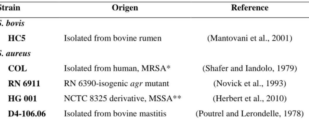
Table 1. Strains used in this study
Strain Origen Reference
S. bovis
HC5 Isolated from bovine rumen (Mantovani et al., 2001)
S. aureus
COL Isolated from human, MRSA* (Shafer and Iandolo, 1979)
RN 6911 RN 6390-isogenic agr mutant (Novick et al., 1993)
HG 001 NCTC 8325 derivative, MSSA** (Herbert et al., 2010)
D4-106.06 Isolated from bovine mastitis (Poutrel and Lerondelle, 1978) *MRSA, methicillin-resistant S. aureus; MSSA**, methicillin-sensitive S. aureus.
2.2 Bacteriocins
Nisin A from L. lactis (2.5% nisin, ≥ 1,000 IU mg-1; Sigma-Aldrich, Germany) was resuspended in sterile sodium phosphate buffer (10 mM, pH 7.2) and stored at 7°C until use.
Extracts of bovicin HC5 were prepared as described by Mantovani et al. (2002). Briefly, stationary-phase S. bovis HC5 were harvested by centrifugation and the cells were washed in sodium phosphate buffer (10 mM, pH 7.2). The cell pellet was re-suspended in acidic NaCl (100 mM, pH 2.0) for 2 h at room temperature. The suspensions were centrifuged to remove cells and the cell-free supernatant was lyophilized. The lyophilized material was suspended in sterile sodium phosphate buffer (10 mM, pH 2.0). Purification of bovicin HC5 was performed by reversed-phase high-performance liquid chromatography (RP-HPLC) using a semi-preparative column (Shimadzu C18, Japan; length, 150 mm; inner diameter, 4.6 mm; particle size, 5 µm). The column was equilibrated with buffer A (0.1% trifluoroacetic acid [TFA] in water), and the peptide was eluted using a linear gradient of 35 to 50% buffer B (80% acetonitrile, 0.1% TFA in water) at 22°C and at a flow rate of 1 ml/min. The absorbance was monitored at 214 and 280 nm, and the eluted fraction corresponding to pure bovicin HC5 was lyophilized (Paiva et al., 2011). Bacteriocin stock solutions were stored at 7°C in sterile sodium phosphate buffer (10 mM, pH 7.2) until use.
was diluted 100 folds with distilled water. To the tubes containing 200 µl of distilled water, 200 µl of the diluted samples and 600 µl of ninhydrin-reagent were added. After incubation at 100ºC for 10 min in the dark, the absorbance was measured at 575 nm. The standard curve was constructed with bovine serum albumin (Sigma-Aldrich, Germany), in a concentration of 0.25-10 µg µl-1 in distilled water.
2.3 Susceptibility to the bacteriocins and adhesion testing
In order to determine the Minimal Inhibitory Concentration (MIC) of bovicin HC5 and nisin, 200 µl of synthetic medium supplemented with increasing bovicin HC5 and nisin concentrations (from 2.0 to 0.2 µM) were transferred to 96-wells microtiter plates, and inoculated with 5 x 10⁵ cfu ml-1 of exponentially growing S. aureus cells (optical density at 500 nm [OD500], 0.5) previously propagated in the same medium
without bacteriocins. The minimal concentration that prevented turbidity of the medium after 18 h incubation at 37ºC was designated as the bovicin HC5 and nisin MIC.
To evaluate the effect of bovicin HC5 and nisin on staphylococcal adhesion, assays were carried out using the same experimental design as previously described for MIC experiments. After 18 h of incubation, the culture supernatant was discarded, and the surface-attached cells were stained with 200 µl of 0.1% (w/v) crystal violet for 30 min. Subsequently, the crystal violet was removed and the plate was washed three times with water. After air drying for 15 min at 40ºC, the attached cells were determined at 590 nm with the microtiter plate reader (Biotek, Germany) by addition of 200 µl of 95% (v/v) ethanol.
MIC and adhesion experiments were conducted in three biological replicates and three technical replicates. Data were expressed as the ratio between the absorbance of violet crystal extract (adhered cells) and the optical density of total cells (planktonic and adhered cells) (Viana et al., 2009).
2.4 Contact angle measurement
2.4.1 Surface
The contact angles between the surface and the ultra-pure water, formamide (LGC Bio, Brazil) and α-bromonaphtalene (Merck, Germany) were determined using a goniometer (Kruss, Germany). Contact angle measurements of 2.0 µl drop were taken each second for 30 s for all liquids. Experiments were conducted in three biological replicates and three technical replicates.
2.4.2 Microorganism
Contact angles of S. aureus COL surface were determined on a layer of vegetative cells (Busscher et al., 1984). First, pre-warmed medium at 37ºC was inoculated with cells from an overnight culture to an initial OD500 of 0.1 and monitored
by measuring the OD until the culture reached an OD500 of 0.5. At that time, the culture
was exposed to 0.4 µM of bovicin HC5 or nisin followed by statically incubation at 37ºC for 18 h. Growth in medium without bacteriocins was performed as a control.
Later, the suspension was centrifuged at 4,000 x g at 4°C for 10 min and then washed three times in 0.1 M phosphate-buffered saline (PBS). The pellet was resuspended in the same buffer and then filtered using acetate cellulose membrane (0.45 μm pore size, 47 mm diameter). During the filtration, γ0 ml of ultra-pure water was added.
To standardize the moisture content, the membranes were transferred into Petri dishes containing 1% (w/v) agar and 10% (v/v) glycerol. The membranes were cut to determine the contact angle with the three different polarities liquids.
2.4.3 Determination of the total interfacial tension (γs tot)
The total interfacial tension was determined by the sum of the apolar and polar components of the respective surfaces (Equation 1):
(1)
where l TOT is the total interfacial tension of the liquid; LW is the interfacial
tension of the interactions of the Lifshitz-van der Waals forces; + is the interfacial tension of the electron acceptor component of the acid-base component; – is the interfacial tension of the electron donor component of the acid-base component, θ is the contact angle, and s and l indicate surface and liquid, respectively (Van Oss and Giese, 1995).
l s l
s LW
l LW s TOT
l
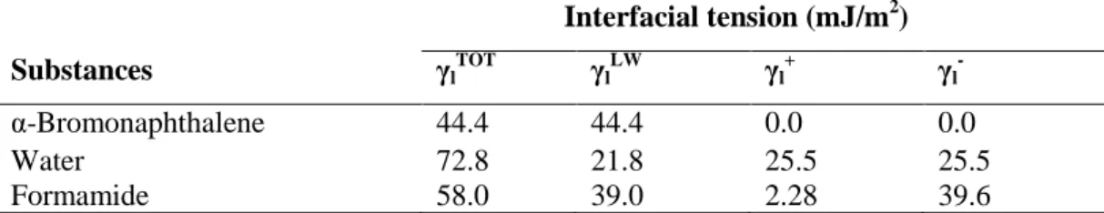
The three components of the interfacial tension of the surfaces were determined from the contact angles obtained from three liquids with different polarities, whose interfacial tensions are known, as shown in Table 2.
Table 2. Components of the interfacial tensions of the substances at 25ºC
Interfacial tension (mJ/m2)
Substances γl
TOT γ l LW γ l + γ l
-α-Bromonaphthalene 44.4 44.4 0.0 0.0
Water 72.8 21.8 25.5 25.5
Formamide 58.0 39.0 2.28 39.6
The interfacial tension is the result of the sum of the two components ( sLW and sAB):
(2) (3) (4)
where sLW is the interfacial tension of the interactions of the Lifshitz-van der
Waals forces; θB is the contact angle obtained with α-bromonaphthalene; sAB is the
polar component of the Lewis acid-base interaction; s+ is the interfacial tension of the
electron acceptor component of the acid-base component; s– is the interfacial tension of
the electron donor component of the acid-base component and stot is the total interfacial
tension of the surface.
2.4.4 Free energy of interaction (∆GswsTOT)
The total free energy of interaction among molecules of the surface (s) immersed in water (w) was determined by the sum of the apolar and polar free energies of interaction, ∆GswsLW and ∆GswsAB, respectively:
(5) (6) (7)
2cos 1 1 . 11 B LW s
s s
AB
s
2 AB s LW s tot
s
AB sws LW sws tot
sws G G
G
LW w LW s LW sws
G
2.
s s w w s w w s
AB sws
2.4.5 Determination of the total free energy of adhesion (∆Gadhesion)
Using the values of the components of the interfacial tensions, it is possible to determine the ∆Gadhesion between two surfaces (microbial cells (b) and polystyrene
surface (s)):
(8) (9) (10)
When free energy is related to the interfacial tension, ∆Gadhesion can then be
represented by the following:
(11) (12) (13)
where bs is the interfacial tension between the bacterial surfaces and the
adhesion surface; bl is the interfacial tension between the bacterial surfaces and the
liquid; and sl is the interfacial tension between the adhesion surfaces and the liquid.
The ∆Gadhesion values allow for evaluation of the thermodynamics of the adhesion
process: if ∆Gadhesion< 0, the process is favorable; if ∆Gadhesion > 0, the process is
unfavorable.
2.5. Impact of bovicin HC5 and nisin on biofilm-related gene expression in S. aureus
2.5.1 Sample preparation and total RNA extraction
Expression of genes related to adhesion and biofilm formation icaD, fnbA, clfB, and rnaIII was evaluated after exponentially growing cells of S. aureus COL (OD500
0.5) have been exposed to 0.4 µM bovicin HC5 or nisin in synthetic medium for 18 h, statically at 37ºC. Untreated cells were also evaluated after 18 h of growth in synthetic medium.
AB bs LW bs
bs
LW s LW b LW s LW b LWbs
2
b b s s b s b sAB
bs
2
AB bls LW
bls
adhesion
G
G
G
LW sl LW bl LW bs LW blsG
AB sl AB bl AB bs AB bls
G
Bacterial cells were collected by centrifugation for 5 min at 6,000 x g. Total RNA was obtained by phenol-chloroform extraction (TRIzol®, Invitrogen, USA) according to the manufacturer’s instructions after a 40 min pre-treatment of the cells with 0.8 µg/µl of lysozyme and 0.00002 U/µl of lysostaphin in 300 µl of TE buffer. Purified RNA was eluted in 30 µl of UltrapureTM distilled water RNase/DNase-free (Invitrogen, USA) and stored at -20ºC. The concentration and purity of the RNA was evaluated by spectrometry at 260 nm and 280 nm. The integrity of the RNA was confirmed by 1% (w/v) agarose gel electrophoresis.
2.5.2 cDNA synthesis and RT-qPCR
Aliquots of 2 µg of total RNA were treated with RQ1 RNase-Free DNase (Promega, USA) for DNA elimination according to the manufacturer’s instructions. cDNA synthesis was performed using Improm-II Reverse Transcription System kit (Promega, USA) following the manufacturer’s recommendations. The mixture was incubated at 25ºC for 5 min, 42ºC for 2 h and 70ºC for 15 min.
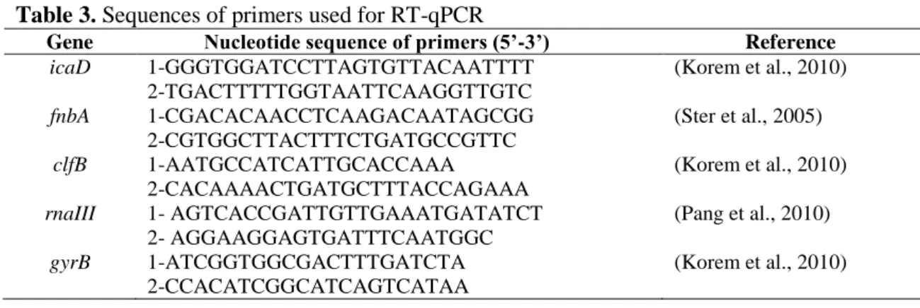
The relative mRNA levels of biofilm-related genes were analyzed by quantitative real-time polymerase chain reaction (RT-qPCR) using SYBR® Green PCR-Master Mix (Applied Biosystems, USA) in 25 µl reaction volumes containing 1 µl of cDNA template, 1 µl of each primer at 0.5 µM (Table 3), 12.5 µl of SYBR® Green PCR-Master Mix and 9.5 µl of UltrapureTM distilled water RNase/DNase-free and the following conditions: 50ºC for 2 min, 95ºC for 10 min, 40 cycles of 95ºC for 15 s and 60ºC for 1 min. Abundance of each specific mRNA was calculated relative to the expression of the housekeeping gene DNA gyrase, B subunit (gyrB) based on 2-ΔΔCt method (Livak and Schmittgen, 2001). The amplifications were performed in 96-well PCR plates using CFX96 TouchTM Real-Time PCR Detection System (Bio-Rad, USA). The fluorescence data for each sample were converted to Cycle threshold (Ct) values using the software Bio-Rad CFX Manager 2.0.
Table 3. Sequences of primers used for RT-qPCR
Gene Nucleotide sequence of primers (5’-3’) Reference
icaD 1-GGGTGGATCCTTAGTGTTACAATTTT
2-TGACTTTTTGGTAATTCAAGGTTGTC
(Korem et al., 2010)
fnbA 1-CGACACAACCTCAAGACAATAGCGG
2-CGTGGCTTACTTTCTGATGCCGTTC
(Ster et al., 2005)
clfB 1-AATGCCATCATTGCACCAAA
2-CACAAAACTGATGCTTTACCAGAAA
(Korem et al., 2010)
rnaIII 1- AGTCACCGATTGTTGAAATGATATCT 2- AGGAAGGAGTGATTTCAATGGC
(Pang et al., 2010)
gyrB 1-ATCGGTGGCGACTTTGATCTA
2-CCACATCGGCATCAGTCATAA
(Korem et al., 2010)
3. RESULTS
3.1 Inhibition of S. aureus by bovicin HC5 and nisin
The MICs of the bacteriocins bovicin HC5 and nisin on four different strains of S. aureus were investigated in synthetic medium and the results are shown in the Table 4. The MIC values average for bovicin HC5 and nisin was 1.05 ± 0.90 and 0.90 ± 0.26, respectively.
Table 4. Minimal Inhibitory Concentration of bovicin HC5 and nisin on S. aureus strains
Bacteriocins (µM)
S. aureus strains Bovicin HC5 Nisin
COL 1.2 1.0
RN 6911 1.2 1.2
HG 001 0.8 0.6
D4-106.06 1.0 0.8
Subinhibitory dosages of bovicin HC5 and nisin were tested for their capacity to inhibit adhesion of S. aureus in polystyrene 96-well microtiter plates after 18 h of incubation using crystal violet method. Adhesion was estimated by calculating the ratio between the absorbance of violet crystal extract and the optical density of total cells. The presence of the bacteriocins clearly reduced adhesion of the strains COL and RN 6911 to polystyrene surface (Figure 1), and the effect of bovicin HC5 and nisin seemed to be similar for COL strain. While only bovicin HC5 was able to reduce adhesion of S. aureus HG 001, only nisin at the highest concentration evaluated reduced adhesion of S. aureus D4-106.06 to polystyrene.
Figure 1. Ratio between the absorbance of violet crystal extract (AVCE) and the optical density
of total cells (OD TC) of S. aureus following bovicin HC5 or nisin treatment. S. aureus COL,
RN 6911, HG001, and D4-106.06 were cultivated in synthetic medium for 18h in the presence
of different concentration of bovicin HC5 (black bars) and nisin (grey bars). Untreated control
(white bars) is shown.
3.2 Contact angle and total free energy of interaction (∆GswsTOT)
As we observed that subinhibitory dosages of bovicin HC5 and nisin were more effective on adhesion reduction than growth reduction, COL strain was selected to investigate if the presence of bovicin HC5 and nisin in low concentration could change the hydrophobicity of the bacterial surface and also the polystyrene surface and thus, interfere with the adhesion process.
The measurement of the water contact angle (ΘW) with the polystyrene surface exposed for 18 h to synthetic medium without bacteriocins was greater than 65º, indicating a hydrophobic profile according to the classification system (Vogler, 1998). The presence of the bacteriocins in the synthetic medium drastically changed the hydrophobicity of the polystyrene surface, since ΘW with the surface was much lower than 65º (Table 5). S. aureus COL cellular surface, hydrophilic in the absence of
COL
Bacteriocins ( M)
0 0.6 0.8
AV CE / O D TC 0 1 2 3 4
5 RN 6911
Bacteriocins ( M)
0 0.6 0.8
AV CE / O D TC 0 1 2 3 4 5 HG 001
Bacteriocins ( M)
0 0.2 0.4
AV CE / O D TC 0 1 2 3 4 5 D4-106.06
Bacteriocins ( M)
0 0.2 0.4
bacteriocins, remained hydrophilic with a small decrease in the contact angle when bacteria grew in the presence of 0.4 µM of bovicin HC5 or nisin.
The ∆GswsTOT values are considered a quantitative criterion for hydrophobicity
evaluation. According to the calculations, polystyrene surface treated with synthetic medium without bacteriocins was classified as hydrophobic (∆GswsTOT < 0) (Table 5).
The presence of bovicin HC5 and nisin decreased the hydrophobicity of the polystyrene surface. The bacterial surface, hydrophilic, remained hydrophilic (∆GswsTOT > 0) even
after 18 h of exposition to the bacteriocins (Table 5).
Table 5. Averages and standard deviation of contact angles measurements with water (ΘW),
formamid (ΘF) and α-bromonaphtalene (ΘB), and total free energy of interaction (∆Gsws TOT
) of
S. aureus COL and polystyrene surface (PS) treated with bovicin HC5 and nisin
Contact angles (º)a ∆GswsTOT
Surface/Bacteria ΘW ΘF ΘB (mJ/m
2)
PS + Synthetic medium 82.4 + 4.4 66.2 + 5.5 22.9 + 1.4 -63.7 PS + Synthetic medium + 0.4 µ M bovicin HC5 33.4 + 7.9 30.2 + 2.9 32.7 + 4.8 22.4 PS + Synthetic medium + 0.4 µ M nisin 40.5 + 1.3 37.0 + 0.8 40.9 + 2.7 18.1
S. aureus + Synthetic medium 25.3 + 2.9 17.7 + 0.3 45.0 + 1.5 20.3
S. aureus + Synthetic medium + 0.4 µM bovicin HC5 21.9 + 3.1 16.6 + 0.7 45.9 + 1.9 22.8
S. aureus + Synthetic medium + 0.4 µM nisin 23.5 + 1.1 25.3 + 3.8 45.8 + 2.3 28.4 aAverage of three repetitions.
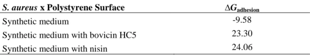
3.2.1 Free energy adhesion (∆Gadhesion)
According to thermodynamic theory, adhesion is considered favorable only if the process results in a decrease in total free energy. Thus, the adhesion process was thermodynamically favorable (∆Gadhesion <0) only when the synthetic medium in contact
with the polystyrene surface contained no bacteriocins (Table 6). In the presence of bovicin HC5 or nisin, the adhesion process was considered thermodynamically unfavorable (∆Gadhesion>0) (Table 6), confirming the previously results that both
bacteriocins decreased the adhesion of S. aureus COL.
Table 6. Free energy of adhesion (mJ/m2) between S. aureus COL and polystyrene surface
treated with bovicin HC5 and nisin
S. aureus x Polystyrene Surface ∆Gadhesion
Synthetic medium -9.58
Synthetic medium with bovicin HC5 23.30
3.3 Effect of bovicin HC5 and nisin on the expression of biofilm-related genes in S. aureus
After verifying that subinhibitory dosages of bovicin HC5 and nisin reduced the adhesion of S. aureus COL to polystyrene microtiter plates been confirmed by decreasing of the hydrophobicity, we investigated the effect of the bacteriocins on the expression of some selected genes related to adhesion and biofilm formation (icaD, fnbA and clfB) and also on the rnaIII involved in the regulation by quorum sensing.
Compared to the control without bacteriocins, low concentrations of bovicin HC5 and nisin increased by 2.62 and 5.79 fold, respectively, the expression of icaD (Figure 2), one of the most studied genes related to biofilm formation by staphylococci. Nisin exert greater influence on increasing icaD expression. Bovicin HC5 and nisin were also able to increase the expression of fnbA, fibronectin A, in 1.34 and 1.94 fold, respectively. Again, the effect of nisin was more pronounced than bovicin HC5. The transcription of clfB, clumping factor B, did not change when S. aureus COL grew for 18 h in the presence of 0.4 µM of nisin. However, bovicin HC5 decreased clfB transcription (Figure 2).
The exposition of S. aureus COL for 18 h to subinhibitory concentration of bovicin HC5 did not cause significant changes in the transcription profile of rnaIII, main effector of agr quorum sensing system in S. aureus. The presence of nisin in the growth medium enhanced 1.64 fold the expression of rnaIII.
Figure 2. Comparison of the expression profiles of selected biofilm formation-related genes
(icaD, fnbA and clfB) and quorum sensing-related (rnaIII) in S. aureus COL. Cells were grown
in synthetic medium and total RNA was extracted from the cells after 18 h of exposition to 0.4
µM of bovicin HC5 (black bars) and nisin (white bars) and the expression profile was analyzed
by RT-qPCR. Untreated control (white bars) is shown.
icaD fnbA clfB rnaIII
Rela
tiv
e
exp
ress
io
n
/
g
yrB
4. DISCUSSION
For long time, S. aureus has caused problems in health care. In order to control S. aureus growth and biofilm formation various natural substances have been tested. In this work, bovicin HC5 and nisin were tested against different strains of S. aureus aiming to determine MIC and also the effects of subinhibitory concentrations on cell adhesion to polystyrene surface. Bovicin HC5 and nisin were effective against all strains of S. aureus tested in synthetic medium and similar inhibitory effect was observed for both bacteriocins. The minimum concentrations of bovicin HC5 and nisin able to inhibit the growth of all tested strains was 1.4 and 1.2 µM, respectively. Recently, our group reported that the bacteriocins bovicin HC5 and nisin were effective to inhibit S. aureus growth in milk (Pimentel-Filho et al., 2013), and in fresh cheese (Pimentel-Filho et al., 2014). These results reinforce the idea that not only nisin, already authorized as a food additive in many countries, but also bovicin HC5 has potential application as a natural food preservative. Other bacteriocins such as enterocin AS-48 (Ananou et al., 2004; Ananou et al., 2010), reuterin (Arques et al., 2008), epidermicin NI01 (Okuda et al., 2013), and lacticin Q (Sandiford and Upton, 2012) have been tested and were effective against S. aureus, even those called drug or multidrug-resistant.
Development of technologies to control microbial adhesion and biofilm formation is desired for food industries. Our study revealed that subinhibitory dosages of bovicin HC5 and nisin reduced the adhesion of the pathogen to the polystyrene
frequently used in trays, utensils and food packing. Low dosages of nisin as 25 IU/ml
were able to reduce staphylococcal biofilm on polypropylene coupons and were even
more effective against planktonic cell of S. aureus (Cabo et al., 2009). Bacteriocin-like substances produced by lactobacilli suppressed biofilm formation on polystyrene surface by S. aureus and S. epidermidis and induced ultrastructural changes leading to their destruction (Sadowska et al., 2010). Davison et al. (2010) verified that nisin accessed the interior of biofilm cells clusters of S. epidermidis on glass faster than other antimicrobial agents as chlorine and glutaraldehyde resulting in a rapid and uniform loss of green fluorescence, indicating cell die, without any removal of biofilm. All these results reinforce the idea that bacteriocins can be a potential strategy to prevent adhesion and to control biofilm formation.
values, a qualitative criterion, we observed that cell surface of S. aureus COL growing in synthetic medium with or without bovicin HC5 and nisin was hydrophilic. Our result was in agreement with reports which found that the S. aureus surface is hydrophilic (Hamadi et al., 2005; Kouidhi et al., 2010). Polystyrene surface conditioned for 18 h with synthetic medium was hydrophobic being considered favorable for bacterial adhesion. Evaluating the hydrophobicity of polystyrene surface, Biazar et al. (2011) found a contact angle of 90.1º confirming its hydrophobic character. However, after 18 h of conditioning with synthetic medium added of bovicin HC5 or nisin, the polystyrene surface became hydrophilic (ΘW < 65º). Our data showed the decrease in the hydrophobicity on treated surfaces indicates one of the reasons for bacterial attachment decrease. Hydrophobicity is an important propriety and represents the wettability of a surface and, in aqueous medium, adhesion is favored between hydrophobic surfaces, which can enter in closer contact by squeezing the water layer between them (Teixeira et al., 2005). After conditioning with surfactant, the polystyrene surface became more hydrophilic (Zeraik and Nitschke, 2010). It has been shown, that Salmonella and Listeria preferably adhere in a higher numbers to hydrophobic surfaces than the hydrophilic ones (Donlan and Costerton, 2002; Sinde and Carballo, 2000).
In order to predict the ability of the pathogen to adhere to those treated or untreated polystyrene surfaces, and further to form biofilm, the free energy of interaction between the micro-organisms and the surface, when immersed in the same synthetic medium condition with or without bacteriocin, was calculated. Only in the medium without bacteriocins the adhesion process was thermodynamically favorable. Since the total free energy of adhesion was positive, in the medium containing bacteriocins, the adhesion process was considered unfavorable. Teixeira et al. (2005) also demonstrated that adhesion of some isolates of Pseudomonas aeruginosa and Staphylococcus sciuri was thermodynamically favorable to stainless steel and rubber, which were considered hydrophobic and unfavorable to glass and polymethylacrylate surfaces, classified as hydrophilic.
understood mechanism of biofilm development, making the ica genes a potential target for biofilm inhibitors (Oduwole et al., 2010). Similar effect was observed by Nuryastuti et al. (2009) when evaluating subinhibitory concentration of cinnamon oil on the expression of icaA in S. epidermidis, another important gene of icaADBC operon evaluated in studies involving biofilm formation by staphylococcal species. They found that even reducing biofilm formation on polystyrene surface, 0.01% of cinnamon oil enhanced icaA expression (Nuryastuti et al., 2009). On the other hand, low dosages of povidone-iodine, a complex of polyvinyl pyrrilidine and triiodine ions widely used as an antiseptic in trauma and orthopaedic surgery, descreased icaA expression and, thus, the biofilm forming capacity of S. aureus RN4220 (Oduwole et al., 2010). According to the reports in the literature, the expression of the ica genes is highly variable and can be induced by variations in the culture conditions, such as an increase in the concentration of sugars or other substances that induce stress (Cho et al., 2002; Oliveira and de Lourdes Cunha, 2010).
Bovicin HC5 and nisin were also able to up-regulate fnbA, meanwhile bovicin HC5 slightly reduced clfB. As found by us, Rasigade et al. (2011) reported that although subinhibitory concentration of rifampicin reduced bacterial adhesion to human
fibronectin, the antibiotic did not affect fnbA/B transcription by five S. aureus strains.
However, low dosages oxacillin, moxifloxacin and linezolid led to the development of a hyper-adhesive phenotype in the fibronectin adhesion assay, increasing also fnbA/B expression (Rasigade et al., 2011). Sublethal dosages of tigecycline were investigated on biofilm formation by 16 methicilin-resistant S. aureus isolates and the transcriptome analysis revealed that the antibiotic was able to reduced expression of icaC, otherwise upregulation of fnbA and clfB, which encode adhesins which attach to human proteins, was observed (Smith et al., 2010).
Biofilm formation can be induced by conditions that are potentially toxic for bacterial cells, such as high levels of osmolarity, detergents, urea, ethanol, oxidative stress, and the presence of sub-MICs of some antibiotics (Nuryastuti et al., 2009).
To our knowledge, this is the first study reporting that bacteriocins act changing the hydrophobicity of polystyrene surfaces. It seems that even prepared to adhere, since biofilm appendages such as IcaD and FnbA are already synthesized by planktonic cells, if the free energy of adhesion is not favorable it is difficult to the bacteria to get in close contact with the surface. This is a very interesting finding since to prevent microbial adhesion to food contact surfaces is much more effective than to remove biofilm already established. Thus, bovicin HC5 and nisin appear as a potential alternative to inhibit the initial step of biofilm formation.
ACKNOWLEDGMENTS
N.J.P.F. was supported by a fellowship (8974/11-0) from Coordenação de Aperfeiçoamento de Pessoal de Nível Superior (CAPES, Brasília, Brazil).
We thank Kathrin Riedel and Stephan Fuchs, from University of Greifswald, for important suggestions and detailed criticism of the manuscript.
REFERENCES
Ananou, S., Baños, A., Maqueda, M., Martínez-Bueno, M., Gálvez, A., Valdivia, E., 2010. Effect of combined physico-chemical treatments based on enterocin AS-48 on the control of Listeria monocytogenes and Staphylococcus aureus in a model cooked ham. Food Control 21, 478-486.
Ananou, S., Valdivia, E., Marínez Bueno, M., Gálvez, A., Maqueda, M., 2004. Effect of combined physico-chemical preservatives on enterocin AS-48 activity against the enterotoxigenic Staphylococcus aureus CECT 976 strain. Journal of Applied Microbiology 97, 48-56.
Antolinos, V., Munoz, M., Ros-Chumillas, M., Aznar, A., Periago, P.M., Fernandez, P.S., 2011. Combined effect of lysozyme and nisin at different incubation temperature and mild heat treatment on the probability of time to growth of Bacillus cereus. Food Microbiology 28, 305-310.
processing surfaces evaluated by the hydrophobicity. International Journal of Food Science & Technology 44, 2519-2525.
Arciola, C.R., Campoccia, D., Baldassarri, L., Donati, M.E., Pirini, V., Gamberini, S., Montanaro, L., 2006. Detection of biofilm formation in Staphylococcus epidermidis from implant infections. Comparison of a PCR-method that recognizes the presence of ica genes with two classic phenotypic methods. Journal of Biomedical Materials
Research Part A 76, 425-430.
Argudín, M.Á., Mendoza, M.C., Rodicio, M.R., 2010. Food poisoning and Staphylococcus aureus enterotoxins. Toxins 2, 1751-1773.
Arques, J.L., Rodriguez, E., Nunez, M., Medina, M., 2008. Antimicrobial activity of nisin, reuterin, and the lactoperoxidase system on Listeria monocytogenes and Staphylococcus aureus in cuajada, a semisolid dairy product manufactured in Spain. Journal of Dairy Science 91, 70-75.
Atshan, S.S., Shamsudin, M.N., Karunanidhi, A., Belkum, A.v., Lung, L.T.T., Sekawi, Z., Nathan, J.J., Ling, K.H., Seng, J.S.C., Ali, A.M., 2013. Quantitative PCR analysis of genes expressed during biofilm development of methicillin resistant Staphylococcus aureus (MRSA). Infection, Genetics and Evolution 18, 106-112.
Biazar, E., Heidari, M., Asefnezhad, A., Montazeri, N., 2011. The relationship between cellular adhesion and surface roughness in polystyrene modified by microwave plasma radiation. International Journal of Nanomedicine 6, 631-639.
Bryers, J.D., Ratner, B.D., 2004. Bioinspired implant materials befuddle bacteria. ASM News-American Society for Microbiology 70, 232-232.
Busscher, H.J., Weerkamp, A.H., Van der Mei, H.C., Van Pelt, A.W., De Jong, H.P., Arends, J., 1984. Measurement of the surface free energy of bacterial cell surfaces and its relevance for adhesion. Applied and Environmental Microbiology 48, 980-983.
Caballero Goméz, N., Abriouel, H., Grande, M.J., Pérez Pulido, R., Gálvez, A., 2013. Combined treatments of enterocin AS-48 with biocides to improve the inactivation of methicillin-sensitive and methicillin-resistant Staphylococcus aureus planktonic and sessile cells. International Journal of Food Microbiology 163, 96-100.
Cabo, M.L., Herrera, J.J., Crespo, M.D., Pastoriza, L., 2009. Comparison among the effectiveness of ozone, nisin and benzalkonium chloride for the elimination of planktonic cells and biofilms of Staphylococcus aureus CECT4459 on polypropylene. Food Control 20, 521-525.
Cho, S.-H., Naber, K., Hacker, J., Ziebuhr, W., 2002. Detection of the icaADBC gene cluster and biofilm formation in Staphylococcus epidermidis isolates from catheter-related urinary tract infections. International Journal of Antimicrobial Agents 19, 570-575.
Chung, E., Yiacoumi, S., Tsouris, C., 2014. Interaction forces between spores and planar surfaces in aqueous solutions. Colloids and Surfaces A: Physicochemical and Engineering Aspects 443, 80-87.
Davison, W.M., Pitts, B., Stewart, P.S., 2010. Spatial and temporal patterns of biocide action against Staphylococcus epidermidis biofilms. Antimicrobial Agents and Chemotherapy 54, 2920-2927.
Donlan, R.M., Costerton, J.W., 2002. Biofilms: survival mechanisms of clinically relevant microorganisms. Clinical Microbiology Reviews 15, 167-193.
Ferreira, C., Pereira, A.M., Melo, L.F., Simões, M., 2010. Advances in industrial biofilm control with micro-nanotechnology. Current Research, Technology and Education Topics in Applied Microbiology and Microbial Biotechnology 2, 845-854.
Gerke, C., Kraft, A., Süßmuth, R., Schweitzer, O., Götz, F., 1998. Characterization of the N-Acetylglucosaminyltransferase activity involved in the biosynthesis of the Staphylococcus epidermidis polysaccharide intercellular adhesin. Journal of Biological
Chemistry 273, 18586-18593.
Gertz, S., Engelmann, S., Schmid, R., Ohlsen, K., Hacker, J., Hecker, M., 1999. Regulation of sigmaB-dependent transcription of sigB and asp23 in two different Staphylococcus aureus strains. Molecular and General Genetics 261, 558-566.
Hamadi, F., Latrache, H., Mabrrouki, M., Elghmari, A., Outzourhit, A., Ellouali, M., Chtaini, A., 2005. Effect of pH on distribution and adhesion of Staphylococcus aureus to glass. Journal of Adhesion Science and Technology 19, 73-85.
Herbert, S., Ziebandt, A.K., Ohlsen, K., Schafer, T., Hecker, M., Albrecht, D., Novick, R., Gotz, F., 2010. Repair of global regulators in Staphylococcus aureus 8325 and comparative analysis with other clinical isolates. Infection and Immunity 78, 2877-2889.
Herrera, J.J.R., Cabo, M.L., Gonzalez, A., Pazos, I., Pastoriza, L., 2007. Adhesion and detachment kinetics of several strains of Staphylococcus aureus subsp. aureus under three different experimental conditions. Food Microbiology 24, 585-591.
Hori, K., Matsumoto, S., 2010. Bacterial adhesion: From mechanism to control. Biochemical Engineering Journal 48, 424-434.
Korem, M., Gov, Y., Rosenberg, M., 2010. Global gene expression in Staphylococcus aureus following exposure to alcohol. Microbial Pathogenesis 48, 74-84.
Le Loir, Y., Baron, F., Gautier, M., 2003. Staphylococcus aureus and food poisoning. Genetics and Molecular Research 2, 63-76.
Livak, K.J., Schmittgen, T.D., 2001. Analysis of relative gene expression data using real-time quantitative PCR and the 2−ΔΔCT Method. Methods 25, 402-408.
Mahalik, N.P., Nambiar, A.N., 2010. Trends in food packaging and manufacturing systems and technology. Trends in Food Science & Technology 21, 117-128.
Mantovani, H.C., Hu, H., Worobo, R.W., Russell, J.B., 2002. Bovicin HC5, a bacteriocin from Streptococcus bovis HC5. Microbiology 148, 3347-3352.
Mantovani, H.C., Kam, D.K., Ha, J.K., Russell, J.B., 2001. The antibacterial activity and sensitivity of Streptococcus bovis strains isolated from the rumen of cattle. FEMS Microbiology Ecology 37, 223-229.
Mantovani, H.C., Russell, J.B., 2003. Inhibition of Listeria monocytogenes by bovicin HC5, a bacteriocin produced by Streptococcus bovis HC5. International Journal of Food Microbiology 89, 77-83.
Nostro, A., Scaffaro, R., Ginestra, G., D'Arrigo, M., Botta, L., Marino, A., Bisignano, G., 2010. Control of biofilm formation by poly-ethylene-co-vinyl acetate films incorporating nisin. Applied Microbiology and Biotechnology 87, 729-737.
Novick, R.P., Geisinger, E., 2008. Quorum sensing in staphylococci. Annual Review of Genetics 42, 541-564.
Novick, R.P., Ross, H.F., Projan, S.J., Kornblum, J., Kreiswirth, B., Moghazeh, S., 1993. Synthesis of staphylococcal virulence factors is controlled by a regulatory RNA molecule. EMBO Journal 12, 3967-3975.
Oduwole, K.O., Glynn, A.A., Molony, D.C., Murray, D., Rowe, S., Holland, L.M., McCormack, D.J., O'Gara, J.P., 2010. Anti-biofilm activity of subinhibitory povidone-iodine concentrations against Staphylococcus epidermidis and Staphylococcus aureus. Journal of Orthopaedic Research 28, 1252-1256.
Okuda, K.-i., Zendo, T., Sugimoto, S., Iwase, T., Tajima, A., Yamada, S., Sonomoto, K., Mizunoe, Y., 2013. Effects of bacteriocins on methicillin-resistant Staphylococcus aureus biofilm. Antimicrobial Agents and Chemotherapy 57, 5572-5579.
Oliveira, A., de Lourdes Cunha, M., 2010. Comparison of methods for the detection of biofilm production in coagulase-negative staphylococci. BMC Research Notes 3, 260.
Paiva, A.D., Breukink, E., Mantovani, H.C., 2011. Role of lipid II and membrane thickness in the mechanism of action of the lantibiotic bovicin HC5. Antimicrobial Agents and Chemotherapy 55, 5284-5293.
Palmer, J., Flint, S., Brooks, J., 2007. Bacterial cell attachment, the beginning of a biofilm. Journal of Industrial Microbiology & Biotechnology 34, 577-588.
Pang, Y.Y., Schwartz, J., Thoendel, M., Ackermann, L.W., Horswill, A.R., Nauseef, W.M., 2010. agr-Dependent interactions of Staphylococcus aureus USA300 with human polymorphonuclear neutrophils. Journal of Innate Immunity 2, 546-559.
Parsek, M.R., Fuqua, C., 2004. Biofilms 2003: emerging themes and challenges in studies of surface-associated microbial life. Journal of Bacteriology 186, 4427-4440.
Pimentel-Filho, N.J., Mantovani, H.C., Carvalho, A.F., Dias, R.S., Vanetti, M.C.D., 2014. Efficacy of bovicin HC5 and nisin combination against Listeria monocytogenes and Staphylococcus aureus in fresh cheese. International Journal of Food Science & Technology 49, 416-422.
Poutrel, B., Lerondelle, C., 1978. Induced staphylococcal infections in the bovine mammary gland. Influence of the month of lactation and other factors related to the cow. Annals of Research Veterinary 9, 119-128.
Rasigade, J., Moulay, A., Lhoste, Y., Tristan, A., Bes, M., Vandenesch, F.o., Etienne, J., Lina, G., Laurent, F., Dumitrescu, O., 2011. Impact of subinhibitory antibiotics on fibronectin-mediated host cell adhesion and invasion by Staphylococcus aureus. BMC Microbiology 11, 263-271.
Rohde, H., Frankenberger, S., Zähringer, U., Mack, D., 2010. Structure, function and contribution of polysaccharide intercellular adhesin (PIA) to Staphylococcus epidermidis biofilm formation and pathogenesis of biomaterial-associated infections. European Journal of Cell Biology 89, 103-111.
Sadowska, B., Walencka, E., Wieckowska-Szakiel, M., Rozalska, B., 2010. Bacteria competing with the adhesion and biofilm formation by Staphylococcus aureus. Folia Microbiologica 55, 497-501.
Sandiford, S., Upton, M., 2012. Identification, characterization, and recombinant expression of epidermicin NI01, a novel unmodified bacteriocin produced by Staphylococcus epidermidis that displays potent activity against staphylococci. Antimicrobial Agents and Chemotherapy 56, 1539-1547.
Shafer, W.M., Iandolo, J.J., 1979. Genetics of staphylococcal enterotoxin B in methicillin-resistant isolates of Staphylococcus aureus. Infection and Immunity 25, 902-911.
Simões, M., Simões, L.C., Vieira, M.J., 2010. A review of current and emergent biofilm control strategies. LWT-Food Science and Technology 43, 573-583.
Smith, K., Gould, K.A., Ramage, G., Gemmell, C.G., Hinds, J., Lang, S., 2010. Influence of tigecycline on expression of virulence factors in biofilm-associated cells of methicillin-resistant Staphylococcus aureus. Antimicrobial Agents and Chemotherapy 54, 380-387.
Solomakos, N., Govaris, A., Koidis, P., Botsoglou, N., 2008. The antimicrobial effect of thyme essential oil, nisin, and their combination against Listeria monocytogenes in minced beef during refrigerated storage. Food Microbiology 25, 120-127.
Starcher, B., 2001. A ninhydrin-based assay to quantitate the total protein content of tissue samples. Analytical Biochemistry 292, 125-129.
Ster, C., Gilbert, F.B., Cochard, T., Poutrel, B., 2005. Transcriptional profiles of regulatory and virulence factors of Staphylococcus aureus of bovine origin: oxygen impact and strain-to-strain variations. Molecular and Cellular Probes 19, 227-235.
Teixeira, P., Lopes, Z., Azeredo, J., Oliveira, R., Vieira, M.J., 2005. Physico-chemical surface characterization of a bacterial population isolated from a milking machine. Food Microbiology 22, 247-251.
Udompijitkul, P., Paredes-Sabja, D., Sarker, M.R., 2011. Inhibitory effects of nisin against Clostridium perfringens food poisoning and nonfood-borne isolates. Journal of Food Science 77, M51-M56.
Van Oss, C.J., Giese, R.F., 1995. The hydrophilicity and hydrophobicity of clay minerals. Clays and Clay Minerals 43, 474-477.
Vázquez-Sánchez, D., Habimana, O., Holck, A., 2013. Impact of food-related environmental factors on the adherence and biofilm formation of natural Staphylococcus aureus isolates. Current Microbiology 66, 110-121.
Vogler, E.A., 1998. Structure and reactivity of water at biomaterial surfaces. Advances in Colloid and Interface Science 74, 69-117.
Winkelströter, L.K., Gomes, B.C., Thomaz, M.R.S., Souza, V.M., De Martinis, E.C.P., 2011. Lactobacillus sakei 1 and its bacteriocin influence adhesion of Listeria monocytogenes on stainless steel surface. Food Control 22, 1404-1407.
Xue, T., You, Y., Shang, F., Sun, B., 2012. Rot and Agr system modulate fibrinogen-binding ability mainly by regulating clfB expression in Staphylococcus aureus NCTC8325. Medical Microbiology and Immunology 201, 81-92.
Yarwood, J.M., Bartels, D.J., Volper, E.M., Greenberg, E.P., 2004. Quorum sensing in Staphylococcus aureus biofilms. Journal of Bacteriology 186, 1838-1850.
CHAPTER 2
PROTEIN EXPRESSION PROFILING OF Staphylococcus aureus IN RESPONSE TO BOVICIN HC5
1. INTRODUCTION
Staphylococcus aureus is an opportunistic pathogen very often multidrug-resistant involved in both hospital and community-associated diseases worldwide (Rivera et al., 2012). S. aureus causes an array of infectious syndromes, ranging from localized skin lesions and tissue damage to systemic infections, such as pneumonia, endocarditis, and exotoxin syndromes (Lowy, 1998). This diversity of S. aureus-associated diseases results from its ability to adapt efficiently to varying environmental conditions, including the production of several secreted and cell wall-associated virulence factors (Majerczyk et al., 2008). Because of the emerging resistance to various antimicrobial agents, S. aureus have become ever more difficult to control.
The development of new therapeutic strategies to overcome S. aureus infections is thus the ambitious goal for the near future (Kohler et al., 2008). One option that cannot be ignored is a group of antimicrobial peptides known as bacteriocins. These are small, bacterially produced, ribosomally synthesized peptides that are active against other bacteria. The producers are immune to their own bacteriocins, a property that is mediated by specific immunity proteins (Cotter et al., 2005). Many bacteriocins have a high specific activity against clinical targets (including antibiotic-resistant strains), have mechanisms of action that are distinct from current chemotherapeutic products and, given their proteinaceous nature, are amenable to gene-based peptide engineering (Cotter et al., 2013).
α-methyllanthionine, dehydroalanine, and dehydrobutyrine (Chen and Hoover, 2003). Bovicin HC5 is a lantibiotic produced by Streptococcus bovis HC5 and first described by Mantovani et al. (2002). This bacteriocin exhibits a broad antimicrobial spectrum including foodborne pathogens and clinical isolates (Mantovani and Russell, 2003; Pimentel-Filho et al., 2013; Prudêncio et al., 2014) and also spoilage bacteria (Carvalho et al., 2007; Carvalho et al., 2008).
The mode of action of bovicin HC5 is similar to nisin, the most studied bacteriocin and the only one approved for food applications (Paiva et al., 2011; Ross et al., 2002). Their amino acid composition, amphipathicity, cationic charge and size allow them to attach to and insert into membrane. The mechanism of action of bovicin HC5 was partially elucidated and, such as nisin, it seems to be based on the specific interaction with lipid II molecule, leading to inhibition of the bacterial cell wall synthesis and eventually to formation (Paiva et al., 2011). Of note, the pore-forming ability of bovicin HC5 seems to be dependent on membrane thickness and composition and determines the inhibitory spectra of these lantibiotic (Paiva et al., 2011).
Global protein expression profiling is an excellent approach to visualize the pattern and the level of the proteins expressed under defined conditions (Wolf et al., 2008). Conventional two-dimensional electrophoresis (2-DE) in combination with advanced mass spectrometric techniques has facilitated the rapid characterization of thousands of proteins in a single polyacrylamide gel, providing a wide range of information on cell physiology, metabolism, and stress responses (Chevalier, 2010). Most of the metabolic pathways are covered by gel-based proteomics offering the possibility to reconstruct the active metabolism at a proteome-wide scale and to analyze the regulation of entire metabolic pathways (Hecker et al., 2010). By labeling newly synthesized proteins with L-[35S]-methionine before and after stress or starvation, protein synthesis patterns can be visualized by 2-DE, allowing the identification of marker proteins whose synthesis is induced or repressed under stress or starvation (Wolf et al., 2008). These representative marker proteins for a single stress condition constitute a so-called proteomic signature.