RESUMO.- [Efeito do hipoclorito de sódio em biofilmes produzidos por Staphylococcus aureus isolados do am-biente de ordenha e de vacas com mastite.] Biofilmes são constituídos de bactérias aderidas a uma superfície e aderi
-das entre si envolvi-das por um polissacarídeo de constitui
-ção proteica, lipídica e glicídica que conferem uma barreira
física às bactérias dentro deste microambiente. O objetivo deste trabalho foi analisar a eficácia do hipoclorito de sódio (NaOCl) contra estirpes de Staphylococcus aureus isoladas de leite cru de vacas com mastite subclínica e Staphylococ-cus aureus isolados do ambiente de ordenha (borrachas de ordenhadeiras e mangueiras condutoras de leite). Os resul
-tados revelaram que, na presença de hipoclorito de sódio (150ppm), o número de células aderidas das 12 estirpes de S. aureus analisadas foi significativamente reduzido. Quan
-do as mesmas estirpes foram avaliadas em condições de biofilme, diferentes resultados foram obtidos. Verificou-se que, após um período de contato de cinco minutos com Na
-OCl (150ppm), quatro estirpes (duas estirpes de leite, uma estirpe das borrachas das ordenhadeiras e uma estirpe de uma mangueira condutora de leite) ainda eram capazes de crescer. Com o aumento do tempo de contato do hipoclorito e as bactérias, cada vez maior, na concentração de 150ppm,
NaOCl effect on biofilm produced by
Staphylococcus aureus
isolated
from the milking environment and mastitis infected cows
1Poliana de Castro Melo2*, Cláudia Sousa3, Cláudia Botelho3, Rosário Oliveira3, and Antonio Nader-Filho4
ABSTRACT.- Melo P.C., Sousa C., Botelho C., Oliveira R. & Nader-Filho A. 2014. NaOCl effect on Staphylococcus aureus biofilm isolated from the milking environment and masti-tis infected cows.Pesquisa Veterinária Brasileira 34(2):109-113.Departamento de Ciên
-cias Agrárias e Ambientais, Universidade Estadual de Santa Cruz, Campus Soane Nazaré de Andrade,Rodovia Jorge Amado, Km 16, Salobrinho, Ihéus, BA 45662-900, Brasil. E-mail: [email protected]
Biofilms constitute a physical barrier, protecting the encased bacteria from detergents and sanitizers. The objective of this work was to analyze the effectiveness of sodium hypo
-chlorite (NaOCl) against strains of Staphylococcus aureus isolated from raw milk of cows with subclinical mastitis and Staphylococcus aureus isolated from the milking environment (blowers and milk conducting tubes). The results revealed that, in the presence of NaOCl (150ppm), the number of adhered cells of the twelve S. aureus strains was significantly reduced. When the same strains were evaluated in biofilm condition, different results were obtained. It was found that, after a contact period of five minutes with NaOCl (150ppm), four strains (two strains from milk , one from the blowers and one from a conductive rub
-ber) were still able to grow. Although with the increasing contact time between the bacteria and the NaOCl (150ppm), no growth was detected for any of the strains. Concerning the efficiency of NaOCl on total biofilm biomass formation by each S. aureus strain, a decrease was observed when these strains were in contact with 150 ppm NaOCl for a total period of 10 minutes. This study highlights the importance of a correct sanitation protocol of all the milk processing units which can indeed significantly reduce the presence of microorganis
-ms, leading to a decrease of cow´s mastitis and milk contamination.
INDEX TERMS: Staphylococcus aureus, mastitis infected cows, biofilm, sodium hypochlorite.
1 Received on August 21, 2013.
Accepted for publication on October 28, 2013.
2 Departamento de Ciências Agrárias e Ambientais, Universidade Estadu
-al de Santa Cruz (UESC), Rod. Jorge Amado, Km 16, S-alobrinho, Ihéus, BA 45662-900, Brasil. *Corresponding author: [email protected]
3 Institute for Biotechnology and Bioengineering (IBB), Centre of Bio -logical Engineering, University of Minho, Campus de Gualtar, 4710-057 Braga, Portugal. E-mail: [email protected]
não foi detectado o crescimento das estirpes. Em relação à eficácia do NaOCl na formação total da biomassa do bio
-filme por cada uma das estirpes de S. aureus, observou-se decréscimo da biomassa dos biofilmes quando estas estir
-pes estavam em contato com o NaOCl na concentração de 150ppm durante um tempo total de 10 minutos. O estudo demonstra a importância de um protocolo de saneamen
-to corre-to de -todas as unidades de processamen-to de leite, que pode, efetivamente, reduzir a presença de microrganis
-mos de forma significativa, conduzindo a uma diminuição da mastite e da contaminação do leite.
TERMOS DE INDEXAÇÃO: Staphylococcus aureus, mastite bovina,
biofilme, hipoclorito de sódio.
INTRODUCTION
Microbial communities attached to a surface and encapsu
-lated in a self-produced polymeric matrix are known as bio
-films. A particular characteristic of biofilms is their extreme tolerance to antimicrobial treatment. This tolerance is me
-diated by several mechanisms that can act together: (i) poor penetration or inactivation of antimicrobials inside the ma
-trix, (ii) an altered bacterial metabolic state, (iii) the forma
-tion of persister cells, and (iv) resistance induced by the anti
-microbial itself following the use of sublethal concentrations and the upregulation of efflux pumps (Mah & O’Toole 2001).
In fact, it is now of general consensus that sessile cells, simply adhered to a surface or already forming a biofilm, are much more tolerant to all common practice antimi
-crobial agents. In food industry, the efficacy of sanitizing protocols is of utmost importance. The most common used disinfectants to control microbial contamination during food processing are chlorine (or its derivatives), peroxy
-gen, quaternary ammonium compounds (QACs) and acids, and their efficiency depends on the presence of waste, wa
-ter hardness, temperature of application and the ability to contact with the microorganisms (Gibson et al. 1999).
The use of chlorine as a disinfecting agent is a common practice in dairy farms worldwide, since it is a good and cheap disinfecting agent. However, its low stability is a sig
-nificant disadvantage as well as the non-compliance of the appropriate use criteria by the milk producers’. This fact can interfere with the quality of the disinfection process of the inner rubber liner, a very important factor in preven
-ting mastitis. Mastitis is an inflammation of the mammary gland resulting from the colonization of pathogenic micro
-organisms, predominantly Staphylococcus aureus, Strepto-coccus agalactiae, coliforms, streptococci and enterococci (Amaral et al. 2004, Heringstad et al. 2000). This disease plays a major role in the microbiological quality of milk as it can modify the milk composition, increasing somatic cell counts and even causing cows’ death, leading to large eco
-nomic losses.
The objective of the present study was to evaluate the effectiveness of NaOCl at a concentration of 150ppm, throu
-ghout two different times of contact (5 and 10 minutes), against S. aureus strains isolated from milk of cows with subclinical mastitis and from sites in the milking environ
-ment, adhered or as biofilms, on polystyrene.
MATERIALS AND METHODS
Bacterial strains
Twelve strains of S. aureus isolated from cows with subclinical
mastitis and milking environment (blowers and milk conducting rubber tubes) in a farm in Brazil were used in this research. The following tests were performed in the Laboratory of Applied Mi -crobiology at the University of Minho, Braga, Portugal (Table 1).
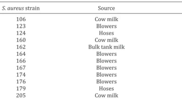
Table 1. Source of the Staphylococcus aureus isolates used
in the study
S. aureus strain Source
106 Cow milk
123 Blowers
124 Hoses
160 Cow milk
162 Bulk tank milk
164 Blowers
166 Blowers
167 Blowers
174 Blowers
176 Blowers
179 Hoses
205 Cow milk
Study design and samplings
In this study we used samples of milk and milking environ -ment (blowers and rubber tubes) collected on a milk farm in the state of Minas Gerais, Brazil. The cows were submitted to the Ca-lifornia Mastitis Test (CMT) to identify the presence of subclinical
mastitis, and then samples were collected from cows reagents to CMT. Swabs were also rubbed on the blowers and rubber tubes after the end of the afternoon milking, in order to isolate strains milking environment that could be correlated with the strains iso -lated from milk.
Swabs were placed in tubes containing sterile saline. Thus, all milk samples and swabs were placed in coolers with dry ice and sent for laboratory analysis at the Federal University of Uberlân -dia, Brazil. It was done the isolation and identification of strains of
Staphylococcus aureus in milk of cows reagents to CMT as the swa -bs in the blowers and rubber conductive milk tubes. The strains identified as Staphylococcus aureus were sent to the laboratory of
Applied Microbiology, University of Minho in Portugal for the tests about biofilm production.
The milking equipment was cleaned after each milking end with soap and water and disinfected with a solution containing 150ppm sodium hypochlorite. In this property was performed two milkings, one on morning and one on afternoon. Sampling was done in afternoon milking, before the process of cleaning and disinfection.
Effect of NaOCl on adhered cells of Staphylococcus aureus
Each S. aureus isolate was cultivated in Triptycase Soy Broth
(TSB) for 24 hours at 37oC, with agitation (120rpm). Cells were
harvested by centrifugation for 5 minutes at 10,500 g and 4oC follo
-wed by a washing procedure with a saline solution (0.9% NaCl in distilled water). After, the cells were homogenized in a vortex and the cellular suspension was adjusted with a saline solution to a final concentration of 1x10⁹ cells/mL determined by optical den
-sity at 640nm. To each well of a 96-well microplate, 200µL of the cellular suspension were added and incubated at 37oC for 2 hours,
for 5 and 10 minutes under agitation at 37oC and 120rpm. The
assay was performed in triplicate and in three different occasions.
Effect of NaOCl on Stgaphylococcus aureus biofilms
A S. aureus cellular suspension at a concentration of 1x10⁶
cells/mL was prepared in TSB supplement with 2% of glucose, ac -cording to the procedure described above. A volume of 200 μL of this suspension was pippeted into the wells of a 96-well microplate, which was then incubated with agitation (120 rpm) for 24h at 37oC.
The biofilm formed on the surface of each well was washed twice with saline solution (0.9% NaCl) to remove planktonic cells. Finally, 200 µL of NaOCl at 150 ppm were added to each well and the micro -plates were incubated at 37oC and 120 rpm for 5, and 10 minutes.
This assay was performed in triplicate, in three different occasions.
Colony forming units (CFU) enumeration
Adhered S. aureus cells and the biofilms formed in the micro -plate wells were washed with 0.9% NaCl followed by the addition of 200μL of the saline solution to each well, to help the scraping of the adhered cells and biofilms from the surface of each well. The resulting suspension was placed in a microtube and was soni -cated for 20s at 30 W (Ultrasonic Processor, Cole-Parmer), with a diameter probe of 0.3cm, followed by a vigorous homogenization for 30s using a vortex. After homogeneization a serial dilution ( 10-1 to 10-6)was performed. Aliquots of 10 μL of each dilution
were drop plated, in triplicate, in Petri dishes containing TSA me -dium (Triptycase Soy Agar). The plates were incubated at 37oC for
48 hours. The resultant colonies were counted in each Petri dish.
Total biomass quantification (crystal violet assay)
To assess total biofilm biomass, after 24h of biofilm formation, and after treatment with NaOCl as described above (Gibson, et al. 1999), the microplate wells were washed with saline solution (0.9% NaCl) and fixed with 200 μL methanol in each well, which was removed after 15 minutes of contact. Then, the biofilm was allowed to dry at room temperature before adding 200 μL of crys -tal violet (1%v/v). After 5 minutes, the biofilm was gently washed with distilled water, and acetic acid (33% v/v) was added to each well (to release and dissolve the stain). The absorbance of the re -sulting solution was read in triplicate in an ELISA reader at 570nm.
Fluorescence microscopy
The bottoms of 6-well plates were cut into squares to be used as coupons for fluorescence microscopy assays. These coupons were placed in 6-well plates with 4mL of S. aureus cell suspension,
at a concentration of 1x10⁹ cells/mL, and incubated at 37oC for 2 h
with agitation (120rpm). Then the coupons were washed in saline solution (0.9% NaCl) and NaOCl was added at a concentration of 150 ppm and incubated at 37oC at 120rpm. The coupons were re
-moved after 5 and 10 minutes of contact with the sanitizing agent. After removal, the coupons were gently washed with 0.9% NaCl and air-dried. The dried coupons were stained with 4´-6-diamidi -no-2-phenylindole (DAPI, Sigma, USA) solution 0.1 (g/L) during 30 minutes. Subsequently, each coupon was rinsed in distilled water in order to remove excess dye and let to air-dry in the dark for 30 minutes. Adhered cells were visualized under an epifluo -rescence microscope (Carl Zeiss, Germany) with a filter sensitive to DAPI fluorescence and coupled with a 3CCD video camera. For each coupon at least 10 images with an 820 x 560 resolution and 1000x magnification were taken.
Pulsed-Field Electrophoresis
The Pulsed-Field Electrophoresis assay was adapted from the McDougal et al. (2003) protocol. Briefly, a colony of S.
au-reus was grown in 5mL of Todd Hewit broth (THB) and incubat -ed at 37°C for 24 hours under vigorous agitation. Afterwards, 1.5 mL of the bacterial suspension was centrifuged for 2 min -utes at a 12 000rpm. The resulting pellet was resuspended in 500μL of TE (10mM Tris HCl, 1mM EDTA [pH 8.0]), heated at 37°C for 10 minutes, and centrifuged again at 12 000rpm for 2 minutes. The resultant pellet was resuspended once more in TE and the solution was heated at 45°C in the presence of 2% low melting agarose. Subsequently, a 20μL drop was placed in a glass slide covered with parafilm. The formed agarose discs were then carefully placed in 3mL of lysing solution (EC - 6mM Tris HCl, 1 M NaCl, 100mM EDTA, 0.5% Brij-58, 0.2% sodium deoxycholate, 0.5% lauroylsarcosine, pH 8.0) and 50μL of lys -ostaphin (1mg/mL), incubated at 37°C for 4 hours. After this period of time, the lysing buffer was removed and 4mL of fresh lysing solution with proteinase K (1mg/mL) and lysozyme (20μg/mL) were added. The new solution was incubated for 18 hours at 50°C.
After four washes with the lysing buffer at 37°C, for 30 min -utes each, and incubation for 30 min-utes at 25°C with a restric -tion buffer, the SmaI enzyme was added and left in solu-tion for a period of 4 hours at 25°C.
After the digestion step, the DNA fragments were placed on the electrophoresis cell (CHEF-DR III - BioRad, Melville, NY) using a voltage of 200 V for 21 hours at 14°C.
The resulting gels were photographed and analyzed using Gel -Doc® (BioRad).
Pulsotype classification
The pulsotype classification was performed by the hierarchic group method, described by Sneath & Sokal (1973), using as simi -larity measurements the Jaccard coefficient and the Ward`s clus -tering algorithm (Ward 1963) to relate the groups. All the data was analyzed using R environment (R Development Core Team, 2010), 2.11.0 version. This assay was performed at the Departa -mento de Ciências Exatas da Faculdade de Ciências Agrárias e Ve -terinárias, Câmpus de Jaboticabal, SP, UNESP.
RESULTS AND DISCUSSION
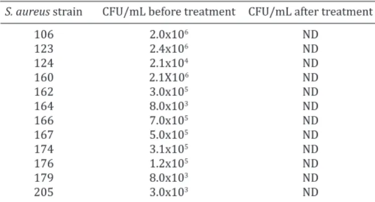
The present study evaluated the effect of NaOCl (150ppm) on twelve strains of Staphylococcus aureus adhered to polystyrene (2h) and as biofilms (24h-old biofilm).
The number of adhered cells to the polystyrene surfa
-ce varied according to the strain, ranging from 3.0x10³ to
2.4x10⁶ CFU/mL, as it can be seen in Table 2.
Table 2. Colony Forming Units (CFU) of 12 strains of
Staphylococcus aureus adhered to polystyrene before and
after treatment with NaOCl (150ppm)
S. aureus strain CFU/mL before treatment CFU/mL after treatment
106 2.0x10⁶ ND
123 2.4x10⁶ ND
124 2.1x10⁴ ND
160 2.1X10⁶ ND
162 3.0x10⁵ ND
164 8.0x10³ ND
166 7.0x10⁵ ND
167 5.0x10⁵ ND
174 3.1x10⁵ ND
176 1.2x10⁵ ND
179 8.0x10³ ND
205 3.0x10³ ND
The results revealed that NaOCl, was able to promote a significant reduction on the number of S. aureus cells adhe
-red to polystyrene wells, regardless of the strain tested, the number of cells adhered, and the time of contact (bellow the detection limit).
The efficacy of 150ppm NaOCl against S. aureus adhered cells was also evaluated by fluorescence microscopy. As it can be seen in Figure 1, there is a significant decrease in the number of bacterial cells adhered to the polystyrene surface.
and where cells are able to develop specific traits like low multiplication rates or additional defense mechanisms, due to the ability of the extracellular polymeric matrix in neutralizing antimicrobial agents, since it consists of or
-ganic matter (Rossi & Porto 2009). This rationale is also in line with the results reported by Norwood & Gilmour (2000), after exposing multispecies biofilms (Listeria mo-nocytogenes, Pseudomonas fragi and Staphylococcus xylo-sus) to increasing concentrations of NaOCl (200, 500 and 1000mg free chlorine/L for 20 minutes). The authors only obtained a 2 log CFU reduction in biofilm L. monocytoge-nes cells with the exposure to 1000mg chlorine/L, while 100% of the corresponding planktonic cultures were kil
-led when exposed to 10mg chlorine/L for 30 seconds, con
-firming the shielding ability of biofilms. Also, according to the study of Amaral et al. (2004), where the efficiency of the disinfection of teatcups and teats during mechanic milking of dairy cows was tested, the disinfection of the teats with a 150ppm NaOCl soaking solution was able to significantly reduce the number of Staphylococcus sp. cells, while the immersion in the same solution was not efficient for the rubber teatcup microorganism reduction number. This demonstrates how biofilm formation on the surface of milking devices increases its resistance to antimicrobial agents.
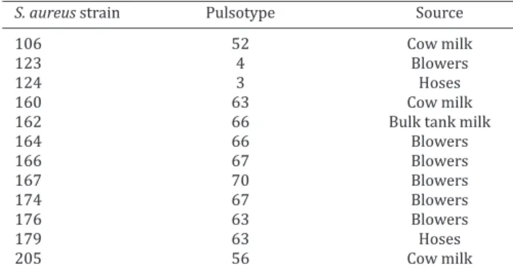
Analyzing the strains whose biofilms were resistant to NaOCl at 150ppm, after 5 minutes treatment, and associa
-ting them to the pulsotypes identified by Pulsed-Field Elec
-trophoresis (PFGE) (Table 4), it was found that three of the four resistant strains belong to the same pulsotype 63.
The strains used in the present study were isolated from cow’s milk, the blower and from a hose. Curiously, the pulsotype 63 was isolated primarily from milk during the month of December 2011 and it was the only isolated from other two points, later, in January 2012. This is a strong indication that the hygiene protocol was failing, probably due to biofilm development, which is more resistant than adhered cells. Actually, after the blowers exchange, no strain with the pulsotype 63 was isolated. It is believed that the pulsotype 63 strain should have acquired resistance to NaOCl. A similar study by Bolton et al. (1988) showed that
Table 3. Colony Forming Units (CFU) obtained from 24h-old biofilms of 12 strains of Staphylococcus aureus before and
after NaOCl (150ppm) treatment
S. aureus strain CFU/mL before treatment CFU/mL after treatement
5 min 10 min
106 3.1x10⁶ ND ND
123 4.3x10⁶ ND ND
124 2.1x10⁶ ND ND
160 3.7x10⁶ 2.0x10⁴ ND
162 4.2x10⁶ 2.5x10⁴ ND
164 3.6x10⁶ ND ND
166 5.1x10⁶ ND ND
167 5.9x10⁶ ND ND
174 3.3x10⁶ ND ND
176 2.8x10⁶ 1.5x10⁴ ND
179 3.2x10⁶ 2.1x10⁴ ND
205 3.0x10⁵ ND ND
ND = no bacterial growth was observed.
Fig.1. Fluorescence microscopy images showing the reduction of the number of Staphylococcus aureus cells adhered to polys -tyrene coupons after contact with 150ppm NaOCl for 5 minu -tes. S. aureus cells adhered to polystyrene coupons, (B) with
and (A) without treatment with NaOCl.
When the sanitizing agent was used to treat already de
-veloped biofilms (24h-old), the degree of surface hygiene attained with 150ppm was dependent on the time of con
-tact - only after 10 minutes no CFU were detected (Table 3). In fact, it was found that in four cases only a 2 log reduction in the number of CFU was obtained after 5 minutes of con
-tact with NaOCl.
The different effect of sodium chloride on adhered cells compared to biofilms, after 5 minutes of contact, confir
-ms that biofilm cells are highly tolerant to antimicrobial agents. In other words, biofilms constitute a shelter habi
strains of S. aureus endemic in equipment used in poultry processing, were eight times more resistant to chlorine than strains of S. aureus isolated from healthy skin.
In the current study, it was also observed an overall re
-duction of the total biofilm biomass, at a sanitizing concen
-tration of 150ppm and contact time of 10 minutes (Table 5), which is accordance with the reduction on the number of viable cells at the same conditions.
A comparison of hypochlorite (alkaline hypochlorite), chlorine and chloramines has been performed for several times. It has been shown that chlorinated agents can pe
-netrate into the biofilm of Pseudomonas aeruginosa and Klebsiella pneumoniae (Stewart et al. 2001)), but these chemicals are not able to inactivate all bacteria because the microorganisms in biofilms carry protective mechanisms against the lethal effect of this type of biocidal agents. In another study, it was observed that chloramines penetrated in the biofilm 6-8 times faster than hypochlorite. However, the bacteria that formed the biofilm were highly tolerant to both agents (Jang et al. 2006). These studies emphasi
-ze that the correct and conscious of cleanliness, hygiene,
sanitation, and health education of workers must be daily and continuous, in addition to equipment maintenance and periodic exchange of rubbers. If these parameters are che
-cked consistently, the installation process of the biofilm is hindered and, thus, the persistence of pathogenic bacteria with the constant contamination of the milk and the pre
-sence of mastitis in animals will be reduced and may even be eradicated.
CONCLUSION
This study shows that a correct sanitation protocol of all the milking installation can indeed significantly reduce the num
-ber of microorganisms’ present, leading to a decrease of cow´s mastitis and milk contamination. Moreover, it is important to emphasize that the sanitation process must be adapted according to the strain of the microorganism detected.
Acknowledgements.- This study was supported by Portuguese Foun -dation for Science and Technology (FCT) through the grant SFRH/ BPD/20987/2004 attributed to Cláudia Botelho and thanks to FAPESP (Fundação de Amparo a Pesquisa do Estado de São Paulo) for the scholar -ship attributed to Poliana de C. Melo.
REFERENCES
Amaral L.A., Isa H., Dias L.T., Rossi-Jr O.D. & Nader-Filho A. 2004. Avaliação da eficiência da desinfecção de teteiras e dos tetos no processo de or -denha mecânica de vacas. Pesq. Vet. Bras. 24(4):173-177.
Bolton K.J., Dadd C.E.R., Mead G.C. & Waites W.M. 1988. Chlorine resistance of strains of Staphylococcus aureus isolated from poultry processing
plants. Lett. Appl. Microbiol. 6:31-34.
Gibson H., Taylor J.H., Hall K.E. & Holah J.T. 1999. Effectiveness of cleaning techniques used in the food industry in terms of the removal of bacterial biofilms. J. Appl. Microbiol. 87:41-48.
Heringstad B., Klemetsdal G. & Ruane J. 2000. Selection for mastitis resis -tance in dairy cattle: a review with focus on the situation in the Nordic countries. Livest. Product. Sci. 64:95-106.
Jang A., Szabo J., Hosni A.A., Coughlin M. & Bishop P.L. 2006. Measurement of chlorine dioxide penetration in dairy process pipe biofilms during disinfection. Appl. Microbiol. Biotechnol. 72(2):368-376.
Mah T.F. & O’Toole G.A. 2001. Mechanisms of biofilm resistance to antimi -crobial agents. Trends Microbiol. 9(1):34-39.
McDougal L.K., Steward C.D., Killgore G.E., Chaitram J.M., McAllister S.K. & Tenover FC. 2003. Pulsed-Field Gel electrophoresis typing of oxacilin-resistant Staphylococcus aureus isolates from the United: establishing a
national database. J. Clin. Microbiol. 41(11):5113-5120.
Norwood D.E. & Gilmour A. 2000. The growth and resistance to NaOCl of
Listeria monocytogenes in a steady-state multispecies biofilm. J. Appl.
Microbiol. 88(3):512-520.
Rossi A.C.R. & Porto E. 2009. A importância da elaboração de procedimen -tos dehigienização considerando a presença de biofilmes. Sociedade Brasileira de Controle de Contaminação. Março/Abril 2009, p.40-41. Sneath P.H. & Sokal R.R. 1973. Numerical Taxonomy: the principles and
practice of numerical classification. W.H. Freeman, San Francisco. 573p. Stewart P.S., Rayner J., Roe F. & Rees W.M. 2001. Biofilm penetration and
disinfection efficacy of alkaline hypochlorite and chlorosulfamates. J. Appl. Microbiol. 91(3):525-532.
Ward J.H. 1963. Hierarchical grouping to optimize an objective function. J. Am. Statist. Assoc. 58:236-244.
Table 4. Pulsotype of the 12 strains of Staphylococcus aureus
S. aureus strain Pulsotype Source
106 52 Cow milk
123 4 Blowers
124 3 Hoses
160 63 Cow milk
162 66 Bulk tank milk
164 66 Blowers
166 67 Blowers
167 70 Blowers
174 67 Blowers
176 63 Blowers
179 63 Hoses
205 56 Cow milk
Table 5. Biofilm biomass of the twelve Staphylococcus aureus
strains, isolated from cases of bovine subclinical mastitis and
milking environment, expressed as crystal violet optical density (O.D.570 nm), before and after exposure to NaOCl (150 ppm), for
two different periods of time (5 minutes, 10 minutes)
S. aureus strain O.D. 570 nm before O.D. 570 nm after
treatment treatment
5 min 10 min
106 0.35 0.51 0.07
123 0.79 0.48 0.05
124 0.70 0.61 0.05
160 0.79 0.77 0.06
162 0.42 0.42 0.04
164 0.55 0.66 0.23
166 0.59 0.67 0.06
167 0.60 0.69 0.08
174 0.59 0.70 0.71
176 0.64 0.63 0.76
179 0.67 0.65 1.30