Is painless synovitis different from painful synovitis?
A controlled, ultrasound, radiographic, clinical trial
Daniele Freitas Pereira,IJamil Natour,I Ana Leticia Pirozzi de Buosi,I Fernando Bernardes Maia Diniz Ferreira,IIArtur da Rocha Correˆa Fernandes,II Rita Nely Vilar FurtadoI
IUniversidade Federal de Sa˜o Paulo, Escola Paulista de Medicina, Disciplina de Reumatologia, Sa˜o Paulo/SP, Brazil.IIUniversidade Federal de Sa˜o Paulo,
Escola Paulista de Medicina, Departamento de Diagno´stico por Imagem, Sa˜o Paulo/SP, Brazil.
OBJECTIVE: This study compares the clinical, ultrasonography, radiography, and laboratory outcomes of painless and painful chronic synovitis in patients with established rheumatoid arthritis.
METHODS: This cross-sectional study involved 60 patients with rheumatoid arthritis and synovitis in the metacarpophalangeal joints; 30 of the patients did not experience pain, and 30 had experienced pain for at least 6 months prior to the study. The radiocarpal, distal radioulnar, and metacarpophalangeal joints were evaluated using the ultrasound gray scale, power Doppler, and radiography. Past and present clinical and laboratory findings were also evaluated.
RESULTS:There were no statistically significant differences between the groups for most of the outcomes. The group with pain scored worse on the disease activity indices (e.g., DAS 28 and SDAI), function questionnaires (HAQ and Cochin), and pinch strength test. A logistic regression analysis revealed that the use of an immunobiological agent was associated with a 3-fold greater chance of belonging to the group that experienced pain. The painless group had worse erosion scores in the second and fifth metacarpophalangeal with odd ratios (ORs) of 6.5 and 3.5, respectively. The painless group had more cartilage with grade 4 damage in the third metacarpophalangeal.
CONCLUSIONS:The rheumatoid arthritis patients with both painless and painful synovitis exhibited similar disease histories and radiographic and ultrasound findings. However, the ultrasonography evaluation revealed worse scores in the second and fifth metacarpophalangeal of the synovitis patients who did not experience pain.
KEYWORDS: Rheumatoid Arthritis; Synovitis; Pain; Ultrasound; Radiographic.
Pereira DF, Natour J, Buosi AL, Ferreira FB, Fernandes AR, Furtado RN. Is painless synovitis different from painful synovitis? A controlled, ultrasound, radiographic, clinical trial. Clinics. 2014;69(2):93-100.
Received for publication onMay 27, 2013;First review completed onJune 24, 2013;Accepted for publication onJuly 23, 2013 E-mail: [email protected]
Tel.: 55 11 5576-4239
& INTRODUCTION
The clinical presentation of rheumatoid arthritis (RA) varies, but most patients have intermittent polyarthritis with swelling and tenderness to palpation (1). Some patients exhibit persistent chronic synovitis, which is marked by joint swelling (2) and may or may not be accompanied by pain. The reason for the absence of pain despite the persistent joint swelling is unknown. Moreover, little is known about the predictive factors of painless synovitis or its relationship to the past progression of the disease, ultrasound inflammatory findings (e.g., power Doppler), or the degree of joint damage (erosion).
Ultrasonography (US) allows the early detection of bone and cartilage damage and the evaluation of synovitis (gray scale [GS-US] and power Doppler [PD-US]). US is more sensitive than a clinical examination and simple radio-graphy (X-ray) (3-8). PD-US enhances the specificity of US (9), assists in the diagnosis of active synovitis (10), and predicts joint damage (11).
No previous studies have addressed the importance of painless chronic synovitis in RA. Thus, the present study compared the clinical, US, radiographic, and laboratory outcomes of patients with established RA and chronic synovitis with or without pain.
& PATIENTS AND METHODS
Study design
This cross-sectional study evaluated RA patients. The study was approved by the Human Research Ethics Committee of the Universidade Federal de Sa˜o Paulo/Escola Paulista de Medicina (Brazil). All patients provided written informed consent.
Copyrightß2014CLINICS– This is an Open Access article distributed under the terms of the Creative Commons Attribution Non-Commercial License (http:// creativecommons.org/licenses/by-nc/3.0/) which permits unrestricted non-commercial use, distribution, and reproduction in any medium, provided the original work is properly cited.
No potential conflict of interest was reported.
The sample size of 30 individuals in each group was considered appropriate, and the PD-US was the primary study outcome. The study had a standard deviation (SD) of 0.4, a power of 90%, and a 5% significance level.
Patients
Adult patients who fulfilled the 1987 American College of Rheumatology criteria for RA (12) were eligible for the study if they also met the following criteria: joint swelling in at least 4 metacarpophalangeal (MCP) joints for at least 6 consecutive months, female gender, and stable use of disease-modifying antirheumatic drugs (DMARDs) in the previous 3 months. The visual analogue scale (VAS), which ranges from 0 to 10 cm, was used for the pain criteria; the VAS score was at least 4 cm in the painful group and 0 in the painless group. The following exclusion criteria were used: overlap syndromes, irreducible deformation and MCP surgery, and comorbidities, such as uncontrolled hypothyr-oidism, uncontrolled fibromyalgia or diabetic neuropathy.
Data collection
Sixty patients were selected from the rheumatology outpatient clinic of the Universidade Federal de Sa˜o Paulo/ Escola Paulista de Medicina (Brazil) between July 2011 and July 2012. The patients were recruited consecutively and assigned to the painful synovitis group or the painless synovitis group. The groups were age matched.
We collected the demographic data, life habits, and information about both the past and present progression of RA using a questionnaire.
Clinical examinations were performed by a rheumatolo-gist who was ‘‘blinded’’ to each patient’s history and the results of the imaging exams. The evaluation included a patient and a medical global assessment (on a 0-100 scale), grip strength using a JamarH, and pinch strength using a Preston Pinch GaugeH; the 28-joint Disease Activity Score (DAS 28) (13), Clinical Disease Activity Index (CDAI) (14), Simplified Disease Activity Index (SDAI) (15), Stanford Health Assessment Questionnaire (HAQ) (16), and Cochin Hand Function Scale (CHFS) were also used (17).
The transverse US exam included the dorsal side of the radiocarpal (RC) and the palmar and dorsal sides of MCPs 1 to 5; the longitudinal US exam included the dorsal radio-ulnar (DRU), according to a quantitative synovitis mea-surement (in mm) in the largest synovial bursa and semiquantitative scores, as described above (Table 1).
Furthermore, a transverse evaluation of the cartilage of the dorsal side of MCPs 1 to 5 was performed with flexion of the fingers.
US was performed bilaterally on the hands and wrists by a ‘‘blinded’’ musculoskeletal sonographer with 5 years of experience, using the ESAOTE MyLab 60 Xvision, with a multi-frequency linear transducer (6-18 MHz). The sonogra-pher followed the guidelines for musculoskeletal US recom-mended by the European League Against Rheumatism (18). The US were measured based on the definitions pub-lished in the Outcome Measures in Rheumatology Clinical Trials (except cartilage) (19).
Semiquantitative synovitis, bone erosion, and PD-US were each evaluated using a 4-grade scale ranging from 0 to 3. The scores were defined as follows.
Grades 0-1 for bone erosion and synovitis were consid-ered normal (Score I), whereas grades 2-3 indicated pathological changes (Score II) (20).
For the PD-US signal, grade 0 was considered to be nor-mal (Score I), whereas grades 1-3 were considered to be pathological (Score II) (20,21).
Joint cartilage was evaluated using a semi-quantitative 5-grade score with the aforementioned categories (22,23).
The inter-observer reliability for the US evaluation was determined based on the image evaluations recorded on 20% of the overall sample in the RC (a total of 52 joint recess). The evaluation was performed by a blinded rheumatologist trained in musculoskeletal US.
The radiological evaluation (plain X-ray of the hands and wrists) was performed by a single experienced radiolo-gist who was unaware of the clinical or US findings. The evaluation used the modified method proposed by van der Heijde and collaborators (24).
& STATISTICAL ANALYSIS
The data were analyzed in SPSS v.17.0. We express the quantitative parameters as the mean, standard deviation, and range. Any value ofp,0.05 was consider significant.
The data were compared using either the Student’s t-test or the Mann-Whitney test.
The categorical variables were measured in percentages and were compared between the groups, using either the chi-squared or Fisher’s exact tests. The correlations be-tween variables were evaluated using either Pearson’s or Spearman’s correlation coefficients. Cohen’s Kappa index and the intraclass correlation coefficient were used to evaluate the inter-observer reliability.
A subanalysis of the 2 groups was performed on the US findings for the joints that exhibited swelling in the clinical examination. Logistic regression analysis was applied to the semi-quantitative US variables to assess the ability of the
Table 1 -Semiquantitative scores for synovitis, PD-US, bone-erosion and cartilage.
Synovitis (19): PD-US (19):
0 no synovial thickening 0 no flow in the synovium
1 minimal synovial thickening 1 single vessel signals
2 moderate synovial thickening with capsule distension 2 confluent vessel signals in less than half of the area evaluated 3 synovial thickening, extending to bone diaphysis 3 vessel signals in more than half of the evaluated area
Bone erosion (19): Cartilage (22):
0 regular bone surface 0 normal hyaline cartilage
1 bone surface irregularity 1 loss of the sharpness of the cartilage margin
2 bone surface defect on 2 planes 2 partial thickness defect of the cartilage layer
3 bone defect with bone destruction 3 full thickness of the cartilage layer
variables to predict painful or painless synovitis and to identify the variables that were likely to be predictive of painless synovitis.
& RESULTS
The sample consisted of 60 patients with established RA, for a total of 120 hands and wrists and 600 MCFs. The mean duration of the absence of joint pain in the painless group and the presence of joint pain in the painful group was, respectively, 30.3¡32.6 months and 50.9¡74.6 months
(p= 0.740). There were no statistically significant differences between the groups for the majority of the demographic variables and laboratory findings or in the disease progres-sion (Tables 2, 3).
Two patients in the painless group (7%) and 9 patients (30%) in the painful group were smokers (p= 0.042) (Table 2). The medical diagnosis for RA took 13.2¡23.6 months in
the painless group and 21.2¡26.1 months in the painful group (p= 0.028) (Table 3). The majority of patients were initially treated with monotherapy consisting of a DMARD associated with a corticosteroid. Furthermore, the majority of patients were primarily treated with a combination of DMARDs (Table 3).
There were statistically significant between-groups differ-ences in the current DAS-28 measurement (using erytrocyte sedimentation rate-ESR) and for all variables influenced by the presence of joint pain, with higher scores in the painful
group (p,0.05) (Tables 2 and 4). However, there was no statistically significant difference between the groups in the number of swollen joints.
Twenty-seven patients (90%) in the painless group and 13 (43%) patients in the painful group were classified as having mild dysfunction (HAQ scores 0 and 1, respectively) (25) (p= 0.001). There were statistically significant between-group differences for the CHFS, lateral pinch, and tripod pinch (p,0.001, p,0.001, and p= 0.039, respectively), with better scores in the painless group. The Jamar and pulp-to-pulp pinch scores were similar between the groups. There were no statistically significant between-group differences in the degree of joint deformities or in the number of prior surgeries (Table 3).
The most frequently used DMARDs in both groups at the time of the study were methotrexate and leflunomide. More than half of the painful group used corticosteroids but at a low mean dose of 5.0 mg¡6.76 mg (Table 2). Eleven
patients in the painful group and 5 patients in the painless group used immunobiological agents (p= 0.080).
In the univariate logistic regression analysis, only the variable ‘‘used an immunobiological agent after one year of the disease’’ was associated with painful synovitis; how-ever, it was associated with a 3-fold increase in the odds of the patient belonging to the painful group (odds ratio [OR] = 3.0, 1.00-9.37,p= 0.049).
A total of 1.560 joint recesses were examined in the US evaluation. In the semi-quantitative analysis, there were no
Table 2 -Group characteristics.
PAINLESS GROUP mean¡SD (%) (N = 30)
PAINFUL GROUP
mean¡SD (%) (N = 30) p-value
Age(in years) 59.9¡11.5 56.8¡14.0 0.441*
Skin colorWhite(%)/Brown(%)/Black(%) 16(55)/10(35)/3(10) 11(41)/11(41)/5(18) 0.496**
Smoking 2 (7) 9 (30) 0.042***
Alcohol use 0 (0) 1 (3) 0.500***
Dominant right hand 28 (93) 27 (90) 1.000***
Arterial hypertension 15 (50) 23 (77) 0.032**
Dyslipidemia 8 (27) 15 (50) 0.063**
Other comorbiditiesOP/Fibromyalgia/Others 8(27)/1(3)/1(3) 5(17)/0(0)/4(13) 0.319**
Disease duration (years) 17.7¡9.4 15.1¡10.2 0.185‘
Duration of the absence or presence of MCP pain(months) 30.3¡32.6 50.9¡74.6 0.740‘
Rheumatoid factor positive 13 (43) 13 (43) 1.000**
Anti-CCP positive 23 (77) 19 (63) 0.269**
Use of MTX 17 (57) 20 (67) 0.426**
Use of Leflunomide 18 (60) 13 (43) 0.196** Use of Hydroxychloroquine 1 (3) 4 (13) 0.353***
Use of CS via oral 11 (37) 16 (53) 0.194**
Dose of CS via oral(in mg) 2.08¡3.09 5.00¡6.76 0.097‘
Use of immunobiological agent 5 (17) 11 (37) 0.080**
DMARD association 9 (30) 11 (37) 0.584**
Morning stiffness (minute) 4.7¡13.3 24.8¡29.0 ,0.001‘
ESR 31.7¡21.8 31.5¡25.9 0.723‘
CRP (mg/dl) 0.75¡1.00 0.68¡0.69 0.706‘
MDGA 33.3¡15.6 48.3¡14.9 ,0.001‘
PGA 30.7¡26.8 60.0¡19.3 ,0.001‘
N painful joints 1.5¡2.3 13.6¡6.1 ,0.001‘
N swollen joints 8.4¡3.0 9.6¡3.9 0.246‘
DAS 28 by ESR 3.75¡0.83 5.57¡0.94 ,0.001* DAS 28 by CRP (mg/L) 3.06¡0.86 4.90¡0.95 ,0.001*
SDAI 16.17¡6.74 30.21¡9.52 ,0.001*
CDAI 15.3¡6.5 28.9¡9.1 ,0.001*
SD: standard deviation; * Student’s t-test; ** chi-squared test;‘Mann-Whitney U-test; *** Fisher’s exact test; OP: osteoporosis;
statistically significant between-group differences for the presence of synovitis (at least grade 2) or positive PD-US (Figure 1) in the majority of the joint recesses studied. Bone erosion (at least grade 2) was similar in the 2 groups, but the painless group had worse scores in some joints (Table 5). Statistically significant differences were found in the cartilage evaluation, with more scores of 4 in the third MCP in the painless group (p,0.004) and more scores of 2 in the second MCP (p,0.022) and 1 in the fourth MCP (p,0.004) in the painful group.
Fifty-eight percent of the wrists in the painful group had pain on clinical examination, and 83% of the wrists in the painless group had no pain.
In the US subanalysis of the joints with swelling, there were no statistically significant between-group differences in the quantitative synovitis, the semi-quantitative synovitis, or the PD-US in the MCPs. The bone erosion scores were worse in the painless group for the palmar and lateral sides
of the second MCP (p,0.022 and p,0.004, respectively). Regarding the joint cartilage, only the third MCP had more 0 scores in the painful group (p,0.030).
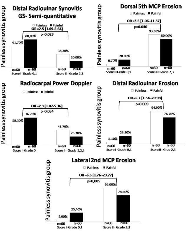
The univariate logistic regression revealed a greater likelihood of patients belonging to the painless group based on worse US scores for the following variables (Figure 2): the semiquantitative synovitis in the DRU, the PD-US in the RC, the erosion on the dorsal face of the 2nd MCP and the dorsal face of the 5th MCP, and the DRU (OR = 2.5, 1.09-5.64,
p= 0.029; OR = 2.3, 1.07-5.16, p= 0.034; OR = 6.5, 1.76-23.77,
p= 0.005; OR = 3.5, 1.06-11.57,p =0.040; and OR = 5.7, 1.54-20.98, p= 0.009, respectively). The interobserver reliability was moderate to strong (Kappa = 0.435 to 1.00;p,0.018) for all US measures and strong for the PD-US (Kappa = 0.655 to 0.783,p= 0.001).
No statistically significant differences were found in the radiographic evaluation, except for the proximal interpha-langeal (PIP) joint, for which the painless group had a worse
Table 3 -Past disease variables of the groups.
PAINLESS GROUP mean¡SD (%) (N = 30)
PAINFUL GROUP
mean¡SD (%) (N = 30) p-value
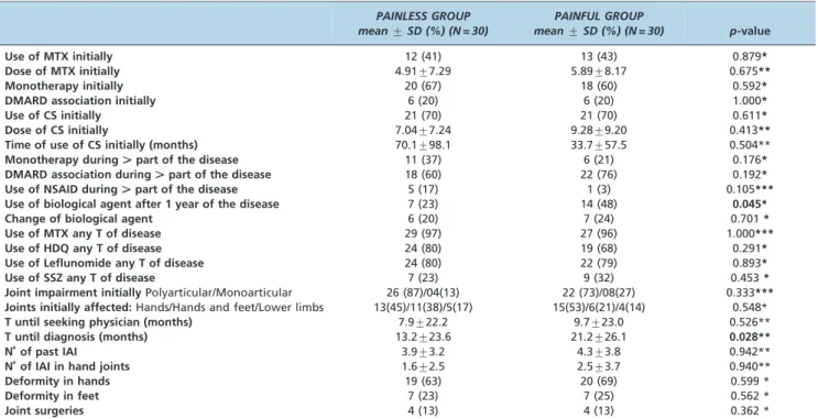
Use of MTX initially 12 (41) 13 (43) 0.879*
Dose of MTX initially 4.91¡7.29 5.89¡8.17 0.675**
Monotherapy initially 20 (67) 18 (60) 0.592*
DMARD association initially 6 (20) 6 (20) 1.000*
Use of CS initially 21 (70) 21 (70) 0.611*
Dose of CS initially 7.04¡7.24 9.28¡9.20 0.413**
Time of use of CS initially (months) 70.1¡98.1 33.7¡57.5 0.504**
Monotherapy during.part of the disease 11 (37) 6 (21) 0.176*
DMARD association during.part of the disease 18 (60) 22 (76) 0.192*
Use of NSAID during.part of the disease 5 (17) 1 (3) 0.105***
Use of biological agent after 1 year of the disease 7 (23) 14 (48) 0.045*
Change of biological agent 6 (20) 7 (24) 0.701*
Use of MTX any T of disease 29 (97) 27 (96) 1.000***
Use of HDQ any T of disease 24 (80) 19 (68) 0.291*
Use of Leflunomide any T of disease 24 (80) 22 (79) 0.893*
Use of SSZ any T of disease 7 (23) 9 (32) 0.453*
Joint impairment initiallyPolyarticular/Monoarticular 26 (87)/04(13) 22 (73)/08(27) 0.333*** Joints initially affected:Hands/Hands and feet/Lower limbs 13(45)/11(38)/5(17) 15(53)/6(21)/4(14) 0.548*
T until seeking physician (months) 7.9¡22.2 9.7¡23.0 0.526**
T until diagnosis (months) 13.2¡23.6 21.2¡26.1 0.028**
N˚of past IAI 3.9¡3.2 4.3¡3.8 0.942**
N˚of IAI in hand joints 1.6¡2.5 2.5¡3.7 0.940**
Deformity in hands 19 (63) 20 (69) 0.599 *
Deformity in feet 7 (23) 7 (25) 0.562 *
Joint surgeries 4 (13) 4 (13) 0.362 *
SD: standard deviation; MTX: methotrexate; * chi-squared test; ** Mann-Whitney U-test; *** Fisher’s exact test; CS: corticosteroid; T: time; NSAID: non-steroidal anti-inflammatory drug; SSZ: sulfasalazine; N: number; IAI: intra-articular injection; HDQ: hydroxychloroquine.
Table 4 -Current functional assessment of the groups.
PAINLESS GROUP mean¡SD % (N = 30)
PAINFUL GROUP
mean¡SD % (N = 30) p-value
HAQ 0.43¡0.41 1.10¡0.56 ,0.001‘
HAQ categorized Mild/mod/severe dysfunction 27 (90)/3 (10)/0 (0) 13 (43)/16 (53)/1(3) 0.001**
Functional Class 1/2/3 17(57)/10(35) 2 (7) 11(37)/19(63)/0 (0) 0.048**
Cochin 8.2¡9.9 24.8¡15.9 ,0.001‘
Jamar 20.35¡12.80 18.42¡13.89 0.284‘
Lateral pinch 4.69¡1.45 3.71¡1.59 ,0.001‘
Pulp-to-pulp pinch 2.92¡1.27 2.56¡1.21 0.069‘
Tripod pinch 3.45¡1.44 2.99¡1.55 0.039‘
joint space reduction score (p= 0.018). The mean total Sharp score was 93.2¡61.6 in the painless group and 65.4¡40.6 in
the painful group (p= 0.114).
& DISCUSSION
Pain control is a priority in 90% of patients with RA. However, in a prospective study, Lee et al. (26) demon-strated that the number of joints with swelling at baseline was negatively associated with the presence of pain in a 1-year follow-up period. The perception of pain is highly subjective and may be influenced by a number of issues, including socio-cultural factors (27-29).
Few studies have assessed painless synovitis in RA. The absence of pain in patients with juvenile idiopathic arthritis can delay the disease diagnosis, which could lead to great-er joint damage and disability (30,31). The significance of painless synovitis for physicians and patients remains unknown.
In the present study, the majority of both the past and present variables were similar in the patients with and without pain. The time until the RA diagnosis was longer for patients in the painful group. Pain or the absence of pain may not be constant for each patient throughout the disease course. Smoking is an aggravat-ing factor for RA (32); this issue was also observed in this study through the association between smoking and painful synovitis.
The painful group had worse disease activity indices (DAS 28, SDAI, and CDAI), a greater number of painful joints, and worse overall evaluations by both the physician and patient. However, the DAS 28 may not be a good measure of disease activity (33) because joint pain is weighted twice as swelling in the DAS 28 score.
Felson et al. (33) have argued that joint swelling is the true predictor of late radiographic progression in RA. In a prospective cohort study, Lukas et al. (34) have found that
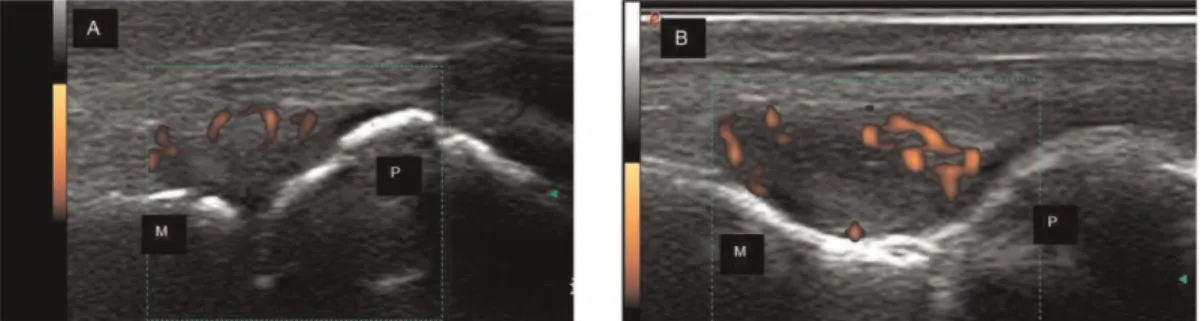
Figure 1 -Dorsal longitudinal image of 2ndMCP; (A) Patient in group without pain with synovial hypertrophy and positive PD signal; (B)
Patient in Painful group with synovial hypertrophy and positive PD signal; M = metacarpal; P = phalange.
Table 5 -Ultrasound findings for each joint.
QUANTITATIVE SYNOVITIS in mm Mean (SD)
ABNORMAL SYNOVITIS SCORES (2-3) N (%)
ABNORMAL POWER DOPPLER SCORES (1-3) N (%)
ABNORMAL BONE EROSION SCORES (2-3)N (%)
JR
PAINLESS N = 60
PAINFUL N = 60 p-value
PAINLESS N = 60
PAINFUL N = 60 p-value
PAINLESS N = 60
PAINFUL
N = 60 p-value
PAINLESS N = 60
PAINFUL
N = 60 p-value
RC 3.0 (2.0) 2.8 (2.2) 0.308 22 (36.7) 18 (30.0) 0.439 25 (41.7) 14 (23.3) 0.032 57 (95.0) 53 (88.3) 0.186
DRU 3.3 (2.3) 3.0 (2.1) 0.542 23 (38.3) 12 (20.0) 0.027 25 (41.7) 16 (26.7) 0.083 56 (94.9) 46 (76.7) 0.004
1st MCP P D 1.7 (1.6) 1.7 (1.7) 1.4 (1.8) 1.4 (1.5) 0.312 0.298 36 (60.0) 35 (58.3) 29 (48.3) 30 (50.0) 0.200 0.360 15 (25.0) 13 (21.7) 8 (13.3) 12 (20.0) 0.104 0.822 51 (85.0) 47 (78.3) 42 (70.0) 40 (66.7) 0.049 0.152 2nd MCP P D L 2.2 (2.0) 2.6 (2.1) 1.9 (1.9) 2.4 (1.9) 0.304 0.404 27 (45.0) 34 (56.7) 19 (31.7) 24 (40.0) 0.133 0.068 13 (21.7) 27 (45.0) 11 (18.3) 17 (28.3) 0.648 0.058 53 (88.3) 54 (90.0) 57 (95.0) 44 (73.3) 51 (85.0) 44 (74.6) 0.037 0.408 0.002 3rd MCP P D 1.5 (1.8) 1.9 (1.8) 1.4 (1.7) 1.9 (1.8) 0.694 0.850 20 (33.3) 21 (35.0) 15 (25.0) 18 (30.0) 0.315 0.559 09 (15.0) 16 (26.7) 08 (13.3) 14 (23.3) 0.793 0.673 42 (70.0) 51 (85.0) 42 (70.0) 43 (71.7) 1.000 0.076 4th MCP P D 1.3 (1.7) 1.4 (1.7) 0.9 (1.3) 1.4 (1.9) 0.274 0.993 14 (23.3) 23 (38.3) 09 (15.0) 19 (31.7) 0.246 0444 03 (5.0) 09 (15.0) 03 (5.0) 10 (16.7) 1.000 0.803 34 (57.6) 33 (55.0) 29 (49.2) 28 (46.7) 0.356 0.361 5th MCP P D 1.4 (2.2) 2.1 (2.0) 1.2 (1.6) 1.9 (2.1) 0.906 0.616 14 (3.7) 23 (38.3) 13 (21.7) 19 (31.7) 0.827 0.444 06 (10.0) 11 (18.3) 6 (10.2) 11 (18.3) 0.976 1.000 33 (55.0) 56 (93.3) 22 (37.3) 48 (80.0) 0.053 0.032
joint swelling was the greatest predictor of ‘‘repair’’ in radiographic erosion. Studying early arthritis, Filler et al. (35) have also found that the progression of RA was more closely associated with the swollen joint count than with the tenderness joint count (35).
Recently, Dougados et al. (36) have emphasized the importance of persistent synovitis (both clinical and US examination) for predicting subsequent structural deteriora-tion in RA patients. In his study, the level of clinical disease activity was defined by the number of swollen joints.
Furthermore, the patients who had synovitis at baseline had more structural progression (OR = 2.01, 1.36-2.98, p,0.001) in a 2-year follow-up period (36).
In this study, worse functional and dynamometric scores were found in the painful group. It is unsurprising that individuals with painful joints at the time of evaluation had worse functional scores than those without joint pain (37). Nonetheless, no statistically significant between-group differences were found with respect to the grip strength or the pulp to pulp pinch strength.
Figure 2 -Probability of belonging to painless synovitis group with presence of Score II semi-quantitative synovial hypertrophy, PD and
Subclinical synovitis may be present in RA remission (8). Furthermore, there is evidence indicating the progression of joint damage in RA patients during clinical remission (11,10), which may be related to residual joint swelling (2). The consideration of persistent joint swelling in RA patients, even in the absence of pain, as in the present study, aligns with the hypothesis that residual swelling causes erosion and is contrary to the notion of ‘‘cold,’’ ‘‘fibrous,’’ and innocuous synovitis.
US has been proven effective at detecting subclinical synovitis (38,8,5), and PD-US is an important tool for detecting active synovitis (10,39). In this study, no differ-ences were found between the painful and painless groups for the majority of US variables in the MCPs and wrists. The detection of the PD-US signal is a predictor of disease evolution in RA (35) and also of the progression of joint damage (11) and the reactivation of the disease (40). In this sample, PD-US was detected in 21% of the joint recesses analyzed, with no significant between-group differences for the majority of joint recesses. This result suggests that there is no association between active synovitis and the presence of joint pain. US also allows for earlier detection of erosion than plain x-rays. In this respect, US is comparable to magnetic resonance imaging (41-43). In the US analysis of erosion, 5 joint recesses were statistically different between groups, with worse scores in the painless group. For the joint cartilage, poorer scores were found more frequently in the painless group, although the between-group difference did not achieve statistical significance.
The similarities between the painful and painless groups in the quantitative analysis of synovial hypertrophy, semi-quantitative analysis of GS-US variables, PD-US, and bone erosion may mean that painless synovitis can still result in joint damage and disability. Moreover, painless synovitis may make the patient and physician more passive in optimizing treatment. This hypothesis is in agreement with the findings that, after the first year of the disease, the painful group were more likely to use immunobiological agents (p= 0.045), and patients with worse US scores in some joint recesses were more likely to be in the painless group.
The radiographic analysis demonstrated that major previous structural damage was similar in both groups, except for IFP scores, which were worse in the painless group. This evidence further indicates that the continual presence of synovitis, instead of joint pain, is an important factor influencing structural damage in RA because both groups had a similar history of radiographic progression. These findings also aligned with the present analysis regarding the identification of joint deformities; there were no statistically significant differences between groups. However, a controlled prospective study with US and radiographic evaluations is needed to compare the evolu-tion of structural joint damage in this sample of patients.
One of the limitations of this study is that was impossible to recruit patients in the painless group who had a complete absence of pain in all joints. The difficulty in obtain-ing individuals with a constant pain status (presence or absence) throughout the progression of RA constitutes another study limitation. This study is the first to compare patients with RA and painless synovitis with those patients with painful synovitis using clinical, ultrasonographic and radiographic variables. The majority of the findings suggest that patients with painless synovitis exhibit a similar profile
to those patients with painful synovitis with respect to the presence of active synovitis and past joint damage.
& ACKNOWLEDGMENTS
Special thanks to the Fundac¸a˜o de Amparo a` Pesquisa do Estado de Sa˜o Paulo for financial support (FAPESP - 2012/04867-6) and to all volunteers from the UNIFESP community and Hospital do Servidor Pu´blico Estadual (IAMSPE).
& AUTHOR CONTRIBUTIONS
Pereira DF recruited the patients, organized the study protocol, and was responsible for the inter-observer reliability for the ultrasonography evaluation. Natour J and Furtado RN coordinated the study and wrote the article. Buosi AL performed the clinical examination. Ferreira FB performed the ultrasonography examination. Fernandes AR performed the radiography evaluation.
& REFERENCES
1. Grassi W, Angelis RD, Lamanna G, Cervini C. The clinical features of rheumatoid arthritis. Eur J Radiol. 1998;27:S18-S24.
2. Bugatti S, Manzo A, Caporati R, Montecucco C. Assessment of synovitis to predict bone erosions in rheumatoid arthritis. Ther Adv Musculo-skeletal Dis. 2012;4(4):235-44, http://dx.doi.org/10.1177/1759720X12 453092.
3. Zordo T, Mlekusch SP, Feuchtner GM, Mur E, Schirmer M, Klauser AS. Value of contrast-enhanced ultrasound in rheumatoid arthritis. Eur J Radiol. 2007;64(2):222-30.
4. Grassi W, Salaffi F, Filippucci E. Ultrasound in rheumatology. Best Pract Res Clin Rheumatol. 2005;19(3):467-85, http://dx.doi.org/10.1016/j.berh. 2005.01.002.
5. Naredo E, Bonilla G, Gamero F, Uson F, Carmona F, Laffon A. Assessment of inflamatory activity in rheumatoid arthritis: a compara-tive study of clinical evaluation with gray scale e power Doppler ultrasonography. Ann Rheum Dis. 2005;64(3):375-81.
6. Cheung PP, Dougados M, Gossec L. Reliability of Ultrasonography to Detect Synovitis in Rheumatoid Arthritis: A Systematic Literature Review of 35 Studies (1,415 Patients). Arthritis Care Res (Hoboken). 2010;62(3):323-34, http://dx.doi.org/10.1002/acr.20102.
7. Kortekaas MC, Kwok WY, Reijnierse M, Watt I, Huizinga TWJ, Kloppenburg M. Pain in hand osteoarthritis is associated with inflammation: the value of ultrasound. Ann Rheum Dis 2010; 69(7):1367-9.
8. Brown AK, Quinn MA, Karim Z, Conaghan PG, Peterfy CG, Hensor E, et al. Presence of significant synovitis in rheumatoid arthritis patients with disease-modifying antirheumatic drug-induced clinical remission – Evidence from an imaging study may explain structural progression. Arthritis Rheum. 2006;54(12):3761-73, http://dx.doi.org/10.1002/art. 22190.
9. Wakefield RJ, Brown AK, O’Connor PJ, Emery P. Power-Doppler Sonography: Improving Disease Activity Assessment in Inflammatory Musculoskeletal Disease. Arthritis Rheum. 2003;48(2):285-8, http://dx. doi.org/10.1002/art.10818.
10. Peluso G, Michelutti A, Bosello S, Gremese E, Tolusso B, Ferraccioli G. Clinical and ultrasonographic remission determines different changes of relapse in early and long standing rheumatoid arthritis. Ann Rheum Dis. 2011;70(1):172-15, http://dx.doi.org/10.1136/ard.2010.129924. 11. Brown AK, Conaghan, Karim Z, Quinn MA, Ikeda K, Peterfy CG, et al.
An Explanation for the Apparent Dissociation Between Clinical Remission and Continued Structural Deterioration in Rheumatoid Arthritis. Arthritis Rheum. 2008;58(10):2958-67, http://dx.doi.org/10. 1002/art.23945.
12. Arnett FC, Edworthy SM, Bloch DA, McShane DJ, Fries JF, Cooper NS, et al. The American Rheumatism Association 1987 revised criteria for the classification of rheumatoid arthritis. Arthritis Rheum. 1988;31(3):315-24, http://dx.doi.org/10.1002/art.1780310302.
13. Prevoo MLL, van’t Hof MA, Kuper HH, van Leeuwen MA, van de Putte LBA, van Riel PLC. Modified disease activity scores that include twenty-eight-joint counts – Development and validation in a prospective longitudinal study of patients with rheumatoid arthritis. Arthritis Rheum. 1995;30(1):44-48, http://dx.doi.org/10.1002/art.1780380107. 14. Aletaha D, Nell VPK, Stamm T, Uffmann M, Pflugbeil S, Machold K, et al.
Acute phase reactants add little to composite disease activity indices for rheumatoid arthritis: validation of a clinical activity score. Arthritis Res Ther. 2005;7(4):R796-R806, http://dx.doi.org/10.1186/ar1740. 15. Smolen JS, Breedveld FC, Schiff MH, Kalden JR, Emery P, Eberl G, et al.
16. Ferraz MB, Liveira LM, Araujo PM, Atra E, Walter D. EPM-ROM Scale: na evaluative instrument to be used in rheumatoid arthritis trials. Clin Exp Rheumatol. 1990;8(5):491-4.
17. Chiari A, Sardim CCS, Natour J. Translation, cultural adaptation and reproducibility ot the Cochin Hand Functional Scale questionnaire for Brazil. Clinics. 2011;66(5):731-6, http://dx.doi.org/10.1590/S1807-593220 11000500004.
18. Backhaus M, Burmester G-R, Gerber T, Grassi W, Machold KP, Swen WA, et al. Guidelines for musculoskeletal ultrasound in Rheumatology. Ann Rheum Dis. 2001;60(7):641–9, http://dx.doi.org/10.1136/ard.60.7. 641.
19. Wakefield RJ, Balint PV, Szkudlarek M, Filippucci E, Backhaus M, D’Agostino M, et al. Proceedings from the OMERACT Special Interest Group for Musculoskeletal Ultrasound including definitions for ultra-sonographic pathology. J Rheumatol. 2005;32(12):2485-7.
20. Szkudlarek M, Court-Payen M, Jacobsen S, Klarlund M, Thomsen HS, Ostergaard M. Interobserver agreement in ultrassonography of the finger and toe joint in rheumatoid arthritis. Arhtitis Rheum. 2003;48(4):955-62, http://dx.doi.org/10.1002/art.10877.
21. Filipucci E, Farina A, Carotti M, Salaffi F, Grassi W. Grey scale and power Doppler sonografic changes induces by intra-articular steroid injection treatment. Ann Rheum Dis. 2004;63(6):740-3, http://dx.doi. org/10.1136/ard.2003.007971.
22. Disler DG, Raymond E, May DA, Wayne JS, McCauley TR. Articular Cartilage Defects: In Vitro Evaluation of Accuracy and Interobserver Reliability for Detection and Grading with US. Radiology. 2000; 215(3):846-51, http://dx.doi.org/10.1148/radiology.215.3.r00jn20846. 23. Filippucci E, da Luz KR, Di Geso L, Salaffi F, Tardella M, Carotti M, et al.
Interobserver reliability of ultrasonography in the assessment of cartilage damage in rheumatoid arthritis. Ann Rheum Dis. 2010;69(10):1845-8, http://dx.doi.org/10.1136/ard.2009.125179.
24. van der Heijde DM. Plain x-rays in rheumatoid arthritis: overview of scoring methods, their rehability and applicability. Baillieres Clin Rheumatol. 1996;10(3):435-53, http://dx.doi.org/10.1016/S0950-3579(96) 80043-4.
25. Bruce B, Fries JF. The Stanford Health Assessment Questionnaire: Dimensions and Practical Applications. Health and Qual Life Outcomes. 2003:1-20.
26. Lee YC, Cui J, Lu B, Frits ML, Iannaccone CK, Shadick NA, et al. Pain persist in DAS28 rheumatoid arthritis remission but not in ACR/EULRA remission: a longitudinal observational study. Arthritis Res Ther. 2011; 13:R83, http://dx.doi.org/10.1186/ar3353.
27. Couvoisier DS, Agoritsas T, Glauser J, Michaud K, Wolfe F, Cantoni E, et al. Pain as an Important Predictor of Psychosocial Health in Patients with Rheumatoid Arthritis. Arthritis Care Res (Hoboken). 2012;64(2): 190-6, http://dx.doi.org/10.1002/acr.20652.
28. Bjo¨rk M, Trupin L, Thyberg I, Katz P, Yelin E. Differences in activity limitation, pain intensity, and global health in patients with rheumatoid arthritis in Sweden and the USA: a 5-year follow-up. Scand J Rheumatol. 2011;40(6):428-32.
29. Ulus Y, Akyol Y, Tander B, et al. Sleep quality in fibromyalgia and rheumatoid arthritis: associations with pain, fatigue, depression, and disease activity. Clin Exp Rheumatol. 2011;29(6 Suppl 69):S92-6.
30. Laaksonen LA, Laine V. A comparative study of joint pain in adult and juvenile rheumatoid arthritis. Ann Rheum Dis. 1961;20:386-7, http://dx. doi.org/10.1136/ard.20.4.386.
31. Sherry DD, Bohnsack J, Salmonson K, Wallace CA, Mellins E. Painless juvenile rheumatoid arthritis. J Pediatr. 1990;116(6):921-3, http://dx.doi. org/10.1016/S0022-3476(05)80652-3.
32. Tehlirian CV, Bathon MJ. Rheumatoid Arthritis. Clinical and Laboratory Manifestations. In: Klippel JH, Stone JH, CroffordLJ, White PH, editors. Primer on the Rheumatic Diseases. 13thed. Springer 2008;114-21. 33. Felson D. Defining remission in rheumatoid arthritis. Ann Rheum Dis.
2012;71(Supp II):i86-i88, http://dx.doi.org/10.1136/annrheumdis-2011-200618. 34. Lukas C, van de Heijde D, Fatenajad S. Repair of erosions occurs almost
exclusively in damaged joints without swelling. Ann Rheum Dis. 2010;69(5):851-5, http://dx.doi.org/10.1136/ard.2009.119156.
35. Filer A, de Pablo P, Allen G, Nightingale P, Jordan A, Jobanputra P, et al. Utility of ultrasound joint counts in the prediction of rheumatoid arthritis in patients with very early synovitis. Ann Rheum Dis. 2011; 70(3):500-7, http://dx.doi.org/10.1136/ard.2010.131573.
36. Dougados M, DEvauchelle-Pensec V, Ferlet JF, Jousse-Joulin S, D’Agostino MA, Backhaus M, et al. The ability of synovitis to predict structural damage in rheumatoid arthritis: a comparative study between clinical examination and ultrasound. Ann Rheum Dis. 2013;72(5):665-71, http://dx.doi.org/10.1136/annrheumdis-2012-201469.
37. Figueiredo IM, Sampaio RF, Mancini MC, Silva FCM, Souza MAP. Teste de forc¸a de preensa˜o utilizando o dinamoˆmetro Jamar. Acta Fisiatr. 2007;14(2):104-10.
38. Saleem B, Brown A, Keen H, Nizam S, Freeston J, Karim Z, et al. Disease remission state in patients treated with the Combination of tumor necrosis factor blockade and Methotrexate or with disease-modifying antirheu-matic drugs. A clinical and imaging comparative study. Arthritis Rheum. 2009;60(7):1915-22, http://dx.doi.org/10.1002/art.24596.
39. Naredo E, Valor L, De la Torre I, Martı´nez-Barrio J, Hinojosa M, Aramburu F, et al. Ultrasound joint inflammation in rheumatoid arthritis in clinical remission: How many and which joints should be assessed? Arthritis Care Res (Hoboken). 2013;65(4):512-7, http://dx.doi.org/10. 1002/acr.21869.
40. Foltz V, Gandjbakhch F, Etchepare F, Carole Rosenberg, et al. Power Doppler Ultrasound, but Not Low-Field Magnetic Resonance Imaging, Predicts Relapse and Radiographic Disease Progression in Rheumatoid Arthritis Patients With Low Levels of Disease Activity. Arthritis Rheum. 2012;64(1):67-76, http://dx.doi.org/10.1002/art.33312.
41. Szkudlarek M, Court-Payen M, Strandberg C, Klarlung M, Klausen T, Ostergaard M. Power Doppler Ultrasonography for Assessment of Synovitis in the Metacarpophalangeal Joints of Patients With Rheumatoid Arthritis. A Comparison With Dynamic Magnetic Resonance Imaging. Arthritis Rheum. 2001;44(9):2018-23.
42. Grassi W and Filippucci E. Ultrasonography and the rheumatologist. Curr Opin Rheumatol. 2007;19(1):55-60, http://dx.doi.org/10.1097/BOR. 0b013e3280119648.