MiR-519d-3p Suppresses Invasion and
Migration of Trophoblast Cells via Targeting
MMP-2
Jie Ding1,2☯, Fei Huang3☯, Gaoyi Wu4☯, Tao Han5☯, Fuqiang Xu6☯, Dan Weng1, Chengli Wu1, Xiaodong Zhang7*, Yuanqing Yao1,8*, Xiaoming Zhu1*
1Department of Obstetrics and Gynecology, Tangdu Hospital, The Fourth Military Medical University, Xi’an 710038, China,2The 307th Hospital of Chinese People’s Liberation Army, the affiliated Hospital of Military Medical Science, Beijing 100071, China,3Department of Stomatology, PLA Navy General Hospital, Beijing 100048, China,4Department of Stomatology, Jinan General Military Hospital, Jinan 250000, China,
5Department of Orthopedics, Hainan Branch of PLA General Hospital, Hainan 572013, China,
6Department of Gynecology and Obstetrics, Shijingshan Hospital, Beijing 100043, China,7Department of Cancer Research, Key Laboratory of Molecular Microbiology and Technology of Ministry of Education, Institute for Molecular Biology, College of Life Sciences, Nankai University, Tianjin 300071, China,
8Department of Obstetrics and Gynecology, PLA General Hospital, Beijing 100853, China
☯These authors contributed equally to this work.
*xiaomingzhu1981@hotmail.com(X. Zhu); rao_yu_zxm@hotmail.com(YY); xiaodongzhang@hotmail.com(X. Zhang)
Abstract
Our study was approved by the Medical Ethics Committee of Tang Du Hospital, Fourth Mili-tary Medical University and complied strictly with national ethical guidelines. Preeclampsia (PE) is a specific clinical disorder characterized by gestational hypertension and proteinuria and is a leading cause of maternal and perinatal mortality worldwide. The miR-519d-3p is upregulated in the maternal plasma of patients with PE which indicates a possible associa-tion between this microRNA and the pathogenesis of PE. No studies to date have ad-dressed the effect of miR-519d-3p on the invasion and migration of trophoblast cells. In our study, we found that miR-519d-3p expression was elevated in placental samples from
pa-tients with PE.In vitro, overexpression of miR-519d-3p significantly inhibited trophoblast
cell migration and invasion, whereas transfection of a miR-519d-3p inhibitor enhanced tro-phoblast cell migration and invasion. Luciferase assays confirmed that matrix metalloprotei-nase-2 (MMP-2) is a direct target of miR-519d-3p. Quantitative real-time PCR and western blot assays showed that overexpression of miR-519d-3p downregulated MMP-2 mRNA and
protein expression. Knockdown ofMMP-2using a siRNA attenuated the increased
tropho-blast migration and invasion promoted by the miR-519d-3p inhibitor. In placentas from pa-tients with PE or normal pregnancies, a negative correlation between the expression of MMP-2and miR-519d-3p was observed using the Pearson correlation and linear
regres-sion analysis. Our present findings suggest that upregulation of miR-519d-3p may contrib-ute to the development of PE by inhibiting trophoblast cell migration and invasion via
targetingMMP-2; miR-519d-3p may represent a potential predictive and therapeutic target
for PE. a11111
OPEN ACCESS
Citation:Ding J, Huang F, Wu G, Han T, Xu F, Weng D, et al. (2015) MiR-519d-3p Suppresses Invasion and Migration of Trophoblast Cells via Targeting MMP-2. PLoS ONE 10(3): e0120321. doi:10.1371/ journal.pone.0120321
Academic Editor:Zhuoli Zhang, Northwestern University Feinberg School of Medicine, UNITED STATES
Received:January 24, 2015
Accepted:February 8, 2015
Published:March 24, 2015
Copyright:© 2015 Ding et al. This is an open access article distributed under the terms of the
Creative Commons Attribution License, which permits unrestricted use, distribution, and reproduction in any medium, provided the original author and source are credited.
Data Availability Statement:All relevant data are within the paper.
Introduction
Preeclampsia (PE) is a specific disorder characterized by gestational hypertension and protein-uria as the main clinical symptoms [1]. Although the exact pathophysiological mechanism lead-ing to PE remains uncertain, it is generally considered that PE is a multifactorial disorder with placental oxygen disruption [2], inappropriate maternal vascular damage [3], anomalous mater-nal-fetal immune interactions [4,5], abnormal trophoblast cell invasion [6], and other processes involved. Recent reports have shown that matrix metalloproteinase-2 (MMP-2), an important zinc-dependent protease in the MMP superfamily that breaks down extracellular matrix compo-nents [7], may be involved in cytotrophoblastic invasion during embryogenesis. Dysregulation of MMP-2 expression and impaired MMP-2 activity are involved in abnormal uteroplacental ar-tery remodeling and trophoblastic invasion in hypertension of pregnancy [8]. Downregulation of MMP-2, in conjunction with inflammation and oxidative factors, causes trophoblastic cell dysfunction in PE [9,10]; however, the underlying molecular mechanism remains unclear.
MicroRNAs (miRNAs) are a class of endogenous, non-coding, small RNAs around 20 to 25 nucleotides long that act as important negative post-transcriptional regulators of gene expres-sion [11–13]. Recently, several studies have used miRNA microarray approaches to identify differentially expressed miRNAs in placentas from patients with normal pregnancies and those with PE [11–14]. The results have revealed numerous differently expressed miRNAs, such as aberrant overexpression of miR-29b [15], miR-16 [16] and miR-222 [17], may play essential roles during the pathogenesis of PE [18]. However, the potential effects and mechanisms by which miRNAs regulate trophoblastic cell function are poorly characterized and need to be investigated further.
MiR-519d-3p can inhibit cell growth by targeting MKi67 to induce DNA hypomethylation in HCC [19–21]. MiR-519d-3p could repress ovarian cancer cell proliferation and promote cell death by targeting X-linked inhibitor of apoptosis protein (XIAP) [19]. The studies revealed that miR-519d-3p is upregulated in the maternal plasma of patients with PE [19–22]. Bioinfor-matic analysis using miRanda, TargetScan and miRBase indicated that MMP-2 is a commonly-predicted target gene of miR-519d-3p. However, there has been no study addressing the impact of miR-519d-3p on trophoblast cell invasion and migration.
In our study, we hypothesized that miR-519d-3p may participate in the regulation of tro-phoblast cell invasion and migration by targeting MMP-2. We compared the expression of miR-519d in placentas from patients with normal pregnancies and those with PE, as well as a normal trophoblast cell line, HTR8/Svneo, and trophoblast tumor cell line, JEG-3. We investi-gated the ability of miR-519d-3p to regulate trophoblast cell invasion and migration, confirmed the predicted binding site for miR-519d-3p in the MMP-2 3`untranslated region (UTR), deter-mined the effect of MMP-2 on miR-519d-3p-regulated trophoblast cell invasion and migra-tion, and analyzed the correlation between the expression of miR-519d-3p and MMP-2 in placentas from patients with PE. The findings of our study confirm that miR-519d-3p can reg-ulate trophoblast cell invasion and migration by targeting MMP-2, and indicate that miR-519d-3p may participate in the pathological processes underlying PE.
Materials and Methods
Ethics statement
Our study was approved by the institutional review board of the Fourth Military Medical Uni-versity. All participants provide their written consent to participate in our study. Additionally, the Ethics Committee of Fourth Military Medical University approved the use of the obtained patient data for our study.
and thank all study participants for their support. The funders had no role in study design, data collection and analysis, decision to publish, or preparation of the manuscript.
Clinical specimen collection
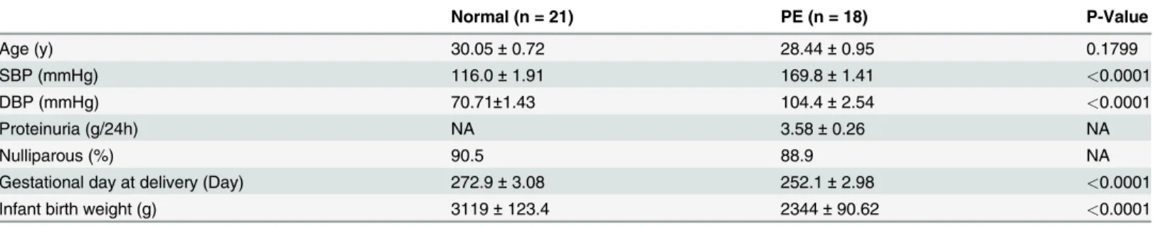
Specimen classification and the inclusion criteria were as follows: normal pregnancy was de-fined as patients with no history of hypertension or proteinuria during weeks 35–40 of preg-nancy who delivered healthily neonates via cesarean section. Severe PE was strictly defined according to the International Society for the Study of Hypertension in Pregnancy [23]. Pa-tients with severe PE were puerperas with no history of chronic hypertension or proteinuria, but who experienced severe hypertension (systolic blood pressure160 mmHG and/or dia-stolic blood pressure110 mmHG) plus severe proteinuria (2.0 g per 24 h or greater than 2+ by dipstick) during pregnancy. Puerperas who had renal disease, hypertension before preg-nancy, placental abruption or placenta praevia, anemia or other hematologic disease, gestation-al diabetes, fetgestation-al distress syndrome or growth retardation were excluded from our study. Placental tissues from 21 women with normal pregnancies and 18 patients with severe PE were collected after cesarean section at the Department of Obstetrics and Gynaecology, Tangdu hos-pital, 2nd Affiliated Hospital of Forth Military Medical University, China. All of the patients in our study were primiparas and the general clinical data, such as age, gestational week, infant birth weight, etc., were matched between groups (Table 1).
Cell culture and transfection
The human immortalized trophoblast cell line HTR8/SVneo and human trophoblast tumor cell line JEG-3 were cultured in DMEM-F12 medium (Hyclone, Logan, USA) supplemented with 10% fetal bovine serum (Gibco, Carlsbad, CA, USA) in a 37°C humidified incubator (5% CO2), and subcultured at ratio of 1:3 when the cells reached 80–90% confluence.
For transient transfection, HTR8/SVneo cells were seeded in complete serum medium the day before transfection, then transfected with miR-519d-3p mimics or inhibitor using Lipofec-tamine 2000 Reagent (Invitrogen, Carlsbad, CA, USA) in Opti-MEM (Hyclone, Logan, USA). The transfection medium was replaced with complete medium 4 h after transfection, and the cells were harvested for subsequent experimental studies 24 h after transfection.
RNA extraction and quantitative real-time PCR
Total RNA was extracted from clinical samples and cells using TRIzol reagent (Invitrogen, Carlsbad, CA, USA) following the manufacturer’s instructions. Reverse transcription of mRNA and miRNAs were performed using the PrimeScript RT Master Mix and SYBR PrimeScript miRNA RT-PCR Kits, respectively (Takara Biotechnology, Dalian, China). Quantitative real-Table 1. Clinical parameters of patients enrolled in our study.
Normal (n = 21) PE (n = 18) P-Value
Age (y) 30.05±0.72 28.44±0.95 0.1799
SBP (mmHg) 116.0±1.91 169.8±1.41 <0.0001
DBP (mmHg) 70.71±1.43 104.4±2.54 <0.0001
Proteinuria (g/24h) NA 3.58±0.26 NA
Nulliparous (%) 90.5 88.9 NA
Gestational day at delivery (Day) 272.9±3.08 252.1±2.98 <0.0001
Infant birth weight (g) 3119±123.4 2344±90.62 <0.0001
Data are shown as Mean±SEM, and significant difference between Normal and PE patients are analyzed with Student’sttest. SBP, systolic blood pressure; DBP, diastolic blood pressure; NA, not available.
time PCR was performed using SYBR Premix Ex Taq (Tli RNaseH Plus; Takara Biotechnology, Dalian, China) using the Applied Biosystems 7500 (Life Technologies);β-actin or U6 were used as internal controls to normalize the relative expression level of the target genes and miRNAs, respectively.
Protein extraction and Western blotting
Total cellular proteins were extracted, subjected to SDS-page electrophoresis and transferred to PVDF membranes using standard procedures. The primary antibodies included rabbit poly-clonal anti-MMP-2 (1:1000; Abcam(Hong Kong) Ltd, HK, China) and mouse monopoly-clonal anti-β-actin (1:1000; Sigma, St. Louis, USA);β-actin was used as an internal loading control. The bands were visualized and imaged using eECL Western Blot Kit (Cwbiotech, Beijing, China) and densitometry was performed using Image J (Version 1.49e, NIH, USA).
Transwell invasion assay
Cell invasion was assessed using Transwell chambers (24-well inserts; 8 um-pore size; Milli-pore, Billerica, MA, USA) that had been pre-coated with Matrigel (200μg/ml, BD Biosciences, Franklin Lakes, New Jersey, USA) as previously described [19]. Briefly, 24 h after transfection, the cells were treated with 10μg/ml mitomycin C for 2 h, trypsinized and seeded into the upper chambers (1 × 10⁵cells/chamber) in serum-free medium, and the lower chambers were filled with DMEM-F12 medium containing 15% FBS as a chemoattractant. The plates were in-cubated for 48 h, then the cells on the upper surface of the chambers were removed gently and the invaded cells on the surface of the lower chambers were fixed in methanol, stained with crystal violet, and the number of cells in four randomly-selected fields of view were imaged using a light microscope and counted.
Wound healing assay
Cells were seeded into 24-well plates, transfected with the miRNA or siRNA (or controls) and cultured for 24 h until about 70% confluent. Prior to the wound healing assay, the cells were treated with 10μg/ml mitomycin C for 2 h to suppress proliferation, then a wound was created in the monolayer using a sterilized 200μl pipette tip, and the width of each wound was mea-sured using Image J at 0 h, 24 h and 48 h after wounding. Cell migration was expressed by sub-tracting the wound width at 24 h or 48 h from that at 0 h.
Luciferase reporter assay
and luciferase activity was measured using the Dual-Luciferase Reporter Assay System (Pro-mega BioSciences, San Luis Obispo, CA, USA) according to the manufacturer’s instructions.
Statistical analysis
GraphPad InStat software (Graphpad, San Diego, USA) was used for statistical analysis. Data from the quantitative real-time PCR, Western blot, Transwell invasion, wound healing and lu-ciferase reporter assays are presented as the mean ± SEM values of three independent experi-ments. Group comparisons were performed using the Student’sttest. The correlation between miR-519d-3p and MMP-2 expression in placentas from patients with PE were analyzed using the Pearson correlation and linear regression analysis. Statistical significance was defined as P<0.05.
Results
MiR-519d-3p is upregulated in the placenta of patients with PE and
downregulated in a trophoblastic tumor cell line
To examine the role of miR-519d-3p in trophoblast cells, we first quantified the expression of miR-519d-3p in placental tissues from 21 women with normal pregnancies and 18 patients with severe PE by qRT-PCR. As shown inFig. 1A, the expression of miR-519d-3p was general-ly higher in placental tissues from patients with PE than those with normal placental tissues. Subsequently, we examined the expression of miR-519d-3p by qRT-PCR in the normal tropho-blast cell line HTR8/SVneo and trophotropho-blastic tumor cell line JEG-3. As shown inFig. 1B, miR-519d-3p was significantly downregulated in JEG-3 cells compared to HTR8/SVneo cells. These data indicate that the expression of miR-519d-3p may be closely related to trophoblast cell function.
MiR-519d-3p suppresses the invasion and migration of trophoblast cells
The Transwell invasion assay was used to evaluate whether miR-519d-3p affects the invasive ability of trophoblast cells. As shown inFig. 2A and 2B, transfection of miR-519d mimic and
Fig 1. Expression of miR-519d-3p is upregulated in the placentas of patients with preeclampsia and a trophoblast cell line.Quantitative real-time PCR analysis of miR-519d expression in placentas from patients with severe preeclampsia (PE) and matched normal pregnancies (Control) (A), and the normal trophoblast cell line HTR8 and trophoblastic tumor cell line JEG-3 (B). Relative expression of miR-519d was normalized to U6.*P<0.05,**P<0.01, ***P<0.001.
Fig 2. MiR-519d-3p suppresses trophoblast cell invasion and migration.(A, B) Quantitative real-time PCR analysis of miR-519d-3p expression in HTR8/SVneo cells transfected with miR-519d-3p mimic (A), 519d-3p inhibitor (B) or the corresponding controls (NC or NC inhibitor). Relative expression of 519d-3p was normalized to U6. (C, D) Transwell invasion assay of HTR8/SVneo cells transfected with miR-519d-3p mimic (C), miR-miR-519d-3p inhibitor (D) or the corresponding controls (NC or NC inhibitor). (E, F) Wound healing assay of HTR8/SVneo cells transfected with miR-519d-3p mimic (E), miR-519d-3p inhibitor (F) or the corresponding controls (NC or NC inhibitor). Scale bar refers to 200um,*P<0.05.
miR-519d inhibitor significantly increased and decreased, respectively, the relative expression of miR-519d-3p in HTR8/SVneo cells. As shown inFig. 2C and 2D, overexpression of miR-519d-3p significantly inhibited HTR8/SVneo cell invasion at 48 h after transfection with the miR-519d-3p mimic compared to the miR-control group. Downregulation of miR-519d-3p by transfection of the miR-519d-3p inhibitor significantly increased the invasive ability of HTR8/ SVneo cells. The wound healing assay was used to evaluate the influence of miR-519d-3p on trophoblast cell migration. As shown inFig. 2E and 2F, overexpression of miR-519d-3p signifi-cantly suppressed HTR8/SVneo cell migration at 24 h and 48 h after transfection with miR-519d-3p mimic, whereas inhibition of miR-miR-519d-3p promoted HTR8/SVneo cell migration at 24 h and 48 h after transfection of the 519d-3p inhibitor. These data indicate that miR-519d-3p suppresses trophoblast cell invasion and migration.
MMP-2 is a direct target of miR-519d-3p
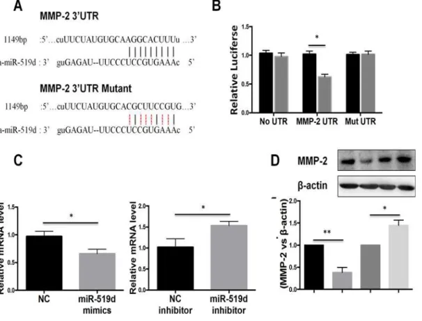
To investigate the mechanism by which miR-519d-3p regulates the invasion and migration of trophoblast cells, we performed bioinformatic analysis of miR-519d-3p. The bioinformatic analysis revealed a putative miR-519d-3p binding site in 3`UTR of MMP-2 (Fig. 3A); this gene is closely associated with the invasion and migration of trophoblast cells [8]. To confirm that miR-519d-3p directly targets MMP-2, we performed luciferase reporter assays in trophoblast cells. As shown inFig. 3B, the luciferase activity of the wild-type MMP-2 3`UTR reporter gene
Fig 3. MMP-2 is a direct target of miR-519d-3p.(A) Schematic illustrating construction of the luciferase reporter constructs (MMP-2 UTR and MMP-2 Mut UTR). (B) Luciferase assay of HTR8/SVneo cells co-transfected with miR-519d-3p mimics (or negative control) and the wild-type MMP-2 3`UTR reporter construct (MMP-2 UTR) or mutated MMP-2 3`UTR reporter construct (Mut UTR). (C, D) Quantitative real-time PCR and Western blot analysis of endogenous MMP-2 mRNA (C) and protein (D) expression in HTR8/SVneo cells transfected with miR-519d-3p mimic or miR-519d-3p inhibitor; data is expressed relative to the corresponding control cells (NC or NC inhibitor),P<0.05.
was markedly lower in cells transfected with miR-519d-3p mimic compared to negative control miRNA transfected-cells; however, this reduction in luciferase activity was abolished by muta-tion of the putative miR-519d-3p binding site in the MMP-2 3`UTR reporter gene. To further validate the association between miR-519d-3p and MMP-2, we quantified endogenous MMP-2 mRNA expression in HTR8/SVneo cells transfected with miR-519d-3p mimic or miR-519d-3p inhibitor. Quantitative RT-PCR demonstrated that transfection of the miR-519d-3p mimic sig-nificantly reduced the expression of endogenous MMP-2 mRNA, whereas the miR-519d-3p in-hibitor significantly increased MMP-2 mRNA expression (Fig. 3C). Western blotting
confirmed that the expression of MMP-2 significantly reduced in cells transfected with miR-519d-3p mimic and increased in cells transfected with the miR-miR-519d-3p inhibitor (Fig. 3D). Taken together, these results indicate that MMP-2 is a direct target gene of miR-519d-3p.
Restoring MMP-2 expression reverses the regulation of miR-519d-3p on
trophoblast cell invasion and migration
MMP-2 has been reported to play a critical role in the migration and invasion of normal tro-phoblast cells and various trotro-phoblastic cancers [8]. To verify whether the function of miR-519d-3p is exerted via regulation of MMP-2, we co-transfected HTR8/SVneo cells with the miR-519d-3p inhibitor and MMP-2 siRNA to perform a rescue experiment. As shown in
Fig. 4A, Western blot analysis showed that simultaneous transfection of HTR8/SVneo cells with the miR-519d-3p inhibitor and MMP-2 siRNA reduced the ability of the miR-519d-3p in-hibitor to upregulate MMP-2 protein expression. Furthermore, the Transwell invasion assay revealed that co-transfection of MMP-2 siRNA significantly reduced the ability of the miR-519d-3p inhibitor to promote cell invasion at 48 h after transfection (Fig. 4B). Additionally, the wound-healing assay showed that co-transfection of MMP-2 siRNA abolished the increased cell migration promoted by transfection of the miR-519d-3p inhibitor at 24 h and 48 h
(Fig. 4C). These results indicate that miR-519d-3p suppresses trophoblast cell invasion and mi-gration via regulating MMP-2.
Expression of MMP-2 correlates negatively with miR-519d-3p in
placental tissues
In order to further elucidate the correlation between MMP-2 and miR-519d-3p, we used qRT-PCR to quantify the expression of MMP-2 and miR-519d-3p in the placentas of patients with PE. As shown inFig. 5A and 5B, the expression of MMP-2 mRNA was reduced in the pla-centas of patients with PE compared to patients with normal pregnancies, and the expression of MMP-2 correlated negatively with the expression of miR-519d-3p. These results indicate that miR-519d-3p can negatively regulate the expression of MMP-2in vivo, indicating that miR-519d-3p may participate in the pathological processes underlying PE.
Discussion
In our study, we found that miR-519d-3p was overexpressed in the placentas of patients with PE, and downregulated in a trophoblast tumor cell line compared to a normal trophoblast cell line. We also proved that miR-519d-3p could suppress the invasion and migration of tropho-blast cells via targeting MMP-2 using in vitro functional and rescue experiments. Furthermore, we demonstrated an inverse correlation exists between the expression of miR-519d-3p and MMP-2 in the placentas of patients with PE.
trophoblast cell functions [26]. MiR-29b participates in the onset of PE by repressing tropho-blast cell invasion and angiogenesis and enhancing cell apoptosis through targeting MMP-2, Integrin beta 1 (ITGB1) and vascular endothelial growth factor A (VEGF-A) [15]. In our study, we observed that the expression of miR-519d was significantly upregulated in the pla-centas of patients with PE compared to those of normal pregnancies, and the expression of miR-519d was downregulated in a trophoblast tumor cell line compared to a normal tropho-blast cell line. MiR-519d-3p is a member of the chromosome 19 miRNA cluster (C19MC), which is exclusively expressed in the placenta and functions as an important regulator of pla-cental–maternal signaling [27]. A recent study showed that circulating C19MC miRNAs play Fig 4. Restoring MMP-2 expression reverses the regulation of miR-519d-3p on trophoblast cell invasion and migration.HTR8/SVneo cells were transfected with miR-519d-3p inhibitor alone, miR-519d-3p inhibitor plus 2 siRNA, and the corresponding control (NC) (A) Western blot assay of MMP-2 protein expression. (B) Transwell invasion assay. (C) Wound healing assay. Scale bar refers to MMP-200um.*P<0.05.
important roles in the pathogenesis of PE, and upregulation of C19MC miRNAs, such as miR-516–5p, miR-517and miR-526a, could be used as a biomarker to diagnose PE [28,29]. To
date, it has been reported that miR-519d-3p promotes cell proliferation and metastasis in a range of pathological processes such as obesity [30], osteosarcoma [31], hepatocellular carcino-ma [20,21] and ovarian cancer [19]; however, a role for miR-519d in PE had not been reported. It is generally accepted that shallow invasion of trophoblast cells into the decidua and subse-quent defects in spiral artery remodeling are involved in the onset of PE [1]. In order to study the function of miR-519d-3p, we examined the effects of miR-519d on the migration and inva-sion of trophoblast cells in vitro by transiently transfecting a trophoblast cell line, HTR8/ SVneo, with a miR-519d-3p mimic or miR-519d-3p inhibitor. Overexpression of miR-519d-3p potently suppressed trophoblast cell migration and invasion in the wound healing and Trans-well invasion assays. In addition, inhibition of endogenous miR-519d-3p significantly promot-ed trophoblast cell migration and invasion. Combinpromot-ed with our findings that the expression of miR-519d-3p is upregulated in the placentas of patients with PE compared to those of normal pregnancies, these observations indicate that miR-519d-3p may play an important role in ab-normal trophoblast migration and invasion during the pathological processes leading to PE.
In our study, we demonstrated that miR-519d-3p interacted with its partially complementa-ry sequence in the 3`UTR of MMP-2 using luciferase reporter assays, and confirmed that miR-519d-3p negatively regulated MMP-2 mRNA and protein expression using qRT-PCR and Western blot analyses. In functional rescue experiments, we found that knockdown of MMP-2 using a siRNA suppressed trophoblast cell migration and invasion; in a similar manner as over-expression of miR-519d-3p. Additionally, the MMP-2 siRNA reversed the ability of miR-519d to promote trophoblast cell migration and invasion. Furthermore, to confirm the regulation of MMP-2 by miR-519d-3p in vivo, we analyzed the expression of miR-519d-3p and MMP-2 in the placentas of patients with PE and those of patients with normal pregnancies, and observed an inverse correlation between the expression of MMP-2 and miR-519d-3p. Consistent with our previous observations described above, these findings demonstrate that miR-519d-3p regu-lates trophoblast cell migration and invasion by targeting MMP-2.
Fig 5. Expression of MMP-2 correlates negatively with miR-519d-3p in placental tissues.(A) Quantitative real-time PCR analysis of MMP-2 mRNA expression in placentas from patients with PE (PE) and normal pregnancies (control). Relative expression of MMP-2 was normalized toβ-actin. Data is mean±SEM of three independent experiments;*,P<0.05 vs. control, Student’st-test. (B) The correlation between miR-519d-3p and MMP-2 expression in placentas from patients with PE was analyzed using the Pearson correlation and linear regression analysis. Relative expression of miR-519d and MMP-2 were normalized to U6 orβ-actin, respectively.
In summary, our study extends our knowledge of the mechanism of action of miRNAs dur-ing the pathogenesis of PE. Upregulation of miR-519d-3p may contribute to the occurrence of PE by inhibiting trophoblast cell migration and invasion via targeting MMP-2, suggesting miR-519d-3p may have potential as a predictive or therapeutic target for PE.
Acknowledgments
This work was supported by grants from the Natural Science Foundation of China (81471474, 31000660, 31301127), Postdoctoral Science Foundation of China (2014T70964, 2013M532203, 2012M521869) and the Tangdu Hospital Reserve Personnel Fund (Xiaoming Zhu). We grate-fully acknowledge the assistance and cooperation of the staffs of the Department of Obstetrics and Gynaecology, Tangdu hospital, 2nd Affiliated Hospital of Forth Military Medical Universi-ty and thank for all study participants for their support. The funders had no role in study de-sign, data collection and analysis, decision to publish, or preparation of the manuscript.
Author Contributions
Conceived and designed the experiments: X. Zhu FH YY X. Zhang. Performed the experiments: JD FH GW TH FX. Analyzed the data: JD FH X. Zhu YY X. Zhang. Contributed reagents/ma-terials/analysis tools: JD FH DW CW. Wrote the paper: JD FH X. Zhu.
References
1. Chaiworapongsa T, Chaemsaithong P, Yeo L, Romero R (2014) Pre-eclampsia part 1: current under-standing of its pathophysiology. Nat Rev Nephrol 10: 466–480. doi:10.1038/nrneph.2014.102PMID: 25003615
2. Matsubara K, Matsubara Y, Hyodo S, Katayama T, Ito M (2010) Role of nitric oxide and reactive oxygen species in the pathogenesis of preeclampsia. J Obstet Gynaecol Res 36: 239–247. doi:10.1111/j. 1447-0756.2009.01128.xPMID:20492372
3. Goel A, Rana S (2013) Angiogenic factors in preeclampsia: potential for diagnosis and treatment. Curr Opin Nephrol Hypertens 22: 643–650. doi:10.1097/MNH.0b013e328365ad98PMID:24076553
4. Perez-Sepulveda A, Torres MJ, Khoury M, Illanes SE (2014) Innate immune system and preeclampsia. Front Immunol 5: 244. doi:10.3389/fimmu.2014.00244PMID:24904591
5. Hsu P, Nanan RK (2014) Innate and adaptive immune interactions at the fetal-maternal interface in healthy human pregnancy and pre-eclampsia. Front Immunol 5: 125. doi:10.3389/fimmu.2014.00125 PMID:24734032
6. Knofler M, Pollheimer J (2013) Human placental trophoblast invasion and differentiation: a particular focus on Wnt signaling. Front Genet 4: 190. doi:10.3389/fgene.2013.00190PMID:24133501
7. Visse R, Nagase H (2003) Matrix metalloproteinases and tissue inhibitors of metalloproteinases: struc-ture, function, and biochemistry. Circ Res 92: 827–839. PMID:12730128
8. Myers JE, Merchant SJ, Macleod M, Mires GJ, Baker PN, Davidge ST. (2005) MMP-2 levels are elevated in the plasma of women who subsequently develop preeclampsia. Hypertens Pregnancy 24: 103–115. PMID:16036395
9. Sankaralingam S, Arenas IA, Lalu MM, Davidge ST (2006) Preeclampsia: current understanding of the molecular basis of vascular dysfunction. Expert Rev Mol Med 8: 1–20.
10. Lockwood CJ, Basar M, Kayisli UA, Guzeloglu-Kayisli O, Murk W, Wang J, et al. (2014) Interferon-γ
Protects First-Trimester Decidual Cells against Aberrant Matrix Metalloproteinases 1, 3, and 9 Expres-sion in Preeclampsia. The American Journal of Pathology 184: 2549–2559. doi:10.1016/j.ajpath.2014. 05.025PMID:25065683
11. Carthew RW, Sontheimer EJ (2009) Origins and Mechanisms of miRNAs and siRNAs. Cell 136: 642–655. doi:10.1016/j.cell.2009.01.035PMID:19239886
12. Cristino AS, Barchuk AR, Freitas FC, Narayanan RK, Biergans SD, et al. (2014) Neuroligin-associated microRNA-932 targets actin and regulates memory in the honeybee. Nat Commun 5: 5529. doi:10. 1038/ncomms6529PMID:25409902
14. Morales-Prieto DM, Chaiwangyen W, Ospina-Prieto S, Schneider U, Herrmann J, Gruhn B, et al. (2012) MicroRNA expression profiles of trophoblastic cells. Placenta 33: 725–734. doi:10.1016/j. placenta.2012.05.009PMID:22721760
15. Li P, Guo W, Du L, Zhao J, Wang Y, Liu L, et al. (2013) microRNA-29b contributes to pre-eclampsia through its effects on apoptosis, invasion and angiogenesis of trophoblast cells. Clin Sci (Lond) 124: 27–40. doi:10.1042/CS20120121PMID:22716646
16. Wang Y, Fan H, Zhao G, Liu D, Du L, Wang Z, et al. (2012) miR-16 inhibits the proliferation and angio-genesis-regulating potential of mesenchymal stem cells in severe pre-eclampsia. FEBS J 279: 4510–4524. doi:10.1111/febs.12037PMID:23083510
17. Hu Y, Li P, Hao S, Liu L, Zhao J, Hou Y. (2009) Differential expression of microRNAs in the placentae of Chinese patients with severe pre-eclampsia. Clin Chem Lab Med 47: 923–929. doi:10.1515/CCLM. 2009.228PMID:19642860
18. Zhu XM, Han T, Sargent IL, Yin GW, Yao YQ (2009) Differential expression profile of microRNAs in human placentas from preeclamptic pregnancies vs normal pregnancies. Am J Obstet Gynecol 200: 661 e661–667. doi:10.1016/j.ajog.2008.12.045PMID:19285651
19. Pang Y, Mao H, Shen L, Zhao Z, Liu R, Liu P. (2014) MiR-519d represses ovarian cancer cell prolifera-tion and enhances cisplatin-mediated cytotoxicity in vitro by targeting XIAP. Onco Targets Ther 7: 587–597. doi:10.2147/OTT.S60289PMID:24790458
20. Fornari F, Milazzo M, Chieco P, Negrini M, Marasco E, Capranico G. et al. (2012) In hepatocellular car-cinoma miR-519d is up-regulated by p53 and DNA hypomethylation and targets CDKN1A/p21, PTEN, AKT3 and TIMP2. J Pathol 227: 275–285. doi:10.1002/path.3995PMID:22262409
21. Hou YY, Cao WW, Li L, Li SP, Liu T, Wan HY, et al. (2011) MicroRNA-519d targets MKi67 and sup-presses cell growth in the hepatocellular carcinoma cell line QGY-7703. Cancer Lett 307: 182–190. doi:10.1016/j.canlet.2011.04.002PMID:21524841
22. Li H, Ge Q, Guo L, Lu Z (2013) Maternal plasma miRNAs expression in preeclamptic pregnancies. Biomed Res Int 2013: 970265. doi:10.1155/2013/970265PMID:24195082
23. Chaiworapongsa T, Chaemsaithong P, Korzeniewski SJ, Yeo L, Romero R (2014) Pre-eclampsia part 2: prediction, prevention and management. Nat Rev Nephrol 10: 531–540. doi:10.1038/nrneph.2014. 103PMID:25003612
24. Santamaria X, Taylor H (2014) MicroRNA and gynecological reproductive diseases. Fertil Steril 101: 1545–1551. doi:10.1016/j.fertnstert.2014.04.044PMID:24882618
25. Bai Y, Yang W, Yang HX, Liao Q, Ye G, Fu G, et al. (2012) Downregulated miR-195 detected in pre-eclamptic placenta affects trophoblast cell invasion via modulating ActRIIA expression. PLoS One 7: e38875. doi:10.1371/journal.pone.0038875PMID:22723898
26. Luo L, Ye G, Nadeem L, Fu G, Yang BB, Honarparvar E, et al. (2012) MicroRNA-378a-5p promotes tro-phoblast cell survival, migration and invasion by targeting Nodal. J Cell Sci 125: 3124–3132. doi:10. 1242/jcs.096412PMID:22454525
27. Donker RB, Mouillet JF, Chu T, Hubel CA, Stolz DB, Morelli AE, et al. (2012) The expression profile of C19MC microRNAs in primary human trophoblast cells and exosomes. Mol Hum Reprod 18: 417–424. doi:10.1093/molehr/gas013PMID:22383544
28. Hromadnikova I, Kotlabova K, Ondrackova M, Kestlerova A, Novotna V, Hympanova L, et al. (2013) Circulating C19MC microRNAs in preeclampsia, gestational hypertension, and fetal growth restriction. Mediators Inflamm 2013: 186041. doi:10.1155/2013/186041PMID:24347821
29. Noguer-Dance M, Abu-Amero S, Al-Khtib M, Lefevre A, Coullin P, Moore GE, et al. (2010) The primate-specific microRNA gene cluster (C19MC) is imprinted in the placenta. Hum Mol Genet 19: 3566–3582. doi:10.1093/hmg/ddq272PMID:20610438
30. Martinelli R, Nardelli C, Pilone V, Buonomo T, Liguori R, CastanòI, et al. (2010) miR-519d overexpres-sion is associated with human obesity. Obesity (Silver Spring) 18: 2170–2176. doi:10.1038/oby.2009. 474PMID:20057369