Signaling Pathway
Tomoko Uehara1, Eriko Kage-Nakadai1¤a, Sawako Yoshina1, Rieko Imae1¤b, Shohei Mitani1,2*
1Department of Physiology, Tokyo Women’s Medical University School of Medicine, Tokyo, Japan,2Tokyo Women’s Medical University Institute for Integrated Medical Sciences, Tokyo, Japan
Abstract
HumanBCL7gene family consists ofBCL7A,BCL7B, andBCL7C. A number of clinical studies have reported thatBCL7family is involved in cancer incidence, progression, and development. Among them,BCL7B, located on chromosome7q11.23, is one of the deleted genes in patients with Williams-Beuren syndrome. Although several studies have suggested that malignant diseases occurring in patients with Williams-Beuren syndrome are associated with aberrations inBCL7B, little is known regarding the function of this gene at the cellular level. In this study, we focused onbcl-7, which is the only homolog ofBCL7
gene family inCaenorhabditis elegans, and analyzedbcl-7deletion mutants. As a result, we found thatbcl-7is required for the asymmetric differentiation of epithelial seam cells, which have self-renewal properties as stem cells and divide asymmetrically through the WNT pathway. Distal tip cell development, which is regulated by the WNT pathway in
Caenorhabditis elegans, was also affected in bcl-7-knockout mutants. Interestingly, bcl-7 mutants exhibited nuclear enlargement, reminiscent of the anaplastic features of malignant cells. Furthermore, in KATOIII human gastric cancer cells,
BCL7B knockdown induced nuclear enlargement, promoted the multinuclei phenotype and suppressed cell death. In addition, this study showed that BCL7B negatively regulates the Wnt-signaling pathway and positively regulates the apoptotic pathway. Taken together, our data indicate that BCL7B/BCL-7 has some roles in maintaining the structure of nuclei and is involved in the modulation of multiple pathways, including Wnt and apoptosis. This study may implicate a risk of malignancies withBCL7B-deficiency, such as Williams-Beuren syndrome.
Citation:Uehara T, Kage-Nakadai E, Yoshina S, Imae R, Mitani S (2015) The Tumor Suppressor BCL7B Functions in the Wnt Signaling Pathway. PLoS Genet 11(1): e1004921. doi:10.1371/journal.pgen.1004921
Editor:Kaveh Ashrafi, University of California San Francisco, United States of America
ReceivedJune 3, 2014;AcceptedNovember 24, 2014;PublishedJanuary 8, 2015
Copyright:ß2015 Uehara et al. This is an open-access article distributed under the terms of the Creative Commons Attribution License, which permits unrestricted use, distribution, and reproduction in any medium, provided the original author and source are credited.
Data Availability:The authors confirm that all data underlying the findings are fully available without restriction. All relevant data are within the paper and its Supporting Information files.
Funding:This work was supported by the Global COE program, Multidiciplinary Education and Research Center for Regeneration Medicine (MERCREM), from the Ministry of Education, Culture, Sports, Science, and Technology (MEXT), Japan. The funders had no role in study design, data collection and analysis, decision to publish, or preparation of the manuscript.
Competing Interests:The authors have declared that no competing interests exist. * Email: [email protected]
¤a Current address: Advanced Research Institute for Natural Science and Technology, Osaka City University, Osaka, Japan ¤b Current address: Graduate School of Pharmaceutical Science, University of Tokyo, Tokyo, Japan
Introduction
Cytogenetic abnormalities of chromosome 7 occur frequently in patients with cancer. Patients with certain types of malignant transformation, such as acute lymphoblastic leukemia, myelodys-plastic syndrome, or juvenile myelomonocytic leukemia, frequent-ly have deletions or abnormalities in chromosome 7 [1–4]. Some of the genes located on chromosome 7 are thought to act as tumor-related genes, with roles in cancer initiation and/or progression. However, few studies have investigated the molecular mechanisms controlled by specific genes located on chromosome 7.
One of the most well-known diseases related to chromosome 7 microdeletions is Williams-Beuren syndrome (WBS), a contiguous gene syndrome with a dominant autosomal inheritance pattern. WBS patients show a variety of phenotypes, including elfin face, mental retardation, reduced spatial reasoning capacity, supravalvular aortic stenosis, and peripheral pulmonic stenosis. In the past three decades, several reports have described the occurrence of malignant diseases in WBS patients [5–9]. These reports have shown that patients with WBS
are at an increased risk of malignant transformation due to aberrations in candidate genes, such asBCL7B.
BCL7Bis a member of theBCL7gene family; members of this gene family, includingBCL7AandBCL7C, located on chromosomes 12 and 16, respectively, have a conserved amino-terminal region as their functional domain [10]. A number of studies have found thatBCL7 family members are involved in cancer initiation, progression, and development. For example, decreased expression ofBCL7Amay be a risk factor for astrocytoma [11], Burkitt lymphoma [12], non-Hodgkin’s lymphoma [13], mycosis fungoides [14], and cutaneous T cell lymphoma [15]. Although theBCL7gene family is thought to have tumor-associated functions, little is known regarding the specific functional roles ofBCL7genes; this may be attributed to the functional redundancy amongBCL7family members, which makes it difficult to analyze the individual roles ofBCL7genes.
homolog inCaenorhabditis elegans(S1A Fig.), was analyzed in the Wnt-signaling pathway and apoptotic pathway. In addition, we also analyzed the function of theBCL7Bgene in both pathways in KATOIII cells, a human gastric cancer cell line [16].
Results
bcl-7is required for normal seam cell development inC. elegans
First, we knocked downbcl-7expression using the feeding RNA interference (RNAi) technique with a bcl-7-specific RNAi clone and observed the phenotypes associated withbcl-7downregulation in wild-typeC. eleganshermaphrodites. Downregulation ofbcl-7 in wild-type worms resulted in the egg-laying defective (Egl) phenotype (S1B and S1E Fig.), the protruding vulva (Pvl) phenotype (S1C Fig.), and the burst phenotype (S1D Fig.), reminiscent of defects in epidermal barrier formation [17]. Therefore, we hypothesized that bcl-7 is involved in the development of the epidermis.
Next, to examine the phenotypes produced bybcl-7knockout, we generated a bcl-7 deletion mutant, tm5268, containing a deletion of 0.7 kbp (Fig. 1A). Because the deletion covered almost allbcl-7 regions, including the amino-terminal domain, which is conserved and considered to be the functional domain [10], tm5268is practically a null mutant. Thetm5268mutant showed a variety of phenotypes, including Pvl (the rate was 61.4%; S1F–I Fig.), the alae morphological variant (Fig. 1B–D, and 1L), and sterility (Ste), which suggest the phenotypes in deletion mutants not only reproduced the RNAi experiments but also indicated additional phenotypes. While the bcl-7 mutants had a normal number of vulval precursor cells at the larval stages, they showed Pvl phenotypes after young adult stages (S1G–H Fig.). In addition, the Pvl phenotype was also observed inbcl-7 heterozygotes at a rate of 14.3% (S1I Fig.). This result suggests that the phenotype of bcl-7deletion mutants is semi-dominant similar to the phenotype of BCL7B deletion in human disease, such as Williams-Beuren syndrome. Furthermore, alae, the cuticle structures considered a hallmark of normal seam cell differentiation, were ‘‘incomplete’’ (alae with only one or two ridges) or absent intm5268worms in contrast to wild-type worms (Fig. 1B and 1C). The Pvl phenotype
and alae malformation are caused by defects in epidermal cells, particularly epidermal stem-like seam cells [17,18]. The presence of these phenotypes inbcl-7deletion mutants suggests that BCL-7 influences the development of seam cells, which have both self-renewal potential and differentiation capability, inC. elegans.
Then, we investigated whether the number of seam cells was altered inbcl-7deletion mutants by analyzing transgenic worms carrying the scm::gfp transgene [19] as a marker of seam cell nuclei. In a wild-type L4-stage hermaphrodite, there were 16 seam cells on each side (Fig. 1E). By contrast, the number of seam cells was significantly reduced in the mutant worms (Fig. 1I), and scm::gfp-negative cells were observed more often in the V cell lineage than in the H and T cell lineages (Fig. 1I). Differences between wild-type worms andtm5268worms were found in most larval stages, except for the early L1 stage (Fig. 1F–I and 1K). The expression pattern of another seam cell marker,cdh-3::gfp[20], which is localized to the cytoplasm of seam cells, also revealed that the number of GFP-positive cells was lower inbcl-7mutants (S2A– D Fig.). Both the defect of alae and the decreased seam cell number were rescued by the introduction ofbcl-7genomic DNA, Pbcl-7::bcl-7::egfp(tm5268;tmEx2966) orPbcl-7::bcl-7::mCherry (tm5268;tmEx3496) (Fig. 1D and 1J–M). The expression of the rescue constructs was ubiquitous, including in the hypodermis, from the embryonic stage to the adult stage, and BCL-7 was localized to the nuclei (S3A–H Fig.). Therefore, we hypothesized that BCL-7 functions cell-autonomously in seam cells. To test this hypothesis, we analyzed whether expressing a seam cell-specific construct rescues decreased seam cell number. The number of seam cells was significantly increased by the introduction of bcl-7 genomic DNA under a seam cell-specific promoter (tmEx4126[scmp::bcl-7, scmp::mCherry]) (Fig. 1M). These results suggest that BCL-7 is involved in the normal development of seam cells and functions cell-autonomously in seam cells.
Next, we addressed whether the observed decrease in seam cells intm5268worms resulted from the hyperactivation of apoptosis. To examine this, we analyzed whether the apoptotic pathway was hyperactivated inbcl-7deletion mutant worms using worms with a mutation in the ced-3 gene, which encodes a member of the caspase family required for the execution of apoptosis inC. elegans [21]. However,bcl-7(III);ced-3(IV) double mutants did not exhibit increased numbers of seam cells compared with bcl-7 single mutants (the average seam cell number in adult, double-mutant hermaphrodites was 3.6 (n = 12)), suggesting that the decrease in the number of seam cells inbcl-7-deficient worms is not caused by hyperactivation of apoptosis.
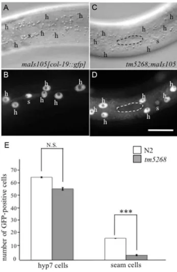
In wild-typeC. elegans, seam cells divide asymmetrically during each larval stage. The anterior daughter cell loses its seam cell properties and differentiates into a hyp7 cell, whereas the posterior cell keeps its self-renewal potential, remaining a seam cell (S4S Fig.) [22,23]. To examine whether the reduction in seam cell numbers in tm5268 is followed by an increase in the hyp7 cell number, we used transgenic worms that expressed an adult-specific hypodermal marker, col-19::gfp [24,25]. In wild-type animals, col-19::gfp was expressed in the hypodermal syncytial hyp7 cells and 16 seam cells (Fig. 2A, 2B and 2E). The number of GFP-positive hyp7 cells intm5268was not increased but tended to decrease compared with the number in the wild-type worms. Additionally, the number of GFP-positive seam cells intm5268 was significantly decreased compared with the number in the wild-type worms (Fig. 2C–E). Interestingly, the nuclei of hyp7 cells from bcl-7 mutants were significantly enlarged and had an irregular shape compared to those from wild-type animals (7.6660.10mm and 6.7760.14mm, p,0.001; S5A–D Fig.).
Author Summary
BCL7B, a member of the human BCL7 gene family, is deleted in patients with Williams-Beuren syndrome. Although several clinical studies have suggested that malignant diseases occurring in patients with Williams-Beuren syndrome are associated with aberrations inBCL7B, little is known regarding the physiological function of this gene. Here, we show thatbcl-7, the only homolog ofBCL7
gene family inCaenorhabditis elegans, regulates asymmet-ric cell differentiation in somatic ‘‘stem-like’’ seam cells through at least the Wnt pathway and promotes the apoptotic pathway. In addition, bcl-7 deletion mutants show enlarged nuclei in epidermis and germ cells. Furthermore, in KATOIII human gastric cancer cells,BCL7B
knockdown induces nuclear enlargement, as observed in
To determine whether the somatic stem-like cells acquire another fate in the cell lineage, we analyzed neural cells, including sensory PVD neurons, PDE neurons, and phasmids,
derived from V- and T-cell lineages. PVD and PDE neurons originate from an asymmetric division of the V5 cell (S4S Fig.) [23], whereas phasmid cells are generated by the asymmetric T-cell division when the posterior daughter T-cell maintains the seam cell phenotype and the anterior daughter cell commits to the neural fate, giving rise to the phasmid (S4T Fig.) [23]. We analyzed the cell fates of PVD and PDE in transgenic worms carrying the PVD and PDE markersdes-2::gfpanddat-1p::gfp, respectively [26,27]. No extra PVD or PDE cells were found in any of thebcl-7mutants (n = 10–15, S4A–J Fig.). In wild-type worms, phasmids can be detected by the uptake of fluorescent dye [28]. Similarly, in allbcl-7mutants, both socket cells in the phasmid, but not other cells, absorbed the fluorescent dye in dye-filling assays (n = 10, S4K–N Fig.). These results suggest that stem-like cells fail to acquire a terminal differentiation fate in bcl-7mutants.
Next, we hypothesized that more undifferentiated cells are present inbcl-7deletion mutants because we observed an increase in the number of cells with enlarged nuclei, as well as the loss of differentiated-cell markers. To test this hypothesis, we analyzed the expression patterns of an undifferentiated state marker, egl-27/ Mta [29], in wild type and tm5268 worms carrying the egl-27p::his-24::mCherrytransgene. The expression pattern ofegl-27 was different between wild type andbcl-7mutant worms. In wild type worms, egl-27 was strongly expressed in the nuclei of intestinal cells (S6A–B Fig.) and weakly expressed in the nuclei of epidermis (S6C–D Fig.) during the L4 stage (n = 10). By contrast, egl-27 was strongly expressed ubiquitously particularly in the epidermal cells including both seam cells and hyp7 cells inbcl-7 mutant worms (n = 10, S6E–H Fig.). Furthermore, we analyzed the expression levels of undifferentiated cell markers using a quantitative real-time polymerase chain reaction (qRT-PCR) analysis. Both egl-27 andceh-6, markers of the undifferentiated state in C. elegans, were significantly increased in the tm5268 mutants compared with the wild type worms (S6I–J Fig.). These results suggest that stem-like cells fail to acquire a terminal differentiation fate inbcl-7mutants.
Loss of BCL-7 function affects gonadal size and germ cell differentiation
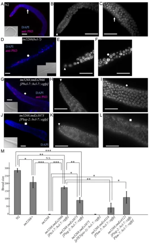
The Ste phenotype was observed in bcl-7 deletion mutants (Fig. 3M); specifically, no oocytes were detected in the homozy-gous mutants, and their gonads were shortened (Fig. 3D). In addition, the brood size ofbcl-7 heterozygotes was significantly reduced compared with that of wild-type worms (Fig. 3M), which indicates that the genetic trait ofbcl-7 mutation inC. elegansis haploinsufficiency, similar to that of human diseases. These results revealed that the loss of BCL-7 function affects not only seam cells but also the development of somatic gonads and/or germ cells. To Fig. 1. Knockout ofbcl-7inhibits normal seam cell development inCaenorhabditis elegans.A: Structure ofbcl-7. Black boxes, exons; bent lines, introns. The region deleted in the straintm5268is shown as a dotted line, below. The numbers indicate the location in the cosmidC28H8.B–D: Nomarski images of adult hermaphrodites. A wild-type hermaphrodite had complete alae (the region between arrows) (B); Alae were incomplete in bcl-7(tm5268) hermaphrodites. A region between dotted arrows indicates partial alae with only two ridges. The area without arrows indicates regions without alae (C); a transgenic rescue line carryingPbcl-7::bcl-7::egfpreporters (tm5268;tmEx2966) with complete alae (three ridges) (D).E–J: Examples of SCM::GFP localization in wild-type,tm5268, andtm5268;tmEx3496hermaphrodites withwIs51(SCM::GFP).tm5268;tmEx3496is a transgenic rescue line carryingPbcl-7::bcl-7::mCherryreporters. A wild-type L4 hermaphrodite expressing SCM::GFP in 16 seam cell nuclei (E),tm5268hermaphrodites carryingscm::gfpreporters (H–G), and atm5268;tmEx3496hermaphrodite carryingscm::gfpreporters (J). Inserts show the Nomarski images of the same animals of the fluorescence images.K: Bar chart showing the average number of seam cells (about the worms at each larval stage, except for the middle L2 stage, and the young adult stage) or daughter cells (about the worms at middle L2 stage) expressing GFP in wild-type andtm5268 hermaphrodites (n = 21–46).L: Percentages of worms with complete alae in wild-type,tm5268, andtm5268;tmEx2966adult hermaphrodites (n = 20– 30).M: Bar chart showing the average number of seam cells expressing GFP in wild-type,tm5268,tm5268;tmEx3496, and tm5268;tmEx4126[SCMp::bcl-7, SCMp::mCherry]adult hermaphrodites withwIs51(SCM::GFP) (n = 15–23). Error bars indicate the standard error of the mean (SEM). Asterisks indicate statistical significance compared with each other. *p,0.05. **p,0.005. ***p,0.001. N.S.: no significance. Scale bar = 25mm.
doi:10.1371/journal.pgen.1004921.g001
further investigate the gonadal and germ cell phenotypes intm5268 worms, we performed diamidinophenylindol (DAPI)-staining and fluorescence immunostaining with an anti-phospho-histone H3 (PH3) antibody as a mitotic marker [30]. In wild-typeC. elegans hermaphrodites, the mitotic region covered approximately 10–15 cell diameters as previously reported (Fig. 3A) [31]. Inbcl-7mutant worms, PH3-positive cells were found, but tended to decrease compared with the wild type worms (Fig. 3D). In addition, they were occasionally observed farther from the distal tip cells (DTCs) than in wild type worms. These results suggest that the shortened gonad observed intm5268mutants was not because of the absence of mitosis but rather because of the decrease of mitosis and may be due to the defects of cell differentiation after mitosis. In addition, the average length of the major axis of germ cell nuclei was 3.1760.03mm in wild type worms (Fig. 3A–C, S7A Fig.). By contrast, bcl-7 mutants exhibited irregularly shaped and signifi-cantly larger germ cell nuclei measuring 4.4260.08mm (Fig. 3D–F, S7B Fig.). Furthermore, the number of germ cells intm5268worms was decreased compared with that in wild-type worms (Fig. 3D–F). Thus, BCL-7 is necessary for gonadal development, particularly for gonadal arm elongation, and for germ cell entry into meiosis. The Ste phenotype, shortened gonads, and enlarged germ cell nuclei in tm5268worms were rescued by the introduction ofbcl-7genomic DNA under the bcl-7 promoter (Fig. 3G–I, 3M and S7C). The rescue construct was strongly expressed in the nuclei of somatic DTCs and weakly expressed in the nuclei of germ cells and gonadal sheath cells (S3I–L Fig.). These results suggest that the expression of BCL-7 in DTCs, germ cells, and/or gonadal sheath cells is necessary for its function.
To further examine the observed phenotypes, we introduced bcl-7 genomic DNA under the DTC-specific lag-2 promoter (Plag-2::bcl-7::egfp). The Ste phenotype of tm5268 worms was partially rescued by the expression of the bcl-7 gene in DTCs (Fig. 3M). In addition, thin gonads and irregularly shaped nuclei of germ cells were also partially rescued by the expression of this construct (Fig. 3J–L and S7D). These results suggest that BCL-7 expression in DTCs is necessary but insufficient for normal gonadal development. Therefore, we hypothesized that restoring BCL-7 expression in germ cells could rescue the Ste phenotype in bcl-7 deletion mutants (tm5268). To test this hypothesis, we expressed bcl-7 genomic DNA fused with the promoter, first intron, and 39-untranslated region (UTR) of thepie-1gene, which is expressed exclusively in germ cells (pTE-5(pie-1)::bcl-7) [30]. However, the Ste phenotype was not rescued by the introduction of pTE5(pie-1)::bcl-7 (tmEx3875) (Fig. 3M). Our study showed that BCL-7 was also expressed in gonadal sheath cells (S3I–L Fig., arrows indicate a part of gonadal sheath cells). Gonadal sheath cells play an important role in embryonic germline amplification and larval gonadal elongation [32]. Therefore, we examined whether the expression of BCL-7 in gonadal sheath cells is sufficient for normal gonadal development by introducing bcl-7 genomic DNA under a sheath cell-specific sequence (lim-7 promoter and first intron; tmEx4116[Plim-7::bcl-7::egfp]) [33,34]. The Ste phenotype of tm5268 worms was partially rescued (Fig. 3M). In addition, we introduced both the DTC-specific rescue construct and the sheath cell-DTC-specific rescue construct (tmEx4121[Plag-2::bcl-7::egfp, Plim-7::bcl-7::egfp]) and found that the brood size was larger than with either the DTC-specific rescue or the gonadal sheath cell-specific rescue alone (Fig. 3M). These results suggest that BCL-7 expression in somatic DTCs and gonadal sheath cells is more important than its expression in germ cells for normal gonadal development.
One of the factors that regulates the size of the gonads and the timing of the entry into meiosis is LAG-2, a homolog of the Notch
receptor ligand that is secreted by DTCs [35,36]. We therefore hypothesized that DTC functions, including LAG-2 secretion, may be impaired in bcl-7 mutants. We analyzed DTCs using worms carrying thelag-2p::gfptransgene (qIs56) [37]. Two GFP-positive DTCs were detected in 100% (17/17) of wild-type worms (S8A and B Fig.), whereas only 19.5% (25/128) ofbcl-7mutant worms were positive for GFP in both DTCs. The remaining 80.5% (103/128) ofbcl-7mutant worms were positive for GFP in only one DTC. Furthermore, 12.5% (16/128) ofbcl-7 mutants showed mispositioning of the DTCs with the expression of lag-2p::gfp(S8E–H Fig. and S1 Table). Interestingly, heterozygous bcl-7mutants exhibited the same phenotypes; i.e., 30% (6/20) of heterozygousbcl-7mutants had expression oflag-2p::gfpon only one DTC and 50% (10/20) showed mispositioning of the DTC with GFP expression (S1 Table, middle row). These differences between homozygous and heterozygous mutants further demon-strate that the phenotype is semi-dominant. These results suggest that BCL-7 partially controls the differentiation of DTCs.
Next, we attempted to determine the time course of BCL-7 function using a chromophore-assisted light inactivation (CALI) assay. We created a light-inactivatable BCL-7::KillerRed fusion protein by substitutingegfpof the rescue construct withKillerRed. We generated transgenic worms (tmEx3878) that expressed the bcl-7 promoter-driven bcl-7::KillerRed construct ( Pbcl-7::bcl-7::KillerRed) in the bcl-7-deficient background (i.e.,tm5268). In the absence of green-light illumination, this transgene rescued the Ste phenotype (similar results were observed independently in three transgenic lines). When the animals were illuminated with a green light, the BCL-7 protein fused to KillerRed was inactivated [38,39], allowing inactivation of BCL-7 function. We exposed tm5268;tmEx3878to a green light starting from the comma stage of the embryo, the early larval L1 stage, or the L2 stage to the young adult stage and analyzed the Ste phenotype. All worms exposed to the green light at the L2 stage were fertile; however, approximately 90% (70/79) of worms illuminated at the comma stage and 54.5% (18/33) of worms illuminated at the early L1 stage retained the Ste phenotype (S7E Fig.). In addition, we performed a pulse experiment to identify the most critical stage in development for the function of BCL-7. We exposed the same transgenic worms to a green light during the comma and early L1 stages and found that only 20% of the worms were fertile (S7E Fig.). These results imply that the expression and function of BCL-7 in DTCs begin during the early L1 stage, which corresponds to the timing of asymmetric cell division in somatic DTCs.
RNAi-based screening for suppressors of thebcl-7 mutant phenotypes
LSY-22 is considered a homolog of Groucho-like protein [43] and is also a member of the noncanonical Wnt pathway,lsy-22-specific RNAi only partially suppressed the Ste phenotype and did not affect the number of seam cells. Subsequently, we focused on WRM-1, which exhibited potent effects in both somatic cells and germ cells and was expressed in both seam cells and SGPs. Interestingly, the suppressor effect ofwrm-1RNAi appeared to be dependent on the strength of the RNAi clone (S3 Table). The effect of the wrm-1c RNAi clone (constructed with a cDNA generated from wild-type worms) on N2 was stronger than that of wrm-1RNAi (from the Ahringer library) and comparable with the phenotypes of thewrm-1mutant strains (WormBase; http://www. wormbase.org/). The effect of diluted wrm-1c RNAi seemed comparable to RNAi by a genomic clone on suppressing thebcl-7 mutant’s phenotypes (Fig. 4E, S3 Table).
Based on the results of the suppressor screening, we hypothe-sized that BCL-7 negatively regulates WRM-1 expression. To test this hypothesis, we analyzed the expression levels ofwrm-1mRNA using qRT-PCR. Additionally, because WRM-1 is one of the three ß-catenin homologs inC. elegans, we also analyzed the expression levels of the other ß-catenin homologs, BAR-1 and SYS-1. Compared with wild type worms, bar-1and sys-1mRNAs were significantly increased inbcl-7deletion mutants (S9A Fig.). There was also a trend for increasedwrm-1 in the mutants (S9A Fig.). These results suggest that BCL-7 functions as a negative regulator of the expression of three ß-catenin homologs inC. elegans.
The phenotypes of thebcl-7 deletion mutants were different from thepop-1deletion mutants andwrm-1(gf) mutants, which are associated with hyperactivation of the Wnt pathway. Therefore, we hypothesized that BCL-7 not only functions as a negative regulator but also affects the Wnt pathway in a different mechanism. Because WRM-1/ß-catenin regulates the levels of POP-1 in the nuclei of target cells during asymmetric cell division, we assumed that the cellular distribution of WRM-1 or POP-1 may be altered inbcl-7 mutants, and therefore we analyzed the localization of WRM-1 and POP-1 using WRM-1::GFP [44] and GFP::POP-1 [45] fusion reporter proteins. In wild-type worms, the cellular localization of WRM-1::GFP in mother seam cells at the L2 stage was observed near the cell cortex in the anterior half of the cells, as shown by previous reports (Fig. 5A–B) [44]. Furthermore, after division, the fluorescence intensity of WRM-1::GFP was stronger in posterior daughter cells than in anterior cells (Fig. 5E–F). In bcl-7 mutants, the cellular localization of WRM-1 before cell division was similar to that in wild-type worms (Fig. 5C–D). However, in daughter cells, the fluorescence intensity in the anterior cells was equal to or stronger than that in the posterior cells in approximately 50% of tm5268 mutant worms (Fig. 5G–I). The asymmetric localization pattern of GFP::POP-1 intm5268worms was also different from that in wild-type worms (Fig. 5J–N), as expected due to the change in WRM-1 localization. We also analyzed the localization of POP-1 in somatic gonadal precursor Z1 and Z4 cells and their daughter cells usinggfp::pop-1 reporter transgenes. In wild-type worms, GFP expression was stronger in the nuclei of Z1.p and Z4.a cells than in Z1.a and Z4.p cells (Fig. 5O–P). By contrast, approximately 60% of tm5268 mutants showed aberrant POP-1 localization (Fig. 5Q–S). In addition, 35.7% of the tm5268 mutants showed both wild type
and aberrant POP-1 asymmetry within the same gonad, as shown in Fig. 5T.
Next, we determined whether the impaired nuclear localization of POP-1 disturbs the asymmetric localization of gene products downstream of POP-1 in DTCs. HLH-2 is a downstream factor in the POP-1/TCF pathway [46,47] and an important transcription factor regulating LAG-2; therefore, it is a determining factor in DTC development. We analyzed the localization of HLH-2 using anhlh-2p::gfp::hlh-2reporter construct (qyIs174) and found that only 28.2% (13/46) of bcl-7 mutants showed hlh-2p::gfp::hlh-2 expression in two DTCs, whereas 100% (10/10) of wild type worms were positive for GFP in two DTCs (S8I–J Fig., and S4 Table). The remaining mutants showed decreased hlh-2p::gfp::hlh-2expression, with either no expression observed or expression observed in only one DTC. In addition, 46% (21/46) of the bcl-7 mutants showed mispositioning of the DTCs (S8K–N Fig. and S4 Table). This result is consistent with the expression pattern oflag-2p::gfpin the mutant worms (S8C–H Fig.). HLH-2 is an important transcription factor that regulates LAG-2 and is thus a determining factor for DTCs; therefore, this result may also partially explain the defects in gonadal development in thebcl-7 mutant (tm5268). Taken together, our data suggest that BCL-7 regulates POP-1 distribution in the Z-cell lineage by controlling WRM-1 activity and therefore affects the expression pattern of HLH-2.
BCL-7 affects the apoptotic pathway inC. elegans BCL-7 has several functions in C. elegans. Therefore, we examined whether BCL-7 regulates the apoptotic pathway by analyzing the number of PLM neurons using transgenic worms carrying the Pmec-4::gfp transgene (bzIs8) [48] as a marker of PLM nuclei. The number of PLM neurons should either decrease or increase if the apoptotic pathway is activated or suppressed, respectively, in tm5268 mutant worms, because one cell in the PLM lineage undergoes to apoptosis. In wild type worms, two PLM neurons were observed in each worm as shown in S4O–P Fig. (n = 10). Similarly, in bcl-7 deletion mutants, two PLM neurons were exhibited in the tail of each worm (n = 12, S4Q–R Fig.). This result showed that extra neurons, which are caused by the suppression of the apoptotic pathway, were not found inbcl-7 mutants. Next, we analyzed whether the levels of apoptosis-related factors were increased intm5268mutant worms. The expression of the anti-apoptotic factor,ced-9, was significantly increased in tm5268mutant worms compared with wild type worms (S9B Fig.). These results suggest that the apoptotic pathway is suppressed in tm5268mutant worms.
BCL7Bregulates the size of nuclei in human gastric cancer cells
As described above, BCL-7 likely affects the morphology of nuclei and functions in the Wnt-signaling pathway in the development of C. elegans. Because bcl-7 is a homolog of the humanBCL7Bgene, we wondered whetherBCL7Bhas similar roles in humans. To examine this, we used KATOIII cells, which are derived from gastric signet-ring cell cancer and express only BCL7B of the BCL7 family members. First, we examined whether siRNA-mediatedBCL7Bknockdown results in nuclear enlargement, DAPI. The arrowhead indicates a DTC. The white squares indicate the corresponding position of the gonad.M: Bar chart indicating the brood size in wild-type, heterozygous and homozygousbcl-7mutants, and transgenic rescue lines. The extrachromosomal arraytmEx3875containspTE5::bcl-7::egfp as a germ cell-specific rescue construct,tmEx4116containslim-7::bcl-7::egfpas a gonadal sheath cell-specific rescue construct, andtmEx4121contains bothPlag-2::bcl-7::egfpandlim-7::bcl-7::egfp. Error bars indicate SEM. Asterisks indicate statistical significance. *p,0.05, **p,0.005, ***p,0.001. N.S.: no significance. Scale bar = 25mm.
as was observed inC. elegans bcl-7mutants. As expected, KATOIII cells transfected with BCL7B siRNA showed enlarged nuclei compared with control cells transfected with nontargeting siRNA (170.867.8mm2and 98.064.5mm2; Fig. 6A–D, and S10A–B Fig.). In addition, the occurrence of multinucleated cells (containing two or more nuclei) was increased inBCL7B-knockdown cells compared
with control cells (the rates of multinucleated cells were 18.8% and 5.7%, respectively; Fig. 6E–H). The average number of nuclei per cell was 1.13 in control cells and 1.41 inBCL7B-knockdown cells (P,0.05, Student’st-test). In general, cell nuclear enlargement and multinucleated cells are observed in undifferentiated cells, such as cancer cells. Therefore, we hypothesized that the downregulation of Fig. 4. Downregulation ofwrm-1orlsy-22suppresses the phenotypes ofbcl-7mutants. A–D: Nomarski (A, C) and GFP images (B, D) of tm5268adult hermaphrodites carrying anscm::gfpreporter treated with empty vector (L4440) (A, B) orwrm-1-specific RNA (C, D).E: Bar chart representing the average number of seam cells in wild-type,tm5268,tm5268;L4440 (RNAi), ortm5268;wrm-1(RNAi) hermaphrodites at the L1, late L2, late L3, or young adult stages.F–H: Nomarski images oftm5268treated with L4440 (RNAi),wrm-1(RNAi), orlsy-22(RNAi). Eggs in the uterus are outlined with dotted lines (G, H).I: Percentages of the Ste phenotypes in wild-type andtm5268adult hermaphrodites treated with L4440 (RNAi), wrm-1(RNAi), orlsy-22(RNAi). Numbers of examined animals were more than 60 for all strains.Error bars indicate SEM. Asterisks indicate statistical significance. *p,0.05. **p,0.005. N.S.: no significance. Scale bar = 50mm.
BCL7Bis involved in cell differentiation. To test this hypothesis, we analyzed the expression levels of undifferentiated markers of human cells using qRT-PCR. We found that the stem cell markers,Nanog, Oct3/4, andSox2, were increased inBCL7B-knockdown KATOIII cells (S11A Fig.). This result suggests that BCL7B affects cell differentiation in KATOIII cells, similar to its role in C. elegans. Because enlarged nuclei are generally a result of uncontrolled DNA synthesis [49], we next tested whether BCL7B-knockdown cells exhibited aneuploidy or cell cycle defects. According to the results of a cell cycle assay,BCL7Bknockdown did not induce aneuploidy (S10C–D Fig.) but did result in a significant accumulation of cells in the G0/G1phase and a decrease in cells in the S phase (Fig. 6J and
S10C–E Fig.). Because the observed nuclear enlargement was not induced by an alteration in DNA synthesis, we hypothesized that this phenomenon was caused by increased RNA levels. To test this hypothesis, we analyzed the RNA content of the nucleus by determining the fluorescence intensity of ethidium bromide-stained cells with or without RNase. We found that the fluorescence intensity of ethidium bromide in cells transfected withBCL7B siRNA was significantly stronger than in control cells. Furthermore, the addition of RNase eliminated the difference between the control cells and BCL7B-knockdown cells (S12A–I Fig.). The expression ofnuclear paraspeckle assembly transcript 1(NEAT1), a noncoding RNA and the core molecule of nuclear paraspeckle [50], was increased in BCL7B-knockdown cells compared with control cells (S12J Fig.). These results suggest that BCL7B plays a role in cell cycle progression and in the maintenance of the nuclear structure.
BCL7B regulates several signaling pathways including Wnt and apoptosis
Next, we examined whether BCL7B is involved in the Wnt signaling pathway in human cells, as observed with BCL-7 inC. elegans. We analyzed the expression of ß-catenin, an important member of the Wnt signaling pathway, and high-mobility group A1(HMGA1), one of the target genes of the Wnt pathway [51]. According to a qRT-PCR analysis, the expression levels of ß-catenin and HMGA1 were significantly increased in BCL7B -knockdown cells (Fig. 6K, S10F and S10G Fig.). These results suggest that BCL7B functions as a negative regulator of the Wnt pathway in KATOIII cells.
We then analyzed the effects of BCL7B overexpression by transfecting KATOIII cells with aBCL7B_EGFPfusion construct or EGFP alone. Our experiments demonstrated that BCL7B overexpression modulated cell proliferation (S13A Fig.). Specifi-cally, 25.5% of BCL7B-overexpressing cells died two days after transfection withBCL7B_EGFP, compared with 12.3% of control EGFP cells (Fig. 6M). Therefore, we hypothesized that overex-pression of BCL7B may promote apoptosis. To test this hypothesis, we analyzed the number of apoptotic cells using flow cytometry. Indeed, overexpression ofBCL7Bresulted in increased apoptosis compared with the control (Fig. 6N, and S13B–C Fig.). To determine whether the apoptotic pathway was conversely inhibited inBCL7B-knockdown cells, we analyzed the survival of BCL7B-knockdown cells and control cells following treatment with actinomycin D. The rate of surviving cells was increased in BCL7B-knockdown cells compared with control cells (Fig. 6I). We next analyzed the expression of apoptosis inhibitors. qRT-PCR
analysis showed that the expression of cellular FLICE-like inhibitory protein(c-FLIP), which inhibits apoptosis by antagoniz-ing caspase-8 and caspase-10 [52,53], was significantly upregu-lated in BCL7B-knockdown cells (Fig. 6L). Another apoptosis inhibitor,Bcl2,was also significantly increased but the apoptotic factor,PTEN, was not increased inBCL7B-knockdown KATOIII cells. Interestingly, Bax, which binds Bcl2 and functions as a positive regulator of apoptosis, was increased in BCL7B -knock-down KATOIII cells (S11B Fig.). However, the degree of increase inBcl2 expression was much larger than the increase in Bax2 expression; therefore, the Bcl2/Bax ratio was increased in BCL7B-knockdown KATOIII cells, similar to what has been observed in other apoptosis-resistant cells [54]. Collectively, these results suggest that BCL7B is a positive regulator of apoptosis in KATOIII cells and support the hypothesis that BCL7B contrib-utes to the apoptotic pathway.
Discussion
Malignant diseases are caused by defects in cell differentiation, cell cycle progression, DNA repair mechanisms, or apoptotic pathways [55–57]. Although previous studies have revealed the genetic backgrounds of certain types of cancer—for example, the MSH2 gene in familial nonpolyposis colon cancer [58], the BRCA1gene in familial breast cancer [59–61], theAPCgene in hereditary adenomatous polyposis [62,63], and theRB gene in retinoblastoma [64,65]—the functional mechanisms leading to cancer initiation, progression and development remain to be elucidated.BCL7B, located on chromosome 7, is a member of the BCL7family of genes and is thought to be a tumor-associated gene [7–9]. In this study, we investigated the functional roles of the bcl-7andBCL7Bgenes in the Wnt signaling pathway and apoptosis inC. elegansand in human gastric cancer cells, respectively. Our results revealed that BCL-7 regulates terminal cell differentiation in somatic ‘‘stem-like’’ seam cells and SGPs via negative regulation of the Wnt pathway in C. elegans. BCL-7 also functions as a positive regulator of apoptosis by inhibiting the expression of an anti-apoptotic factor. Additionally, similar to its role inC. elegans, human BCL7B functions as a negative regulator of Wnt signaling, presumably upstream of ß-catenin, and induces apoptosis in gastric cancer cells. Furthermore, this study revealed that BCL-7 and BCL7B are also involved in the mechanisms of nuclear enlargement, which is an important signature of malignancy. Collectively, our data suggest that the members of the BCL7 family and their homolog proteins may function as tumor suppressors by affecting multiple pathways (S14 Fig.). Therefore, patients with WBS who have a heterozygous deletion inBCL7B may be at risk of malignancy. This study may also be clinically significant in terms of the long-term medical care of WBS patients.
BCL-7 and BCL7B are involved in the Wnt signaling pathway
Fig. 6. BCL7B functions as a tumor suppressor via multiple pathways. A–D: Nomarski (A, C) and DAPI (B, D) images of KATOIII cells transfected with control siRNA (A, B) andBCL7B-specific siRNA (C, D).E–H: Fluorescence images of KATOIII cells stained with WGA-Alexa Fluor 488 (E, G) and DAPI (F, H). These cells were transfected with control siRNA (E, F) orBCL7B-specific siRNA (G, H). The arrowheads indicate the presence of three nuclei in one cell.I: The percentages of the surviving KATOIII cells after transfection with control siRNA orBCL7B-specific siRNA and addition of actinomycin D. The number of living cells was counted 5 h after the addition of actinomycin D using trypan blue dye exclusion; this number was then divided by the total cell number.J: Percentages of KATOIII cells transfected with control siRNA orBCL7B-specific siRNA in the G0/G1, S, and G2phases. More than twenty thousand cells were counted for each analysis.K, L: The expression levels of ß-catenin (K) and c-FLIP (L) by the qRT-PCR analysis. M: Survival rates of KATOIII cells transfected withEGFP, as a control, orBCL7B_EGFP. The number of living cells was counted using trypan blue dye exclusion, and this number was then divided by the total cell number.N: The rates of apoptotic KATOIII cells transfected withEGFP, as a control, or BCL7B_EGFP. Apoptotic cells were stained for Annexin-V and/or 7-AAD and monitored by flow cytometric analysis. More than ten thousand cells were counted. Each experiment was performed three times independently. Error bars indicate SEM. The asterisks indicate the statistical significance of differences between the two groups. *p,0.05, **p,0.005, ***p,0.001. N.S.: no significance. Scale bar = 25mm.
Wnt/ß-catenin pathway in C. elegans. In humans, BCL7B is a component of the SWI/SNF complex [66], which has multiple functions, including regulation of the Wnt pathway through transcriptional control of related signaling molecules [67]. InC. elegans, the SWI/SNF complex regulates asymmetric cell division and may be associated with Wnt signaling [68,69]. Although there is currently no evidence of an association between BCL-7 and the SWI/SNF complex inC. elegans,our data are consistent with a recent report [68]. Therefore, the SWI/SNF complex may represent the link between BCL-7 and the Wnt pathway.
The Wnt pathway is known to regulate the asymmetric division of most somatic cells in C. elegans through the asymmetric localization of Wnt components [70]. Our results showed that bcl-7knockout also induced defects in the localization of WRM-1 and POP-1 in the target cell nuclei. Specifically,bcl-7deletion caused a reversal of cell polarity and occasionally a loss of cell polarity in seam cells and SGPs. Yamamoto et al. (2011) [71] found that the knockout of multiple Wnt ligands resulted in the randomization, but rarely the loss, of cell polarity. By contrast, knockout ofapr-1/ APCresulted in the symmetrical localization of WRM-1 to both the anterior and posterior nuclei by inhibiting the export of WRM-1 from the anterior nuclei [72,73]. Thus, the functions of BCL-7 and multiple Wnt ligands or APR-1 may be similar. BCL-7 may affect the translocation of WRM-1 from the cortex to the nucleus or the export of WRM-1 from the nucleus. In addition, multiple molecules, including WRM-1 itself, regulate the asym-metric localization of WRM-1. Therefore, BCL-7 may also interact, either directly or indirectly, with WRM-1 itself to regulate its asymmetric localization in the nuclei of target cells and maintain both nuclear WRM-1 and nuclear POP-1 at appropriate levels inC. elegans.
As described above, the suppressor screening supported the idea that BCL-7 functions mostly as a negative regulator of the Wnt pathway in C. elegans. A weak downregulation of WRM-1 had little effect on wild type worms but was sufficient to partially suppress the phenotype induced bybcl-7deficiency (S3 Table). In general, activating the expression of Wnt-related molecules, such aswrm-1(gf) mutants and pop-1mutants, results in an increased number of seam cells, as indicated by downregulation of terminal differentiation markers and upregulation of stem cell markers (Fig. 2, S2 Fig., S6 Fig., S11 Fig.) [74,75]. However, knockout of BCL-7, which is thought to result in the activation of the Wnt pathway, results in a decreased number of seam cells. This discrepancy may be caused by an additional function of BCL-7 other than its role in the Wnt pathway. For example, BCL-7 can affect seam cell development not only by suppressing the Wnt pathway but also by regulating the terminal cell differentiation of seam cells. The phenotype of fewer seam cells observed intm5268 mutants is caused by the disturbance of these two mechanisms.
In this study, we show that BCL-7 suppresses the expression of theß-cateninhomologs,bar-1,sys-1, andwrm-1. However, RNAi-based screening showed that onlywrm-1was a suppressor of the tm5268mutant phenotype. This discrepancy may be the result of differences in the degree of BCL-79s effect on the mRNA expression level of these genes intm5268mutants. The expression of bothbar-1andsys-1is markedly increased intm5268mutant worms (S9A Fig.); therefore, the moderate downregulation of these genes caused by feeding RNAi may not be enough to affect the phenotypes in bcl-7 deletion mutants. By contrast, the mRNA level ofwrm-1is only approximately doubled intm5268mutants, therefore, even weak downregulation ofwrm-1may be sufficient to partially suppress the phenotypes oftm5268mutants.
Interestingly, our findings showed that knockdown oflsy-22also partially suppressed the Ste phenotype inbcl-7-knockout mutants.
LSY-22 is a homolog of the Groucho-like protein and is thought to promote expression of the Groucho homolog UNC-37 [43], thereby repressing the transcription of target genes inC. elegans. In our experiments, knockdown of lsy-22 by RNAi partially suppressed the phenotype of the bcl-7 mutant (tm5268). This result is inconsistent with a previous study [43] and suggests that the downregulation oflsy-22inhibits the Wnt pathway.
BCL-7 may affect the differentiation of DTCs and the development of gonads
LAG-2/Notch is secreted from differentiated DTCs and regulates gonadal development. The Wnt signaling pathway is an important regulator of DTC differentiation. In this study, the DTC-specific expression of BCL-7 rescued both the Ste phenotype and the defects in gonadal development observed inbcl-7mutant worms. Althoughlag-2temperature-sensitive (ts) mutants or glp-1(ts) mutants exhibited defects in gonadal development, these mutants had only meiotic cells (no mitotic cells) in their gonads and regular germ cell sizes, unlike thebcl-7mutant (tm5268) [76– 78]. This result suggests that BCL-7 is involved in the normal cell differentiation of SGPs and subsequent gonadal development in bcl-7 mutants is affected by the impairment of normal LAG-2 secretion from differentiated DTCs.
Our study found that BCL-7 was also expressed in gonadal sheath cells, as shown in S3I–L Fig. Differentiated gonadal sheath cells are crucial for gonadal elongation, meiotic maturation of germ cells, and embryogenesis [32]. In this study, gonadal sheath cell-specific expression of BCL-7 partially rescued the Ste phenotype of thebcl-7deletion mutant. This result suggests that BCL-7 functions not only in DTCs but also in gonadal sheath cells as a regulator of terminal cell differentiation. Furthermore, BCL-7 was also expressed in germ cells, although we did not demonstrate any cell-autonomous functions of BCL-7 in germ cells (Fig. 3M).
Taken together, these data suggest that BCL-7 functions cell-autonomously at least in DTCs and gonadal sheath cells, similar to in seam cells, and its functions are necessary for terminal cell differentiation and normal gonadal development.
bcl-7/BCL7Bshares characteristics with other tumor-suppressor genes
colorectal cancer [84]. The role of BCL7B in the Wnt pathway is similar to APC. However, there are some differences between BCL7B and other tumor suppressors. For example, theRbgene, which encodes the retinoblastoma (RB) protein, primarily functions to modulate the G1/S-phase cell cycle checkpoint
[64,65,85], whereasBCL7Bknockdown increased the rate of G1
arrest. This dissimilarity is further reflected by the finding that BCL7Bknockdown does not induce hyperproliferation. Further-more, the accumulation of cells in the G0/G1phase observed in
BCL7B-knockdown KATOIII cells is similar to the specific profile of quiescent cancer stem cells, which also accumulate in the G0/ G1 phase [86]. Collectively, these findings suggest thatBCL7Bis a novel tumor suppressor gene and is required for the terminal cell differentiation.
Downregulation ofBCL7Bin KATOIII cells induced nuclear enlargement, which is considered a hallmark of undifferentiated cells, such as cancer cells, and is associated with the grade of malignancies in neoplastic diseases [49]. This phenotype was similar to the phenotype of the bcl-7 mutant (tm5268) in C. elegans. Because the mechanisms mediating nuclear enlargement are not clearly understood, it is difficult to compare the function of BCL7 with the functions of other tumor-related genes. However, we demonstrated that the mRNA expression of specific genes, e.g., NEAT1, is significantly increased in BCL7B-knockdown cells. Although a direct interaction between nuclear enlargement and an increase in RNA has not been clearly established, a variety of noncoding RNAs have been shown to play various roles in many types of diseases, including cancer. Additionally, the increase in mRNA expression may indicate that transcriptional hyperactivity occurs in BCL7B-knockdown cells, potentially through the loosening of chromatin structure [87–90]. Although our experi-ments did not evaluate the chromatin structure of BCL7B -knockdown cells, changes in the chromatin structure may explain the observed nuclear alterations in these cells. Furthermore, the mRNA expression levels of undifferentiated markers were significantly increased in both bcl-7 knockout worms and BCL7B-knockdown cells, similar to observations made in some types of malignant cancer cells [91]. Thus, understanding the roles of BCL7B may provide insights into malignant alterations in nuclei.
It should be noted that nuclear changes are not the only alterations that occur in malignant diseases. Poor differentiation, autonomous growth, unlimited proliferation, immortalization, metastatic ability, angiogenic ability, and other phenotypes also contribute significantly to the development of malignancies. Our study demonstrated that BCL7 is involved in mediating nuclear defects, an indicator of poor differentiation and immortalization, but our study did not provide evidence of an association between BCL7 and other malignant phenotypes. Furthermore, BCL7B -knockdown cells did not show hyperproliferation compared with control cells, but they did demonstrate phenotypes similar to cancer stem cells [86]. These results suggest that BCL7B activity contributes only to some malignant phenotypes and that cancer initiation may be caused by a combination of aberrations of BCL7 and abnormalities in other tumor-related genes. Therefore, further investigation of the role of BCL7 in malignant transformation and cancer progression and of molecules that associate with BCL7 family proteins is necessary.
Materials and Methods
Strains
All strains ofC. eleganswere seeded withEscherichia coliOP50. All experiments were performed at 20uC using standard
techniques [92]. The wild-type strain Bristol N2 and some transgenic animals (wIs51,maIs105,osIs5,qIs74,arIs51,hmIs4, baIs4, bzIs8,stIs10165, qIs56and qyIs174) were obtained from the Caenorhabditis Genetics Center (CGC, Minneapolis, MN). Strains carrying the following mutations were obtained from the trimethylpsoralen/ultraviolet-mutagenized library, as described previously [93]. Mutated strains were identified by polymerase chain reaction (PCR) amplification with primers spanning the deletion regions:bcl-7 (tm5268) III andced-3(tm1196) IV [48]. Straintm5268was backcrossed twice with N2 and balanced with hT2 [bli-4 (e937) let-? (q782) qIs48](I;III). The primers used in this study for nested PCR screening were as follows:tm5268_1st round; 59-TCC GGA TGA GTT GGA TTG TC-39, 59-TGT CAT TTC AGC GTC GCG CA-39; 2ndround; 59-GCT CCG TCA GAC TCG TAG AT-39, 59-AGT GGC TCC ACC TTG ATA GT-39.
Constructs and transgenic worms
To determine the expression pattern of the rescue plasmid in the wild-type hermaphrodite, the bcl-7 genomic sequence, comprising a 0.3-kbp sequence upstream from the ATG initiation codon ofbcl-7,as well as the full-lengthbcl-7(0.7 kbp), was PCR amplified from N2 genomic DNA using the following primers: bcl-7_sense, 59-GGT TCC GCG TGG ATC CCA TTT TGA CGC AAG ATT TGA GAG-39; bcl-7_antisense, 59-GCT CAC CAT GCG GCC GCA TGG TTG TTT TGA TGT CAT TTC A-39. ThepFX_egfpor thepFX_mCherryexpression vector was digested withBamHI andNotI, and thebcl-7fragment (Pbcl-7::bcl-7) was cloned into the distinct vectors to generatePbcl-7::bcl-7::egfpand Pbcl-7::bcl-7::mCherry, respectively [94].
For the seam cell rescue experiment, full-lengthbcl-7(0.7 kbp) was amplified from N2 genomic DNA; the pPD95.77_ Pscm_mCherry expression vector was digested with SmaI and NotI, and the full-lengthbcl-7sequence was cloned into the vector to generatePscm::bcl-7.
For the gonadal sheath cell rescue experiment, the first intron of lim-7and the full-lengthbcl-7(0.7 kbp) were amplified from N2 genomic DNA using the following primers: lim-7(1stintron)_sense, 59-TTC TGG TTC CGC GTG GAT CCG TGA GTG TTT TTT TTT TAA TTT G-39 and lim-7(1st intron)_antisense, 59 -ATT TGC TGA GTA CAT ACG TTC TGA AAA ATG AAA GCT CGA-39; bcl-7_sense, 59-TCA TTT TTC AGA ACG TAT GTA CTC AGC AAA TAG ATC TCA-39 and bcl-7_antisense, 59-GCT CAC CAT GCG GCC GCT GGT TGT TTT GAT GTC ATT TCA G-39. The pFX_egfp expression vector was digested withBamHI andBglII, and thelim-7(1stintron)::bcl-7 fragment was cloned into the vector to generate lim-7(1st intron)::bcl-7::egfp.
For the somatic DTC rescue experiment, the 3-kbp sequence upstream from the ATG initiation codon oflag-2and the full-lengthbcl-7(0.7 kbp) were amplified using the following primers: Plag-2_sense, 59-GGT TCC GCG TGG ATC CTC TTA CAG GTT ATA TTA AAT TCT C-39and Plag-2_antisense, 59-GCT GAG TAC ATA AGG CAA ATT TG-39; bcl-7_sense, 59-TGC CTT ATG TAC TCA GCA AAT AG-39, and bcl-7_antisense, 59 -TCA AAA ATA GAG ATC TTG GTT GTT TTG ATG -TCA TTT CAG-39. ThepFX_egfpexpression vector was digested with BamHI andBglII, and thePlag-2::bcl-7fragment was cloned into the vector to generatePlag-2::bcl-7::egfp.
For the chromophore-assisted light inactivation (CALI) of BCL-7, we prepared Pbcl-7::bcl-7::KillerRed. To generate this construct, a bcl-7 genomic sequence, composed of a 0.3-kbp sequence upstream from the ATG initiation codon ofbcl-7 and the full-length bcl-7 (0.7 kbp), was PCR amplified from N2 genomic DNA using the following primers: bcl-7-KR_sense, 59 -GGT TCC GCG TGG ATC CCA TTT TGA CGC AAG ATT TGA GAG-39, and bcl-7-KR_antisense, 59-GAA CAG GGC GGG GCC GCC CTC CAT TTA TGG TTG-39. TheKillerRed coding sequence was amplified from the commercially available pKillerRed-C vector (Evrogen, Moscow, Russia) using the following primers: KillerRed_sense, 59-TAA ATG GAG GGC GGC CCC GC-39, and KillerRed_antisense, 59-GCT CAC CAT GCG GCC GCT CCT CGT CGC TAC CGA TGG CGC-39. Pbcl-7::bcl-7andKillerRedwere cloned into thepFXvector at the BamHI andNotI sites to producePbcl-7::bcl-7::KillerRed.
To generate the transgenic lines, constructs were injected into worms at 20–100 ng/mL along with Pmyo-2::DsRed, Plin-44::gfp, or scmp::mCherry as an injection marker (100 ng/mL). The transgenic strains constructed for this study weretmEx2966 [Pbcl-7::bcl-7::egfp, Pmyo-2::DsRed], tmEx3496 [Pbcl-7::bcl-7::mCherry, Plin-44::gfp], tmEx3873 [Plag-2::bcl-7::egfp, Pmyo-2::DsRed],tmEx3875 [pTE5::bcl-7::egfp, Pmyo-2::DsRed], tmEx3878 [Pbcl-7::bcl-7::KillerRed, Plin-44::gfp], tmEx4126 [scmp::bcl-7, scmp::mCherry], tmEx4116 [Plim-7::bcl-7::egfp, Pmyo-2::DsRed], and tmEx4121[Plag-2::bcl-7::egfp, Plim-7::bcl-7::egfp, Pmyo-2::DsRed].
The integrated arrays wIs51, containing scm::gfp, maIs105, containing col-19::gfp, and arIs51, containing cdh-3::gfp, were used to assay the numbers of seam cells and hyp7 cells as well as the differentiation of seam cells. The integrated arrays qIs74, containing pop-1::gfp, and osIs5, containing scm::wrm-1::gfp, were used to assay the distribution of POP-1 and WRM-1, respectively, in the seam cells. The integrated arrays hmIs5, containing Pdes-2::gfp, and baIs4, containingPdat-1::gfp, were used to assay the formation of PVD and PDE sensory neurons, respectively. The integrated arraybzIs8, containingPmec-4::gfp, was used to assay the number of PLM neurons. The integrated arraystIs10165, containingegl-27p::his-24::mCherry, was used to analyze the expression pattern of egl-27 as an undifferentiated marker. The integrated arrayqIs56, containingPlag-2::gfp, and qyIs174, containingPhlh-2::gfp::hlh-2, was used to assay devel-opment of DTCs. These transgenic strains were obtained through CGC.
Microscopy
Differential interference contrast and fluorescence images were obtained using a BX51 microscope equipped with a DP30BW CCD camera (Olympus Optical Co., Ltd, Tokyo, Japan).
Analysis of seam cells
To characterize seam cell phenotypes, we scored the number of seam cell nuclei and observed alae formation. The numbers of GFP-positive seam cell nuclei were counted at each larval stage (early L1, middle L2, late L3, and L4) and the young adult stage, and staging was assessed by vulval shapes and gonadal morphol-ogies. Alae formation was observed through a Nomarski micro-scope at the young adult stage.
Dye-filling assay inC. elegans
We performed a dye-filling assay to observe the phenotype of the phasmid inbcl-7mutants as described in a previous report [95]. The assay was performed by incubating the wild-type andbcl-7(tm5268) hermaphrodites in the DiI solution (10mg/mL DiI in M9 buffer) for
2 h at room temperature. Thereafter, these specimens were observed under a fluorescence microscope as above.
Self-brood size ofC. elegans
To determine the average number of progeny produced by each strain, as well as by the transgenic worms, L4 worms were placed on individual NGM plates. Worms were transferred daily until egg laying ceased, and the total number of produced live progeny was then counted.
DAPI staining and immunostaining inC. elegans
Immunostaining of the gonads was performed as described previously [96]. Transgenic or wild-type animals were placed on a subbed slide in 5.0mL of M9 buffer containing 1 mM levamisole. The heads or tails of these worms were cut off using a 27-gauge needle to extrude their gonads. The dissected gonads were fixed with 1.0% paraformaldehyde in phosphate-buffered saline (PBS) for 10 min and permeabilized with PBS containing 0.1% Triton X-100 for 3 min. Samples were blocked in PBS containing 2.5% bovine serum albumin (BSA) for at least 30 min. Fixed worms were incubated with an anti-PH3 antibody overnight at 4uC. Samples were washed three times with PBS containing 0.5% BSA, incubated with a secondary antibody conjugated to Alexa 594 (Invitrogen, San Diego, CA) for 1 h, and washed at least twice. Then, samples were suspended in 0.1mg/mL Prolong Gold containing DAPI (Invitrogen) for more than 15 min and observed under a fluorescence microscope.
Chromophore-Assisted Light Inactivation (CALI) of BCL-7 For CALI of BCL-7,bcl-7 (tm5268)mutants expressing Pbcl-7::bcl-7::KillerRed were exposed to a light-emitting diode array (572 nm) from the comma-stage, early L1 stage, and L2 stage. At the adult stage, we observed the worms through a microscope and calculated the rates of worms exhibiting the Ste phenotype. In addition, to determine whenbcl-7function is most critical during development, we also performed the pulse experiments using the CALI method. bcl-7 (tm5268) mutants expressing Pbcl-7::bcl-7::KillerRed were exposed to a light-emitting diode array (572 nm) only during the comma and early L1 stages (the exposure period was 6 hrs). The mutants grew in a dark room at all other stages until they became adults. At the adult stage, the percent of worms with Ste phenotypes was calculated.
RNA interference
RNA interference analyses (RNAi) were performed by feeding animals with dsRNA-producing bacteria as described previously [97]. Briefly, the RNAi clones were transformed into E. coli HT115(DE3), and then, approximately 10–20 P0 animals at the early L1 stage were transferred to plates containing RNAi-bacteria grown on 100mg/mL ampicillin and 1 mmol/L isopropyl-beta-D-thiogalactopyranoside (IPTG).
For the analysis of the phenotypes ofbcl-7-knockdown worms, the Egl-grade was scored in the F1 generation at the young adult larval stage. More specifically, we observed the worms under a microscope and categorized them as follows: stage 1: 1- to 8-cell stage embryos present in the uterus; stage 2: 16-cell stage to comma-stage embryos present in the uterus; or stage 3: postcomma-stage embryos present in the uterus [98]. We compared the frequencies ofbcl-7-knockdown and control worms in each category.
remission of the mutant phenotype, we identified the correspond-ing gene as a candidate suppressor gene.
All RNAi clones, except forsys-1, glp-1, cki-1, bro-1, lin-17, mig-14, andcwn-1, were taken from the Ahringer RNAi library. Thesys-1,glp-1,cki-1,bro-1,lin-17,mig-14,cwn-1, andwrm-1c RNAi clones were constructed with the cDNAs generated from wild-type worms using the primers listed in S5 Table. These cDNA fragments were cloned into the L4440 (pPD129.36) vector.
Cell culture and transfection
KATOIII cells obtained through ATCC were maintained in Dulbecco’s modified Eagle’s medium (DMEM) supplemented with 20% fetal bovine serum at 37uC in a humidified 5% CO2
incubator. Transfection of 100 nM siRNA (ON-TARGETplus SMARTpool L-017228-00 or ON-TARGET plus siCONTROL non-targeting siRNA; Dharmacon RNAi Technologies, Lafayette, CO) into cells was performed using Lipofectamine 2000 reagent (Invitrogen) according to the manufacturer’s protocol.
Nuclear size assays
KATOIII cells were grown on 2-well chamber slides (Lab-Tek, Campbel, CA) at 16105cells/well for approximately 24 h prior to transfection. Forty-eight hours after transfection with control-siRNA or BCL7B-siRNA, the cells were fixed with 4% paraformaldehyde for 10 min at room temperature and permea-bilized with 0.1% Triton X-100 for 3 min. Then, the cells were washed and incubated in 0.1mg/mL DAPI stain (Life Technol-ogy, Carlsbad, CA) overnight at room temperature in the dark. Samples were observed under a fluorescence microscope (with a UV filter), and acquired images were digitally analyzed with ImageJ software (National Institutes of Health, Bethesda, MA).
Analysis of the number of the nuclei
KATOIII cells were grown on 2 well chamber slides (Lab-Tek) at 16105cells/well for approximately 24 h prior to transfection. Forty-eight hours after transfection ofcontrol-siRNAor BCL7B-siRNA, 1 mg/l wheat germ agglutinin (WGA)-Alexa Fluor 488 (Invitrogen) was added to the cells to stain the plasma membrane, which were then incubated for 10 min at 37uC in the dark. Subsequently, the cells were washed twice, fixed with 4% paraformaldehyde for 10 min at room temperature, and permea-bilized with 0.1% Triton X-100 for 3 min. Then, the cells were washed and suspended in 0.1mg/mL DAPI overnight at room temperature in the dark. The samples were then observed under a fluorescence microscope, and the numbers of the cells and the nuclei were counted; the number of nuclei per cell was also calculated for each sample.
RNA detection assays in KATOIII cells
KATOIII cells were grown on 2 well chamber slides (Lab-Tek) at 16105cells/well for approximately 24 h prior to transfection. Forty-eight hours after transfection with control-siRNA (slide-a and slide-b) orBCL7B-siRNA(slide-a and slide-b), the cells were fixed with 4% paraformaldehyde for 10 min at room temperature and permeabilized with 0.1% Triton X-100 for 3 min. Thereafter, 0.3 mg/mL RNase (Qiagen, Valencia, CA) was added to the cells on slide-a for all samples, while the cells on slide-b were not treated with RNase; all cells were incubated overnight at 4uC. Then, the cells were washed, incubated with the ethidium bromide (Life Technologies, Gaithersburg, MD) for more than 1 h, washed twice, and suspended in 0.1mg/mL DAPI overnight at room temperature in the dark. Samples were observed under a fluorescence microscope (DAPI: UV filter; ethidium bromide:
594 nm filter). To adjust the focus of the DAPI image, a 25% neutral density (ND) filter was used, and the exposure time was 300 msec at each data acquisition point; for the EtBr image, a 25% ND filter was also used, and the exposure time was 100 msec at each data. Thereafter, acquired images were digitally analyzed with ImageJ software (National Institutes of Health).
Cell viability assays
To analyze the effects ofBCL7Bknockdown on cell viability, KATOIII cells were transfected withBCL7B-siRNA or control-siRNA (day 0) and incubated until day 2 at 37uC. Then, 0.1% actinomycin D was added to the medium, and the cells were incubated for more than 5 h at 37uC. Cells were then collected, supplemented with 0.2% trypan blue, transferred to a plastic disposable counting chamber, and counted with an automated cell counter (TC20, Bio-Rad laboratories, Hercules, CA).
qRT-PCR analysis
For worms, total RNA was isolated from young adult animals using TRIzol reagent (Invitrogen) according to the manufacturer’s instructions. For KATOIII cells, total RNA was extracted using TRIzol (Invitrogen). Total RNA was used for reverse transcription with the Superscript III reverse transcriptase (Invitrogen) using an oligo(dT) primer (for worms) or random primers (for KATOIII cells), according to the manufacturer’s instructions, and was subsequently diluted with nuclease-free water (Sigma-Aldrich, St. Louis, MO). qRT-PCR amplification mixtures (25mL) contained 25 ng of template cDNA, 12.5mL of 26SYBR Green I Master Mix buffer (Applied Biosystems, Framingham, MA), and 300 nM of each forward and reverse primers. Reactions were performed on an ABI PRISM 7500 Sequence Detector (Applied Biosystems). The cycling conditions comprised 10 min polymerase activation at 95uC. All data was normalized toama-1gene (for the worms) or GAPDHgene (for KATOIII cells). The primer pairs used in this study are listed in S6 Table and S7.
Cell cycle analysis
After 48 h of BCBL7B-siRNA or control-siRNA transfection, cells were applied to a CycleTEST PLUS DNA Reagent Kit (Becton Dickinson Biosciences, San Diego, CA) according to the manufacturer’s instructions. Thereafter, cells were subjected to flow cytometry with a Cell Lab Quanta SC flow cytometer (Beckman-Coulter, Fullerton, CA). For the cell cycle analysis, unstained non-treated samples were used to set the EV gain (set at 0.25) and the FL1 photomultiplier tube (PMT) voltages (set at 3.26). These experiments were repeated three times independently.
Analysis of apoptotic cells