A Polymorphism in the Processing Body
Component Ge-1 Controls Resistance to
a Naturally Occurring Rhabdovirus in
Drosophila
Chuan Cao*☯, Michael M. Magwire¤a☯, Florian Bayer¤b, Francis M. Jiggins
Department of Genetics, University of Cambridge, Cambridge, United Kingdom
☯These authors contributed equally to this work.
¤a Current address: Syngenta Biotechnology, Durham, North Carolina, United States of America
¤b Current address: Environment and Sustainability Institute, University of Exeter, Cornwall, United Kingdom *[email protected]
Abstract
Hosts encounter an ever-changing array of pathogens, so there is continual selection for novel ways to resist infection. A powerful way to understand how hosts evolve resistance is to identify the genes that cause variation in susceptibility to infection. Using high-resolution genetic mapping we have identified a naturally occurring polymorphism in a gene called
Ge-1that makesDrosophila melanogasterhighly resistant to its natural pathogen Drosoph-ila melanogastersigma virus (DMelSV). By modifying the sequence of the gene in trans-genic flies, we identified a 26 amino acid deletion in the serine-rich linker region of Ge-1 that is causing the resistance. Knocking down the expression of the susceptible allele leads to a decrease in viral titre in infected flies, indicating thatGe-1is an existing restriction factor whose antiviral effects have been increased by the deletion. Ge-1 plays a central role in RNA degradation and the formation of processing bodies (P bodies). A key effector in antivi-ral immunity, the RNAi induced silencing complex (RISC), localises to P bodies, but we found thatGe-1-based resistance is not dependent on the small interfering RNA (siRNA) pathway. However, we found that Decapping protein 1 (DCP1) protects flies against sigma virus. This protein interacts with Ge-1 and commits mRNA for degradation by removing the 5’cap, suggesting that resistance may rely on this RNA degradation pathway. The serine-rich linker domain of Ge-1 has experienced strong selection during the evolution of Dro-sophila, suggesting that this gene may be under long-term selection by viruses. These find-ings demonstrate that studying naturally occurring polymorphisms that increase resistance to infections enables us to identify novel forms of antiviral defence, and support a pattern of major effect polymorphisms controlling resistance to viruses inDrosophila.
OPEN ACCESS
Citation:Cao C, Magwire MM, Bayer F, Jiggins FM (2016) A Polymorphism in the Processing Body Component Ge-1 Controls Resistance to a Naturally Occurring Rhabdovirus inDrosophila. PLoS Pathog 12(1): e1005387. doi:10.1371/journal.ppat.1005387
Editor:David S. Schneider, Stanford University, UNITED STATES
Received:July 24, 2015
Accepted:December 17, 2015
Published:January 22, 2016
Copyright:© 2016 Cao et al. This is an open access article distributed under the terms of theCreative Commons Attribution License, which permits unrestricted use, distribution, and reproduction in any medium, provided the original author and source are credited.
Data Availability Statement:All relevant data are within the paper and its Supporting Information files.
Funding:This work was funded by the European Research Councilhttp://erc.europa.eu/(281668 to FMJ) and the Wellcome Trusthttp://www.wellcome. ac.uk/(WT081279 to FMJ). CC’s PhD was funded by the Cambridge Commonwealth, European & International Trusthttps://www.cambridgetrust.org/
Author Summary
Hosts and their pathogens are engaged in a never-ending arms race, and hosts must con-tinually evolve new defences to protect themselves from infection. In the fruit fly
Drosoph-ila melanogasterwe show that virus resistance can evolve through a single mutation. In
flies that are highly resistant to a naturally occurring virus called sigma virus we identified a deletion in the protein-coding region of a gene calledGe-1. We experimentally con-firmed that this was the cause of resistance by deleting this region in transgenic flies. Fur-thermore, we show that even the susceptible allele of Ge-1 helps protect flies against the virus, suggesting that this mutation has made an existing antiviral defence more effective. Ge-1 plays a central role in RNA degradation in regions of the cytoplasm called P bodies, and our results suggest that this pathway has been recruited during evolution to protectD.
melanogasteragainst sigma virus. The protein domain that contains the deletion has
expe-rienced strong selection during its evolution, suggesting that it may be involved in an ongoing arms race with viruses.
Introduction
Hosts and their pathogens are engaged in a never-ending arms race, where the evolution of new defences in turn selects for pathogens that can overcome those defences. In order to understand how hosts are evolving resistance, it is necessary to investigate the genes that cause variation in the susceptibility to infection in the wild. Resistance can evolve not only by altering the host immune defences, but also by changing host factors that are hijacked by the pathogen for its own benefit. For example, bacteria commonly evolve resistance to phages by modifying surface receptors needed to bind and enter cells [1]. Therefore studying natural variation can identify novel forms of host defence that are not apparent from classical immunology.
Viruses are important pathogens of insects, but the antiviral defences of insects are still comparatively poorly understood. The most important immune defence against RNA viruses is RNAi, whereby double-stranded viral RNA is cleaved into short RNAs by Dicer 2, and these then guide the degradation of viral RNA by Argonaut 2 [2]. In response, many insect viruses have evolved Viral Suppressors of RNAi (VSRs) that block RNAi in a variety of different ways [2–5]. Several other pathways and the endosymbiotic bacteriumWolbachiahave been impli-cated in the antiviral immunity inDrosophila[6–16], but these are mostly relatively poorly understood.
In insect populations there is extensive genetic variation in susceptibility to viral infection.
InDrosophilagenome-wide association studies have shown that much of this genetic variation
can be explained by a small number of major effect polymorphisms [17,18]. Interestingly, these major effect polymorphisms were only seen when natural coevolved viral pathogens of
Dro-sophilawere used (Drosophila melanogastersigma virus (DMelSV) and Drosophila C virus
(DCV), but not when flies were infected with viruses from other species. Haplotypes carrying the resistant alleles of two genes–ref(2)PandCHKov1–carry very little genetic variation, indi-cating that they have been driven to a high frequency by natural selection [17,19–21]. It has been estimated thatref(2)Phas experienced a selective sweep within the last 1000–7000 years, whileCHKov1swept to a higher frequency within the last few hundred years (although the allele may be much older) [20,22,23]. Therefore, it appears that selection for resistance to the sigma virus is driving major-effect resistance alleles through populations, and the genetic varia-tion observed in nature is the result of these transient polymorphisms.
InDrosophila melanogasterthree of these major-effect polymorphic resistance genes have been identified at the molecular level, although their mode of action remains uncertain. The first of the genes to be cloned is known asref(2)Porp62and confers resistance to sigma virus. Resistance arose through a mutation from a Gln-Asn to a single Gly in the PB1 domain (a pro-tein interaction module) of the propro-tein [19,21,22,24]. P62 is an adaptor propro-tein which, among other functions, selectively targets polyubiquitinated substrates for degradation by autophagy. In mammals it targets bacteria for degradation by autophagy [25] [26], and autophagy is known to protect flies from infection of vesicular stomatitis virus (VSV), a relative of sigma viruses [11]. Thereforep62may contribute to sigma virus defence through its role in autop-hagy. The second gene to have been identified wasCHKov1[17]. Three alleles ofCHKov1
genes occur in natural populations ofD.melanogaster, each conferring a different level of pro-tection to DMelSV infection. The ancestral allele has the most susceptible phenotype, and a transposable element (Doc1420) insertion into the protein coding sequence ofCHKov1has dramatically increased flies’resistance to DMelSV. The insertion results in truncation of mRNA, leading to four different transcripts [20]. This allele is the most common allele in North American populations, being found at a frequency of 0.82 in North Carolina. The third allele is the most resistant and is the result of two duplications, with rearrangement of three copies of both the truncatedCHKov1allele andCHKov2(one of which is truncated) [17]. The mechanism by whichCHKovgenes confer protection is unclear. The third gene to have been identified ispastrel, which provides resistance to DCV [18,27].
DMelSV infects up to 18% of flies in natural populations [28–30]. It is a single-stranded, negative-sense RNA virus from the Rhabdovirus family. It is only transmitted vertically from parent to offspring, and is therefore a host-specific pathogen ofD.melanogaster. Compared to some viruses that cause high levels of mortality, infection by sigma virus appears relatively benign. However it can reduce egg viability and infected adults may be less likely to survive to overwinter than uninfected ones [31]. Both field and laboratory studies have estimated that sigma virus infected flies suffer a reduction in fitness of approximately 20–30% [32,33]. There-fore as sigma viruses are common and costly parasites ofD.melanogaster, there is selection for flies to evolve resistance.
Aside from the two polymorphic genes known to affect sigma virus–P62andCHKov1– three other naturally polymorphic genes that alter the replication or transmission of sigma virus have been mapped to fairly large regions of theDrosophilagenome:ref(1)H,ref(2)Mand
ref(3)O[34]. Among themref(2)M, a naturally polymorphic resistance gene, was mapped to a
region of the left arm of the second chromosome between two visible markers in 1978[34]. There is little study of this gene since. Here we combine linkage mapping, association studies and reverse genetics to map this gene and identify the polymorphism causing resistance.
Results
High resolution genetic mapping locates a major-effect polymorphism
controlling resistance to DMelSV
The susceptibility ofD.melanogasterto DMelSV is affected by several naturally polymorphic genes that have been roughly mapped in the genome but have not been identified at the molec-ular level [34,35]. One of these genes, which has previously been calledref(2)M, has been approximately mapped to a region of chromosome 2 [34]. Starting with two fly stocks known to carry different alleles of this gene: EME (resistant) [34] and 22a (susceptible) [17], we found that its effect on the DMelSV was dramatic—10 days after injecting the virus, 94% of flies car-rying the susceptible allele showed symptoms of infection after exposure to CO2compared to
To map the location of this gene to a smaller region, we crossed the resistant and susceptible fly lines, and then used balancer chromosomes to generate stocks that carried a homozygous recombinant 2ndchromosome. As we knew the approximate location of the gene, we only retained 31 lines that had recombined between two molecular markers at 36 and 52 cM on the left arm of chromosome 2. Further molecular markers were then scored in this region, and these lines were then injected with DMelSV and tested for infection using the CO2assay. These
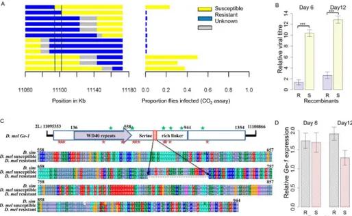
recombinant lines had a bimodal distribution of infection rates, with most lines having infec-tion rates of either 0% or greater than 40%. There was a region of approximately 735kb where there was a perfect correspondence between the genotype of the fly and infection rate (Fig 1B; Wilcoxon Rank Sum Test:W= 225.5,P= 8.4x10⁻⁶).
To further refine the location of the gene, we repeated this experiment to generate informa-tive recombinants in the candidate region. To reduce any effects of the genetic background, in this experiment we crossed a susceptible line 22a to one of the resistant recombinants from the previous experiment. Out of 2112 individuals, 133 were recombinants in a 6cM interval contain-ing this region. Initially 28 of these were used to create lines homozygous for the recombinant chromosomes. Again individuals from each line were injected with the virus and genotyped, which reduced the region to 298kb (Fig 1C; Wilcoxon Rank Sum Test:W= 176,P= 3.8x10⁻⁵). We then returned to the remaining recombinant lines, and used these to generate 8 homozygous recombinants in this reduced region. Again these lines (along with 7 lines from before) were gen-otyped and phengen-otyped. This new information allowed the identification of an 89kb region con-taining the candidate gene (Fig 1D; Wilcoxon Rank Sum Test:W= 56,P= 0.0013).
Generating recombinants in an 89kb region using this approach was not feasible, thus we turned to transposable elements carrying visible markers to select new recombinants. We chose two transposable element lines, each homozygous for an EP element that flanked our region of interest. These elements were combined on a single chromosome to generate a‘2EP’ line [36] which was susceptible to the DMelSV (100% infected). As these elements carry visible eye-colour markers, recombinants between our resistant chromosome and the 2EP line can be detected from their light orange eye colour (the non-recombinant resistant and susceptible flies have white or dark orange eyes respectively). This approach was used to generate 12 recombi-nant lines. Again the lines were assayed for resistance to DMelSV and genotyped for several markers across the 89kb region, which reduced the region to just under 8kb (2L: 11094733– 11102848, BDGP5;Fig 2A, Wilcoxon Rank Sum Test:W= 32,P= 0.0063). This region con-tains a whole gene calledGe-1and the flanking non-coding regions upstream of the genes CG4705 andReps. Ge-1 has been shown involved in RNA degradation and the formation of processing bodies (P bodies) in animals and plants [37–40]. It acts as a bridge between Decap-ping protein 1 and DecapDecap-ping protein 2, which remove the 5’cap from mRNA (“decapping”). This results in the RNA molecule being rapidly degraded by exonucleases [39].
In all of the experiments above we have used the symptom of paralysis after CO2exposure
to map resistance. To check thatGe-1is affecting the viral load rather than CO2sensitivity
itself, we used quantitative PCR to measure viral titres in 20 of the resistant and 16 of the sus-ceptible recombinant lines after they had been injected with the DMelSV. We found that six days after the injection, there was approximately a 471–fold lower viral load in resistant lines compared to susceptible lines (Fig 2B;F1,76= 137.5,p<2.2e-16), and after 12 days this rose to a
1168–fold difference (Fig 2B;F1,97= 125.8,p<2.2e-16).
and susceptible in yellow. There is a near-perfect association between genotype and phenotype in the regions between the vertical lines. Experiment (B) narrowed down the region to 735 kb using 31 recombinant lines. (C) Further narrowed down the region to 298 kb using 28 recombinant lines. (D) Narrowed down the region to 89 kb using 15 recombinant lines.
The resistant allele of
Ge-1
contains a 26 amino acid deletion
To identify the mutations that could be responsible for resistance, we sequenced this entire region (2L: 11094733–11102848) in the original resistant and susceptible line. The two sequences differed by 47 SNPs and 1 indel. The indel is 78bp long in the 5thexon ofGe-1(2L: 11097925–11098002), and reduces the length of the serine-rich linker region of the protein by 26 amino acids, with the resistant allele being the shorter of the two (Fig 2C). The homolog of this gene inD.simulansencodes the full length protein, suggesting that a deletion mutation has occurred in this region on the resistant chromosome. The SNPs are spread across the 3’UTR
ofRepsandCG4705and all ofGe-1, with only two found in intergenic regions (S1 Table). To
examine whether the difference seen in viral resistance could be due to a change in the expres-sion ofGe-1, we used quantitative PCR to measure its expression in 20 resistant and 16 suscep-tible recombinant lines that were used in the viral load measurement. We found that there was no significant difference in gene expression in the resistant and susceptible lines both six days and twelve days after they had been injected with the virus (Fig 2D; day6:F1,76= 0.0189,
p= 0.891; day12:F1,97= 6.109,p= 0.015).
We estimated the frequency of the 26 amino acid deletion in natural populations from the USA and Africa. We scored the presence or absence of the deletion by PCR in the 189 inbred lines from North Carolina that comprise the DGRP panel [41]. We found that only two of the lines contained the deletion. We also looked for the deleted allele in genome sequences from Fig 2. Resistance assay of resistant and susceptible recombinants.(A) Recombinants (left) were selected in the region mapped inFig 1using two eye-colour markers, injected with sigma virus and tested for paralysis after exposure to CO2. This identified an ~8 kb region containing a whole gene calledGe-1and the
flanking 3’untranslated regions of the genesCG4705andReps. (B) relative viral titre in 20 resistant and 16 susceptible recombinants at 6 days post infection and 12 days post infection. (C) Domain organization of Ge-1(adapted from [38]). The C-terminal domain is highlighted in blue with the highly conserved C-terminal region in dark blue. Red indicates a 26 amino acid deletion that differs between the resistant and susceptible alleles. Stars represent non-synonymous differences betweenD.melanogasterandD.simulans. Red stars represent changes that occurred on the lineage leading toD.melanogasterand green stars represent changes on the lineage leading toD.simulans. Serine-rich linker sequences fromD.simulansandD.
melanogastersusceptible line 22a and resistant line EME are shown. (D)Ge-1expression in 20 resistant and 16 susceptible recombinants (described in Fig 2B) at 6 days post infection and 12 days post infection. Error bars are standard errors. Viral RNA loads and gene expression were measured by quantitative PCR and standardised to the reference geneActin5c.
several African populations (319 alleles ofGe-1) [42], and none of the lines contained the deletion.
In a separate experiment, we have injected all of the DGRP lines with the DMelSV and mea-sured the proportion of flies that were infected 13 days later using the CO2assay [17]. We
found that the two lines with the deletion lines were both very resistant to the DMelSV (4% of flies in line 153 and 8% in line 361 were infected after injection, compared to an average of 40.6%), but this difference was not significant (S1 Fig; MCMCglmm:p= 0.092). We also tested all of the other polymorphisms in the 8kb region for an association with resistance and found that only one SNP, located in the 3’UTR ofReps, was significant (MCMCglmm:p<0.001).
Knocking down
Ge-1
expression increases susceptibility to DMelSV
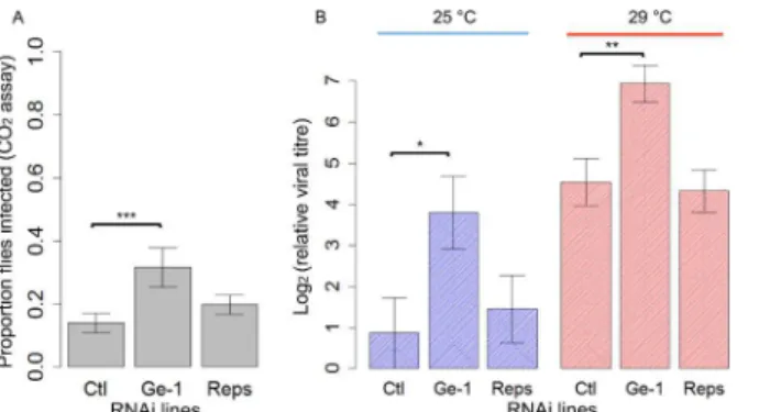
In order to test ifGe-1orRepsis involved in sigma virus resistance inD.melanogaster, we used RNAi to knock down the expression of the two genes. These flies did not have theGe-1 dele-tion. As we found that knock-downs in flies reared at 25°C are lethal, the flies were reared at a low temperature (18°C) whereGal4drivers are normally inefficient before being transferred to a higher temperature (25°C or 29°C).Ge-1-RNAi flies showed a higher proportion of flies infected after exposure to CO2than the control (Fig 3A; Generalized Linear Model:|z|= 3.391,P<0.001) whileReps-RNAi flies had a similar proportion of flies infected to the control
(Gen-eralized Linear Model: |z| = 0.951,P= 0.34). We also measured the viral titres to see whether the resistance is due to reduction in viral replication in flies. We found that the viral load in the
Ge-1-RNAi line was ~8-fold higher than in the control (|t| = 2.336,P= 0.02) whileReps-RNAi flies had a similar viral load to the control (Fig 3Bblues bars; |t| = 0.490,P= 0.627). We repeated the RNAi test and kept injected flies at 29°C. This time the viral titre inGe-1-RNAi line was ~5-fold higher than in the control (|t| = 3.281,P= 0.002) while theReps-RNAi flies had similar viral loads to the control (Fig 3Bred bars; |t| = 0.282,P= 0.779). The knockdowns in all these flies were likely inefficient, as qPCR onRepsandGe-1RNA levels was not signifi-cantly different from the controls.
Deleting the 26 amino acid region of
Ge-1
in transgenic flies makes them
resistant to DMelSV
To test whether the 26 amino acid deletion inGe-1is causing flies to be resistant to DMelSV, we generated transgenic flies that only differ by this deletion. To do this we first took a BAC clone of the region that lacked the deletion, and used recombineering [43] to seamlessly delete
Fig 3. The effect of knocking downGe-1andRepsby RNAi on susceptibility to DMelSV.(A) Proportion of flies that were paralysed after exposure to CO2in control,Ge-1-RNAiandReps-RNAilines. Flies were kept
at 25°C. (B) Viral titres in control,Ge-1-RNAiandReps-RNAilines standardised to reference gene
Ef1alpha100E. This experiment was repeated at 25°C and 29°C. Error bars are standard errors.
the 26 amino acids inE.coli. We then inserted two forms of the BAC into identical positions on the 3rdchromosome using thephiC31integrase system [44], to generate flies that express the two alleles of the gene under the control of the same natural promoter. In total we con-structed four independently transformed transgenic lines, one with the deletion ( CH322-Ge-1Δ78C) and three without (CH322-Ge-1+A,CH322-Ge-1+BandCH322-Ge-1+C). These trans-genic lines were then crossed to aGe-1null mutant,dGe-1Δ5/Cyo, so that only the transgene is expressed. ThedGe-1Δ5mutation is homozygous lethal [37], and the insertion of either allele of
Ge-1on the 3rdchromosome complemented this lethal effect, allowingdGe-1Δ5homozygotes to be generated (dGe-1Δ5;CH322-Ge-1Δ78C,dGe-1Δ5;CH322-Ge-1+A,dGe-1Δ5;CH322-Ge-1+B
anddGe-1Δ5;CH322-Ge-1+C). Transgenic flies carrying the deletion inGe-1are highly resistant
to DMelSV. Following injection with DMelSV, only 1.6% of flies from the line with the deletion showed the symptoms of paralysis and death after exposure to CO2compared to 69.6% of the
flies in the three lines without the deletion (Fig 4A; Generalized Linear Mixed Model: |z|= 7.350,
P<<0.001). Similarly, viral titres were 512-fold higher in the transgenic line with the deletion
than the three lines without the deletion (Fig 4B;F1,56= 156.9,P<<0.001).
In order to test whetherGe-1expression differs in transgenic lines, we measuredGe-1
expression indGe-1Δ5;CH322-Ge-1Δ78,dGe-1Δ5;CH322-Ge-1+(mix of A, B and C) flies. We didn’t detect any significant difference inGe-1expression in these two genotypes (S1 Fig;
F1,37= 0.008,P= 0.92). In addition, we measuredGe-1expression in DMelSV injected flies and
controls that were injected with Ringer’s solution. In both transgenic lines, we found no signifi-cant difference between virus injected and Ringer’s injected flies (S1 Fig;Ge-1+:F1,18= 2.32,
P= 0.15;Ge-1Δ78C:F1,17= 0.403,P= 0.53). These results indicate thatGe-1expression does not
differ in the resistant and susceptible transgenic flies and DMelSV infection does not induce
Ge-1expression.
Ge-1
resistance to DMelSV is not dependent on the siRNA pathway
The RNAi effector protein Argonaute 2, which is a key antiviral defence inDrosophila, localises to some P bodies and this localisation depends on Ge-1 [37,38,40]. To test whetherGe-1 resis-tance to sigma virus is mediated by RNAi, we crossed the mutant alleleAgo251Binto lines car-rying the susceptible and resistant alleles ofGe-1. This allele ofAgo2deletes the first two exons and is known to abolish its slicer activity [45], but it does still produce the shortestAgo2 tran-script and this may have some function [46]. In the CO2sensitivity assay, the two alleles ofGe-Fig 4. Transgenic flies with the 26 amino acid deletion inGe-1are resistant to DMelSV.Flies were transformed with BAC clones carryingGe-1that differs only in the presence (red) or absence (blue) of the deletion. The four genotypes are independent transformants. (A) The proportion of flies that was paralysed or dead after exposure to CO2. Each point is a vial of flies. Horizontal bars represent the mean of each line. (B)
Mean viral titre in transgenic lines. Viral RNA loads were measured by quantitative PCR and standardised to the reference geneRpL32. Error bars are standard errors.
1significantly affected susceptibility regardless of whether there was a functional allele ofAgo2
(Fig 5A; Wilcoxon Rank Sum Test withAgo2mutant: |Z|= 4.9,P= 8.99e-7; Wilcoxon Rank Sum Test withAgo2wild-type: |Z| = 5,P= 4.7e-7). The same pattern was found when we mea-sured viral titre in these lines–Ge-1had a large effect on viral titre (Fig 5B;Ge-1:F1,37= 100.82,
P<0.0001), but this was not affected byAgo2(interactionGe-1Ago2:F
1,37= 0.3,P= 0.59). The
Ago251Bmutants did have higher titres of DMelSV (Fig 5B;Ago2:F1,37= 35.69,P<0.0001).
Therefore, siRNA pathway does defend flies against DMelSV butGe-1does not rely on the siRNA pathway to provide resistance.
Decapping protein 1 protects flies against DMelSV
Ge-1is essential for forming P bodies, so we investigated the role of eight other P-body compo-nents in DMelSV resistance. As mutants tend to be lethal, we used RNAi to knock down the expression of these genes in adult flies. We found that knocking down the expression of
Decap-ping protein 1(DCP1) resulted in increased viral load (Fig 5C;Ge-1susceptible background:
Wilcoxon Rank Sum Test:W= 27,P= 0.009;Ge-1heterozygous background: Wilcoxon Rank Sum Test:W= 87,P= 0.02). The effect of knocking downDCP1was greater inGe-1susceptible flies than in flies that were heterozygous for the twoGe-1alleles (Fig 5C; interaction between
Ge-1andDCP1: |t| = 2.15,P= 0.03). However, we would interpret this interaction cautiously, as theGe-1susceptible and heterozygous flies did not differ in their viral load, perhaps due to genetic background effects or dominance (Fig 5C). Knocking down the other genes did not have a significant effect on DMelSV titres (S2 Fig;Edc3,DCP2,DCP1,GW182,pcm,me31B,
Part-1andstau; note the efficiency of these knockdowns was not checked).
Ge-1
does not affect susceptibility to DAV or DCV in
D
.
melanogaster
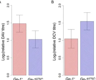
We tested whetherGe-1resistance was specific to DMelSV by infectingGe-1transgenic flies
withDrosophilaA virus (DAV) andDrosophilaC virus (DCV). Both DAV and DCV are
natu-ral pathogens ofD.melanogaster, and DAV infects flies in nature and laboratories widely Fig 5. The effect ofAgo2andDCP1on susceptibility to DMelSV.(A) and (B) are the effect ofGe-1on susceptibility inAgo251Bmutantflies. Blue represents flies with the resistantGe-1allele andAgo2mutant, black the resistantGe-1allele andAgo2wild-type, red theGe-1susceptible allele andAgo2mutant, and orange the susceptibleGe-1allele andAgo2wild-type. (A) CO2sensitivity assay. (B) Relative viral titre
estimated by quantitative PCR relative toRpL32was used as reference gene. (C) The effect of knocking downDCP1on viral titre inGe-1susceptible backgrounds (red bar) andGe-1heterozygous backgrounds (blue bar). Grey bars are controls for the RNAi. Error bars are standard errors.
[30,47]. Transgenic flies carrying resistant (Ge-1Δ78C) or susceptible allele (Ge-1+) ofGe-1have similar viral titres after DAV infection (Fig 6A;F1,16= 1.685,P= 0.21) and after DCV infection
(Fig 6B;F1,13= 1.871,P= 0.19). This indicates that this polymorphism inGe-1does not have
an effect on DAV or DCV infections and its antiviral function is likely to be specific to sigma virus.
The serine-rich linker of Ge-1 has evolved under positive selection in
D
.
melanogaster
and
D
.
simulans
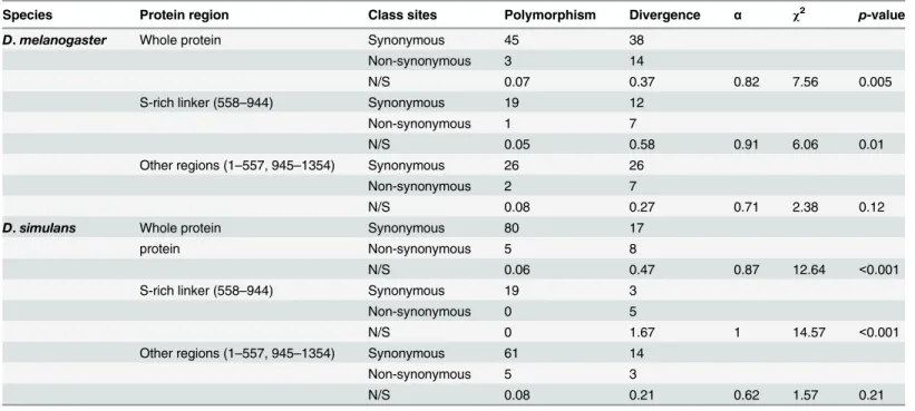
As changes inGe-1can make flies resistant to viral infection, it is possible that it has been the tar-get of sustained selection by viruses during evolution. To investigate this we used a McDonald-Kreitman Test to detect beneficial amino acid changes that have been fixed by natural selection during the evolution ofGe-1[48]. In bothD.melanogasterandD.simulanswe estimate that over 80% of the amino acid substitutions were fixed by natural selection (αinTable 1) and therefore had a beneficial phenotypic effect. Virus resistance is caused by a change to the serine-rich linker, and 13 of the 22 non-synonymous changes betweenD.melanogasterandD.simulansare located in this region (stars inFig 2B). When we repeated the test on just the serine-rich linker we found evidence of selection on this domain in both theD.melanogasterandD.simulanslineages and no evidence of selection in the remainder of the protein (Table 1).
Discussion
We have identified a naturally occurring polymorphism in a gene calledGe-1that makesD.
melanogasterhighly resistant to the naturally occurring rhabdovirus DMelSV. When we
knocked-down the expression of the susceptible allele ofGe-1by RNAi the flies became even more susceptible to infection, indicating thatGe-1is an existing restriction factor whose antivi-ral effects have been increased by the deletion. By modifying the sequence of the gene in trans-genic flies, we identified a 26 amino acid deletion in the serine-rich linker region ofGe-1that is causing the resistance. This polymorphism has no effect on resistance to DAV or DCV, indi-cating resistance is likely to be specific to sigma or related viruses. Ge-1 acts as a bridge bring-ing together DCP1 and Decappbring-ing Protein 2 (DCP2), which can then remove the 5’cap from mRNA leading to its degradation [38]. We found that DCP1 also restricts sigma virus infection
Fig 6. Titres of DAV and DCV in infectedGe-1transgenic flies.(A) Relative viral titres in DAV infected flies. (B) Relative viral titres in DCV infected flies. Error bars are standard errors.
in flies, suggesting that this pathway may underlie resistance. The serine-rich linker of Ge-1 has experienced strong selection during its evolution, suggesting that it may be involved in an ongoing arms race with viruses.
Ge-1 plays a central role in RNA degradation and the formation of processing bodies (P bodies) in animals and plants [37–40]. It is required for the removal of the 5’cap from mRNA, which results in the subsequent degradation of the RNA molecule by an exonuclease in the 5’ to 3’direction [49]. It is thought to act as a bridge between two of the key molecules involved the decapping process, with its C-terminal domain interacting with the protein DCP2 and its N-terminal domain interacting with DCP1 [39]. The C-terminal domain of Ge-1 also results in the localization of the protein to P bodies, which are sites in the cytoplasm where many of the enzymes involved in RNA degradation are localized. Ge-1 not only localises to P bodies, but it is also required for P-body stability and formation, probably due to its role as a scaffold protein [38,39].
P bodies can play both pro- and anti-viral roles [50]. In some cases viruses disrupt P body formation, presumably to prevent viral RNA being degraded. For example, P body components act as restriction factors for influenza A virus, and the viral NS1 protein in turn disrupts the formation of P bodies [51]. Similar interactions also occur between insects and their viruses. In
Drosophilathe dicistrovirus CrPV disrupts P bodies [52]. Furthermore, several P body
compo-nents involved in mRNA decapping have an antiviral effect against the bunyavirus Rift Valley fever virus (RVFV) [53]. This is thought to be because bunyaviruses“cap-snatch”their 5’cap from host mRNAs, and the decapping process reduces the availability of 5’caps for viral Table 1. McDonald Kreitman test onGe-1.
Species Protein region Class sites Polymorphism Divergence α χ2 p-value
D.melanogaster Whole protein Synonymous 45 38
Non-synonymous 3 14
N/S 0.07 0.37 0.82 7.56 0.005
S-rich linker (558–944) Synonymous 19 12
Non-synonymous 1 7
N/S 0.05 0.58 0.91 6.06 0.01
Other regions (1–557, 945–1354) Synonymous 26 26
Non-synonymous 2 7
N/S 0.08 0.27 0.71 2.38 0.12
D.simulans Whole protein Synonymous 80 17
protein Non-synonymous 5 8
N/S 0.06 0.47 0.87 12.64 <0.001
S-rich linker (558–944) Synonymous 19 3
Non-synonymous 0 5
N/S 0 1.67 1 14.57 <0.001
Other regions (1–557, 945–1354) Synonymous 61 14
Non-synonymous 5 3
N/S 0.08 0.21 0.62 1.57 0.21
Divergence was polarised to the lineage leading toD.melanogasterandD.simulans. As well as whole-protein analyses, the serine-rich region only and other domains were analysed separately. For the polymorphism data we usedGe-1sequences from 197 ZambianD.melanogastergenomes (only site with minor allele frequencies over 10%) [42] and 6D.simulansgenomes [66]. So that we can look separately at evolution occurring on the lineage leading toD.melanogasterandD.simulans, we reconstructed the most recent common ancestor of the sequences ofGe-1that are found inD.simulansandD.
melanogasterusing outgroups.αis the estimated proportion of amino acid changes that werefixed by natural selection.
replication [53]. As Rhabdoviruses do not cap-snatch, this mechanism cannot explain our results. Other viruses have co-opted P bodies for their own benefit. This is the case for West Nile virus, which recruits various P body components to its replication centres, and knocking down the expression of these proteins results in lower viral titres [54].
There are two possible ways in which Ge-1 could confer resistance. First, it could be used in some unknown way by DMelSV during the viral replication cycle, and the resistant allele of the gene may interfere with this process. Second, it could play an existing antiviral function that is made more efficient. We found that knocking down the expression of the susceptible allele of
Ge-1further increased the susceptibility of flies, indicating that the polymorphism that we identified is increasing the effectiveness of an existing restriction factor.
We demonstrated that a 26 amino acid deletion inGe-1is the cause of increased resistance to DMelSV by modifying the gene in transgenic flies. We ensured the gene was under the con-trol of its natural promoter by modifying BAC clones of this region of theDrosophilagenome, and these BACs were then inserted into a fly line that carried a null allele ofGe-1. This deletion removes 26 amino acids from the middle of the protein, which is a flexible linker that lies between two structured domains. It does not alter the sequence of the domains that interact with DCP1 and DCP2, or the region required for P body formation [38]. It is therefore possible that the deletion may affect the protein’s role as a scaffold by changing the conformation of the protein complex.
We investigated two possible ways in which Ge-1 might be affecting the susceptibility of flies. The primary antiviral defence of insects is RNAi, and the RNAi-Induced Silencing Com-plex (RISC) localises to P bodies [55,56]. However, our results show thatGe-1-based resistance does not require Ago2, and therefore is independent of RNAi. Therefore, a likely hypothesis is that resistance relies on the destruction of viral genomic or messenger RNA in an RNAi-inde-pendent way in P bodies. Consistent with this we found that knocking down the expression of DCP1 increases DMelSV titres in flies. As the polymorphism affecting resistance is in the ser-ine-rich linker that forms a bridge between DCP1 and DCP2, it seems likely that Ge-1-based resistance involves the decapping complex.
By looking at theGe-1sequence inD.simulans, we found the ancestral state ofGe-1is the susceptible allele. This fits with an evolutionary arms race in which hosts are continually evolv-ing new defences against pathogens. Consistent with this, we found that the protein sequence of Ge-1 has been subject to long term positive selection. While our analysis demonstrates that many of the amino acid substitutions that have occurred in theD.melanogasterlineage pro-vided a selective advantage to flies, further work would be needed to demonstrate this was linked to the antiviral function of Ge-1 (especially given the mutation that gave rise to resis-tance was a deletion not a substitution). Nonetheless, studies of other genes involved in antivi-ral immunity, such as those in the siRNA pathway, have found that they are also evolving rapidly under positive selection [57,58]. Therefore, selection by viruses may frequently be an important force in the molecular evolution of genes involved in antiviral defence.
Our results show that studying naturally occurring polymorphisms that increase resistance to infection provides a valuable alternative to studying immunity through artificial mutations that reduce resistance. Not only can this approach identify novel forms of antiviral defence, but it also allows us to understand how resistance to infection evolves. This is important as evolved defences may not always involve the classical immune response.
Materials and Methods
Recombinant mapping fly lines and crosses
Susceptible (22a) and resistant (EME) fly lines were provided by Didier Contamine. The 2nd chromosome in the EME stock carries the resistant allele of the generef(2)M[34], and the other two chromosomes are from a susceptible ebony stock. To map the resistance genes we crossed the resistant and susceptible parental stocks and created lines that carried homozygous recombinant chromosomes. The female F1 progeny of a cross between EME and 22a were crossed to a balancer stockSM5,Cy/Pm. In the next generation we selected individualSM5,
Cy/+males and crossed these back to the balancer. A few days after setting up this cross we removed the male parents from the vial and genotyped them using molecular markers flanking the region that we knew contained the resistance gene (S2 Table). This allowed us to discard all the lines that had not recombined in this region and only retain the informative recombinants. In the next generation we crossed siblingSM5,Cy/+flies, and then selected for homozygous recombinants in the subsequent generation. Using this approach we initially generated 33 lines that had recombined between insertion/deletion (indel) markers at 36cM and 52cM on the 2nd chromosome. We then repeated the experiment to produce recombinants between markers at 42cM and 47cM.
To select recombinants in even smaller regions we used phenotypic markers flanking the region of interest rather than molecular markers. First, mapping lines were generated by cross-ing twoP-element lines that flanked the region of interest to create a chromosome carrying bothP-elements. These elements both carried the mini-white gene, and flies that carry a single heterozygous element have lighter coloured eyes than flies carrying two heterozygous elements [36].The linesw;P{EP}2377andw;P{EP}2478were combined to the same chromosome to
generate the 2ndchromosome mapping line (2EP-2). This was then crossed to one of the resis-tant recombinant lines (M34) that had been generated in the experiment described above. 12 homozygous 2ndchromosome recombinant lines were generated using the balancer SM5, Cy/+ and the crossing scheme described above.
Genotyping
Sequencing and genotyping candidate regions
DNA for sequencing was extracted using either Tissue Genomic DNA kit (Metabion, Munich) or DNeasy 96 Blood & Tissue Kit (Qiagen). The 8 kb region identified on chromosome 2 (2L: 11094733–11102848, BDGP5) was sequenced for lines EME and 22a. Primer pairs were designed to amplify these regions in overlapping fragments (S2 Table), and the sequencing was performed as described above. Diagnostic PCR primers were designed to genotype flies for a deletion inGe-1. The forward primerGe-1Indel 1F (5’AGCGTCAAGCTTTTCCTTCA 3’) and the reverse primerGe-1Indel 1R (5’CACCAGCGGTCAGGATAGAT 3’) were used to establish presence or absence of the 78bp deletion inGe-1.
D
.
melanogaster
sigma virus (DMelSV)
The Hap23 strain of DMelSV was extracted from an infected line ofD.melanogaster(Om) using the protocol described in Magwireet al. 2011[17].
Fly infection and measuring resistance
FemaleD.melanogasterwere injected in the abdomen with sigma virus either by blowing virus through a glass needle connected with a hose until slight extension of the proboscis was observed (only for recombinant mapping experiment) or by using Nanoject (Drummond Sci-entific) to inject 69nl virus suspension (all other experiments). Injected flies were tipped onto new media every two days before they were tested for infection. Flies infected with the DMelSV become paralysed and die on exposure to CO2. To test for this symptom of infection, the flies
were exposed to CO2for 15 minutes at 12°C 10–16 days post infection. The exact gassing date
for each experiment was carefully picked by pilot CO2exposure. Flies were given 2 hours to
recover from the CO2and then the number of dead or paralyzed individuals was counted as
well as the total number of individuals in each vial.
In the recombinant mapping experiment, four replicate vials each containing approximately 20 flies were used in each experiment except for the first round of recombinant assay (one rep-licate) and were used in the CO2sensitivity assay. In the 25°C RNAi experiment, 29–40
repli-cate vials for each cross were injected with virus. 14–25 were used in the CO2sensitivity assay
and the remaining 15 vials were used to measure viral titre (see details below). In the 29°C RNAi experiment, 24 copies of each cross were injected with sigma virus and all of them were used to measure viral titre. In the transgenic fly experiment and theAgo2dependency experi-ment, 30–32 replicate vials each containing approximately 15 flies were injected with virus. 15– 18 vials per line were assayed for resistance to sigma virus by measuring CO2sensitivity and
the remaining 15 vials were used to measure viral titre.
Quantitative RT-PCR
ATGG 3’) and the reverse primer qGe-1R3 (5’GAGCAAGCAATTTCTGGATACTT 3’). Expression ofActin 5Cwas used as a reference using the primers qActin5c_for2 (5’GAGCGC GGTTACTCTTTCAC 3’) and qActin5c_rev2 (5’aagcctccattcccaagaac 3’) [17]. In RNAi and transgenic experiments, viral load was measured using a QuantiTect Virus+ROX Vial Kit (QIAGEN) on RNA template. Dual-labelled probe [FAM] TGTGCCAAGTCTGTAATCC TGCTA [NFQ-MGB] and primers DMelSV_F (5’CCGACTACAAATGCTATATG 3’), DMelSV_R (5’CAGGTATTAGAGGCTTCTTA 3’) were used to amplify DMelSV genomic RNA. The amount of viral RNA and gene expression were standardised to twohousekeeping genes,Ef1alpha100EorRPL32, using theΔΔCt (critical threshold) method (see below).
Ef1al-pha100Ewas amplified using probe [FAM] ATCGGAACCGTACCAGTAGG [BHQ3] and
primers Ef1a100E_FW (5’GGACGTCTACAAGATC 3’) and Ef1a100E_RV (5’TCTCCACA GACTTTAC 3’).RPL32was amplified using probe [VIC] ACAACAGAGTGCGTCGCCGCT TCAAGG [NFQ-MGB] and primers RPL32_FW (5’TGCTAAGCTGTCGCACAAATGG 3’) and RPL32_RV (5’TGCGCTTGTTCGATCCGTAAC 3’). We performed two or three techni-cal replicates of each PCR and used the mean of these in subsequent analyses. We techni-calculated the PCR efficiency by using a dilution series. Using this approach we found that the actin PCR is 93% efficient, the virus PCR using fluorescein is 96% efficient, theGe-1PCR is 102% effi-cient, the virus PCR using probe is 98% and theRPL32PCR is 96%.
RNAi
Fly strains UAS-Ge-1-RNAi (y,w1118;P{KK102275}VIE-260B, KK 106687), UAS-Reps-RNAi (y,w1118;P{KK101677}VIE-260B, KK 110704) as well as a control strainy,w1118;P{attP,y+,w3-}
(60100) were bought from VDRC Stock Center [60]. A ubiquitously expressedGal4driver under the control of thedaughterlesspromoterw;P{GAL4-da.G32}UH1was crossed to the
UAS strains and the control strain to induce the knock down effect. Four replicates were set up for each cross and flies were reared in cornmeal bottles with live yeast at 18°C whereGal4 driv-ers are inefficient (efficient knock-downs are lethal). Two- to three-day old mated F1 females were injected with DMelSV for each cross and injected flies were kept at 25°C to induce an effi-cient knock-down effect before assaying for resistance. We later repeated this experiment and kept the injected flies at 29°C for a more efficient knock-down effect. TheGe-1-RNAi construct is 345 bp long and targets exon 9 ofGe-1. TheReps-RNAi construct is 360 bp long and targets exon 4 ofReps.
SinceGe-1is essential for forming P bodies, we also tested the effect of knocking down other P-body genes by RNAi on DMelSV susceptibility [60]. We knocked down 8 P-body genes:Edc3(CG6311,w1118;P{GD11886}v30149, GD30149),DCP2(CG6169, P{KK101790} VIE-260B, KK105130),DCP1(CG11183,P{KK101204}VIE-260B, KK105638),GW182
(CG31992,P{KK101472}VIE-260B, KK103581),pcm(CG3291,P{KK108511}VIE-260B,
KK105739),me31B(CG4916, w1118;P{GD11470}v49378, GD49378),Part-1(CG5208,
P{KK104961}VIE-260B, KK100872) andstau(CG5753,P{KK108121}VIE-260B, KK106645) in
Ge-1susceptible background andGe-1heterozygous background separately.Daughterless Gal4
driver was crossed toGe-1resistant and susceptible recombinants to generate twoGal4drivers with resistant or susceptibleGe-1backgrounds. UAS lines were then crossed to two
daughter-less Gal4drivers and kept at 18°C whereGal4drivers are inefficient (efficient knock-downs are
Note that the design of the crosses means that there are 4 different genetic backgrounds (the GD and KK RNAi lines crossed to the two different Gal4 driver lines). Within these four genetic backgrounds, the flies should be genetically identical except for the RNAi construct.
Generating transgenic flies using recombineering
DrosophilaP[acman] Bacteria Artificial Chromosomes (BACs) clone CHORI-322-120M19
covering fly genome 2L: 11090119–11111928 was obtained from BACPAC Resources Centre (BPRC) [61]. This BAC contains the susceptible allele ofGe-1which doesn’t have the deletion. Recombineering was carried out to replace the susceptible allele ofGe-1in the BAC with a resistant allele containing the deletion through homologous recombination [43]. DNA was extracted from the resistant stain EME to use as a template for amplifying the resistant allele of
Ge-1. A fragment containing a region ofGe-1with the deletion was amplified using the primers Ge-1_newrc_F1 (5’TATCTCCTGCACCTCTCGAC 3’) and Ge-1_newrc_R1 (5’CTGCCTGC ACGAG TGGAA 3’). TheGalKtargeting cassette was amplified from bacterial strain pgalK. Phusion High Fidelity polymerase (NEB) and the primers: Ge-1_galK_F (5’CGTCCTGTGTG
GCCATTATCTCCTGCACCTCTCGACTCGGACTCGAACTGCCGCT-CCTGTTGACAATTA
ATCATCCGCA 3’), Ge-1_galK_R (5’GGAAGCCACATGATGGCAAAAA-AGTCTGCTCTCT
CTTTTTCCTCGACAATATCAACAAGTCAGCACTGTCCTGCTCCTT 3’) were used in
PCRs. The sequences in italics were homologous sequences to theGe-1gene.
To transform flies with the BAC clone, the BAC carrying the resistant allele ofGe-1was injected into embryos of a fly strain containing the 3rdchromosomeattpsite:M(eGFP,vas-int,
dmRFP)ZH-2A;;M(attP)ZH-86Fb[62]. The original BAC was also injected into the same strain
as a control. Since the BAC contains a mini-white gene, flies emerged from injected embryos were crossed to a white-eye double balancerw-;If/Cyo;TM6B/MRKSand successful transfor-mants were picked by their red-eye phenotype. Balanced transfortransfor-mants were crossed to their siblings and generated homozygotes with balanced 2ndchromosome.
Testing whether
Ge-1
-based resistance is
Ago2
dependent
Ago2mutant Ago251Bwas kindly provided by Dr. Maria Carla Saleh (Institute Pasteur). Two
recombinants 47 (resistant) and 16 (susceptible) generated above were chosen to cross to an
Ago2mutant strain Ago251Band generated two homozygous lines:47; Ago251Band16;
Ago251B. 30 vials containing 15 two- to five-day old females were infected with sigma virus for each line. The same number of flies were infected for strain 47 and strain 16 as controls. Infected females were kept at 25°C and maintained on cornmeal food.
Infecting
Ge-1
transgenic flies with other viruses
We also infected the transgenic flies carrying resistant allele (Ge-1Δ78C) and susceptible allele
(Ge-1+) ofGe-1with two other viruses extracted fromD.melanogaster: DAV and DCV (TCID50= 5x10⁸) [63]. For lineGe-1Δ78C, 10 vials of 15 mated females were pricked with either
DAV or DCV and kept in 25°C. 10 vials of infected females ofGe-1+(Ge-1+A,Ge-1+Band Ge-1+C) were also pricked with viruses and kept in 25°C. DAV infected flies were collected 3 days post infection and DCV infected flies were collected 2 days post infection for RNA extraction.
Statistical analysis
using the R library MCMCglmm [64], which uses Bayesian Markov chain Monte Carlo (MCMC) techniques. To test for an association between resistant genes and resistance to sigma virus in DGRP, we fit the model:
vi;j ¼logit 1ðX T
ibþajþεi;jÞ
Whereνi,jis the probability of flies in vialifrom linejbeing infected.βis a vector of the fixed effects ofref(2)Pgenotype,doc1420ofCHKov1genotype (two genes known to affect sigma virus resistance [17,19]), and XiTis a row vector relating the fixed effects to viali.αjis a
random effect of linej. The residual,εi,j,allows over-dispersion due to unaccounted for hetero-geneity between vials in the probability of infection. We tested for the effects of a 78bp deletion inGe-1and SNPs by including this as an additional fixed effect inβ.
For each fly line in which we measured viral titres or gene expression by quantitative RT-PCR, we first calculatedΔCtas the difference between the cycle thresholds of the gene of interest and the endogenous control. In recombinant mapping and transgenic lines’assays, the viral titre or gene expression in resistant flies relative to susceptible flies was calculated as 2-ΔΔCt, whereΔΔCt=ΔCt
resistant—ΔCtsusceptible, whereΔCtresistantandΔCtsusceptible, are the
means of theΔCtvalues of the resistant and susceptible lines. In RNAi, the viral titre or gene expression in RNAi lines relative to the control was calculated as 2-ΔΔCt, whereΔΔCt=
ΔCtRNAi—ΔCtcontrol, whereΔCtRNAiandΔCtcontrol, are the means of theΔCtvalues of RNAi
lines and the control. To assess whether these differences were statistically significant, we com-pareΔCtin the resistant lines (RNAi lines) and the susceptible lines (control) by fitting theΔCt
values in a linear model.
In P-body genes RNAi experiment, the viral titre data was not normally distributed. We first Box-Cox transformed the data using R function“boxcox”from package“MASS”[65]. Then we fitted the transformed data in a linear model to test whetherΔΔCtof RNAi lines were significantly different from controls.
To test for positive selection, we used a McDonald and Kreitman (MK) test [48] applied to
197D.melanogastersequences from Zambian lines from Drosophila Population Genomics
Project 3 (DPGP3) [42] and 6D.simulanssequences [66]. Substitutions were polarised along
theD.melanogasterandD.simulanslineages. Consensus sequences of 197D.melanogaster
samples were downloaded fromhttp://www.dpgp.org/.Ge-1sequences (2L: 11095353– 11100866) were pulled out from all lines using the scripts“breaker.pl”and“dataslice.pl” writ-ten by the authors (Masking package available fromhttp://www.dpgp.org/). Script“breker.pl” inserts line breaks every 1000 bp in all files and script“dataslice.pl”returns locus-specific FastA files when given a subset of individuals and locus information. Coding sequences ofGe-1
from all the lines were manually aligned using BioEdit. Vertical multiple alignment (vma) files
of 6D.simulanslines were downloaded from the DPGP website. Vma files were converted into
FastA files by script“VMA2FASTA”provided by the authors.Ge-1coding sequences were pulled out and manually aligned.Drosophila yakubaandDrosophila erectasequences were used to infer the sequence of the most recent common ancestor ofGe-1inD.melanogasterand
D.simulans. For any polymorphic codon, if the ancestral state is ambiguous (ie. both theD.
melanogasterandD.simulansnucleotides were present in the outgroup species), we simply
Supporting Information
S1 Fig. Ge-1 expressions in transgenic flies.Blue bars are transgenic flies carrying susceptible Ge-1 allele and red bars are flies carrying Ge-1 resistant allele (with deletion).Error bars are standard errors.
(TIF)
S2 Fig. RNAi knock-downs of P-body components.Eight genes encoding P-body compo-nents were knocked down by RNAi:Edc3(CG6311),DCP2(CG6169),DCP1(CG11183),
GW182(CG31992),pcm(CG3291),me31B(CG4916),Part-1(CG5208) andstau(CG5753)).
Left 10 boxes (red) are RNAi knock-downs in flies withGe-1susceptible background. Right 10 boxes (blue) are RNAi knock-downs of flies that were heterozygous for the resistant and sus-ceptibleGe-1alleles. Each dot represents one sample which is a pool of 10–15 flies. There were two different genetic backgrounds: round dots represent KK library RNAi lines and triangles GD library RNAi lines. The flies were reared at 18C where expression of the RNAi construct is inefficient and then transferred as adults to 25C. There was a significant heterogeneity among the KK library RNAi lines (F= 7.29,P= 3.6x10⁻⁶) but not among the GD lines.
(TIF)
S1 Table. SNPs and indel found in the region (2L: 11094733–11102848) by comparing between resistant and susceptible flies.
(XLS)
S2 Table. Primers used for genotyping and sequencing.
(XLS)
Acknowledgments
Thanks to Yuk-Sang Chan for embryo microinjection, Dr. Anne Ephrussi for providing the
Ge-1mutant fly, Dr. Maria Carla Saleh for providing theAgo2mutant fly and Dr. Darren Obbard for sharingGe-1sequences of manyDrosophilaspecies.
Author Contributions
Conceived and designed the experiments: CC MMM FMJ. Performed the experiments: CC MMM FB. Analyzed the data: CC MMM FMJ. Wrote the paper: CC MMM FMJ.
References
1. Meyer JR, Dobias DT, Weitz JS, Barrick JE, Quick RT, et al. (2012) Repeatability and contingency in the evolution of a key innovation in phage lambda. Science 335: 428–432. doi:10.1126/science. 1214449PMID:22282803
2. Wang XH, Aliyari R, Li WX, Li HW, Kim K, et al. (2006) RNA interference directs innate immunity against viruses in adult Drosophila. Science 312: 452–454. PMID:16556799
3. Aliyari R, Wu Q, Li HW, Wang XH, Li F, et al. (2008) Mechanism of induction and suppression of antivi-ral immunity directed by virus-derived small RNAs in Drosophila. Cell Host Microbe 4: 387–397. doi:
10.1016/j.chom.2008.09.001PMID:18854242
4. van Rij RP, Berezikov E (2009) Small RNAs and the control of transposons and viruses in Drosophila. Trends Microbiol 17: 163–171. doi:10.1016/j.tim.2009.01.003PMID:19299135
5. van Rij RP, Saleh MC, Berry B, Foo C, Houk A, et al. (2006) The RNA silencing endonuclease Argo-naute 2 mediates specific antiviral immunity in Drosophila melanogaster. Genes Dev 20: 2985–2995. PMID:17079687
7. Imler J-L, Eleftherianos I (2009) Drosophila as a model for studying antiviral defences. Insect Infection and Immunity. Oxford: Oxford University Press,Chapter 4.
8. Kemp C, Mueller S, Goto A, Barbier V, Paro S, et al. (2013) Broad RNA interference-mediated antiviral immunity and virus-specific inducible responses in Drosophila. J Immunol 190: 650–658. doi:10.4049/ jimmunol.1102486PMID:23255357
9. Moy RH, Gold B, Molleston JM, Schad V, Yanger K, et al. (2014) Antiviral Autophagy Restricts Rift Val-ley Fever Virus Infection and Is Conserved from Flies to Mammals. Immunity 40: 51–65. doi:10.1016/j. immuni.2013.10.020PMID:24374193
10. Nakamoto M, Moy RH, Xu J, Bambina S, Yasunaga A, et al. (2012) Virus Recognition by Toll-7 Acti-vates Antiviral Autophagy in Drosophila. Immunity 36: 658–667. doi:10.1016/j.immuni.2012.03.003
PMID:22464169
11. Shelly S, Lukinova N, Bambina S, Berman A, Cherry S (2009) Autophagy is an essential component of Drosophila immunity against vesicular stomatitis virus. Immunity 30: 588–598. doi:10.1016/j.immuni. 2009.02.009PMID:19362021
12. Teixeira L, Ferreira A, Ashburner M (2008) The bacterial symbiont Wolbachia induces resistance to RNA viral infections in Drosophila melanogaster. PLoS Biol 6: e2.
13. Tsai CW, McGraw EA, Ammar ED, Dietzgen RG, Hogenhout SA (2008) Drosophila melanogaster mounts a unique immune response to the Rhabdovirus sigma virus. Appl Environ Microbiol 74: 3251– 3256. doi:10.1128/AEM.02248-07PMID:18378641
14. Xu J, Grant G, Sabin LR, Gordesky-Gold B, Yasunaga A, et al. (2012) Transcriptional Pausing Controls a Rapid Antiviral Innate Immune Response in Drosophila. Cell Host & Microbe 12: 531–543.
15. Zambon RA, Nandakumar M, Vakharia VN, Wu LP (2005) The Toll pathway is important for an antiviral response in Drosophila. Proceedings of the National Academy of Sciences of the United States of America 102: 7257–7262. PMID:15878994
16. Zhu F, Ding HJ, Zhu BN (2013) Transcriptional profiling of Drosophila S2 cells in early response to Dro-sophila C virus. Virology Journal 10:210. doi:10.1186/1743-422X-10-210PMID:23803447
17. Magwire MM, Bayer F, Webster CL, Cao C, Jiggins FM (2011) Successive increases in the resistance of Drosophila to viral infection through a transposon insertion followed by a Duplication. PLoS Genet. 2011/10/27 ed. pp. e1002337. doi:10.1371/journal.pgen.1002337PMID:22028673
18. Magwire MM, Fabian DK, Schweyen H, Cao C, Longdon B, et al. (2012) Genome-wide association studies reveal a simple genetic basis of resistance to naturally coevolving viruses in Drosophila mela-nogaster. PLoS Genet 8: e1003057. doi:10.1371/journal.pgen.1003057PMID:23166512
19. Wayne ML, Contamine D, Kreitman M (1996) Molecular population genetics of ref(2)P, a locus which confers viral resistance in Drosophila. Mol Biol Evol 13: 191–199. PMID:8583891
20. Aminetzach YT, Macpherson JM, Petrov DA (2005) Pesticide resistance via transposition-mediated adaptive gene truncation in Drosophila. Science 309: 764–767. PMID:16051794
21. Bangham J, Obbard DJ, Kim KW, Haddrill PR, Jiggins FM (2007) The age and evolution of an antiviral resistance mutation in Drosophila melanogaster. Proc Biol Sci 274: 2027–2034. PMID:17550883 22. Carre-Mlouka A, Gaumer S, Gay P, Petitjean AM, Coulondre C, et al. (2007) Control of sigma virus
mul-tiplication by the ref(2)P gene of Drosophila melanogaster: an in vivo study of the PB1 domain of Ref(2) P. Genetics 176: 409–419. PMID:17409092
23. Longdon B, Jiggins FM (2012) Vertically transmitted viral endosymbionts of insects: do sigma viruses walk alone? Proc Biol Sci 279: 3889–3898. doi:10.1098/rspb.2012.1208PMID:22859592
24. Dru P, Bras F, Dezelee S, Gay P, Petitjean AM, et al. (1993) Unusual variability of the Drosophila mela-nogaster ref(2)P protein which controls the multiplication of sigma rhabdovirus. Genetics 133: 943– 954. PMID:8462852
25. Nezis IP, Simonsen A, Sagona AP, Finley K, Gaumer S, et al. (2008) Ref(2)P, the Drosophila melano-gaster homologue of mammalian p62, is required for the formation of protein aggregates in adult brain. J Cell Biol 180: 1065–1071. doi:10.1083/jcb.200711108PMID:18347073
26. Zheng YT, Shahnazari S, Brech A, Lamark T, Johansen T, et al. (2009) The adaptor protein p62/ SQSTM1 targets invading bacteria to the autophagy pathway. J Immunol 183: 5909–5916. doi:10. 4049/jimmunol.0900441PMID:19812211
27. Martins NE, Faria VG, Nolte V, Schlotterer C, Teixeira L, et al. (2014) Host adaptation to viruses relies on few genes with different cross-resistance properties. Proc Natl Acad Sci U S A 111: 5938–5943. doi:10.1073/pnas.1400378111PMID:24711428
29. Carpenter JA, Obbard DJ, Maside X, Jiggins FM (2007) The recent spread of a vertically transmitted virus through populations of Drosophila melanogaster. Mol Ecol 16: 3947–3954. PMID:17725574 30. Webster CL, Waldron FM, Robertson S, Crowson D, Ferrari G, et al. (2015) The Discovery, Distribution,
and Evolution of Viruses Associated with Drosophila melanogaster. PLoS Biol 13: e1002210. doi:10. 1371/journal.pbio.1002210PMID:26172158
31. Fleuriet A (1981) Comparison of various physiological traits in flies (Drosophila melanogaster) of wild origin, infected or uninfected by the hereditary Rhabdovirus sigma. Arch Virol 69: 261–272. PMID:
6794546
32. Wilfert L, Jiggins FM (2013) The dynamics of reciprocal selective sweeps of host resistance and a para-site counter-adaptation in Drosophila. Evolution 67: 761–773. doi:10.1111/j.1558-5646.2012.01832.x
PMID:23461326
33. Yampolsky LY, Webb CT, Shabalina SA, Kondrashov AS (1999) Rapid accumulation of a vertically transmitted parasite triggered by relaxation of natural selection among hosts. Evolutionary Ecology Research 1: 581–589.
34. Gay P (1978) Drosophila Genes Which Intervene in Multiplication of Sigma Virus. Molecular & General Genetics 159: 269–283.
35. Bangham J, Knott SA, Kim KW, Young RS, Jiggins FM (2008) Genetic variation affecting host-parasite interactions: major-effect quantitative trait loci affect the transmission of sigma virus in Drosophila mela-nogaster. Mol Ecol 17: 3800–3807. doi:10.1111/j.1365-294X.2008.03873.xPMID:18665899 36. Chen D, Ahlford A, Schnorrer F, Kalchhauser I, Fellner M, et al. (2008) High-resolution, high-throughput
SNP mapping in Drosophila melanogaster. Nat Methods 5: 323–329. doi:10.1038/nmeth.1191PMID:
18327265
37. Fan SJ, Marchand V, Ephrussi A (2011) Drosophila Ge-1 promotes P body formation and oskar mRNA localization. PLoS One 6: e20612. doi:10.1371/journal.pone.0020612PMID:21655181
38. Jinek M, Eulalio A, Lingel A, Helms S, Conti E, et al. (2008) The C-terminal region of Ge-1 presents con-served structural features required for P-body localization. Rna 14: 1991–1998. doi:10.1261/rna. 1222908PMID:18755833
39. Xu J, Yang JY, Niu QW, Chua NH (2006) Arabidopsis DCP2, DCP1, and VARICOSE form a decapping complex required for postembryonic development. Plant Cell 18: 3386–3398. PMID:17158604 40. Yu JH, Yang WH, Gulick T, Bloch KD, Bloch DB (2005) Ge-1 is a central component of the mammalian
cytoplasmic mRNA processing body. RNA 11: 1795–1802. PMID:16314453
41. Mackay TF, Richards S, Stone EA, Barbadilla A, Ayroles JF, et al. (2012) The Drosophila melanogaster Genetic Reference Panel. Nature 482: 173–178. doi:10.1038/nature10811PMID:22318601 42. Pool JE, Corbett-Detig RB, Sugino RP, Stevens KA, Cardeno CM, et al. (2012) Population Genomics
of sub-saharan Drosophila melanogaster: African diversity and non-African admixture. PLoS Genet 8: e1003080. doi:10.1371/journal.pgen.1003080PMID:23284287
43. Warming S, Costantino N, Court DL, Jenkins NA, Copeland NG (2005) Simple and highly efficient BAC recombineering using galK selection. Nucleic Acids Res 33: e36. PMID:15731329
44. Venken KJ, He Y, Hoskins RA, Bellen HJ (2006) P[acman]: a BAC transgenic platform for targeted insertion of large DNA fragments in D. melanogaster. Science 314: 1747–1751. PMID:17138868 45. Xu K, Bogert BA, Li W, Su K, Lee A, et al. (2004) The fragile X-related gene affects the crawling
behav-ior of Drosophila larvae by regulating the mRNA level of the DEG/ENaC protein pickpocket1. Curr Biol 14: 1025–1034. PMID:15202995
46. Hain D, Bettencourt BR, Okamura K, Csorba T, Meyer W, et al. (2010) Natural variation of the amino-terminal glutamine-rich domain in Drosophila argonaute2 is not associated with developmental defects. PLoS One 5: e15264. doi:10.1371/journal.pone.0015264PMID:21253006
47. Brun GP, N. (1980) The viruses of Drosophila. In: Wright MATRF, editor. The genetics and biology of Drosophila. New York: NY: Academic Press. pp. 625–702.
48. McDonald JH, Kreitman M (1991) Adaptive protein evolution at the Adh locus in Drosophila. Nature 351: 652–654. PMID:1904993
49. Coller J, Parker R (2004) Eukaryotic mRNA decapping. Annu Rev Biochem 73: 861–890. PMID:
15189161
50. Reineke LC, Lloyd RE (2013) Diversion of stress granules and P-bodies during viral infection. Virology 436: 255–267. doi:10.1016/j.virol.2012.11.017PMID:23290869
52. Khong A, Jan E (2011) Modulation of stress granules and P bodies during dicistrovirus infection. J Virol 85: 1439–1451. doi:10.1128/JVI.02220-10PMID:21106737
53. Hopkins KC, McLane LM, Maqbool T, Panda D, Gordesky-Gold B, et al. (2013) A genome-wide RNAi screen reveals that mRNA decapping restricts bunyaviral replication by limiting the pools of Dcp2-accessible targets for cap-snatching. Genes Dev 27: 1511–1525. doi:10.1101/gad.215384.113PMID:
23824541
54. Chahar HS, Chen S, Manjunath N (2013) P-body components LSM1, GW182, DDX3, DDX6 and XRN1 are recruited to WNV replication sites and positively regulate viral replication. Virology 436: 1–7. doi:
10.1016/j.virol.2012.09.041PMID:23102969
55. Ding SW, Voinnet O (2007) Antiviral immunity directed by small RNAs. Cell 130: 413–426. PMID:
17693253
56. Sen GL, Blau HM (2005) Argonaute 2/RISC resides in sites of mammalian mRNA decay known as cyto-plasmic bodies. Nat Cell Biol 7: 633–636. PMID:15908945
57. Bernhardt SA, Simmons MP, Olson KE, Beaty BJ, Blair CD, et al. (2012) Rapid intraspecific evolution of miRNA and siRNA genes in the mosquito Aedes aegypti. PLoS One 7: e44198. doi:10.1371/ journal.pone.0044198PMID:23028502
58. Obbard DJ, Jiggins FM, Halligan DL, Little TJ (2006) Natural selection drives extremely rapid evolution in antiviral RNAi genes. Curr Biol 16: 580–585. PMID:16546082
59. Jiggins FM, Tinsley MC (2005) An ancient mitochondrial polymorphism in Adalis bipunctata linked to a sex-ratio-distorting bacterium. Genetics 171: 1115–1124. PMID:16079227
60. Dietzl G, Chen D, Schnorrer F, Su KC, Barinova Y, et al. (2007) A genome-wide transgenic RNAi library for conditional gene inactivation in Drosophila. Nature 448: 151–156. PMID:17625558
61. Venken KJ, Carlson JW, Schulze KL, Pan H, He Y, et al. (2009) Versatile P[acman] BAC libraries for transgenesis studies in Drosophila melanogaster. Nat Methods 6: 431–434. doi:10.1038/nmeth.1331
PMID:19465919
62. Bischof J, Maeda RK, Hediger M, Karch F, Basler K (2007) An optimized transgenesis system for Dro-sophila using germ-line-specific phiC31 integrases. Proc Natl Acad Sci U S A 104: 3312–3317. PMID:
17360644
63. Martinez J, Longdon B, Bauer S, Chan YS, Miller WJ, et al. (2014) Symbionts commonly provide broad spectrum resistance to viruses in insects: a comparative analysis of Wolbachia strains. PLoS Pathog 10: e1004369. doi:10.1371/journal.ppat.1004369PMID:25233341
64. Hadfield JD (2009) MCMC Methods for Multi-Response Generalized Linear Mixed Models: The MCMCglmm R Package. Journal of Statistical Software 33: 1–22.
65. Venables WN, Ripley BD, Venables WN (2002) Modern applied statistics with S. New York: Springer. xi, 495 p. p.
66. Begun DJ, Holloway AK, Stevens K, Hillier LW, Poh YP, et al. (2007) Population genomics: whole-genome analysis of polymorphism and divergence in Drosophila simulans. PLoS Biol 5: e310. PMID:
17988176
67. Egea R, Casillas S, Barbadilla A (2008) Standard and generalized McDonald-Kreitman test: a website to detect selection by comparing different classes of DNA sites. Nucleic Acids Res 36: W157–162. doi:
10.1093/nar/gkn337PMID:18515345