Associated Invariant T (MAIT) Cells Are Found in HIV and
HIV/TB Co-Infection
Emily B. Wong1,2*, Ngomu Akeem Akilimali1,3, Pamla Govender1,3, Zuri A. Sullivan1,4, Cormac Cosgrove5, Mona Pillay3, David M. Lewinsohn6,7, William R. Bishai1,4, Bruce D. Walker3,8, Thumbi Ndung’u1,3,8,9, Paul Klenerman5, Victoria O. Kasprowicz1,3,8
1KwaZulu-Natal Research Institute for Tuberculosis and HIV, Durban, South Africa, 2Division of Infectious Diseases, Massachusetts General Hospital, Boston, Massachusetts, United States of America,3HIV Pathogenesis Programme, University of KwaZulu-Natal, Durban, South Africa,4Division of Infectious Diseases, Johns Hopkins School of Medicine, Baltimore, Maryland, United States of America,5Peter Medawar Building for Pathogen Research, University of Oxford, Oxford, United Kingdom,6Division of Pulmonary and Critical Care Medicine, Oregon Health & Science University, Portland, Oregon, United States of America,7Portland Veterans Administration Medical Center, Portland, Oregon, United States of America,8The Ragon Institute of MGH, MIT, and Harvard, Harvard Medical School, Cambridge, Massachusetts, United States of America,9Max Planck Institute for Infection Biology, Berlin, Germany
Abstract
Background:High expression of CD161 on CD8+T cells is associated with a population of cells thought to play a role in
mucosal immunity. We wished to investigate this subset in an HIV andMycobacterium tuberculosis(MTB) endemic African setting.
Methods:A flow cytometric approach was used to assess the frequency and phenotype of CD161++CD8+ T cells. 80
individuals were recruited for cross-sectional analysis: controls (n = 13), latent MTB infection (LTBI) only (n = 14), pulmonary tuberculosis (TB) only (n = 9), HIV only (n = 16), HIV and LTBI co-infection (n = 13) and HIV and TB co-infection (n = 15). The impact of acute HIV infection was assessed in 5 individuals recruited within 3 weeks of infection. The frequency of CD161++CD8+T cells was assessed prior to and during antiretroviral therapy (ART) in 14 HIV-positive patients.
Results:CD161++CD8+T cells expressed high levels of the HIV co-receptor CCR5, the tissue-homing marker CCR6, and the
Mucosal-Associated Invariant T (MAIT) cell TCR Va7.2. Acute and chronic HIV were associated with lower frequencies of CD161++CD8+T cells, which did not correlate with CD4 count or HIV viral load. ART was not associated with an increase in
CD161++CD8+T cell frequency. There was a trend towards lower levels of CD161++CD8+T cells in HIV-negative individuals
with active and latent TB. In those co-infected with HIV and TB, CD161++CD8+T cells were found at low levels similar to
those seen in HIV mono-infection.
Conclusions:The frequencies and phenotype of CD161++CD8+T cells in this South African cohort are comparable to those
published in European and US cohorts. Low-levels of this population were associated with acute and chronic HIV infection. Lower levels of the tissue-trophic CD161++CD8+T cell population may contribute to weakened mucosal immune defense,
making HIV-infected subjects more susceptible to pulmonary and gastrointestinal infections and detrimentally impacting on host defense against TB.
Citation:Wong EB, Akilimali NA, Govender P, Sullivan ZA, Cosgrove C, et al. (2013) Low Levels of Peripheral CD161++CD8+Mucosal Associated Invariant T (MAIT)
Cells Are Found in HIV and HIV/TB Co-Infection. PLoS ONE 8(12): e83474. doi:10.1371/journal.pone.0083474
Editor:Thomas Jens Scriba, University of Cape Town, South Africa
ReceivedJuly 10, 2013;AcceptedNovember 2, 2013;PublishedDecember 31, 2013
This is an open-access article, free of all copyright, and may be freely reproduced, distributed, transmitted, modified, built upon, or otherwise used by anyone for any lawful purpose. The work is made available under the Creative Commons CC0 public domain dedication.
Funding:The authors would like to thank the following funders: the Howard Hughes Medical Institute (HHMI), the KwaZulu-Natal Research Institute for Tuberculosis and HIV (K-RITH), the Harvard University CFAR grant (P30 AI060354), the National Institutes of Health (NIH) (grant AI 097138 and AI 007387), the Wellcome Trust (WT019663MA), the Oxford Martin School, the NIAID U19 Bio-defense Program (NIH NIAID 1U19AI082630-01), and the NIHR Biomedical Research Centre, Oxford. PK is an NIHR Senior Investigator. The funders had no role in study design, data collection and analysis, decision to publish, or preparation of the manuscript.
Competing Interests:The authors have declared that no competing interests exist. * E-mail: [email protected]
Introduction
CD161++ CD8+ T cells have recently been brought to the
forefront of research on the cellular immune response to a number of infectious diseases [1] [2,3]. Northfield et al reported that
CD161 expression indicates a unique pattern of CD8+ T cell
differentiation, tightly linked to co-expression of CXCR6 (a
chemokine receptor with a major role in liver homing)[1]. Other studies reported two sub-populations of CD161 cells based on
staining intensity [2]. The CD161++CD8+ T cell population,
[2,4–7]. In addition this population, that expresses tissue-homing markers including CCR6 and CXCR6, is enriched in tissue samples including the liver and the joints [2]. More recently, an important overlap between expression of CD161 and the antimicrobial Mucosal Associated Invariant T cells (MAITs) has
been reported with up to 80–90% of CD161++cells co-expressing
a semi-invariant T cell receptor that features Va7.2 [8–10].
MAITs recognize bacterial riboflavin metabolite ligands presented by the MHC-related protein 1 (MR1) and are activated by
pathogens includingEscherichia coli, Candida albicans and
Mycobac-terium tuberculosis(MTB) [10,11]). Importantly, these cells have been shown to provide protection against bacterial infection [12,13]. For example, Chua et al demonstrated that MAIT cells inhibited
bacterial growth ofBacillus Calmette–Gue´rin(BCG) in macrophages
in an MR1-dependent manner [12]. This group also reported higher bacterial loads in MAIT-cell deficient mice infected with BCG compared to wild-type mice, highlighting their significant role in anti-bacterial immunity [12]. Reports from humans from our group and others indicate that this cell population is depleted from the blood of tuberculosis (TB) patients [13–15].
HIV and MTB mono and co-infection are significant and interconnected problems in Africa. Between 1990 and 2005, the TB incidence rate in Africa was estimated to have increased by an annual average of 6%, driven predominantly by the HIV epidemic [16]. South Africa has the greatest burden of HIV infected individuals with at least 5.7 million infected people [17]. It is estimated that HIV has led to a 3-fold increase in TB prevalence in South Africa over the last decade, and the country now has the second highest estimated TB incidence per capita, with TB being the leading cause of death [18,19]. The province of KwaZulu-Natal (KZN) is the epicenter of the HIV and TB co-epidemic in South Africa. KZN has an estimated 1.2 million HIV positive individuals, an antenatal HIV prevalence of 37.4%, a TB notification rate of 12,900/100,000 population, and an HIV co-infection rate of 70–80% of TB cases [20,21]. A striking feature of these two infections is that there is an increased risk of MTB infection and progression to TB in HIV infected individuals, even before their immune system is compromised to levels at which other opportunistic infections occur [22]. In addition, even on effective ART, this TB vulnerability is not fully repaired;
suggesting that loss of the CD4+ T cell population is not the
sole mechanism responsible[22,23].
Recent data from two groups have indicated that CD161++/
MAIT cells are lost from the blood in patients infected with HIV [9,24,25]. Although both studies identified a very clear impact of
HIV on circulating CD161++frequencies, they were discrepant
on the relationship with CD4 count and the impact of
co-infections was not assessed. Therefore, while loss of CD161++/
MAIT cells could contribute to the excess risk of TB in early-stage HIV infection, further examination is required, especially in a setting where the risk is very high.
We therefore wished to further investigate the CD161++CD8+
T cell population in the context of HIV and MTB mono and co-infection. As this population is present in cord blood and thought to expand post-natally in response to the gut microbiome, regional differences in frequency and phenotype of this population may
exist [11,26–28]. We therefore investigated how the CD161++
CD8+T cell subset is affected by infection with HIV and/or MTB
in KZN, South Africa, where HIV and TB are devastating co-epidemics.
Methods
Patients
Ethical approval was obtained form the University of KwaZulu-Natal Biomedical Research Ethics Committee (BFC 115/09
‘Characterization ofM.tb-specific immunity in HIV infected and
uninfected South Africa Adults’) and written informed consent was obtained from all patients.
A total of 80 individuals in six different states of HIV and MTB mono- and co-infection were recruited for cross-sectional analysis in KwaZulu-Natal, South Africa: Healthy subjects (HIV negative and TB negative, HNTN (n = 13), HIV negative with latent TB infection (HNLTBI) (n = 14), HIV negative with active tubercu-losis (TB) (HNTP) (n = 9), HIV positive TB negative (HPTN, n = 16), HIV positive with LTBI (HPLTBI, n = 13) and HIV positive with active TB (HPTP, n = 15) (Table 1). All patients were participants in the iThimba cohort based in KZN, South Africa. All patients were ARV and TB therapy naı¨ve at baseline. All active TB subjects were pulmonary TB cases identified by a positive sputum AFB or sputum culture. All individuals defined as having LTBI were asymptomatic, had a positive ESAT-6 and/or CFP-10 (RD1)-specific IFN-gamma ELISPOT result, and were smear and culture negative for MTB on induced sputum[29]. All individuals defined as negative for TB were asymptomatic, RD-1 IFN-gamma ELISPOT negative and sputum smear and culture negative for MTB on induced sputum.
In addition, we recruited 5 individuals in the acute stage of HIV infection (within 2–3 weeks of presumed infection date) who were assessed regularly (for a maximum of 23 weeks (as detailed in [30]).
To assess the impact of ART, the frequency of CD161++CD8+
cells was assessed prior to and during ART in 14 HIV-positive patients (median duration of treatment was 172 days (range = 92– 282), all patients suppressed their HIV viral load to undetectable by the time of the ‘‘on ART’’ measurement) (Table 2).
Flow cytometric studies
All antibodies were pre-titrated to determine appropriate working concentrations. For the phenotypic characterization of
CD161++CD8+T cells, whole blood was stained directly and the
erythrocytes subsequently lysed with Fix/VersaLyse (Beckman Coulter) and stained with the following antibodies: CD3 Pacific orange (Invitrogen, clone UCHT1), CD4 Qdot 605 (Invitrogen, clone S3.5), CD8 V450 (BD Bioscience, clone RPA-T8) and CD161 APC (Mitenyi Biotech, clone 191B8). Surface staining for specific receptors was performed using monoclonal antibodies directed against: CD103 FITC (clone Ber-ACT8), CCR6 PerCP Cy5.5 (clone 11A9), CXCR4 PE Cy-7 (clone 12G5), or CCR5 PE (clone 2D7/CCR5, all antibodies from BD Biosciences).
Addi-tional analyses (including staining for Va7.2 expression and
longitudinal assessment of response to ART) were performed on cryopreserved PBMC using the following monoclonal antibodies: CD3 Alexafluor 700 (BD, clone UCHT1), CD8 APC-H7 (BD, clone SK1) CD4 PerCP Cy5.5 (Biolegend, clone RPA-T4),
CD161 PECy7 (Biolegend, clone HP-3G10), Va7.2 APC (courtesy
of Gold laboratory, OHSU), CD14 PE (Biolegend). Surface stains were incubated for 20 minutes at 4C (PBMC) and room temperature (whole blood). Cells were analyzed on an LSRII cytometer using FACSDiva software. Data were analyzed using FloJo (v9.3.1 and v10). Doublets were excluded based on FSC-H and FSC-A, lymphocytes were identified based on FSC and SSC, and dead cells were excluded based on Infra-Red or Aqua viability
dye (Invitrogen). CD3+ and CD8+ lymphocytes were identified.
Statistics
Two-sided Mann-Whitney tests for non-parametric data were used to compare groups. Threshold for significance was 0.05 with adjustment using the Bonferroni correction in cases of multiple comparisons. Pairwise longitudinal comparisons were performed using Wilcoxon matched pairs signed rank test. Correlations
between CD161++CD8+ T cell frequency and markers of HIV
disease progression were assessed using Spearman’s rank.
Results
CD161++CD8+T cells display a distinct phenotype in healthy individuals from KwaZulu-Natal
In 13 healthy individuals (HIV negative, TB negative),
CD161++CD8+ T cells were observed to express high levels of
the tissue-homing markers CXCR4, CCR5 and CCR6, and low-level expression of CD103 (liver/gut homing marker) [31,32]. Significantly higher expression of CCR5 (median CCR5
expres-sion on CD161++CD8+ T cells = 98.7%), and CCR6 (median
CCR6 expression on CD161++ CD8+ T cells = 56.6%) were
observed on CD161++CD8+T cells compared to CD161+CD8+
T cells (CCR5: p,0.0001 and CCR6: p,0.0001) and
CD161-CD8+T cells (CCR5: p,0.0001 and CCR6: p,0.0001) (Fig. 1B,
Fig. 1C). Significantly lower levels of expression of CD103 were
observed on CD161++CD8+T cells (median CD103 expression
on CD161++CD8+T cells = 1.38%) compared to CD161+CD8+
T cells (median CD103 expression on CD161+ CD8+ T
cells = 7.46%, p,0.0001), similar to expression levels observed
on CD161- CD8+T cells (Fig. 1C). CXCR4 was expressed at high
levels in CD161++, CD161+ and CD161- CD8+ T cells, and
expression levels were not able to differentiate between the different groups (data not shown). In another subset of healthy
subjects, a large proportion of CD161++ CD8+ T cells were
observed to co-express the semi-invariant T cell receptor Va7.2
(median = 83.8%, range: 47.7–92.9), a marker of the anti-bacterial Mucosal Associated Invariant T cells (MAITs). This compared to
a median of 19.4% (p,0.001) for CD161+ CD8+ T cells and
6.2% of CD161- CD8+T cells (p,0.001) (Fig. 1D and Fig. 1E).
Overall these data are consistent with previous studies of this cell subset performed in healthy individuals from the US and Europe [2,8,9,24,26,33].
HIV and TB are associated with low levels of anti-microbial CD161++ CD8+T cells
We investigated the effects of HIV and MTB mono- and
co-infection on the CD161++CD8+ T cell frequency using the 6
groups described above (their demographic and clinical charac-teristics are summarized in Table 1). In ‘healthy’ (HNTN)
individuals the median CD161++CD8+ T cell frequency was
4.14% (range 0.96–25.5) of all CD8+T cells (Fig. 2B). HIV
mono-infection (HPTN) was associated with significantly lower levels of
CD161++ CD8+ T cells compared to healthy individuals
(median = 0.59%, range = 0.19–4.89; p,0.0001). Among
HIV-negative subjects, individuals with active TB infection
demon-strated a trend towards lower CD161++CD8+ T cell frequency
(median = 2.33%, range = 0.75–7.97; p = 0.0429). In HIV-nega-tive subjects, those with latent TB infection also demonstrated a
trend towards lower CD161++ CD8+ T cell frequency
(medi-an = 2.41%, r(medi-ange = 0.92–17.0; p = 0.0679). The above
observa-tions were not seen for the CD161+CD8+T cell frequency, which
did not show changes across the different clinical groups (data not shown). Among those subjects with HIV, TB co-infection (either
active TB or LTBI) did not further lower CD161++CD8+T cell
population compared to those with HIV mono-infection (medi-an = 0.75%, r(medi-ange = 0.07–6.3; p = 0.97 (medi-and medi(medi-an = 1.22%, range = 0.095–3.63; p = 0.35 respectively, Fig. 2B).
CD161++CD8+ T cell frequency in HIV positive individuals
did not correlate with either CD4 count (cells/mL) or HIV viral
load (copies/mL) (figure 2C). Interestingly, both TB and HIV mono-infection were associated with statistically significant lower-level expression of the mucosal-homing receptor CCR6 on the
CD161++CD8+T cell population compared to that observed in
healthy individuals (median CCR6 expression in healthy subjects was 56.6%, in HIV mono-infection 37.3%; p = 0.0125, and in TB mono-infection 30.2%; p = 0.0284). All other phenotypic markers remained stable, including CCR5 (data not shown).
Acute HIV infection is associated with lower frequencies of CD161++CD8+T cells
CD161++CD8+ T cells were measured in five subjects with
acute HIV who were observed very early in acute infection (first observed 15–21 days post presumed date of infection, 20% female,
median age 36 years). Significantly lower levels of CD161++
CD8+T cells (comparable to those seen in chronic HIV infection)
were found in these individuals in the acute stage of HIV infection
compared to healthy controls (HNTN) (median = 0.900%,
range = 0.03–3.89; p = 0.0098; Fig. 3A). Similar decreases in the
CD161+CD8+ T cell population were not observed (Fig. 3B).
When acutely HIV-infected patients were followed longitudinally over the first 24 weeks post-infection, small fluctuations in the
CD161++CD8+ population were observed but population
per-centages remained within the low range observed in those with chronic HIV infection (Fig. 3C shows one characteristic example).
Table 1.Clinical and demographic characteristics of the individuals whose samples were used to assess the frequency and phenotype of the CD161++CD8+T cell population.
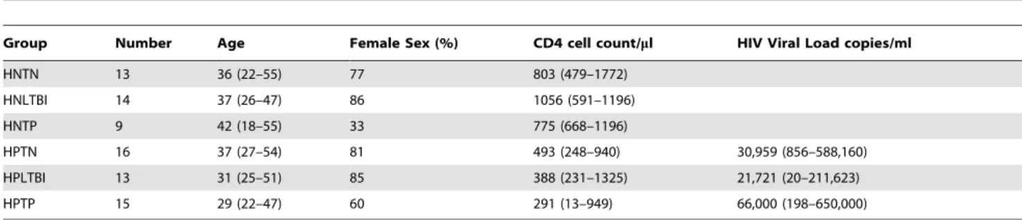
Group Number Age Female Sex (%) CD4 cell count/ml HIV Viral Load copies/ml
HNTN 13 36 (22–55) 77 803 (479–1772)
HNLTBI 14 37 (26–47) 86 1056 (591–1196)
HNTP 9 42 (18–55) 33 775 (668–1196)
HPTN 16 37 (27–54) 81 493 (248–940) 30,959 (856–588,160)
HPLTBI 13 31 (25–51) 85 388 (231–1325) 21,721 (20–211,623)
HPTP 15 29 (22–47) 60 291 (13–949) 66,000 (198–650,000)
HNTN: HIV negative, tuberculosis (TB) negative; HNLTBI: HIV negative, latent tuberculosis infection (LTBI)); HNTP: HIV negative, TB positive; HPTN: HIV positive, TB negative; HPLTBI: HIV positive, LTBI; HPTP: HIV positive, TB positive. Except where designated, all values are expressed as median (range).
Impact of ART on CD161++/MAIT cell frequency
We next investigated the impact of ART on our population of interest, using fourteen subjects from the iThimba cohort who initiated ART and achieved viral suppression during the period of observation. The demographic and clinical description of these subjects is summarized in Table 2. As we were using cryopreserved
PBMC we first determined if the pattern of lower CD161++CD8+
T cell frequency in HIV infected (untreated) compared to healthy individuals was confirmed in this sample type. Figure 4A shows
that we again saw significantly lower levels of the CD161++CD8+
T cell population in HIV infected individuals compared to healthy controls (p = 0.0095). ART that effectively suppressed HIV viral load (median duration 172 days, range 92–282) was not associated
with an increase in CD161++CD8+T cell frequency (pre-ART
median = 0.665, on ART median = 0.605; p = 0.7354) (Fig. 4B).
The median fold change in CD161++CD8+T cell frequency was
0.97 (range = 0.37–3.71). Fig. 4C shows representative FACS plots
of the CD161++CD8+population (co-stained for Va7.2) prior to
and after approximately 6 months of ART.
Discussion
In our group of individuals based in Durban, South Africa we
identified a population of CD8+T cells that expressed very high
levels of the C-type lectin CD161. This population (between
1-26% of CD8+ T cells in healthy (HIV and TB negative)
individuals) had a distinct phenotypic profile and expressed distinctly high levels of the HIV co-receptor CCR5 and the tissue-homing marker CCR6. In addition, the vast majority of this
population expressed the Va7.2 TCR, consistent with a MAIT cell
population. The CD161++CD8+T cell frequency and phenotype
observed in our cohort is in line with recent reports in populations
based in Europe and the US ([2,8,9,24,26]. This CD161++/
MAIT cell subset may play a significant role in antimicrobial immunity in both HIV and MTB infections. This is the first study
to investigate this CD161++/MAIT cell population in HIV
infected, MTB infected (both LTBI and TB), and co-infected individuals in an HIV- and TB-endemic setting.
Chronic HIV infection was associated with significantly lower
levels of CD161++CD8+ T cells. This finding is supported by
recent reports by Cosgrove et al and Leeansyah et al [9,24]. Interestingly, in all of the data sets there are healthy individuals
who have low CD161++CD8+ T cell frequencies, in the same
range as individuals with HIV. This raises the question of whether low-levels of this cell subset make individuals more susceptible to HIV infection, or if HIV infected individuals undergo a decrease of this subset in the bloodstream. Cosgrove et al investigated the
frequency of CD161++ CD8+ T cells in early HIV infection
(baseline sample was taken within 6 months of HIV infection),
reporting that the frequency of CD161++ CD8+ T cells was
already reduced in this phase of infection [9]. Our data from individuals in the acute phase of HIV infection reveal that low-level frequencies of this MAIT cell population are present even 2-3 weeks post presumed HIV infection date, indicating either that a rapid change occurs, or that these low levels were present in these individuals prior to infection. To clarify this issue, this question needs to be addressed with a larger population of individuals, with
both pre- and post-infection time-points. The CD161++/MAIT
cell subset did not show further declines in frequency in longitudinal follow-up of our acutely infected population (up to 24 weeks post-presumed infection date). Interestingly, this is in contrast to Leeansyah et al, who reported that the decline of
MAIT cells (defined as CD161+, Va7.2+, and primarily CD8+) in
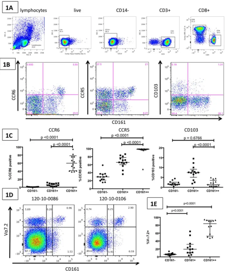
Figure 1. CD161++ CD8+MAIT cells have a distinct phenotypic profile. Figure 1A: Depiction of the gating strategy used throughout: lymphocytes were selected on the basis of forward- and side-scatter characteristics, dead cells excluded, then CD14 negative, CD3 positive, CD4 negative, CD8 positive cells selected. Figure 1B: FACS plots (gated on the CD8+T cell population) show CD161++cells demonstrating high expression
of CCR6 and CCR5 and low expression of CD103 in a representative example from an HIV negative TB negative subject (HNTN). Figure 1C: Aggregate data from 13 HNTN individuals demonstrate that CD161++CD8+T cells express significantly higher levels of CCR5 and CCR6 and lower levels of
CD103 compared to CD161+and CD161- CD8+T cells. Figure 1D: FACS plots (gated on the CD8+T cell population) show high expression of the
Va7.2 semi-invariant T-cell receptor by CD161++ T cells from two representative HNTN individuals. Figure 1E: Aggregate data from 12 HNTN individuals demonstrate that CD161++CD8+T cells express significantly higher levels of the Va7.2 TCR than either CD161+CD8+T cells or
CD161-CD8+T cells. All graphs show median and intra-quartile range; p-values are reported for two-sided Mann-Whitney tests with threshold for significance p = 0.025 after Bonferroni correction for 2 comparisons.
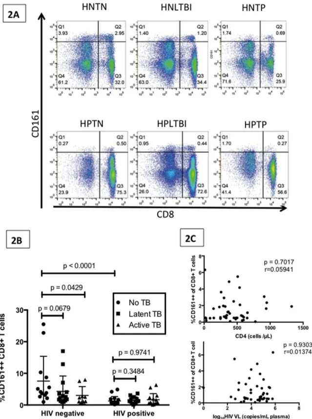
Figure 2. Impact of HIV and TB mono- and co-infection on CD161++CD8+T cell frequencies.Figure 2A: FACS plots (gated on the CD3+T cell population) show CD161 and CD8 co-staining on representative individuals from 6 different clinical groups: HIV negative and TB negative (HNTN), HIV negative with latent TB infection (HNLTBI), HIV negative with active TB (HNTP), HIV positive TB negative (HPTN), HIV positive with latent TB infection (HPLTBI) and HIV positive with active TB (HPTP). Figure 2B: Aggregate data demonstrating CD161++CD8+T cell frequency (median and
interquartile range) in 6 different groups: HNTN (n = 13), HNLTBI (n = 14), HNTP (n = 9), HPTN (n = 16), HPLTBI (n = 13) HPTP (n = 15). P-values reported for two-sided Mann-Whitney tests, with a p-value threshold for significance of 0.01 after Bonferroni correction for 5 comparisons. Figure 2C: CD161++CD8+T cell frequency in all subjects with HIV-infection showed no significant correlation with either CD4 count (cells/ml) or HIV viral load
(copies/ml).
HIV infected individuals is associated with time since diagnosis [24].
It is unlikely that the ‘reduced frequencies’ of this MAIT cell population is merely a result of dilution due to expansion of
HIV-specific subsets following infection as virus-HIV-specific CD8+ T cells
do not contribute significantly to the CD161++CD8+T cell pool
(data not shown and [9]) and MAIT cells are activated by a range of bacteria and fungi (but not viruses) in a MR1-dependent manner. In addition the low frequency of this population in
HIV-infected subjects is only observed for CD161++CD8+T cells and
not CD161+CD8+T cells, which appear to be stably maintained
in HIV infected individuals. Cosgrove et al providein vitrosupport
for the possibility that the CD161++CD8+T cell population is
depleted by activation-induced apoptosis on encounter with translocated bacteria [9]. Other potential scenarios are that this cell population is redistributed to tissues upon infection, which could explain in part the low levels of CCR6 expression on this subset in HIV and TB infected individuals. This area requires further work as to date only one report has emerged from the literature, which found no enrichment of this population in colon tissue from HIV infected individuals [9]. We investigated if ART
was able to increase the observed frequency of the CD161++
CD8+MAIT cell population and did not find a significant change
following up to 282 days of treatment (a finding supported by
[9,24]). Interestingly, in our study the CD161++ CD8+ T cell
frequency did not correlate with CD4 count or HIV viral load in HIV positive individuals.
Individuals with active pulmonary TB displayed a trend towards
lower levels of CD161++CD8+ T cells compared to ‘healthy’
individuals, however the significance of the finding (p = 0.043) did not survive the stringency of a 5 comparison Bonferroni correction. Since our finding supports previous reports that the MAIT cell population is depleted from the blood of tuberculosis (TB) patients, it is likely that the borderline significance is the result of our relatively small sample sizes [13–15]. Further investigation on a larger cohort needs to be performed, especially to dissect the impact of LTBI and subclinical infection. Interestingly, HIV-TB co-infection was not associated with a synergistic decrease in frequency when compared to HIV mono-infected individuals. It
will be interesting to monitor the CD161++CD8+ MAIT cell
population in individuals who progress from LTBI to active disease and those who undergo TB. Further work in tissues also needs to be performed.
In conclusion, low levels of CD161++ CD8+ MAIT cells in
peripheral blood samples were associated with HIV infection and HIV/TB co-infection. Interestingly, low levels are found in acute HIV infection (2–3 weeks post presumed infection) suggesting that either low frequencies of this population predispose individuals to
Figure 3. Acute HIV infection is associated with a lower frequency of CD161++CD8+T cells.Figure 3A: Graph displays CD161++CD8+T
cell frequency in HIV negative and TB negative subjects (HNTN, n = 13), individuals with chronic HIV infection (HIV positive TB negative HPTN, n = 16), and individuals in the acute phase of HIV infection (n = 5, sample taken within 2–3 weeks of presumed infection date). CD161++CD8+T cells are
significantly lower in HPTN individuals and those with acute HIV. Figure 3B: Graph displays CD161+CD8+T cell frequency in the same three groups of subjects; CD161+CD8+T cells do not show any differences between these three groups. Figure 3C: A representative FACS plot displaying the
CD161++CD8+T cell population over time in an individual acutely infected with HIV and assessed at 5 timepoints. Both graphs show median and
interquartile range; p-values are reported for two-sided Mann-Whitney tests with threshold for significance p = 0.025 after Bonferroni correction for 2 comparisons.
HIV infection, or that this population of cells is either destroyed as a consequence of infection or is redistributed to the site of infection. ART was unable to increase the frequency of this MAIT cell population, however TB therapy in HIV uninfected individ-uals may offer this possibility. Lower levels of the tissue-homing
CD161++CD8+T cell population may contribute to a weakened
mucosal immune defense in subjects infected with HIV and TB, making HIV subjects more susceptible to diseases like TB, and detrimentally impacting on the host’s battle with TB.
Acknowledgments
We would like to thank the staff and patients of the iThimba cohort based at McCord Hospital (Durban, KZN, South Africa) and the HPP Acute
Infection Study (Durban, KZN, South Africa). In particular we wish to thank the efforts of Dr. Henry Sunpath. Open access publication of this article has been made possible through support from the Victor Daitz Information Gateway, an initiative of the Victor Daitz Foundation and the University of KwaZulu-Natal.
Author Contributions
Conceived and designed the experiments: EBW CC BDW TN PK VOK. Performed the experiments: EBW NAA PG ZS MP VOK. Analyzed the data: EBW NAA PG ZS DML PK VOK. Contributed reagents/ materials/analysis tools: DML WRB BDW TN PK VOK. Wrote the paper: EBW VOK.
References
1. Northfield JW, Kasprowicz V, Lucas M, Kersting N, Bengsch B, et al. (2008) CD161 expression on hepatitis C virus-specific CD8+T cells suggests a distinct pathway of T cell differentiation. Hepatology 47: 396–406.
2. Billerbeck E, Kang YH, Walker L, Lockstone H, Grafmueller S, et al. (2010) Analysis of CD161 expression on human CD8+ T cells defines a distinct
Figure 4. Impact of Antiretroviral treatment (ART) on CD161++CD8+T cell frequency.Figure 4A: Graph displaying CD161++CD8+T cell frequency (median and interquartile range) from 14 HIV negative TB negative individuals (HNTN) and 14 HIV positive TB negative individuals (HPTN) prior to ART initiation (pre-ART) and on virally suppressive ART (on ART). P-value reported from two-sided Mann-Whitney tests with threshold for significance p = 0.025 after Bonferroni correction for 2 comparisons. Figure 4B: Paired CD161++CD8+T-cell frequencies in HIV-positive individuals
prior to and following virally suppressive ART (p-value reported for Wilcoxon matched pairs test). Figure 4C: FACS plots (gated on the CD8+T cell population) displaying the lack of change in the CD161++CD8+population (co-stained with the MAIT cell semi-invariant TCR, Va7.2) prior to and
functional subset with tissue-homing properties. Proc Natl Acad Sci U S A 107: 3006–3011.
3. Nigam P, Kwa S, Velu V, Amara RR (2011) Loss of IL-17-producing CD8 T cells during late chronic stage of pathogenic simian immunodeficiency virus infection. J Immunol 186: 745–753.
4. Chung DR, Kasper DL, Panzo RJ, Chitnis T, Grusby MJ, et al. (2003) CD4+T cells mediate abscess formation in intra-abdominal sepsis by an IL-17-dependent mechanism. J Immunol 170: 1958–1963.
5. Puel A, Cypowyj S, Bustamante J, Wright JF, Liu L, et al. (2011) Chronic mucocutaneous candidiasis in humans with inborn errors of interleukin-17 immunity. Science 332: 65–68.
6. Huang W, Na L, Fidel PL, Schwarzenberger P (2004) Requirement of interleukin-17A for systemic anti-Candida albicans host defense in mice. J Infect Dis 190: 624–631.
7. Khader SA, Bell GK, Pearl JE, Fountain JJ, Rangel-Moreno J, et al. (2007) IL-23 and IL-17 in the establishment of protective pulmonary CD4+T cell responses after vaccination and during Mycobacterium tuberculosis challenge. Nat Immunol 8: 369–377.
8. Dusseaux M, Martin E, Serriari N, Peguillet I, Premel V, et al. (2011) Human MAIT cells are xenobiotic-resistant, tissue-targeted, CD161hi IL-17-secreting T cells. Blood 117: 1250–1259.
9. Cosgrove C, Ussher JE, Rauch A, Gartner K, Kurioka A, et al. (2013) Early and nonreversible decrease of CD161++/MAIT cells in HIV infection. Blood 121: 951–961.
10. Walker LJ, Kang YH, Smith MO, Tharmalingham H, Ramamurthy N, et al. (2012) Human MAIT and CD8alphaalpha cells develop from a pool of type-17 precommitted CD8+T cells. Blood 119: 422–433.
11. Treiner E, Duban L, Bahram S, Radosavljevic M, Wanner V, et al. (2003) Selection of evolutionarily conserved mucosal-associated invariant T cells by MR1. Nature 422: 164–169.
12. Chua WJ, Truscott SM, Eickhoff CS, Blazevic A, Hoft DF, et al. (2012) Polyclonal mucosa-associated invariant T cells have unique innate functions in bacterial infection. Infect Immun 80: 3256–3267.
13. Gold MC, Cerri S, Smyk-Pearson S, Cansler ME, Vogt TM, et al. (2010) Human mucosal associated invariant T cells detect bacterially infected cells. PLoS Biol 8: e1000407.
14. Le Bourhis L, Martin E, Peguillet I, Guihot A, Froux N, et al. (2010) Antimicrobial activity of mucosal-associated invariant T cells. Nat Immunol 11: 701–708.
15. Georgel P, Radosavljevic M, Macquin C, Bahram S (2011) The non-conventional MHC class I MR1 molecule controls infection by Klebsiella pneumoniae in mice. Mol Immunol 48: 769–775.
16. Dye C, Williams BG (2008) Eliminating human tuberculosis in the twenty-first century. J R Soc Interface 5: 653–662.
17. UNAIDS G (2008) Report on the global AIDS epidemic.
18. Bateman C (2010) The protracted TB struggle - SA ups the intensity. S Afr Med J 100: 207–209.
19. Bateman C (2009) Collaborative push to address TB crisis on mines. S Afr Med J 99: 852–855.
20. Barnighausen T, Tanser F, Gqwede Z, Mbizana C, Herbst K, et al. (2008) High HIV incidence in a community with high HIV prevalence in rural South Africa: findings from a prospective population-based study. AIDS 22: 139–144. 21. Houlihan CF, Mutevedzi PC, Lessells RJ, Cooke GS, Tanser FC, et al. (2010)
The tuberculosis challenge in a rural South African HIV programme. BMC Infect Dis 10: 23.
22. Sonnenberg P, Glynn JR, Fielding K, Murray J, Godfrey-Faussett P, et al. (2005) How soon after infection with HIV does the risk of tuberculosis start to increase? A retrospective cohort study in South African gold miners. J Infect Dis 191: 150– 158.
23. Sutherland R, Yang H, Scriba TJ, Ondondo B, Robinson N, et al. (2006) Impaired IFN-gamma-secreting capacity in mycobacterial antigen-specific CD4 T cells during chronic HIV-1 infection despite long-term HAART. AIDS 20: 821–829.
24. Leeansyah E, Ganesh A, Quigley MF, Sonnerborg A, Andersson J, et al. (2013) Activation, exhaustion, and persistent decline of the antimicrobial MR1-restricted MAIT-cell population in chronic HIV-1 infection. Blood 121: 1124– 1135.
25. Sandberg JK, Dias J, Shacklett BL, Leeansyah E (2013) Will loss of your MAITs weaken your HAART? AIDS.
26. Turtle CJ, Delrow J, Joslyn RC, Swanson HM, Basom R, et al. (2011) Innate signals overcome acquired TCR signaling pathway regulation and govern the fate of human CD161(hi) CD8alpha(+) semi-invariant T cells. Blood 118: 2752– 2762.
27. Martin E, Treiner E, Duban L, Guerri L, Laude H, et al. (2009) Stepwise development of MAIT cells in mouse and human. PLoS Biol 7: e54. 28. Round JL, Mazmanian SK (2009) The gut microbiota shapes intestinal immune
responses during health and disease. Nat Rev Immunol 9: 313–323. 29. Day CL, Mkhwanazi N, Reddy S, Mncube Z, van der Stok M, et al. (2008)
Detection of polyfunctional Mycobacterium tuberculosis-specific T cells and association with viral load in HIV-1-infected persons. The Journal of infectious diseases 197: 990–999.
30. Radebe M, Nair K, Chonco F, Bishop K, Wright JK, et al. (2011) Limited immunogenicity of HIV CD8+T-cell epitopes in acute Clade C virus infection. J Infect Dis 204: 768–776.
31. Morgan AJ, Guillen C, Symon FA, Huynh TT, Berry MA, et al. (2005) Expression of CXCR6 and its ligand CXCL16 in the lung in health and disease. Clin Exp Allergy 35: 1572–1580.
32. Morgan AJ, Guillen C, Symon FA, Birring SS, Campbell JJ, et al. (2008) CXCR6 identifies a putative population of retained human lung T cells characterised by co-expression of activation markers. Immunobiology 213: 599– 608.