Association of Physical Training with Beta-Blockers in Heart Failure
in Mice
Andréa Somolanji Vanzelli
1,2, Alessandra Medeiros
3, Raquel de Assis Sirvente
2, Vera Maria Cury Salemi
2, Charles
Mady
2, Patricia Chakur Brum
1Escola de Educação Física da Universidade de São Paulo1, Faculdade de Medicina da Universidade de São Paulo (FMUSP)2, Universidade
Federal de São Paulo (UNIFESP)3, São Paulo, SP - Brazil
Mailing address: Andréa Somolanji Vanzelli •
Rua Arnaldo Simões Pinto, 166 - Vila Bonilha - 02917-110 - São Paulo, SP - Brazil
E-mail: vanzelli@usp.br, andreasvanzelli@gmail.com
Manuscript received September 29, 2009; revised manuscript received February 11, 2010; accepted March 16, 2010.
Abstract
Background: Currently there are several types of interventions for the treatment of heart failure (HF). Among these are beta-blocker therapy (BB) and physical training (PT). However, the effects of the combination of these therapies are poorly studied.
Objective: To investigate the effects of BB treatment with metoprolol (M) and carvedilol (C) associated with PT in mice with HF.
Methods: We used a genetic model of sympathetic hyperactivity-induced heart failure in mice. Initially, we divided the HF animals into three groups: sedentary (S); trained (T); treated with M (138 mg/kg) (M); or C (65 mg/kg) (C). In the second part, we divided the groups into three subgroups: sedentary (S); trained and treated with M (TM); and trained and treated with C (CT). The PT consisted of aerobic training on a treadmill for 8 weeks. Exercise tolerance was assessed by maximal graded test, and fractional shortening (FS) was assessed by echocardiography. Cardiomyocyte diameter and collagen volume fraction were evaluated by histological analysis. Data were compared by one way ANOVA and post hoc
Duncan test. The significance level was set at p ≤ 0.05.
Results: As to FS and cardiac remodeling, we found that, in isolation, T, M, and C showed an improvement of the variables analyzed. As to therapy combination, after the intervention period, we observed an increase in exercise tolerance in MT and CT (43.0% and 33.0% respectively). There was also a reduction in cardiomyocyte diameter (10.0% and 9.0% respectively) and in collagen volume fraction (52.0% and 63.0%) after the intervention. However, only CT significantly improved FS.
Conclusion: The association of PT with M or C therapies provided benefits on cardiac function and remodeling in HF mice. (Arq Bras Cardiol 2010; 95(3): 373-380)
Key words: Heart failure; training; exercise; sympathetic nervous system; b-blockers e mice.
in the treatment of HF. The introduction of beta blockers and renin-angiotensin-aldosterone inhibitors made a considerable difference in the clinical evolution of patients, offering a more promising prospect for greater survival. Relevant clinical trials showed an unprecedented mortality reduction in the treatment of HF6-10.
There are increased possibilities of therapeutic and clinical optimization when non-pharmacological measures, such as diet and regular physical activity, are associated with conventional pharmacological treatment. Actually, physical training has been increasingly adopted, and its benefits include improved quality of life and tolerance to physical exertion, besides a reduction in the hyperactivity of the neurohumoral system, which is directly related to improved survival rates11 and autonomic balance12-14.
In previous laboratory studies we demonstrated that a therapy with the beta-blocker carvedilol significantly reduced sympathetic nerve activity in patients with HF15. More recently, we demonstrated that a non-pharmacological
Introduction
Heart failure is the final common pathway for many cardiovascular diseases. It is a syndrome characterized by signs and symptoms related to inadequate tissue perfusion1 and cathecolamine excess2. According to the hemodynamic view, heart failure syndrome is associated with low cardiac output, with consequent low renal flow, resulting in sodium and water retention and the appearance of peripheral and pulmonary edema3. Another common feature of heart failure is cardiac remodeling associated with loss of cardiomyocytes and increased collagen fraction4,5.
therapy based on exercise training resulted in additional reduction in sympathetic activity in HF patients who were already under therapy with the beta-blocker carvedilol16. These results suggest that the association of physical training with beta-blocker therapy can optimize the reduction in the hyperactivity of the neurohumoral system.
In this study, we used a genetic model of sympathetic hyperactivity-induced HF in mice to study the effect of the combination of the beta blockers metoprolol or carvedilol with aerobic exercise training on cardiac function and structure.
Material and methods
Sample
We used male mice of the C57/BL6 strain with gene inactivation of the α2A/α2C adrenergic receptor (α2A/α2CARKO mice), aged between 5-7 months, from the Laboratory Animal Facility of the School of Physical Education and Sports of the University of São Paulo (n = 35). These mice develop clinical signs of HF such as pulmonary edema associated with severe ventricular dysfunction at 7 months of age, when the mortality rate is 50.0%12,17. The gene inactivation of the
α2A and α2C adrenergic receptors leads to an increase in the release of circulating noradrenaline18,19. Therefore, the HF observed in this model results from sympathetic nervous system hyperactivity.
This study was approved by the Ethics Committee of the Medical School of the University of São Paulo (897/06).
As a first step in our study, in order to evaluate the effect of isolated therapies on the variables analised, the animals were randomly divided into four groups: α2A/α2CARKO sedentary mice (S); α2A/α2CARKO trained mice (T); α2A/
α2CARKO mice treated with metoprolol (M); and α2A/
α2CARKO mice treated with carvedilol (C). After observing the isolated effect of the treatments, the effects of combined therapies were evaluated and, for that end, the animals were randomly divided into the following subgroups: α2A/
α2CARKO sedentary mice (S); α2A/α2CARKO mice trained and treated with metoprolol (MT); and α2A/α2CARKO mice trained and treated with carvedilol (CT).
Control mice without HF (WT) are shown in the figures of the study with a dashed line to indicate the expected value in a control group without heart failure.
The treatments were carried out by gavage, and the doses used were previously tested in order to adjust them and to obtain equipotent doses for reducing the heart rate of animals, which corresponded to 135 mg/kg and 68 mg/kg for metoprolol and carvedilol, respectively20,21.
Body weight was monitored weekly in a semi-analytical scale (Gehaka, BG 400 - São Paulo, Brazil).
Maximum capacity of physical exercise
Intolerance to exertion was estimated in the groups studied by quantifying the maximum capacity to perform physical exercise (total test time in seconds) using a phased progressive exercise test to exhaustion, which was evidenced
when the animal could not continue running on the treadmill (manufactured by the Federal University of São Carlos, São Carlos, Brazil) with an initial velocity of 3 m/min, and an increase of 3 m/min every 3 min12.
Aerobic exercise training protocol
The groups of trained animals underwent a program of aerobic exercise training on treadmill for 8 weeks (from 5 to 7 months of age). The exercise sessions were conducted 5 times per week, lasting 60 minutes per session, with an intensity of 60.0% of the maximum speed recorded in the progressive test to exhaustion, which, as noted in a previous study, corresponds to the maximum lactate steady state22 ie, corresponds to the maximum exercise intensity in which there is a balance between production and removal of blood lactate during prolonged exercise.
Indirect measurement of blood pressure and heart rate We performed an indirect measurement of blood pressure using the tail plethysmographic method over the 8 weeks of intervention (Kent Scientific, CODA - Torrington, CT, USA). By using the pressure pulses recorded, we calculated the heart rate of the mice, as had already been done in previous studies13,20,21,23.
Echocardiographic assessment
The transthoracic echocardiography was performed before and after the intervention period in all groups. The echocardiographic examination was performed with the animals anesthetized with the inhalation of the anesthetic halothane 1.0%, mixed with an O2 flow of 1 l/min. We used an echocardiograph (Acuson Corporation, Sequoia 512 - Mountain View, CA, USA) with a 15 MHz transducer. Images were taken at a frequency of 14 MHz. From the visualization of the left ventricle (cross section) at the level of the papillary muscles, M-mode was performed, and the diastolic diameter (LVDD) and the systolic diameter (LVSD) of the left ventricle were obtained and used to calculate fractional shortening (FS). The formula used was: FS = (LVDD - LVSD)/LVDD. The echocardiographer (R. S.) was blinded to the genotype and the type of treatment of the animals.
Cardiac morphological and morphometric analyses After 48 hours of formalin fixation (4.0%), the left ventricle was subjected to the usual histological processing, with 2 micron slices and hematoxylin-eosin or picrosirius
red staining for the quantification of the cardiomyocyte cross-sectional diameter and the cardiac collagen fraction, respectively. These measurements were performed in a computerized system (Leica Imaging Systems Ltd., Quantimet 500 - Cambridge, UK, England), with a magnification of 400x and 200x, respectively. The cardiomyocyte diameter was determined as the mean of 10 measured values for each animal.
area of myocardial tissue in each ventricular region examined (absolute amount of collagen and myocytes), field by field. About 15-20 fields were examined for each animal13,20,21,23,24.
Statistical analysis
All variables showed normal distribution, when analyzed using the Shapiro-Wilk normality test, and therefore, the parametric statistical analysis was used. Data were expressed as mean ± standard error of mean.
The variables were compared among groups by one-way analysis of variance (ANOVA) (cardiomyocyte cross-sectional diameter and collagen fraction volume), or one way repeated measures (exercise tolerance, fractional shortening, and heart rate). For mortality, log rank analysis was used. For cases in which some significance was shown, post hoc Duncan’s test was used. For all analyses, we adopted the significance level of p < 0.05. The software used for statistical analysis was
Statistica version 6.0.
Results
Isolated effect of treatments (physical training, metoprolol, and carvedilol on exercise tolerance, cardiac structure, and contractile function)
In Figure 1, Panel A, which highlights the isolated effects of the treatments on exercise tolerance, we observed that exercise training alone leads to a significant improvement in exercise tolerance, which is not observed in groups treated with the beta blockers metoprolol and carvedilol. As to ventricular function, we observed that all treatments (PT, M, or C) similarly improved the fractional shortening (Figure 1B).
Regarding the effects on cardiac structure, physical training alone did not result in a significant reduction of the cardiomyocyte cross-sectional diameter; however, it had effects similar to the pharmacological therapies in reducing the cardiac collagen fraction (Figures 1C and 1D).
Figure 1 -Isolated effects of treatment on exercise tolerance (Panel A), fractional shortening (Panel B), cardiomyocyte diameter (Panel C), and collagen volume fraction
Effects of the combination of physical training with treatment with beta blockers
During the experimental period, the body weight of the animals was measured weekly. We observed that α2A/α2CARKO animals initially had lower body mass than those of the WT control group (22.2 ± 1.3 g vs 28.3 ± 2.2 g, p ≤ 0.05). Neither
group showed a change in mass during the intervention period. Additionally, we evaluated the mortality curve during the intervention period and found no significant difference among groups (40.0% vs 20.0% and 18.0% for groups S, MT, and CT,
respectively, p ≤ 0.05).
Maximum exercise tolerance
In Figure 2, we observe that the association of physical training with beta-blocker therapy significantly improved the exercise tolerance when compared to the group treated with saline (S).
It should be emphasized that exercise tolerance in α2A/
α2CARKO animals reached the values observed in control mice without HF (Figure 2, dashed line); also, the groups that were trained and treated with the beta blockers metoprolol and carvedilol were equally effective in improving exercise tolerance (43.0% and 33.0% increase, respectively).
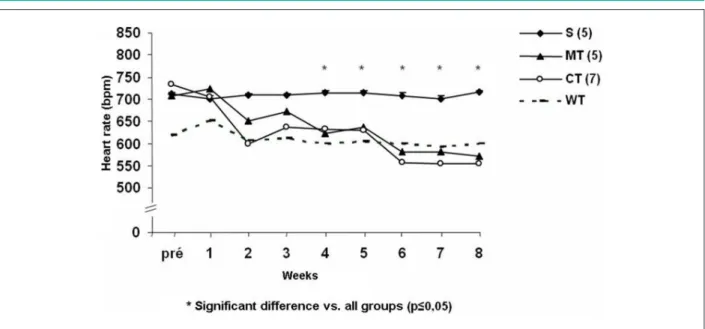
Heart rate and blood pressure
As shown in Figure 3, α2A/α2CARKO mice have resting tachycardia compared to control WT mice (711 ± 3 vs 617
± 24 bpm, respectively, p ≤ 0.05). The association of physical
training with therapy with beta blockers similarly reduced
the heart rate of α2A/α2CARKO mice, from the 4th week of intervention, to values similar to the heart rate observed in control mice without HF (WT, dashed line). There was no difference in blood pressure between groups over the eight weeks of intervention (114 ± 7.3 mmHg at week 8 of study).
Fractional shortening
By 8 weeks after the intervention, only the α2A/α2CARKO mice group trained and treated with carvedilol (CT) significantly improved in values of fractional shortening (Figure 4) compared to the α2A/α2CARKO mice group treated with saline (20 ± 0.8 vs 15 ± 0.4%, respectively, p ≤ 0.05),
reaching values similar to those of the control group without HF (WT). Interestingly, the association of metoprolol with PT did not improve fractional shortening. This result was unexpected because both metoprolol alone and PT alone were able to increase fractional shortening (Figure 1B).
Cardiomyocyte diameter and collagen volume fraction The morphometric assessment showed that both cardiac beta-blockers associated with exercise training were equally effective in significantly reducing cardiomyocyte cross-sectional diameter (10.0% and 9.0% for the trained groups treated with metoprolol and carvedilol, respectively) and cardiac collagen fraction (52.0% and 63.0% for the trained groups treated with metoprolol and carvedilol, respectively), suggesting a possible effect of reverse cardiac remodeling associated with the combined effects of both beta-blockers to physical training (Figure 5).
Figure 2 -Total time (seconds) of the progressive test to exhaustion in α2A/α2CARKO control mice (S), and in α2A/α2CARKO mice undergoing the association of
In mice with sympathetic hyperactivity-induced cardiomyopathy as well as in clinical HF, the manifestation of clinical signs, such as intolerance to exertion, pulmonary edema, decreased cardiac function, and cardiac remodeling, is very common. It is important to emphasize that, even though a cause-effect on sympathetic hyperactivity has not been established in clinical heart failure, the autonomic imbalance plays an important role in the patient’s prognosis,
Figure 3 -Heart rate over the intervention period in α2A/α2CARKO control mice (S), and in α2A/α2CARKO mice undergoing the association of pharmacological treatment
with metoprolol (MT) or carvedilol (CT) to physical training in the pre-intervention (5 months age) and the post-intervention (7 months) periods. WT - wild type.
Figure 4 -Fractional shortening before and after the intervention period in α2A/α2CARKO control mice (S), and in α2A/α2CARKO mice undergoing the association of
pharmacological treatment with metoprolol (MT) or carvedilol (CT) to physical training in the pre-intervention (5 months age) and the post-intervention (7 months) periods. WT - wild type.
which makes the mice model used in this study even more interesting, because it allows for studies evaluating the direct effect of sympathetic hyperactivity in HF.
Following this addendum, the main results of this study in mice with sympathetic hyperactivity-induced HF were:
Figure 5 -Cardiomyocyte diameter (Panel A) and collagen volume fraction (panel B) evaluated in α2A/α2CARKO control mice (S), and in α2A/α2CARKO mice undergoing
the association of pharmacological treatment with metoprolol (MT) or carvedilol (CT) to physical training in the pre-intervention (5 months age) and the post-intervention (7 months) periods.
and reduced resting tachycardia in an equipotent manner; 2) Both physical and pharmacologic therapies, individually, and the association of carvedilol with physical training significantly improved fractional shortening;
3) Both metoprolol and carvedilol associated with physical training similarly reduced collagen volume fraction and cardiomyocyte cross-sectional diameter.
We demonstrated previously that exercise training improved fractional shortening in heart failure by improving the calcium transient and the expression profile of proteins that regulate intracellular Ca2+ flow13,23. The association of metoprolol with physical training seems to be less effective, probably because metoprolol has a cardiac effect similar to that of PT, i.e. it is more associated with the calcium transient and not with reducing oxidative stress.20
Although carvedilol and metoprolol in combination with exercise training produced different effects on ventricular contractility, both were equally effective in improving tolerance to exertion when combined with exercise training.
The increased tolerance to physical exertion is not associated only with an improved cardiac function, but with the combination of better cardiac performance with beneficial adaptations in non-cardiac target organs. This may include the vasodilator response in the vascular endothelium, the cardiac output distribution, the ventilatory response25, and changes in skeletal muscles.
Skeletal muscle structure analyses, conducted in the same genetic model used in this study, with α2A/α2C-ARKO mice of 7 months of age, using the technique of staining for myosin ATPase, showed a decreased cross-sectional area of muscle fibers, a phenotypic change of fiber type I to type II fibers, and capillary rarefaction. Besides, the changes described appear to be accompanied by a change in the metabolic profile, with a reduced activity of oxidative enzymes26. This may be
associated with the progressive reduction in exercise tolerance observed in this study.
With regard to the effects of beta blockers in tolerance to exertion, several studies in the literature have examined their use in patients with various heart diseases27,28. Clinical trials that have lasted for more than a month have observed significant improvements in ventricular function, but very subtle improvement in exercise tolerance27,28. Therefore, physical training seems to play a key role in improving tolerance to exertion in mice with heart failure , corroborating the results of other studies that show the same effect29,30.
Similarly to what was observed for tolerance to exertion, heart rate reduction had the same outcome pattern in the groups treated with metoprolol and carvedilol associated with physical training. Both had equipotent effects on HR, reducing it to values similar to those observed in WT mice (dashed line), and thus showing the effectiveness of the treatment with both beta-blockers in reducing tachycardia in this model of cardiomyopathy, which is related to an increased activity of the sympathetic nervous system. Therefore, these bradycardic effects in mice, may suggest an improvement in the autonomic balance, which, in turn, is associated with an improved prognosis and survival in heart failure16,31. Moreover, high values of heart rate are also independent predictors of mortality in the general population32,33.
Besides changes in ventricular function, our results also suggest the presence of cardiac remodeling in mice with heart failure (Figure 4). Both beta-blockers in combination with exercise training had beneficial effects on cardiac structure and prevented cardiomyocyte hypertrophy and cardiac collagen increase.
physiological hypertrophy35. Therefore, the association of PT to beta-blockers is an approach to be adopted, because remodeling is highly related to death in patients with HF36-38. Other effects of physical training on minimizing the process of myocardial failure have recently been shown and may be associated with a reduction in proteins associated with fibrosis and cardiac remodeling, among other factors39,40.
Conclusion
The association of physical training to therapy with metoprolol or carvedilol similarly improved tolerance to exertion and also improved the cardiac structure in mice with HF induced by sympathetic hyperactivity. However, only carvedilol, when associated with PT, improved contractility.
Acknowledgements
The authors would like to express their appreciation to
the School of Medicine and the School of Physical Education and Sports of the University of São Paulo. Also to FAPESP for funding the study (06/56123-0, 05/59740-7). PCB has been awarded an ID level researcher scholarship by CNPq (301519/2008-0).
Potential Conflict of Interest
No potential conflict of interest relevant to this article was reported.
Sources of Funding
This study was funded by FAPESP.
Study Association
This article is part of the thesis of doctoral submitted by Andréa Somolanji Vanzelli, from Faculdade de Medicina da Universidade de São Paulo.
References
1. Adamopoulos S, Parissis JT, Kremastinos DT. New aspects for the role of physical training in the management of patients with chronic heart failure. Int J Cardiol. 2003; 90 (1): 1-14.
2. Opie LH, Walpoth B, Barsacchi R. Calcium and catecholamines: relevance to cardiomyopathies and significance in therapeutic strategies. J Mol Cell Cardiol. 1985; 17 (Suppl 2): 21-34.
3. Katz AM. Evolving concepts of heart failure: cooling furnace, malfunctioning pump, enlarging muscle--Part I. J Card Fail. 1997; 3 (4): 319-34.
4. Grimm D, Huber M, Jabusch HC, Shakibaei M, Fredersdorf S, Paul M, et al. Extracellular matrix proteins in cardiac fibroblasts derived from rat hearts with chronic pressure overload: effects of beta-receptor blockade. J Mol Cell Cardiol. 2001; 33 (3): 487-501.
5. Kunst G, Kress KR, Gruen M, Uttenweiler D, Gautel M, Fink RH. Myosin binding protein C, a phosphorylation-dependent force regulator in muscle that controls the attachment of myosin heads by its interaction with myosin S2. Circ Res. 2000; 86 (1): 51-8.
6. Rationale, design, and organization of the Metoprolol CR/XL Randomized Intervention Trial in Heart Failure (MERIT-HF). The International Steering Committee. Am J Cardiol. 1997; 80 (9B): 54J-8J.
7. Carvedilol saves lives--new data from landmark trials prove survival benefits in heart failure and post myocardial infarction. Cardiovasc J S Afr. 2001; 12 (2): 122-3.
8. Anderson JL, Lutz JR, Gilbert EM, Sorensen SG, Yanowitz FG, Menlove RL, et al. A randomized trial of low-dose beta-blockade therapy for idiopathic dilated cardiomyopathy. Am J Cardiol. 1985; 55 (4): 471-5.
9. Cesario DA, Fonarow GC. Beta-blocker therapy for heart failure: the standard of care. Rev Cardiovasc Med. 2002; 3 (1): 14-21.
10. Metra M, Nodari S, Bordonali T, Milani P, Lombardi C, Bugatti S, et al. Bisoprolol in the treatment of chronic heart failure: from pathophysiology to clinical pharmacology and trial results. Ther Clin Risk Manag. 2007; 3 (4): 569-78.
11. Pereira MG, Ferreira JC, Bueno CR Jr, Mattos KC, Rosa KT, Irigoyen MC, et al. Exercise training reduces cardiac angiotensin II levels and prevents cardiac dysfunction in a genetic model of sympathetic hyperactivity-induced heart failure in mice. Eur J Appl Physiol. 2009; 105 (6): 843-50.
12. Brum PC, Kosek J, Patterson A, Bernstein D, Kobilka B. Abnormal cardiac function associated with sympathetic nervous system hyperactivity in mice.
Am J Physiol Heart Circ Physiol. 2002; 283 (5): H1838-45.
13. Medeiros A, Rolim NP, Oliveira RS, Rosa KT, Mattos KC, Casarini DE, et al. Exercise training delays cardiac dysfunction and prevents calcium handling abnormalities in sympathetic hyperactivity-induced heart failure mice. J Appl Physiol. 2008; 104 (1): 103-9.
14. Stickland MK, Miller JD. The best medicine: exercise training normalizes chemosensitivity and sympathoexcitation in heart failure. J Appl Physiol. 2008; 105 (3): 779-81.
15. De Matos LD, Gardenghi G, Rondon MU, Soufen HN, Tirone AP, Barretto AC, et al. Impact of 6 months of therapy with carvedilol on muscle sympathetic nerve activity in heart failure patients. J Card Fail. 2004;10 (6): 496-502.
16. Fraga R, Franco FG, Roveda F, de Matos LN, Braga AM, Rondon MU, et al. Exercise training reduces sympathetic nerve activity in heart failure patients treated with carvedilol. Eur J Heart Fail. 2007; 9 (6-7): 630-6.
17. Ferreira JC, Bacurau AV, Evangelista FS, Coelho MA, Oliveira EM, Casarini DE, et al. The role of local and systemic renin angiotensin system activation in a genetic model of sympathetic hyperactivity-induced heart failure in mice. Am J Physiol Regul Integr Comp Physiol. 2008; 294 (1): R26-32.
18. Davies MF, Tsui JY, Flannery JA, Li X, DeLorey TM, Hoffman BB. Augmentation of the noradrenergic system in alpha-2 adrenergic receptor deficient mice: anatomical changes associated with enhanced fear memory. Brain Res. 2003;986 (1-2): 157-65.
19. Kurnik D, Muszkat M, Friedman EA, Sofowora GG, Diedrich A, Xie HG, et al. Effect of the alpha2C-adrenoreceptor deletion322-325 variant on sympathetic activity and cardiovascular measures in healthy subjects. J Hypertens. 2007; 25 (4): 763-71.
20. Bartholomeu JB, Vanzelli AS, Rolim NP, Ferreira JC, Bechara LR, Tanaka LY, et al. Intracellular mechanisms of specific beta-adrenoceptor antagonists involved in improved cardiac function and survival in a genetic model of heart failure. J Mol Cell Cardiol. 2008; 45 (2): 240-9.
21. Medeiros A, Vanzelli AS, Rosa KT, Irigoyen MC, Brum PC. Effect of exercise training and carvedilol treatment on cardiac function and structure in mice with sympathetic hyperactivity-induced heart failure. Braz J Med Biol Res. 2008; 41 (9): 812-7.
23. Rolim NP, Medeiros A, Rosa KT, Mattos KC, Irigoyen MC, Krieger EM, et al. Exercise training improves the net balance of cardiac Ca2+ handling protein expression in heart failure. Physiol Genomics. 2007; 29 (3): 246-52.
24. Xu R, Lin F, Zhang S, Chen X, Hu S, Zheng Z. Signal pathways involved in reverse remodeling of the hypertrophic rat heart after pressure unloading. Int J Cardiol. 2009. [Epub ahead of print].
25. Pina IL, Daoud S. Exercise and heart failure. Minerva Cardioangiol. 2004; 52 (6): 537-46.
26. Bacurau AV, Jardim MA, Ferreira JC, Bechara LR, Bueno CR Jr, Alba-Loureiro TC, et al. Sympathetic hyperactivity differentially affects skeletal muscle mass in developing heart failure: role of exercise training. J Appl Physiol. 2009; 106 (5): 1631-40.
27. Al-Nasser F, Yousufuddin M, Al-Nozha F, Anker SD, Coats AJ, Piepoli MF, et al. Effect of carvedilol on exercise tolerance in patients with chronic heart failure and a restrictive left ventricular filling pattern. Am J Cardiol. 2003; 91 (10): 1281-3.
28. Gullestad L, Dolva LO, Soyland E, Kjekshus J. Difference between beta-1-selective and non-beta-1-selective beta-blockade during continuous and intermittent exercise. Clin Physiol. 1988; 8 (5): 487-99.
29. Experience from controlled trials of physical training in chronic heart failure. Protocol and patient factors in effectiveness in the improvement in exercise tolerance. European Heart Failure Training Group. Eur Heart J. 1998; 19 (3): 466-75.
30. Tyni-Lenne R, Gordon A, Europe E, Jansson E, Sylven C. Exercise-based rehabilitation improves skeletal muscle capacity, exercise tolerance, and quality of life in both women and men with chronic heart failure. J Card Fail. 1998; 4 (1): 9-17.
31. Barretto AC, Santos AC, Munhoz R, Rondon MU, Franco FG, Trombetta IC, et al. Increased muscle sympathetic nerve activity predicts mortality in heart failure patients. Int J Cardiol. 2009; 135 (3): 302-7.
32. Dyer AR, Persky V, Stamler J, Paul O, Shekelle RB, Berkson DM, et al. Heart rate as a prognostic factor for coronary heart disease and mortality: findings in three Chicago epidemiologic studies. Am J Epidemiol. 1980; 112 (6): 736-49.
33. Kannel WB, Kannel C, Paffenbarger RS Jr, Cupples LA. Heart rate and cardiovascular mortality: the Framingham Study. Am Heart J. 1987; 113 (6): 1489-94.
34. Oliveira RS, Ferreira JC, Gomes ER, Paixao NA, Rolim NP, Medeiros A, et al. Cardiac anti-remodelling effect of aerobic training is associated with a reduction in the calcineurin/NFAT signalling pathway in heart failure mice. J Physiol. 2009; 587 (Pt 15): 3899-910.
35. Kemi OJ, Ceci M, Wisloff U, Grimaldi S, Gallo P, Smith GL, et al. Activation or inactivation of cardiac Akt/mTOR signaling diverges physiological from pathological hypertrophy. J Cell Physiol. 2008; 214 (2): 316-21.
36. Meris A, Amigoni M, Uno H, Thune JJ, Verma A, Kober L, et al. Left atrial remodelling in patients with myocardial infarction complicated by heart failure, left ventricular dysfunction, or both: the VALIANT Echo study. Eur Heart J. 2009; 30 (1): 56-65.
37. Udelson JE, Konstam MA. Relation between left ventricular remodeling and clinical outcomes in heart failure patients with left ventricular systolic dysfunction. J Card Fail. 2002; 8 (6 Suppl): S465-71.
38. Yu CM, Bleeker GB, Fung JW, Schalij MJ, Zhang Q, van der Wall EE, et al. Left ventricular reverse remodeling but not clinical improvement predicts long-term survival after cardiac resynchronization therapy. Circulation. 2005; 112 (11): 1580-6.
39. Garciarena CD, Pinilla OA, Nolly MB, Laguens RP, Escudero EM, Cingolani HE, et al. Endurance training in the spontaneously hypertensive rat: conversion of pathological into physiological cardiac hypertrophy. Hypertension. 2009; 53 (4): 708-14.