Radiol Bras. 2014 Set/Out;47(5):310–316 310
Uncommon hepatic tumors: iconographic essay – Part 1
*
Tumores hepáticos incomuns: ensaio iconográfico – Parte 1
Pedrassa BC, Rocha EL, Kierszenbaum ML, Bormann RL, Torres LR, D’Ippolito G. Uncommon hepatic tumors: iconographic essay – Part 1. Radiol Bras. 2014 Set/Out;47(5):310–316.
Abstract
R e s u m o
Most malignant liver tumors are represented by hepatocellular carcinoma and cholangiocarcinoma; however a variety of other uncommon hepatic lesions might also be found. Common lesions such as hemangioma, focal nodular hyperplasia and metastases are well known and have already been extensively documented in the literature. The diagnosis of typical hepatic lesions may be done with some reliability by means of several imaging methods; on the other hand, uncommon lesions normally represent a diagnostic challenge for the radiologist. In this first part of the study, the authors will approach five uncommon liver tumors – angiosarcoma, angiomyolipoma, cystadenoma/biliary carcinoma, epithelioid hemangioendothelioma, and fibrolamellar hepatocellular carcinoma –, describing their main characteristics and image findings with focus on computed tomography and magnetic resonance imaging.
Keywords: Neoplasms; Liver; Atypical; Computed tomography; Magnetic resonance imaging.
A maioria dos tumores hepáticos primários malignos é representada pelo carcinoma hepatocelular e pelo colangiocarcinoma, entretanto, uma variedade de outras lesões hepáticas incomuns pode ser encontrada. Lesões comuns como o hemangioma, a hiperplasia nodular focal e as metástases são bem conhecidas e já foram extensamente documentadas na literatura. O diagnóstico das lesões hepáticas típicas pode ser feito com alguma segurança utilizando-se os diversos métodos de imagem; por outro lado, as lesões incomuns são geralmente um desafio diagnóstico para o radiologista. Nesta primeira parte do estudo abordaremos cinco tumores hepáticos incomuns – o angiossarcoma, o angiomiolipoma, o cistoadenoma/carcinoma biliar, o hemangioendotelioma epitelioide e o carcinoma hepatocelu-lar fibrolamehepatocelu-lar –, suas principais características e achados de imagem, com foco na tomografia computadorizada e na ressonância magnética.
Unitermos: Neoplasias; Fígado; Atípicos; Tomografia computadorizada; Ressonância magnética.
* Study developed at Department of Imaging Diagnosis – Escola Paulista de Medicina da Universidade Federal de São Paulo (EPM-Unifesp), São Paulo, SP, Brazil. 1. MDs, Radiologists, Trainees at the Unit of Abdomen, Department of Imaging Diagnosis – Escola Paulista de Medicina da Universidade Federal de São Paulo (EPM-Unifesp), São Paulo, SP, Brazil.
2. Master, Fellow PhD degree, Department of Imaging Diagnosis – Escola Paulista de Medicina da Universidade Federal de São Paulo (EPM-Unifesp), MD, Radiologist, Hospital São Luiz, São Paulo, SP, Brazil.
3. Private Docent, Professor, Department of Imaging Diagnosis – Escola Paulista de Medicina da Universidade Federal de São Paulo (EPM-Unifesp), São Paulo, SP, Brazil.
Mailing Address: Dr. Giuseppe D’Ippolito. Departamento de Diagnóstico por Ima-gem – EPM-Unifesp. Rua Napoleão de Barros, 800, Vila Clementino. São Paulo, SP, Brazil, 04024-012. E-mail: [email protected].
Received March 28, 2013. Accepted after revision October 14, 2013.
Among tumors considered uncommon, one highlights epithelial tumors such as cystadenomas and biliary cystadeno-carcinomas, non epithelial tumors including angiomyoli-pomas, epithelioid hemangioendotheliomas and angiosarco-mas, besides other such as lymphoangiosarco-mas, inflammatory pseudo-tumors (myofibroblastic pseudo-tumors) and some sarcomas(3).
Few studies are available in the literature providing a comparative and comprehensive analysis of imaging findings of uncommon liver tumors.
The objective of this study is, by means of a pictorial es-say, to describe the main imaging findings of several uncom-mon benign and malignant liver tumors observed at computed tomography (CT) and magnetic resonance magnetic (MRI).
ANGIOSARCOMA
It is the most common primary liver sarcoma, represent-ing 2% of the malignant hepatic neoplasms, frequently af-fecting patients in the sixth and seventh decades of life, with prevalence in men (4 men:1 woman)(4). Patients with liver
angiosarcoma present a poor prognosis. At the moment of the diagnosis, most of them have metastatic lesions, gener-ally in the lungs and spleen (60% of cases), and a mean sur-vival of six months(5).
The development of such tumors is associated with ex-posure to chemical substances such as thorium dioxide, vi-Bruno Cheregati Pedrassa1, Eduardo Lima da Rocha1, Marcelo Longo Kierszenbaum1, Renata Lilian Bormann1,
Lucas Rios Torres2, Giuseppe D’Ippolito3
INTRODUCTION
A wide range of tumors affect the liver, and 90% of the focal liver lesions are benign. Among benign liver tumors, hemangiomas and focal nodular hyperplasias are the most common non-cystic lesions(1). Metastases are the most
fre-quently found malignant neoplasms; hepatocellular carcino-mas being responsible for 80–85% of primary malignant tumors, followed by intrahepatic cholangiocarcinomas(2).
nyl chloride and arsenic and, in some cases is related to hemochromatosis and use of anabolic steroids(5,6).
The pathological evaluation of angiosarcomas may dem-onstrate four growth patterns, as follows: multiple nodules; dominant mass; mixed pattern (dominant mass with nodules);
and diffuse micronodular infiltration. Among such patterns, the multinodular and the diffuse micronodular infiltration patterns are most commonly observed(4).
Imaging findings vary, which reflects the diverse histo-logical composition of the lesions(7). At ultrasonography
(US), one may observe a heterogeneous mass (Figure 1), eventually with hyperechogenic areas with posterior acous-tic shadowing, indicating the presence of calcifications(8). At CT, most lesions are hypodense at the non contrast-en-hanced phase, but some of them may be hyperattenuating due to the presence of areas of intralesional bleeding. After intravenous contrast injection, at the arterial and portal phases, they may present focal areas of central and ring-shaped heterogeneous enhancement persistent and progres-sive at delayed phases, in addition to possible calcifications(4)
(Figure 2). Recent studies have demonstrated that angiosa-rcomas do not present the same progressive centripetal en-hancement of cavernous hemangiomas, differently from de-scriptions in earlier studies, so both entities can be safely differentiated by imaging methods(4,5).
At MRI, such tumors may demonstrate areas with hypersignal on T1-weighted sequences, and heterogeneous
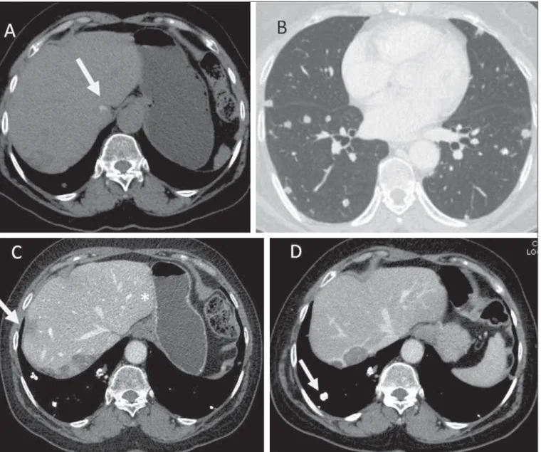
Figure 2. Angiosarcoma. Abdominal CT. At the non-contrast-enhanced phase, the presence of hypodense and heterogeneous liver masses is identified, intermingled with foci of calcifications. The largest of such masses occupies the whole right liver lobe (A). At the arterial phase, the mass presents intense enhancement by the iodinated contrast agent, representing hypervascularization (B), which remains at the portal (C) and delayed (D) phases.
Figure 1. Angiosarcoma. Ultrasonography demonstrates the presence of a pre-dominantly hyperechogenic, heterogeneous hepatic mass occupying the whole right liver lobe.
aspect on T2-weighted sequences, with foci of hemorrhage, fibrotic septa or intermixed calcifications, presenting early and progressive enhancement(5).
Main differential diagnoses include epithelioid heman-gioendothelioma, intrahepatic cholangiocarcinoma and hypervascular metastasis(4).
Main aspects favoring the diagnosis of hepatic angiosa-rcoma include:
• Men at the sixth and seventh decades of life.
• History of exposure to chemical substances such as tho-rium dioxide, vynil chloride and arsenic, and anabolic steroids.
• Heterogeneous, hypervascular masses with persistent en-hancement.
ANGIOMYOLIPOMA
Angiomyolipoma is a rare mesenchymal tumor with varied histological components such as fat tissue, smooth muscle and vessels(7). Recent studies suggest that
angiomyo-lipoma is not a mesenchymal lesion, but a neoplasia origi-nating from perivascular epithelioid cells, classified by the World Health Organization as PEComa (“perivascular epi-thelioid cells” tumor)(8). It occurs more frequently in middle
aged women and 50% of patients are asymptomatic. Symp-toms are observed in cases where intralesional bleeding is present, which generally occurs in lesions > 5.0 cm. There is neither association with tuberous sclerosis nor with renal angiomyolipomas. Most patients do not present hepatopa-thy and tumor markers (CEA and CA 19-9) are negative(7,8).
Due to the presence of different histological components, hepatic angiomyolipoma presents variable imaging features, many times resembling those of hepatocarcinoma. The diag-nosis is preoperatively suspected in only 11% to 25% of cases, respectively by CT and MRI(9). At US, the lesion may
ap-pear hyperechogenic and homogeneous, similar to heman-gioma (Figure 3). At CT and MRI, in the presence of a sufficient amount of intralesional fat, one can identify it with basis on the negative CT density and signal drop on
out-of-phase gradient-echo sequence (Figure 4). Main differential diagnoses include fatty liver lesions such as adenoma, hepa-tocellular carcinoma, lipoma and liposarcoma(9).
Main aspects to be considered in the diagnosis of angiomyolipomas include:
• Middle aged women.
• CT and MRI findings similar to those of hepatocellular carcinoma, without hepatopathies and with negative tu-mor markers.
• Presence of intralesional fat.
CYSTADENOMA AND BILIARY CYSTADENOCARCINOMA
Biliary cystadenomas are uncommon and represent less than 5% of all uni- or multilocular cysts of biliary origin(10).
Generally, most of these lesions are intrahepatic (85% of cases), with diameter ranging from 1.5 cm to 35 cm and predilection for the right liver lobe(9). Predominantly, such
lesions affect middle aged women and in less than 5% of cases are considered to be pre-malignant lesions(11). Symptoms, if present, are associated with mass-effect and consist in in-termittent pain or jaundice(12).
Macroscopically, the lesion is cystic, with fluid or mu-cinous content. Hemorrhagic, biliary, mixed and even pu-rulent content may be found. At histological analysis, the epi-thelium shows papillary projections or polyps, besides ova-rian stromal cells in female patients(9).
At CT and MRI, such lesions present as a solitary cystic mass with a well defined fibrous capsule, mural nodules, in-ternal septa, calcifications and fluid-fluid level(9) (Figures 5
and 6).
Main differential diagnoses include hydatid cyst, cystic metastasis and abscess(9).
Main aspects to be considered in the diagnosis of bil-iary cystadenoma include:
• Middle aged women.
• Solitary cystic mass with internal septa, mural nodules and calcifications.
EPITHELIOID HEMANGIOENDOTHELIOMA
It is a malignant tumor of vascular origin affecting adult individuals, particularly women since estrogen is an associ-ated risk factor. It is considered an insidious neoplasm of intermediate to low grade malignancy and, on average, a five-year survival rate is about 50%(13,14).
Clinical findings are nonspecific and include right hy-pochondrium pain and weight loss, and in some cases may progress with fulminant hepatitis or Budd-Chiari syndrome(13). Most common metastasis sites include abdominal lymph nodes, peritoneum, lungs and bones(14).
Epithelioid hemangioendotheliomas have the following imaging presentations: nodular and diffuse. The multinodu-lar presentation represents the initial phase of the disease, when frequently subcapsular nodules with varied sizes and peripheral distribution are found. On the other hand, in late
phases, the nodules grow and coalesce, forming extensive and infiltrative lesions(6).
After intravenous contrast injection, the lesions may present peripheral and central enhancement, corresponding to capsular retraction and calcifications within the lesion can
be noted (Figure 7). Alomari have described the so called “lollipop sign” that can be observed at CT and MRI, corre-sponding to abrupt termination of the portal vein or hepatic artery in the mass periphery and representing a specific find-ing of this entity(15).
Figure 5. A 42-year-old female patient with no previous history of disease, com-plaining of a painful, palpable mass in the epigastrium. Contrast-enhanced CT portal phase demonstrates cystic multiloculated lesion, with thick internal septa enhanced after contrast injection. The diagnosis was biliary cystadenocarcinoma.
Figure 6. Biliary cystadenoma. Contrast-enhanced CT portal phase demonstrates cystic lesion with fine internal septa intermingled with foci of calcification.
Pulmonary metastases generally calcify and bone me-tastases are osteolytic, insufflating and with marginal scle-rosis (Figures 7 and 8).
Main aspects to be considered in the diagnosis of epi-thelioid hemangioendothelioma include:
• Adult women undergoing estrogen therapy.
• Presence of multiple peripheral nodules with capsular retraction.
• Calcified pulmonary metastasis. • Lollipop sign.
FIBROLAMELLAR HEPATOCARCINOMA
It is a primary malignant tumor corresponding to 1–9% of all tumors of hepatocellular origin. Clinical, laboratory, histopathological and imaging findings differentiate this tumor from the conventional hepatocellular carcinoma. It
occurs preferentially in adolescent patients or in young adults with neither underlying hepatopathy nor increasing in tumor markers such as alpha-fetoprotein(16).
Almost all cases are incidentally diagnosed at late stages of the disease, since it is asymptomatic. However, pain, pal-pable abdominal mass and ascites are most commonly ob-served in symptomatic cases (40% of cases)(16).
Frequent CT and MRI findings of fibrolamellar hepa-tocellular carcinomas include large solitary lobulated and well defined mass (80% of cases) in non cirrhotic liver which, in 50% of the cases cause biliary tract dilatation. After intra-venous contrast agent injection, such lesions present hetero-geneous hypervascular enhancement, with radiating septa and central scar (70% of cases) with a fibrotic component that presents delayed enhancement, which is helpful to differen-tiate this tumor from focal nodular hyperplasia. Metastasis
occurs in 30% of cases, most frequently in lungs and adre-nal glands(17).
Calcifications are found in about 50% of cases investi-gated by CT and almost exclusively in the region of the cen-tral scar, differently from focal nodular hyperplasia where calcifications are extremely rare(17) (Figure 9).
Main aspects to be considered in the diagnosis of fibrolamellar hepatocellular carcinoma include:
• Young individuals with no history of hepatopathy and negative tumor markers.
• Presence of large hepatic, hypervascular mass with central scar and calcifications.
Figure 8. Epithelioid hemangioendothelioma. A: Abdominal CT portal phase demonstrates the presence of hypovascular peripheral hepatic nodules. MRI demonstrates that the hepatic nodule presents low signal intensity at T1-weighted sequences (B) and high signal intensity at T2-weighted with target sign (C). At pelvic CT, insufflating osteolytic lesions are observed, with marginal sclerosis in the left iliac bone and scrum at right (D).
A
A
B
C
D
Figure 9. A 23-year-old woman with no hepatopathy and negative tumor markers and with fibrolamellar hepatocellular carcinoma. Non-contrast-enhanced CT (A) arterial (B) and equilibrium (C) phases demonstrate the presence of a subtly hypodense large mass in the right liver lobe, with central punctiform calcifications presenting heterogeneous hypervascular enhancement with progressive appearance in its central region, intermingled with necrotic areas.
CONCLUSION
Radiological findings of the most common liver lesions are well known and widely described in the literature. On the other hand, scarce studies are found in the literature con-solidating the main information regarding more rare liver tumors and lesions(18–22).
In this first part of the present study, the authors describe the main clinical-radiological characteristics of these rare tumors. Although in the great majority of cases, the defini-tive diagnosis must be confirmed by histopathological analy-sis, the imaging findings can raise the diagnostic suspicion, hence the relevance of familiarity with such radiological find-ings to reduce the list of differential diagnoses and increase the chances for a more accurate radiological evaluation.
REFERENCES
1. Tiferes DA, D’Ippolito G. Neoplasias hepáticas: caracterização por métodos de imagem. Radiol Bras. 2008;41:119–27.
2. Walther Z, Jain D. Molecular pathology of hepatic neoplasms: clas-sification and clinical significance. Patholog Res Int. 2011;2011: 403929.
3. Hamilton SR, Aaltonen LA. World Health Organization Classifi-cation of Tumours. Pathology and genetics of tumours of the di-gestive system. Lyon, France: IARC Press; 2000.
4. Yu RS, Chen Y, Jiang B, et al. Primary hepatic sarcomas: CT find-ings. Eur Radiol. 2008;18:2196–205.
5. Koyama T, Fletcher JG, Johnson CD, et al. Primary hepatic an-giosarcoma: findings at CT and MR imaging. Radiology. 2002; 222:667–73.
6. Kim KA, Kim KW, Park SH, et al. Unusual mesenchymal liver tu-mors in adults: radiologic-pathologic correlation. AJR Am J Roentgenol. 2006;187:W481–9.
7. Zhou YM, Li B, Xu F, et al. Clinical features of hepatic angio-myolipoma. Hepatobiliary Pancreat Dis Int. 2008;7:284–7. 8. Chang Z, Zhang JM, Ying JQ, et al. Characteristics and treatment
strategy of hepatic angiomyolipoma: a series of 94 patients collected from four institutions. J Gastrointest Liver Dis. 2011;20:65–9. 9. Mortelé KJ, Ros PR. Cystic focal liver lesions in the adult: differential
CT and MR imaging features. Radiographics. 2001;21:895–910.
10. Billington PD, Prescott RJ, Lapsia S. Diagnosis of a biliary cystad-enoma demonstrating communication with the biliary system by MRI using a hepatocyte-specific contrast agent. Br J Radiol. 2012; 85:e35–6.
11. Tsepelaki A, Kirkilesis I, Katsiva V, et al. Biliary cystadenoma of the liver: case report and systematic review of the literature. Annals of Gastroenterology. 2009;22:278–83.
12. Lewin M, Mourra N, Honigman I, et al. Assessment of MRI and MRCP in diagnosis of biliary cystadenoma and cystadenocarcinoma. Eur Radiol. 2006;16:407–13.
13. Lyburn ID, Torreggiani WC, Harris AC, et al. Hepatic epithelioid hemangioendothelioma: sonographic, CT, and MR imaging appear-ances. AJR Am J Roentgenol. 2003;180:1359–64.
14. Makhlouf HR, Ishak KG, Goodman ZD. Epithelioid hemangioen-dothelioma of the liver: a clinicopathologic study of 137 cases. Cancer. 1999;85:562–82.
15. Alomari AI. The lollipop sign: a new cross-sectional sign of he-patic epithelioid hemangioendothelioma. Eur J Radiol. 2006;59: 460–4.
16. Ichikawa T, Federle MP, Grazioli L, et al. Fibrolamellar hepatocel-lular carcinoma: pre- and posttherapy evaluation with CT and MR imaging. Radiology. 2000;217:145–51.
17. Ichikawa T, Federle MP, Grazioli L, et al. Fibrolamellar hepatocel-lular carcinoma: imaging and pathologic findings in 31 recent cases. Radiology. 1999;213:352–61.
18. Gossling PAM, Alves GRT, Silva RVA, et al. Bilioma espontâneo: relato de caso e revisão da literatura. Radiol Bras. 2012;45:59–60. 19. Guimarães Filho A, Carneiro Neto LA, Palhtea MS, et al. Doença de Caroli complicada com abscesso hepático: relato de caso. Radiol Bras. 2012;45:362–4.
20. Hollanda ES, Torres US, Gual F, et al. Perfuração espontânea da vesícula biliar com formação de biloma intra-hepático: sinais ultras-sonográficos e correlação com tomografia computadorizada. Radiol Bras. 2013;46:320–2.
21. Galvão BVT, Torres LR, Cardia PP, et al. Prevalência de cistos sim-ples e hemangiomas hepáticos em pacientes cirróticos e não cirróti-cos submetidos a exames de ressonância magnética. Radiol Bras. 2013;46:203–8.