Case 1873
Mucinous cystadenoma of the appendix
T.M. Cunha*, C. Couceiro*, A.P. Vasconcelos*, A. Félix**
Section: Abdominal Imaging
Published: 2003, Jan. 31
Patient: 53 year(s), female
Clinical Summary
A cystic mass in the right iliac fossa of an asymptomatic patient.
Clinical History and Imaging Procedures
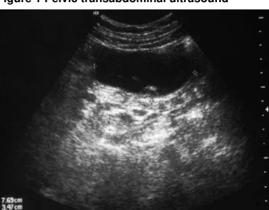
A cystic mass was found in the right iliac fossa of this asymptomatic patient during a routine
gynaecological ultrasound scan by the transabdominal approach (Fig. 1). The mass measured 7.7cm x 3.5cm, with no internal echoes and without solid areas. It was painful with pressure of the probe.
Transvaginal ultrasound was not able to identify the mass.
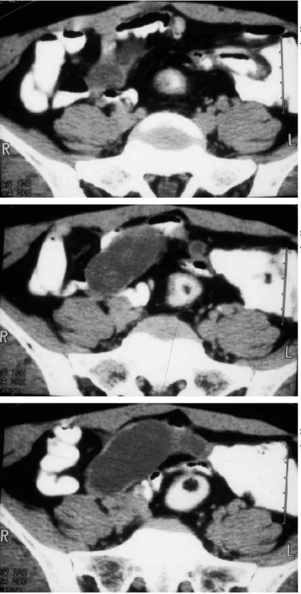
The patient underwent a CT scan, which confirmed that the cystic lesion was roughly sausage shaped
and seemed to be contiguous with the ileo-caecal complex. The cystic mass was thin walled, showed
uniform enhancement (after endovenous contrast), and was low-attenuation with no parietal nodules (Fig. 2).
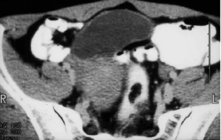
An exploratory laparotomy was performed, for a suspected cystic lesion of the right adnexa, and an intraoperative diagnosis of mucocele of the ileo-caecal appendix was made (Fig. 3). The final
pathological result was mucinous cystadenoma.
Discussion
Cystic lesions of the appendix are rare (being found in 0.25% of appendectomies), although slightly more
common in female patients. These lesions are not always neoplastic and include different entities, such as mucocele, and many other aetiological processes, such as inflammation, hyperplasia, benign
The preoperative imaging diagnosis of these lesions is difficult and needs extensive differential diagnosis with other intra- or retro-peritoneal lesions. In postmenopausal women, in whom cystic appendiceal
masses are slightly more common, the differential diagnosis with adnexal lesions is particularly difficult.
In many cases in this age group the ovaries can no longer be individualised by sonography. Until recently, the diagnosis was most often made incidentally during surgery, such as genitourinary tract
operations, cholecystectomy, appendectomy or exploratory laparotomy procedures.
In the literature, the imaging criteria for appendicular cystic lesions are well known. Early in 1947,
Euphrat described the signs on conventional radiological examination (internal displacement of the
caecum, calcium deposits on the walls, non-filling of the appendix). On ultrasonography, the main
characteristics of these cystic lesions are good delineation of the mass, a size of around 5-6cm in the largest dimension, hypoechogenicity, heterogenicity, calcifications and a wall thickness of less than
6mm. On CT these cystic lesions usually have a low-attenuation value, are well encapsulated,
sometimes have septations or calcifications and may be adherent to the small bowel loops. An enhancing mural nodule within the wall of the cystic lesion has been described as suggestive of
malignancy.
Final Diagnosis
Mucinous cystadenoma of the appendix
Figures
Figure 1 Pelvic transabdominal ultrasound
Figure 2 Pelvic CT scan
Pelvic CT scan identifies a
sausage-shaped cystic lesion, contiguous with
the ileo-caecal complex.
Pelvic CT scan identifies a
sausage-shaped cystic lesion, contiguous with
the ileo-caecal complex.
Pelvic CT scan identifies a
sausage-shaped cystic lesion, contiguous with
the ileo-caecal complex.
Figure 3 Gross specimen
The ileo-caecal appendix.
The ileo-caecal appendix, in longitudinal
section, filled with a gelatinous substance.
MeSH
A multilocular tumor with mucin secreting epithelium. They are most often found in the ovary, but are also found in the pancreas, appendix, and rarely, retroperitoneal and in the urinary bladder. They are
considered to have low-grade malignant potential.
Cystadenoma, Mucinous [C04.557.470.590.485.225]
A multilocular tumor with mucin secreting epithelium. They are most often found in the ovary, but are also found in the pancreas, appendix, and rarely, retroperitoneal and in the urinary bladder. They are
considered to have low-grade malignant potential.
References
[1] McGinnis HD, Chew FS. Mucin-producing adenoma of the appendix. AJR Am J Roentgenol. 1993 May;160(5):1046.
[2] Higa E, Rosai J, Pizzimbono CA, Wise L. Mucosal hyperplasia, mucinous cystadenoma and mucinous cystadenocarcinoma of the appendix: a re-evaluation of appendiceal "mucocele". Cancer
1973;32:1525-41.
[3] Madwed D, Mindelzun R, Jeffrey RB Jr. Mucocele of the appendix: imaging findings. AJR Am J
Roentgenol. 1992 Jul;159(1):69-72.
[4] Kim SH, Lim HK, Lee WJ, Lim JH, Byun JY. Mucocele of the appendix: ultrasonographic and CT
findings. Abdom Imaging 1998 May-Jun;23(3):292-6.
Citation
T.M. Cunha*, C. Couceiro*, A.P. Vasconcelos*, A. Félix** (2003, Jan. 31)
Mucinous cystadenoma of the appendix {Online}