Fracture Healing Is Delayed in
Immunodeficient NOD/scid-IL2R
γ
c
null
Mice
Anna E. Rapp1☯*, Ronny Bindl1☯, Stefan Recknagel1, Annika Erbacher2, Ingo Müller3, Hubert Schrezenmeier4,5, Christian Ehrnthaller6, Florian Gebhard6, Anita Ignatius1
1Institute of Orthopaedic Research and Biomechanics, University of Ulm, Ulm, Germany,2Department of General Paediatrics, Haematology and Oncology, University Children’s Hospital Tübingen, Tübingen, Germany,3Clinic for Paediatric Haematology and Oncology, Bone Marrow Transplantation Unit, University Medical Centre Hamburg-Eppendorf, Hamburg, Germany,4Institute of Clinical Transfusion Medicine and Immunogenetics, German Red Cross Blood Transfusion Service and University Hospital Ulm, Baden Wuerttemberg-Hessen, Ulm, Germany,5Institute of Transfusion Medicine, University of Ulm, Ulm, Germany,6Department of Traumatology, Hand-, Plastic, and Reconstructive Surgery, Centre of Surgery, University of Ulm, Ulm, Germany
☯These authors contributed equally to this work. *[email protected]
Abstract
Following bone fracture, the repair process starts with an inflammatory reaction at the frac-ture site. Fracfrac-ture healing is disturbed when the initial inflammation is increased or pro-longed, whereby, a balanced inflammatory response is anticipated to be crucial for fracture healing, because it may induce down-stream responses leading to tissue repair. However, the impact of the immune response on fracture healing remains poorly understood. Here, we investigated bone healing in NOD/scid-IL2Rγcnullmice, which exhibit severe defects in innate and adaptive immunity, by biomechanical testing, histomorphometry and micro-computed tomography. We demonstrated that NOD/scid-IL2Rγcnullmice exhibited normal skeletal anatomy and a mild bone phenotype with a slightly reduced bone mass in the tra-becular compartment in comparison to immunocompetent Balb/c mice. Fracture healing was impaired in immunodeficient NOD/scid-IL2Rγcnullmice. Callus bone content was unaf-fected during the early healing stage, whereas it was significantly reduced during the later healing period. Concomitantly, the amount of cartilage was significantly increased, indicat-ing delayed endochondral ossification, most likely due to the decreased osteoclast activity observed in cells isolated from NOD/scid-IL2Rγcnullmice. Our results suggest that—under aseptic, uncomplicated conditions—the immediate immune response after fracture is non-essential for the initiation of bone formation. However, an intact immune system in general is important for successful bone healing, because endochondral ossification is delayed in immunodeficient NOD/scid-IL2Rγcnullmice.
a11111
OPEN ACCESS
Citation:Rapp AE, Bindl R, Recknagel S, Erbacher A, Müller I, Schrezenmeier H, et al. (2016) Fracture Healing Is Delayed in Immunodeficient NOD/scid-IL2RγcnullMice. PLoS ONE 11(2): e0147465.
doi:10.1371/journal.pone.0147465
Editor:Martijn van Griensven, Klinikum rechts der Isar - Technical University Munich - TUM, GERMANY
Received:October 23, 2015
Accepted:January 4, 2016
Published:February 5, 2016
Copyright:© 2016 Rapp et al. This is an open access article distributed under the terms of the
Creative Commons Attribution License, which permits unrestricted use, distribution, and reproduction in any medium, provided the original author and source are credited.
Data Availability Statement:All relevant data are within the paper.
Funding:This study was funded by the 7th Framework Programme (FP7) of the European Commission through the REBORNE Project, grant no. 241879 and the German Research Foundation (DFG) in the framework of the Collaborative Research Center 1149 (CRC1149). The funders had no role in study design, data collection and analysis, decision to publish, or preparation of the manuscript.
Introduction
A close relationship exists between the bone and immune systems. Both systems share a large number of regulatory molecules, macrophages and osteoclasts develop from the same progeni-tor and inflammaprogeni-tory disorders can be associated with bone loss [1–4]. The immune system also appears to play an important role in bone healing, because the fracture repair process starts with an inflammatory response [5,6]. The fracture leads to tissue damage and blood vessel rup-ture, initiating acute inflammation and the development of a haematoma, which is character-ized by low pH, hypoxia and a high concentration of inflammatory mediators and chemokines being released from resident immune cells after sensing injury associated danger signals [7,8]. Polymorphonuclear neutrophils (PMNs), which are rapidly recruited at the early stage of inflammation, act against endogenous and exogenous pathogens by secreting reactive oxygen species, proteases and cytokines, and phagocytize debris and dead cell remnants. By releasing chemokines, PMNs attract macrophages, which further remove pathogens and initiate tissue repair by producing pro-angiogenic and trophic factors [9]. Later, the immune response shifts towards adaptive immunity, reflected by the invasion of lymphocytes into the fracture zone [10]. The inflammatory phase is orchestrated by many pro- and anti-inflammatory mediators (e.g. interleukin (IL)-1, IL-6, tumour necrosis factor-α), pro-angiogenic mediators and growth factors (e.g. of the bone morphogenetic protein superfamily) [11]. With the resolution of acute inflammation, mesenchymal progenitor cells are attracted and new bone is formed by intra-membranous and endochondral ossification [5,6].
A balanced inflammation at the fracture site restricts tissue damage and initiates tissue repair by providing pro-angiogenic mediators and attracting mesenchymal progenitors cells, and is, therefore, anticipated to be crucial for fracture healing [6,12]. In contrast, fracture heal-ing is disturbed when the inflammatory response is increased or prolonged. For example, excessive inflammation associated with complex local tissue injury results in delayed healing [13,14]. Systemic inflammatory conditions, including polytrauma, which induce an acute sys-temic immune response, significantly increase the risk for non-union [13,15]. There is also clinical evidence that fracture healing is disturbed in patients with chronic immune disorders, including rheumatoid arthritis, and diabetes [16,17].
Currently, it is poorly understood how much inflammation may be too much, and whether an immune response is actually crucial for bone repair. Here, we investigated bone healing in NOD/scid-IL2Rγcnullmice, which exhibit severe defects in innate and adaptive immunity, and hypothesized that fracture healing would be considerably disrupted when a balanced immune response, which is proposed to have a positive effect on regeneration, is disturbed.
Materials and Methods
Mouse model
All experiments were performed according to national and international regulations for the care and use of laboratory animals and were approved by the Local Ethics Committee (No. 1000,Regierungspräsidium Tübingen, Germany). NOD/scid-Il2Rγcnull(NOD.Cg-Prkdcscid
Il2Rgtm1Wjl/SzJ) and Balb/cByJ (referred to as Balb/c) mice were purchased from Jackson Labo-ratories (Bar Harbor, ME, USA). The immunedeficient NOD/scid-Il2Rγcnullmouse is
common gamma-chain of the IL2 receptor (γc). Therefore, the signalling of 2, 4, 7,
IL-9, IL-15 and IL-21 is defective [19].
BALB/c mice were chosen as the immunocompetent control, because the spontaneous mutation for scid (severe combined immunodeficiency) in thePrkdc-locus (protein kinase,
DNA-activated, catalytic polypeptide) originally occurred in this strain.
Animal studies
To investigate whether the immune defect of NOD/scid-Il2Rγcnullmice influences bone forma-tion, the skeleton of Balb/c and NSG (male, 12 weeks old, n = 10 for both genotypes) mice was comparatively analysed as described below. Fracture healing was investigated in NOD/scid-Il2Rγcnulland BALB/c mice of the same age (n = 24 per genotype). Surgery was performed as
previously described [20]. Briefly, an osteotomy was created at the mid-shaft of the right femur and stabilized using an external fixator that was fitted to the bone using four mini-Schanz screws (axial stiffness 18.1 N/mm, RISystem, Davos, Switzerland). To reduce pain, an analgesic (tramadol hydrochloride, 15 mg/kg) was administered subcutaneously before the operation and via the drinking water (25 mg/L) for the first 3 postoperative days. The mice received daily subcutaneous injections of clindamycin-2-dihydrogenphosphate (45 mg/kg) until the third postoperative day. During the first three post-operative days, the animals were monitored daily, afterwards twice a week until sacrification. Specifically, the use of the operated limb was assessed as well general health condition (bodyweight, grooming, overall behaviour). Eight ani-mals per group were sacrificed by CO2asphyxiation after 21, 28 and 35 days and the
osteoto-mized bone was analysed as described below.
Biomechanical testing
To determine the mechanical quality of the intact and osteotomized femurs, the flexural rigid-ity was assessed by a non-destructive three-point bending test using a material testing machine as described previously [20,21]. Briefly, the proximal end of the femur was fixed to an alumin-ium cylinder using SelfCem (Heraeus Kulzer, Hanau, Germany). The cylinder was fixed in a hinge joint, serving as the proximal support for the bending test. The femoral condyles rested on the bending support, the distance between both supports being 20 mm (l). The bending load F was applied on the mid-shaft and continuously recorded vs. sample deflection (d) up to a maximum force of 5 N. Because the callus was not always located exactly in the middle of the supports (l/2), the distances between the load vector and the proximal (a) and distal (b) sup-ports were considered when calculating the flexural rigidity EI = k((a2b2)/3l) [20].
Micro-computed tomography (
μ
CT)
The intact and osteotomized femurs were scanned using aμCT-device (Skyscan 1172, Bruker,
Belgium) at a resolution of 8μm using a peak voltage of 50 kV and 200μA. In the cortical bone
of the non-osteotomized femurs, a volume of interest (VOI) of 168μm height was defined in
the mid-diaphysis. The trabecular bone was evaluated in the distal part of the femur in a 280μm high VOI with its lower end 200μm above the growth plate. To assess the
Histology and histomorphometry
The osteotomized and intact femora were processed for undecalcified histology. The specimens were fixed in 4% formaldehyde, dehydrated, and embedded in methyl methacrylate. Sections of 10μm of fractured femurs were stained using Giemsa. To determine the osteoblast numbers,
10-μm thick sections of intact femurs were stained using toluidine blue for better visualization
of osteoblasts. To visualize osteoclasts, tartrate resistant acid phosphatase (TRAP) staining was performed using naphthol AS-MX phosphate (Sigma-Aldrich, Taufkirchen, Germany) and Fast Red TR-Salt (Sigma-Aldrich) in 0.2 M acetate buffer pH 5.0. For histomorphometric anal-ysis of the fracture callus, the sections were examined using light microscopy (Leica
DMI6000B, Leica, Switzerland) at 50-fold magnification. In the fracture callus, the relative car-tilage fraction was analysed using the software Leica MMAF 1.4.0 (MetaMorph1
, Leica, Swit-zerland). The osteoblasts and osteoclasts were counted using the OsteoMeasure
histomorphometry system (OsteoMetrics, Decatur, USA). The analyses were performed according the recommendations of the ASBMR [23].
Ex vivo
analyses of osteoblast- and osteoclast-like cells
To investigate whether osteoblasts and osteoclasts exhibit cell-autonomous defects in the NOD/scid-Il2Rγcnullmouse, cell function was analysedex vivo. Osteoblasts were isolated from
cortical bone of both Balb/c and NSG mice. Briefly, the bones were minced and digested for 2 h using 300 U/ml collagenase type IV (Sigma-Aldrich) inα-minimum essential eagle medium (α-MEM, Biochrom AG, Berlin, Germany). The bone chips were cultivated inα-MEM supple-mented with 15% foetal calf serum (FCS), 4 nM L-glutamine, 100 U/mL penicillin, 0.1 mg/mL streptomycin (all Biochrom AG) and 0.25 mg/mL amphotericin-B (Fungizone1
, Gibco, Darm-stadt, Germany) at 37°C under 5% CO2, until colonies started to form. The colonies were
sub-cultivated and cells in passages 3 to 5 were seeded for osteogenic differentiation at a density of 10,000 cells/cm2in medium further supplemented with 10 mM disodiumβ-glycerophosphate and 0.2 mM ascorbate-2-phosphate (all Sigma-Aldrich). After 21 days, matrix mineralization was analysed by von-Kossa-staining. Briefly, incubation with silver nitrate leads to the replace-ment of calcium with silver ions. Reduction of the silver ions results in a dark staining of min-eralized areas. Alkaline phosphatase activity was detected by staining using a commercially available kit (Sigma, Germany).
Osteoclast-like cells (OCL) were generated from bone marrow that was flushed from humeri and tibiae. The cells were plated at 3x10⁵cells/cm2and cultivated inα-MEM supplemented with 10% FCS (Gibco), 4 nM L-glutamine, 100 U/mL penicillin and 0.1 mg/mL streptomycin (all Biochrom AG). To stimulate osteoclast fusion, 10 ng/mL recombinant human macro-phage-colony stimulating factor (rh-MCSF, Chemicon, Limburg, Germany) were added for 3 days. The non-adherent cell fraction was plated at a density of 5x10⁵cells/cm2on normal tissue culture plastic and in plates with a synthetic calcium phosphate coating (BD BioCoat™ Osteolo-gic™Bone Cell Culture System plates; Becton Dickinson GmbH, Germany) and cultivated for 7 days. To assess OCL formation, cells were stained for TRAP using a commercial kit (Sigma, Germany). TRAP-positive cells with3 nuclei were counted as OCL. To analyse the resorption activity of the OCL, BD BioCoat™Osteologic Bone Cell Culture System™slides were treated using 6% sodium hypochloride and von-Kossa stained to visualise the resorbed areas in the cal-cium phosphate coating.
Statistics
using student’s T-test. (GraphPad Prism6, GraphPad Software, Inc., La Jolla, CA, USA). The level of significance was p<0.05.
Results
Immunodeficiency in NOD/scid-IL2R
γ
cnullmice induces a moderate
bone phenotype
To assess the skeletal phenotype of NOD/scid-IL2Rγcnullmice, whole body X-rays were evaluated
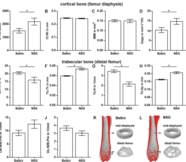
in comparison to Balb/c mice. We observed no gross abnormalities when comparing the skeleton of both strains (data not shown). The flexural rigidity of the femurs assessed by three-point bend-ing was significantly higher (+47%, p = 0.0495) in NOD/scid-IL2Rγcnullcompared to Balb/c mice (Fig 1A). Micro-computed tomography analysis of the femur diaphysis revealed no significant alterations in cortical width (Ct.Wi), moment of inertia (MMI) or mineralization represented by the mean grey value (115.76±1.43 for Balb/c vs. 112.67±1.40 for NOD/scid-IL2Rγcnull). The
apparent Young’s Modulus (Eapp), representing the mechanical properties of the bone matrix, was significantly increased by 43% in NOD/scid-IL2Rγcnull(p = 0.0448) (Fig 1B–1D).
Analysis of trabecular bone at the distal femur revealed a moderately reduced bone mass in NOD/scid-IL2Rγcnullcompared to Balb/c mice (Fig 1E–1H). The bone per tissue volume was
significantly decreased due to a reduced trabecular number and increased trabecular spacing, whereas trabecular thickness was increased (all parameters p<0.0001) (Fig 1). Representative
3D-reconstructions of cortical and trabecular bone are depicted inFig 1K and 1L. Osteoblast and osteoclast numbers were not significantly different (Fig 1I and 1J).
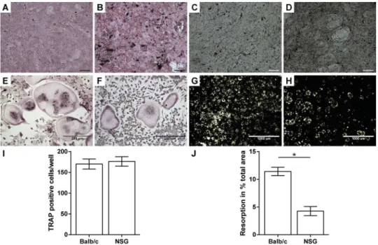
Primary osteoblasts isolated from NOD/scid-IL2Rγcnullmice exhibited a strongerin vitro
differentiation capacity compared to Balb/c mice, as demonstrated by increased alkaline phos-phatase activity and mineral deposition (Fig 2A–2D). Thein vitroformation of osteoclast-like
cells was unaffected in NOD/scid-IL2Rγcnullmice (Fig 2E, 2F and 2I); however, the resorption activity of the cells was significantly reduced (Fig 2G, 2H and 2J).
Immunodeficiency in NOD/scid-IL2R
γ
cnullmice delays fracture healing
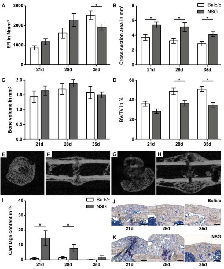
Biomechanical testing of healed femurs in Balb/c and NOD/scid-IL2Rγcnullmice revealed no statistically significant differences after a healing period of both, 21 and 28 days. On day 35, flexural rigidity was significantly lower in NOD/scid-IL2Rγcnullmice (p = 0.0430) (Fig 3A).
At all time points,μCT analysis demonstrated a significant increase in callus cross-section
area in NOD/scid-IL2Rγcnullmice compared to Balb/c mice (p = 0.0115–0.015) (Fig 3B and 3E–
3H). Although the absolute bone volume was not significantly different (Fig 3C), the relative bone content of the fracture callus was significantly lower in NOD/scid-IL2Rγcnullmice on days 28 and 35 (p = 0.0197 and p<0.0006, respectively) due to the increased callus size (Fig 3D).
Histomorphometric analysis revealed a significantly increased cartilage content in the calli of NOD/scid-IL2Rγcnullmice on days 21 and 28 (p = 0.048 and 0.0297, respectively) (Fig 3I, 3J and 3K). Therefore, the longer persistence of cartilage and the reduced bone fraction in the fac-ture callus of the immunodeficient mice suggest delayed endochondral bone formation. The reduced callus tissue quality in the NOD/scid-IL2Rγcnullmice did not affect the flexural rigidity on days 21 and 28, because the callus was larger. However, on day 35, the greater callus size no longer compensated for the poor quality.
Discussion
was impaired in the immunodeficient mice compared to immunocompetent Balb/c mice, as demonstrated by a significantly reduced bone content of the fracture callus in the late healing phase. Concomitantly, the amount of cartilage was significantly increased, indicating delayed endochondral ossification.
We chose NOD/scid-IL2Rγcnullmice for this study, because they have severe defects in
innate and adaptive immunity, but do not exhibit obvious dysfunctions of other organs and have a normal life expectancy. Therefore, these mice are an established model, for example, for xenogeneic transplantation [25]. NOD/scid-IL2Rγcnullmice lack functional leucocytes, macro-phages, dendritic cells, natural killer cells and lymphocytes. They also display no detectable Fig 1. Skeletal phenotype of Balb/c and NOD/scid-IL2Rγcnull(NSG) mice.Cortical bone was analysed by a three-point bending test and micro-computed
tomography (μCT) (A-D); trabecular bone in the distal femur (E–H) was analysed byμCT. The osteoblast number per bone perimeter (Ob.N/B.Pm) was assessed in toluidine-blue-stained sections (I). The number of osteoclasts per bone perimeter (Oc.N/B.Pm) was evaluated in sections stained for tartrate resistant acid phosphatase (TRAP) (J). K and L depict representative 3-dimensional reconstructions of femurs from Balb/c (K) and NSG mice (L). Data is presented as the mean±standard error of the mean. n = 9–10. Asterisks denote significant differences; p<0.05.
activity of haemolytic complement. The mice exhibit deficiencies in cytokine signalling due to the lack of the common gamma chain (γc) of the IL-2 receptor [18]. This receptor subunit is described as important, if not essential, for the binding and signalling of various interleukins, including IL-2, IL-4, IL-7, IL-9, IL-15 and IL-21 [26,27], which also partially play a role in bone homeostasis [28,29].
Because—to our best knowledge—the bone phenotype was not previously described in detail, we first analysed the skeleton of 12-week-old NOD/scid-IL2Rγcnullmice. Gross
inspec-tion of the skeleton revealed no obvious abnormalities, indicating that bone development was not disturbed. Furthermore, immunodeficient mice developed only a mild bone phenotype. Structural parameters of the cortical bone were not significantly affected. In contrast, mechani-cal properties were increased, indicating alterations to the bone matrix that we could not detect by the applied methods. In the trabecular compartment, bone mass was slightly reduced in NOD/scid-IL2Rγcnullmice due to a diminished trabecular number; however, the existing bone
trabeculae were thicker. Bone cell numbers were unalteredin vivo; however, osteoblasts
iso-lated from immunodeficient mice and cultivatedex vivodisplayed an increased differentiation
capacity, whereas osteoclast resorption activity was reduced, indicating cell-autonomous defects. These results explain the increased trabecular thickness in immunodeficient mice. As stated above, cells of the hematopoietic lineage are affected by the defects of the NOD/scid-IL2Rγcnullmouse. As osteoclasts derive from the myeloid lineage, the disturbed osteoclast
func-tion observedex vivomight be caused by the deletion of theγc-subunit of the IL-2 receptor, which affects the signalling of various interleukins [26,27]. Cytokine signalling plays a central role in osteoclastogenesis and defects in this signalling pathway could cause osteoclast dysfunc-tions [30]. The increased differentiation capacity of osteoblasts is difficult to account for by a Fig 2.Ex vivoanalysis of osteoblast and osteoclast function.Osteogenic differentiated primary osteoblasts from Balb/c (A, C) and NOD/scid-IL2Rγcnullmice (NSG; B, D) were stained for alkaline phosphatase activity (A, B) and mineral deposition (C, D) using von-Kossa-stain, respectively. Osteoclast-like (OCL) cell fusion from mononuclear cells was analysed using tartrate resistant acid phosphatase (TRAP)-staining (E, F, I). The resorption activity of the OCL was analysed on calcium-phosphate-coated discs. (G, H, J). Data is depicted as the mean±standard error of the mean. I: n = 4; J: n = 7. Scale bar in A– D = 100μm.
Fig 3. Fracture healing is moderately altered in NOD/scid-IL2Rγcnull(NSG) mice.Assessment of the fracture-healing outcome by three-point bending
test (A), micro-computed tomography (B-D) and histomorphometry (I). Representative cross sections (E, G) and longitudinal sections (F, H) on day 21 of calli from Balb/c (E, F) and NSG mice (F, G). Representative micrographs of fracture calli of Balb/c (J) and NSG (K) mice on day 21, 28 and 35. Data is presented as the mean±standard error of the mean; n = 6–8. Asterisks indicate significant differences between Balb/c and NSG mice at the indicated time-point; p<0.05. Scale bars in J and K = 200μm.
single underlying factor. Some of the cytokines affected by theγcnullmutation also play a role
in bone homeostasis [28,29,31]; however, the impact on isolated osteoblasts is unclear. In con-clusion, 12-weeks-old NOD/scid-IL2Rγcnullmice exhibit only a mild bone phenotype in
com-parison to Balb/c mice.
A possible limitation of our study is that we could not use immunocompetent wild-type mice with the same genetic background as the NOD/scid-IL2Rγcnullmice, because these mice were generated by intercrossing several mouse strains with different immune deficiencies [18]. Also, using the founder strains as controls was not possible, as they display abnormalities that NOD/scid-IL2Rγcnullmice do not display like wound healing disorders in the NOD/ShiLt
mouse. So, as a compromise, we used Balb/c mice as an immunocompetent control because the spontaneous mutation for scid (severe combined immune deficiency) in thePrkdc-locus
(pro-tein kinase, DNA-activated, catalytic polypeptide) originally occurred in this strain and thus the genetic background may have a high degree of similarity. However, because bone pheno-type and healing characteristics in mice depend on the genetic background [32,33], an influ-ence on bone cell function cannot be completely excluded. To overcome this problem, one could think about a rescue-approach, where bone marrow of immunocompetent mice is trans-planted into NOD/scid-IL2Rγcnullmice in order to generate a functional immune system.
However, irradiation of the mice is necessary for such an approach, thus, other problems arise. Our results demonstrated that fracture healing was impaired in immunodeficient NOD/ scid-IL2Rγcnullmice. The callus bone content was unaffected on day 21, but it was significantly reduced in the later healing phase. The longer persistence of cartilage indicates delayed carti-lage-to-bone transformation during endochondral ossification, most likely through reduced osteoclast activity. Impaired osteoclast activity is likely to arise from cell-autonomous defects, as suggested by ourex vivoexperiments, or by the absence of functional immune cells,
includ-ing T-lymphocytes, which support osteoclast activity by producinclud-ing receptor activator of nuclear factorκ-B ligand [34]. Reduced osteoclast activity could also account for the increased callus size in NOD/scid-IL2Rγcnullmice, which indicates delayed callus remodelling.
Osteo-clasts are crucial for the degradation of hypertrophic cartilage and for callus remodelling in the later healing period, whereas periosteal primary bone formation during the earlier healing phase is osteoclast independent [35,36]. Therefore, delayed fracture healing in NOD/scid-IL2Rγcnullmice was rather caused by impaired osteoclast function than disturbed osteoblast
precursor cell recruitment and differentiation during the early healing period. Therefore, our results suggest that—under sterile, uncomplicated conditions—the immediate immune response after fracture is nonessential for the initiation of bone formation.
Because we aimed to study the general impact of the immune system on fracture healing, we used a mouse model of severe immune deficiency. Other authors investigated the impact of specific immune cell subsets on fracture healing using transgenic mouse models or antibody induced cell depletion [37,38]. Thereby, the impact of immune cells, which are classically attributed to innate immunity and act primarily as first line of defence, is intensely discussed. Increased recruitment of neutrophils induced by granulocyte-colony stimulating factor was shown to accelerate bone formation [39]. In contrast, neutrophil depletion impaired fracture healing [40]. Therefore, it appears that a balanced activation of PMNs is necessary for regular bone regeneration [41]. Macrophages are generally considered to contribute to the resolution of inflammation and initiation of tissue repair through the clearance of apoptotic neutrophils and secretion of anti-inflammatory factors, including IL-10 and transforming growth factor-β
macrophage-deficient NOD/scid-IL2Rγcnullmouse, possibly because the deficiencies in this
model are much more complex.
The role of other innate immune cells, including natural killer cells and mast cells in bone healing remains unclear. Cells attributed to adaptive immunity are believed to play an impor-tant role mainly in later stages of fracture healing [10]. Bone healing was improved in lymphocyte-deficient, recombination activating gene-1 (RAG-1)-knockout mice, indicating a negative effect of these cells [37]. In agreement with this, depletion of CD8-positive cells improved fracture healing, whereas transplantation of CD8-positive cells resulted in delayed healing [38]. Furthermore, mice lacking functional B-lymphocytes displayed enhanced bone formation [45]. These studies suggest that lymphocytes may provoke negative effects on bone regeneration. However, the studies mentioned above investigated the effect of single cell types and did not consider the delicate interplay of cells and factors of the immune system.
In conclusion, this study described, for the first time, fracture healing in a mouse model with severe immune deficiency. Fracture healing was delayed due to impaired endochondral ossification. However, primary bone formation in the early healing stage was unaffected in the model used for uncomplicated fracture healing. Further studies are necessary to unravel the multifaceted interactions between immune cells and bone cells in fracture healing.
Acknowledgments
We thank Ursula Maile, Marion Tomo, Sevil Essig and Helga Bach for their excellent technical assistance. This study was supported by the 7thFramework Programme (FP7) of the European Commission through the REBORNE Project, grant no. 241879.
Author Contributions
Conceived and designed the experiments: AER RB AI HS FG IM. Performed the experiments: AER RB SR CE. Analyzed the data: AER RB SR CE AI HS. Contributed reagents/materials/ analysis tools: AE IM. Wrote the paper: AER RB AI.
References
1. Nakashima T, Takayanagi H. Osteoimmunology: crosstalk between the immune and bone systems. J Clin Immunol. 2009; 29(5):555–67. doi:10.1007/s10875-009-9316-6PMID:19585227
2. Takayanagi H. New immune connections in osteoclast formation. Annals of the New York Academy of Sciences. 2010; 1192(1):117–23. doi:10.1111/j.1749-6632.2009.05303.x
3. Takayanagi H. Osteoimmunology and the effects of the immune system on bone. Nature Reviews Rheumatology. 2009; 5(12):667–76. doi:10.1038/Nrrheum.2009.217PMID:19884898
4. Danks L, Takayanagi H. Immunology and bone. Journal of biochemistry. 2013; 154(1):29–39. doi:10. 1093/jb/mvt049PMID:23750028
5. Claes L, Recknagel S, Ignatius A. Fracture healing under healthy and inflammatory conditions. Nature reviews Rheumatology. 2012; 8(3):133–43. doi:10.1038/nrrheum.2012.1PMID:22293759
6. Schmidt-Bleek K, Kwee BJ, Mooney DJ, Duda GN. Boon and Bane of Inflammation in Bone Tissue Regeneration and Its Link with Angiogenesis. Tissue engineering Part B, Reviews. 2015; 21(4):354–64. doi:10.1089/ten.TEB.2014.0677PMID:25742724
7. Kolar P, Gaber T, Perka C, Duda GN, Buttgereit F. Human early fracture hematoma is characterized by inflammation and hypoxia. Clinical orthopaedics and related research. 2011; 469(11):3118–26. doi:10. 1007/s11999-011-1865-3PMID:21409457
8. Schmidt-Bleek K, Schell H, Kolar P, Pfaff M, Perka C, Buttgereit F, et al. Cellular composition of the ini-tial fracture hematoma compared to a muscle hematoma: a study in sheep. Journal of orthopaedic research: official publication of the Orthopaedic Research Society. 2009; 27(9):1147–51. doi:10.1002/ jor.20901
10. Konnecke I, Serra A, El Khassawna T, Schlundt C, Schell H, Hauser A, et al. T and B cells participate in bone repair by infiltrating the fracture callus in a two-wave fashion. Bone. 2014; 64:155–65. doi:10. 1016/j.bone.2014.03.052PMID:24721700
11. Gerstenfeld LC, Cullinane DM, Barnes GL, Graves DT, Einhorn TA. Fracture healing as a post-natal developmental process: molecular, spatial, and temporal aspects of its regulation. Journal of cellular biochemistry. 2003; 88(5):873–84. doi:10.1002/jcb.10435PMID:12616527
12. Kolar P, Schmidt-Bleek K, Schell H, Gaber T, Toben D, Schmidmaier G, et al. The early fracture hema-toma and its potential role in fracture healing. Tissue engineering Part B, Reviews. 2010; 16(4):427–34. doi:10.1089/ten.TEB.2009.0687PMID:20196645
13. Bhandari M, Tornetta P 3rd, Sprague S, Najibi S, Petrisor B, Griffith L, et al. Predictors of reoperation following operative management of fractures of the tibial shaft. Journal of orthopaedic trauma. 2003; 17(5):353–61. PMID:12759640
14. Bunn RJ, Burke G, Connelly C, Li G, Marsh D. Inflammation—A double edged sword in high-energy fractures? The Journal of bone and joint surgery British volume. 2005; 87-B(SUPP_III):265-c-6.
15. Karladani AH, Granhed H, Karrholm J, Styf J. The influence of fracture etiology and type on fracture healing: a review of 104 consecutive tibial shaft fractures. Arch Orthop Trauma Surg. 2001; 121(6): 325–8. PMID:11482464
16. Hernandez RK, Do TP, Critchlow CW, Dent RE, Jick SS. Patient-related risk factors for fracture-healing complications in the United Kingdom General Practice Research Database. Acta Orthop. 2012; 83(6): 653–60. doi:10.3109/17453674.2012.747054PMID:23140093
17. Niikura T, Lee SY, Sakai Y, Nishida K, Kuroda R, Kurosaka M. Causative factors of fracture nonunion: the proportions of mechanical, biological, patient-dependent, and patient-independent factors. J Orthop Sci. 2014; 19(1):120–4. doi:10.1007/s00776-013-0472-4PMID:24081392
18. Shultz LD, Lyons BL, Burzenski LM, Gott B, Chen X, Chaleff S, et al. Human Lymphoid and Myeloid Cell Development in NOD/LtSz-scid IL2R{gamma}null Mice Engrafted with Mobilized Human Hemopoi-etic Stem Cells. Journal of immunology. 2005; 174(10):6477–89.
19. Kovanen PE, Leonard WJ. Cytokines and immunodeficiency diseases: critical roles of the gamma(c)-dependent cytokines interleukins 2, 4, 7, 9, 15, and 21, and their signaling pathways. Immunol Rev. 2004; 202:67–83. IMR203 [pii] doi:10.1111/j.0105-2896.2004.00203.xPMID:15546386
20. Röntgen V, Blakytny R, Matthys R, Landauer M, Wehner T, Gockelmann M, et al. Fracture healing in mice under controlled rigid and flexible conditions using an adjustable external fixator. Journal of ortho-paedic research: official publication of the Orthoortho-paedic Research Society. 2010; 28(11):1456–62. doi:
10.1002/jor.21148
21. Heilmann A, Schinke T, Bindl R, Wehner T, Rapp A, Haffner-Luntzer M, et al. Systemic treatment with the sphingosine-1-phosphate analog FTY720 does not improve fracture healing in mice. Journal of orthopaedic research: official publication of the Orthopaedic Research Society. 2013; 31(11):1845–50. doi:10.1002/jor.22426
22. Bouxsein ML, Boyd SK, Christiansen BA, Guldberg RE, Jepsen KJ, Müller R. Guidelines for assess-ment of bone microstructure in rodents using micro–computed tomography. Journal of bone and min-eral research: the official journal of the American Society for Bone and Minmin-eral Research. 2010; 25(7): 1468–86. doi:10.1002/jbmr.141
23. Parfitt. Bone Histomorphometry. Journal of bone and mineral research: the official journal of the Ameri-can Society for Bone and Mineral Research. 1987; 2(6):595–610.
24. Dempster DW, Compston JE, Drezner MK, Glorieux FH, Kanis JA, Malluche H, et al. Standardized nomenclature, symbols, and units for bone histomorphometry: A 2012 update of the report of the ASBMR Histomorphometry Nomenclature Committee. Journal of bone and mineral research: the offi-cial journal of the American Society for Bone and Mineral Research. 2013; 28(1):2–17. doi:10.1002/ jbmr.1805
25. Ito M, Hiramatsu H, Kobayashi K, Suzue K, Kawahata M, Hioki K, et al. NOD/SCID/gamma cnull mouse: an excellent recipient mouse model for engraftment of human cells. Blood. 2002; 100(9): 3175–82. doi:10.1182/blood-2001-12-0207PMID:12384415
26. Sugamura K, Asao H, Kondo M, Tanaka N, Ishii N, Ohbo K, et al. The interleukin-2 receptor gamma chain: its role in the multiple cytokine receptor complexes and T cell development in XSCID. Annu Rev Immunol. 1996; 14:179–205. doi:10.1146/annurev.immunol.14.1.179PMID:8717512
27. Asao H, Okuyama C, Kumaki S, Ishii N, Tsuchiya S, Foster D, et al. Cutting edge: the common gamma-chain is an indispensable subunit of the IL-21 receptor complex. Journal of immunology. 2001; 167(1): 1–5.
29. Djaafar S, Pierroz DD, Chicheportiche R, Zheng XX, Ferrari SL, Ferrari-Lacraz S. Inhibition of T cell-dependent and RANKL-cell-dependent osteoclastogenic processes associated with high levels of bone mass in interleukin-15 receptor-deficient mice. Arthritis Rheum. 2010; 62(11):3300–10. doi:10.1002/ art.27645PMID:20617528
30. Lee Y, Kim HH. The Role of Jak/STAT Pathways in Osteoclast Differentiation. Biomol Ther. 2011; 19(2):141–8. doi:10.4062/Biomolther.2011.19.2.141
31. Mori G, D'Amelio P, Faccio R, Brunetti G. The Interplay between the bone and the immune system. Clinical & developmental immunology. 2013; 2013:720504. doi:10.1155/2013/720504
32. Li X, Gu W, Masinde G, Hamilton-Ulland M, Rundle CH, Mohan S, et al. Genetic variation in bone-regenerative capacity among inbred strains of mice. Bone. 2001; 29(2):134–40. PMID:11502474
33. Manigrasso MB, O'Connor JP. Comparison of fracture healing among different inbred mouse strains. Calcified tissue international. 2008; 82(6):465–74. doi:10.1007/s00223-008-9144-3PMID:18528610
34. Pacifici R. T cells: critical bone regulators in health and disease. Bone. 2010; 47(3):461–71. doi:10. 1016/j.bone.2010.04.611PMID:20452473
35. Schindeler A, McDonald MM, Bokko P, Little DG. Bone remodeling during fracture repair: The cellular picture. Semin Cell Dev Biol. 2008; 19(5):459–66. doi:10.1016/j.semcdb.2008.07.004PMID:
18692584
36. Marsell R, Einhorn TA. The biology of fracture healing. Injury. 2011; 42(6):551–5. doi:10.1016/j.injury. 2011.03.031PMID:21489527
37. Toben D, Schroeder I, El Khassawna T, Mehta M, Hoffmann JE, Frisch JT, et al. Fracture healing is accelerated in the absence of the adaptive immune system. Journal of bone and mineral research: the official journal of the American Society for Bone and Mineral Research. 2011; 26(1):113–24. doi:10. 1002/jbmr.185
38. Reinke S, Geissler S, Taylor WR, Schmidt-Bleek K, Juelke K, Schwachmeyer V, et al. Terminally Differ-entiated CD8(+) T Cells Negatively Affect Bone Regeneration in Humans. Science translational medi-cine. 2013; 5(177):177ra36. doi:10.1126/scitranslmed.3004754PMID:23515078
39. Bozlar M, Aslan B, Kalaci A, Baktiroglu L, Yanat AN, Tasci A. Effects of human granulocyte-colony stim-ulating factor on fracture healing in rats. Saudi Med J. 2005; 26(8):1250–4. PMID:16127524
40. Chan JK, Glass GE, Ersek A, Freidin A, Williams GA, Gowers K, et al. Low-dose TNF augments frac-ture healing in normal and osteoporotic bone by up-regulating the innate immune response. EMBO molecular medicine. 2015; 7(5):547–61. doi:10.15252/emmm.201404487PMID:25770819
41. Butterfield TA, Best TM, Merrick MA. The dual roles of neutrophils and macrophages in inflammation: a critical balance between tissue damage and repair. J Athl Train. 2006; 41(4):457–65. PMID:17273473
42. Ortega-Gomez A, Perretti M, Soehnlein O. Resolution of inflammation: an integrated view. EMBO molecular medicine. 2013; 5(5):661–74. doi:10.1002/emmm.201202382PMID:23592557
43. Alexander KA, Chang MK, Maylin ER, Kohler T, Muller R, Wu AC, et al. Osteal macrophages promote in vivo intramembranous bone healing in a mouse tibial injury model. Journal of bone and mineral research: the official journal of the American Society for Bone and Mineral Research. 2011; 26(7): 1517–32. doi:10.1002/jbmr.354
44. Raggatt LJ, Wullschleger ME, Alexander KA, Wu AC, Millard SM, Kaur S, et al. Fracture healing via periosteal callus formation requires macrophages for both initiation and progression of early endochon-dral ossification. The American journal of pathology. 2014; 184(12):3192–204. doi:10.1016/j.ajpath. 2014.08.017PMID:25285719