NCAM1 Polysialylation: The Prion
Protein’s Elusive Reason for Being?
Mohadeseh Mehrabian
1,2, Herbert Hildebrandt
3, and
Gerold Schmitt-Ulms
1,2Abstract
Much confusion surrounds the physiological function of the cellular prion protein (PrPC). It is, however, anticipated that knowledge of its function will shed light on its contribution to neurodegenerative diseases and suggest ways to interfere with the cellular toxicity central to them. Consequently, efforts to elucidate its function have been all but exhaustive. Building on earlier work that uncovered the evolutionary descent of the prion founder gene from an ancestral ZIP zinc transporter, we recently investigated a possible role of PrPCin a morphogenetic program referred to as epithelial-to-mesenchymal transition (EMT). By capitalizing on PrPCknockout cell clones in a mammalian cell model of EMT and using a comparative proteomics discovery strategy, neural cell adhesion molecule-1 emerged as a protein whose upregulation during EMT was perturbed in PrPCknockout cells. Follow-up work led us to observe that PrPCregulates the polysialylation of the neural cell adhesion molecule NCAM1 in cells undergoing morphogenetic reprogramming. In addition to governing cellular migration, polysialyla-tion modulates several other cellular plasticity programs PrPChas been phenotypically linked to. These include neurogenesis in the subventricular zone, controlled mossy fiber sprouting and trimming in the hippocampal formation, hematopoietic stem cell renewal, myelin repair and maintenance, integrity of the circadian rhythm, and glutamatergic signaling. This review revisits this body of literature and attempts to present it in light of this novel contextual framework. When approached in this manner, a coherent model of PrPCacting as a regulator of polysialylation during specific cell and tissue morphogenesis events comes into focus.
Keywords
neural cell adhesion molecules, polysialic acid, prion protein, polysialyltransferases, signaling, protein function
Received April 18, 2016; Received revised September 8, 2016; Accepted for publication October 2, 2016
Background
Arguably the most successful strategies for elucidating the function of a protein are (a) to extrapolate it from the known function of its binding partners, (b) to inter-pret the phenotypic consequences of deleting the gene coding for it, or (c) to derive it by studying the function of homologous proteins and place its role in the context of evolution. For the cellular prion protein (PrPC), all three approaches have been pursued with moderate suc-cesses summarized in comprehensive earlier reviews (Steele et al., 2007; Watts and Westaway, 2007; Aguzzi et al., 2008; Ehsani, Huo, et al., 2011). Once the predom-inant function of a protein is known, data gathered by all three approaches tend to merge in ways that make sense. Furthermore, as insights into the mechanism of action underlying the function of a protein are refined, useful predictions can often be made about how it will behave
when placed into a hitherto unexplored experimental paradigm.
The elusive nature of the function of PrPCcombined
with the large number of reports dedicated to this topic has led to a widespread sentiment that PrPCharbors sev-eral independent activities and its function may be
1Tanz Centre for Research in Neurodegenerative Diseases, University of Toronto, Toronto, Ontario, Canada
2Department of Laboratory Medicine and Pathobiology, University of Toronto, Toronto, Ontario, Canada
3Institute for Cellular Chemistry, Hannover Medical School, Hannover, Germany
Corresponding Author:
Gerold Schmitt-Ulms, Tanz Centre for Research in Neurodegenerative Diseases, University of Toronto, Krembil Discovery Tower, 6th Floor, Rm 447, 60 Leonard Avenue, Toronto, ON M5T 2S8, Canada.
Email: [email protected]
ASN Neuro
November-December 2016: 1–25 !The Author(s) 2016 DOI: 10.1177/1759091416679074 asn.sagepub.com
Creative Commons CC-BY: This article is distributed under the terms of the Creative Commons Attribution 3.0 License
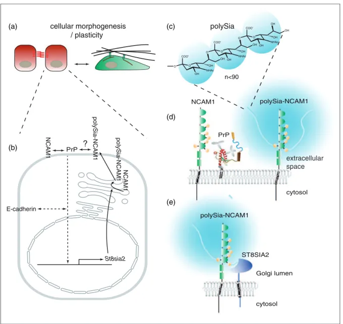
context-dependent, multifaceted, or impossible to know with certainty. Although this perspective has merit—and perhaps represents a truism that applies to any pro-tein—adapting this position is not productive for efforts to identify the key role of a protein, which most often eventually comes to the fore. Here, we will take a differ-ent view: We will point toward data which suggest that PrPCmay operate in several cellular plasticity events as a highly specialized partner of neural cell adhesion
mole-cule-1 (NCAM1). According to this model, PrPC acts
upstream of a signaling loop, which modulates the
expression of the polysialyltransferase (polyST)
ST8SIA2 (and perhaps its paralog ST8SIA4), thereby
determining levels of polysialylation of NCAM1 (and, possibly, other polyST substrates), during the execution of specific cell plasticity programs (Figure 1).
A full account of the function of a protein requires an understanding of both the broader cellular context it tributes to and the precise mechanism by which it con-tributes. It therefore should be stressed that even if the
model we are proposing for PrPCwill hold up upon close
scrutiny, more work is needed to define precisely how PrPCexerts its role in this signaling loop. Viewed in this manner, its contribution to NCAM1 polysialylation is
not the function of PrPC but may well represent the
broader cellular context its function serves.
St8sia2
E-cadherin
NCAM1
cellular morphogenesis / plasticity
COO
-OH O OH O
OH
NHAc COO
-OH O OH O
OH
NHAc OH
COO
-OH O OH O
OH
NHAc n<90
NCAM1
polySia-NCAM1
?
polySia-NCAM1
ST8SIA2
polySia
PrP NCAM1
(c) (a)
(b)
(d)
(e)
PrP
polySia-NCAM1
cytosol Golgi lumen
cytosol
polySia-NCAM1
extracellular space
In the following paragraphs, we chose to loosely group pertinent information available in a way that reflects the principal approaches for studying a protein’s function. By doing so, we will show that the proposed role is consistent with evidence gathered with any of the three aforemen-tioned methods for studying the physiological role of a protein.
In light of the sheer volume of work published on both
PrPC function and NCAM1 polysialylation (for recent
comprehensive reviews, see Mouillet-Richard and
Vilotte, 2015 and Schnaar et al., 2014), no attempt was made to cover all facets of the pertinent literature. Instead, next to brief introductions into the biology of
the key players of the PrPC-ST8SIA2-NCAM1 signaling
loop, we will review manuscripts selected with the intent to distill common themes in the literature surrounding
PrPC and NCAM1 polysialylation and point the reader
toward interesting facets that warrant further
investigation.
The Cellular Prion Protein
The prion protein is notorious for its causative role in rare and invariably fatal neurodegenerative diseases, known as prion diseases (Prusiner, 1995). In prion dis-eases, the cellular form of the prion protein (PrPC) under-goes profound physicochemical changes that give rise to
rogue conformers known as PrP Scrapie (PrPSc), a term
derived from Scrapie disease in sheep, the first known prion disease. The human prion gene is coded on the short arm of Chromosome 20 (Basler et al., 1986; Lee et al., 1998). The open reading frame of the prion gene is confined to a single exon, consistent with proposed retrogene origins (Ehsani, Tao, et al., 2011) and preclud-ing the existence of splice-isoforms. Upon removal of an N-terminal signal sequence during its translocation into the endoplasmic reticulum and replacement of a hydro-phobic C-terminal signal sequence with a glycosylpho-sphatidylinositol-anchor (Cashman et al., 1990), the mature prion protein has a length of 208 amino acids in humans. The protein is characterized by a disordered N-terminal domain comprising a short basic motif, five imperfect octarepeats known for their ability to bind copper or other divalent cations, and a hydrophobic domain. A globular domain present at the C-terminus
of PrPC is stabilized by a highly conserved disulfide
bridge and carries up to two N-glycans (Haraguchi et al., 1989; Riek et al., 1996; Wuthrich and Riek, 2001).
Neural Cell Adhesion Molecules
NCAMs are members of the immunoglobulin (Ig) super-family. The human genome codes for two NCAM para-logs, known as NCAM1 (commonly referred to as NCAM or N-CAM) and NCAM2 (also known as
OCAM). The human NCAM1 gene maps to
Chromosome 11—Chromosome 9 in mice—and com-prises 24 exons (Kolkova, 2010). Expression and
alterna-tive splicing of NCAM1 gene transcripts give rise to
several splice isoforms. The three most often encountered NCAM1 protein isoforms migrate under denaturing gel
electrophoresis conditions with apparent molecular
masses of 180, 140, and 120 kDa, hence their designation as NCAM-180, NCAM-140, and NCAM-120 (Edelman, 1984). Whereas NCAM-180 and NCAM-140 adapt a Type I transmembrane topology, NCAM-120 is, like
PrPC, inserted into the outer leaflet of the membrane
bilayer by a glycosylphosphatidylinositol-anchor. The
N-terminal ectodomains of all three isoforms of
NCAM1 consist of five Ig-like domains and two fibro-nectin Type III domains and can form homophilic (Soroka et al., 2010) and heterophilic (Nielsen et al., 2010) interactions in cis or trans.
PolySTs ST8SIA2 and ST8SIA4
ST8SIA2 (also known as ST8SiaII, SIAT8B, or STX) and ST8SIA4 (also known as ST8Sia IV, SIAT8D, or polyST-1 [PST-polyST-1]; Eckhardt et al., polyST-1995; Nakayama et al., polyST-1995; Scheidegger et al., 1995) are the only enzymes in humans and mice that can synthesize polysialic acid (polySia) with a degree of polymerization greater than 8 (Weinhold et al., 2005; Galuska et al., 2006). In the human genome,
ST8SIA2 and ST8SIA4 genes map to Chromosome 15, Band q26 and Chromosome 5, Band q21, respectively (Angata et al., 1997). The function of these proteins is
to catalyze the polycondensation of a2,8-linked
N-acet-ylneuraminic acid building blocks to assemble linear polySia homopolymers on NCAM1 and on a very limited number of other acceptor proteins (Mu¨hlenhoff et al., 2013). Whereas both ST8SIA2 and ST8SIA4 are strongly expressed during embryogenesis and in newborn mam-mals (Ong et al., 1998; Oltmann-Norden et al., 2008), ST8SIA4 represents the predominant polyST in adult brains (Hildebrandt et al., 1998; Eckhardt et al., 2000; Schiff et al., 2009). Both polySTs are Type II transmem-brane proteins that carry their catalytic domains at the distal end of their ectodomain. Other domains found within polySTs are a membrane-proximal stalk domain, a transmembrane domain, and a short cytosolic domain (Nakata et al., 2006; Foley et al., 2009; Zapater and Colley, 2012).
NCAM Polysialylation
domain 5 (Ig5; Nelson et al., 1995; Box 2). NCAM2 is not naturally polysialylated despite sharing the overall modu-lar organization and conserved N-glycan acceptor sites with NCAM1 and being 37% identical to NCAM1 in sequence (Yoshihara et al., 1997).
Although NCAM1 represents by far the most prom-inent polySia protein acceptor, polySia modifications in mammals are also found on a small number of other
proteins, namely, the a-subunit of the voltage-gated
sodium channel (Zuber et al., 1992), CD36 (Yabe et al., 2003), neuropilin-2 (NRP2; Curreli et al., 2007), the syn-aptic cell adhesion molecule SynCAM1 (Galuska et al., 2010), the chemokine receptor CCR7 (Kiermaier et al.,
2016), the E-selectin ligand 1 (ESL-1, gene name GLG1;
Werneburg et al., 2016), and the two polySTs themselves (Mu¨hlenhoff et al., 1996).
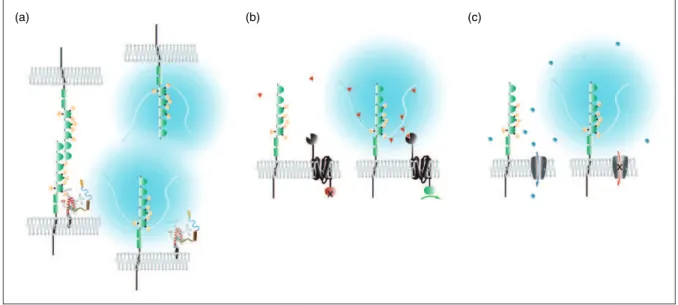
How does polySia contribute to cellular function? The polySia modification of NCAM1 alters its hydrodynamic radius and confers negative charge. These properties are critical for phenotypes that have been linked to polySia-NCAM1, which can be broadly grouped into those that (a) facilitate cellular morphogenesis programs ranging from proliferation and migration to neuritogenesis and fasciculation by affecting protein interactions that medi-ate cell-cell or cell-substrmedi-ate contacts; (b) refine cellular responses to guidance cues, or (c) modulate inputs of brain circuitry or activity of ion channels (Figure 2). Although not formally recognized as categories by
which PrPC-related phenotypes can be grouped, examples
for each of the aforementioned three types of phenotypes can also be identified in the literature surrounding PrPC. Structural requirements, sequence elements, and key residues for substrate recognition and polySia transfer within polySTs are less clear. A conserved histidine resi-due within a C-terminal domain referred to as sialyl motif VS has been shown to be essential for activity (Kitazume-Kawaguchi et al., 2001). A polybasic region within ST8SIA4 that is expected to reside in proximity to the outer face of the lipid bilayer was proposed to contribute to NCAM1 substrate recognition. Interestingly, replace-ment of a particular arginine amino acid within this
domain also prevented in vitro polysialylation of NRP2
and SynCAM1 (Zapater and Colley, 2012), an unex-pected observation given that the membrane-adjacent domains of these two other known polySia carrier pro-teins differ fundamentally from the FN1 and 2 domains found in the respective location within NCAM1.
PrP and polySia-NCAM1
Protein–Protein Interactions
Several excellent reviews on protein–protein interactions
that PrPC (Watts and Westaway, 2007; Aguzzi et al.,
2008; Rubenstein, 2012) or NCAM1 (Nielsen et al., 2010) engage in have been published before. Here, the scope will be limited to describing evidence in support
of an interaction between PrPC and NCAM1.
Formaldehyde crosslinking of mouse neuroblastoma Neuro-2a cells, followed by affinity purification, led in 2001 to a first report on a next-neighbor relationship of
PrPCand NCAM1 (Schmitt-Ulms et al., 2001). Of note,
the PrPC-NCAM1 crosslink product was not only readily
detectable by denaturing gel electrophoresis but it also constituted by far the strongest crosslink signal when detection was based on PrP-directed antibodies. This
Box 1. PolySia.
Sialic acids (Sia) are 9-carbon-carboxylated sugars derived from N-acetylneuraminic acid (Neu5Ac). PolySia is a linear homopolymer of Neu5Ac present on the cell surface of a subset of Gram-negative bacteria and observed attached to a small number of cell surface proteins in eukaryotic cells (Mu¨hlenhoff et al., 1998, 2013). In mam-mals, polySia is made ofa2,8-glycosidically linked N-acetylneurami-nic acid and has been observed to vary in the degree of polymerization between 8 (the minimum number of Sia molecules required for distinguishing polySia chains from shorter oligoSia chains) and up to 90 or more sialic acid residues (Galuska et al., 2008).
Box 2. Mechanism of NCAM1 polysialylation.
observation is worth highlighting because it represents one of only a small number of data points in the literature
on PrPC interactions, which can be used to draw
infer-ences on the relative abundance of proteins in proximity to PrPC.In vitrointerface mapping experiments based on
recombinant PrPC and an NCAM1 peptide array not
only corroborated the notion that the PrPC–NCAM1
interaction can occur through direct binding but also
sketched out an interface that comprises b-strands C
and C’ in the two fibronectin Type III modules within NCAM1 and a nonlinear binding epitope that included the N-terminus, Helix A, and the adjacent loop domain within PrPC(Schmitt-Ulms et al., 2001).
Taken together, these results were screaming for a physiological role of this interaction and suggested that
NCAM1 and PrPC may profoundly influence each
other’s biology. Enthusiasm for this line of investigation waned, however, when prion infection studies in
wild-type and Ncam1-deficient mice revealed conclusively
that NCAM1 plays no role in PrPSc replication. In
sub-sequent years, proteomics-based discovery projects
con-firmed the PrPC–NCAM1 interaction in mouse brain
(Schmitt-Ulms et al., 2004), Neuro-2a cells (Watts et al., 2009), and rat cerebellar granule cells (Farina et al., 2009) but also revealed several additional proteins, including the 37-kDa/67-kDa laminin receptor (RP21; Gauczynski et al., 2001), Na/K ATPases (Watts et al.,
2009), cyclic-nucleotide phosphodiesterase (CNP;
Rutishauser et al., 2009), and oligomeric forms of the
amyloid precursor protein-derived Ab-peptide (Lauren
et al., 2009), in proximity to PrPC.
A first indication of the physiological significance of
the PrPC–NCAM1 interaction emerged when it was
reported that this interaction can occur in cis or trans, leads to the recruitment of the tyrosine kinase FYN into
lipid rafts, and stimulates neuritic outgrowth
(Santuccione et al., 2005). Perhaps because this work was undertaken by a preeminent NCAM research
group, NCAM1 polysialylation in PrPC-deficient mice
was also investigated at that time. Although differences in levels of polySia-NCAM1 were noted between
wild-type and Prnp/
mice in total brain homogenates and raft fractions purified from brain or synaptic growth cones, these observations were interpreted to reflect
PrPC effects on the distribution of NCAM1 isoforms
rather than an influence of PrPC on the polysialylation
of NCAM1 per se.
A new angle for investigating the significance of the
PrPC–NCAM1 interaction came to the fore when
NCAM1 emerged from an unbiased global proteomics screen as one of three proteins whose levels were not only profoundly altered in cells undergoing
epithelial-to-mesenchymal transition (EMT) but were most
impacted by the depletion of PrPC (Mehrabian et al.,
2015). This study was undertaken in NMuMG cells, a well-defined mouse model routinely used for studying this cellular transdifferentiation program. Close inspec-tion of NCAM1 signals by Western blot analyses revealed that NCAM1 undergoes polysialylation during
EMT and that PrPCdeficiency interferes with this
post-translational modification. The fact that polysialylated NCAM1 only exists in a small number of brain cells
(c) (b)
(a)
and is entirely absent in Neuro-2a cells, which are devoid of endogenous polySTs (Kojima et al., 1997), serves as a plausible explanation why this PrPC-deficiency phenotype was not observed in prior studies, which focused on these models.
NCAM1 being a direct interactor of PrPC, one might
predict that PrPC directs ST8SIA2, the polyST
respon-sible for NCAM1 polysialylation during EMT in NMuMG cells, to its NCAM1 substrate. However, the ectopic expression of ST8SIA2 in Neuro-2a cells could restore NCAM1 polysialylation even in cell clones made
PrPC-deficient by CRISPR-Cas9 technology. This
indi-cated that PrPC is not essential for directing ST8SIA2
to its NCAM1 substrate. Closer investigations revealed
that PrPCcontrols NCAM1 polysialylation in NMuMG
cells by acting onSt8sia2gene transcription. The precise steps involved in the PrP-dependent signaling that con-trols transcription of theSt8sia2gene are not yet under-stood but may involveb-catenin (Mehrabian et al., 2015) as well as members of the MARCKS protein family (Mehrabian et al., 2016). Morevoer, we observed that distinct cell models exhibit a PrP-dependent upregulation or downregulation ofSt8sia2gene transcription, suggest-ing that input from other signalsuggest-ing pathways must exist (Mehrabian et al., 2015). Such an observation may, for example, be caused by subtle differences in the compos-ition of the transcriptional complexes that assemble on the St8sia2 promoter in response to PrP signaling. The
net effect on St8sia2 transcription may depend on
whether these complexes are dominated by a repressor or activator. Data available to date also do not rule out
a modulating direct effect of PrPC on NCAM1
polysia-lylation. Finally, currently unaddressed remains the
ques-tion whether the interacques-tion between PrPCand NCAM1
is essential for initiating the signaling cascade that con-trols the transcription of theSt8sia2gene.
Phenotypes of polySia-NCAM1 or PrP
C-Deficient
Models
It has been posited that an inverse correlation exists between the severity of a given gene knockout phenotype and the number of articles that will be published to describe it, a not so seriously intended statement that appears to have some merit when considering the
expan-sive literature on PrPCand polySia-NCAM1 phenotypes.
Therefore, instead of attempting to provide a comprehen-sive review of this body of literature, we will shine a light on selected observations that illustrate possible
connec-tions between PrPC and the biology of NCAM1
polysialylation.
From the wide range of examples we present, it will be apparent that expression levels of PrP and polySia-NCAM are coinciding in several cell types and anatom-ical structures. Having stated this, it is also important to
recognize that the expression of these proteins is not strictly correlated, suggesting their expression is at least partially independently regulated in other cells.
Whereas phenotypes in PrP-deficient models were observed following PrP knockout or knockdown, there are several ways in which polySia-NCAM1 deficiency was generated in specific studies, including knockdown or knockout (a) of NCAM1 or (b) of one or both polySTs, as well as (c) digestion of polySia by endosiali-dases. Because NCAM1 is the predominant polySia
accep-tor in the brain (accounting for >95% of this
modification), phenotypes related to polysialylation in neuronal paradigms are most likely NCAM1 related. The situation is more complicated in certain hematopoietic stem cell (HSC) lineages, where polySia might be attached to one of the other polySia acceptor proteins (see earlier). Note also that due to the passenger mutations con-founder that applies to virtually all experimentally gener-ated mouse models (Vanden Berghe et al., 2015), phenotypes observed need to be considered tentative, unless confirmed or refuted in co-isogenic models. That this concern is not merely a theoretical construct is appar-ent from recappar-ent reports, which drew into question previ-ously reported roles of PrPCin certain stroke paradigms (Sakurai-Yamashita et al., 2005; Spudich et al., 2005; Weise et al., 2006; Striebel et al., 2013) and the hyperpha-gocytosis of apoptotic cells (de Almeida et al., 2005; Nuvolone et al., 2013, 2016). In the subsequent
compari-son of reports on polySia-NCAM1- and PrPC-deficient
models, we have specified the type of deficiency studied and grouped them by predominant phenotypes.
EMT and gastrulation. NCAM1 has been shown to be a crit-ical regulator of EMT in a transdifferentiation paradigm of human embryonic stem cells (ESCs) acquiring mesoderm-like properties (Lehembre et al., 2008; Evseenko et al., 2010). More specifically, increases in the levels of NCAM1 and a redistribution of NCAM1 that involves its detachment from fibroblast growth factor receptors and recruitment into caveolae or raft-like domains have been recognized as early steps during EMT. The redistribution of NCAM1 coincides with stimulation of src family kinase p59Fyn, activation of focal adhesion kinase, and the assem-bly of integrin-mediated focal adhesions (Lehembre et al., 2008). Whereas the aforementioned study did not address the possible role of polySia in this paradigm, a related manu-script suggests that its presence may be a critical factor that determines the balance between cell-cell and cell-matrix adhesion and modulates cell migration. Thus, in a neuro-blastoma tumor cell migration paradigm, the loss of polySia at sites of cell-cell contacts has been observed to translate into increased focal adhesion and attenuated cell migration (Eggers et al., 2011).
and migration of blastomeres during mesenchyme
forma-tion. The most dramatic PrPC deficiency phenotype
reported to date has been the observation of a gastrula-tion arrest in zebrafish embryos made to express reduced levels of PrP-1, one of two PrPCorthologs in this organ-ism, through a morpholino-based knockdown approach (Malaga-Trillo et al., 2009). Interestingly, the PrP-1-defi-cient embryos did not appear to suffer from an inability to initiate the EMT program. Instead, blastodermal margin cells in PrP-1-deficient embryos migrated slower during epiboly. Transplanted morphant PrP-1-deficient blastomeres also were impaired in their ability to re-establish cellular contacts and functional adherens junc-tions, leading to the conclusion that they cannot fully execute a cell migration program. Whereas in normal embryos, PrP-1 was shown to promote accumulation of p59Fyn at cell contacts and to increase phosphotyrosine 416 levels (a conserved epitope shared by activated src family kinases), as well as modulate the posttranscrip-tional E-cadherin biology, these activities were perturbed in PrP-1-deficient embryos. Importantly, the phenotype does not appear to reflect a functional specialization or idiosyncrasy of zebrafish PrP-1 because it could be
res-cued by the introduction of mammalian PrPC
(Malaga-Trillo et al., 2009).
Currently missing is information on whether the NCAM1 zebrafish ortholog plays a related role in this
gastrulation paradigm. However, at least in the
NMuMG mouse model of EMT, the expression of
NCAM1 and PrPC appears to be coordinated, with
levels of both proteins substantially increasing during the first day of TGFB1-induced EMT. Moreover, in
this model, PrPC can be observed to enhance NCAM1
polysialylation during EMT through a signaling loop that modulates ST8SIA2 transcription (Mehrabian et al., 2015).
Subventricular zone, rostral migratory system, and olfactory bulb. Within the subventricular zone (SVZ), neurogenesis gives rise to glia or neuroblasts, which upon further differ-entiation migrate rostrally to the olfactory bulb (OB; Rousselot et al., 1995), before converting into mature neu-rons. PolySia-NCAM1 became a marker of adult neurogen-esis research (Bonfanti, 2006; Gascon et al., 2010) ever since it emerged that its expression is not restricted to embryogen-esis and early neurodevelopment but extends into adulthood within brain regions that demonstrate increased plasticity (Theodosis et al., 1991). Whereas within the adult SVZ the expression of NCAM1 can be observed in neuroblasts and glia, polySia-NCAM1 has only been observed in neuroblasts (Doetsch et al., 1997).
The first indication that NCAM1 plays a role in the rostral migratory system (RMS) was provided by the observation that the OB was reduced in size by 30% in
Ncam1-deficient mice (Tomasiewicz et al., 1993; Cremer
et al., 1994). Studies in mice deficient for both polySTs have since corroborated that polySia-NCAM1 is critical for a well-functioning RMS (Weinhold et al., 2005; Angata et al., 2007). In these mice, the cross-sectional diameter of the RMS was observed to be thicker, possibly due to cellular blockage. Consequently, the OBs were seen to be smaller, a phenotype that had also been reported following injection of the polySia-degrading enzyme endosialidase (a.k.a. endoneuraminidase; Ono et al., 1994). A more detailed characterization of the under-lying structural and functional defects revealed that the smaller OBs of NCAM1 null mice are mainly due to the loss of granule cells and manifested as an impairment in the discrimination of odors, as opposed to unaltered detection threshold and short-term olfactory memory of odors (Gheusi et al., 2000).
Interestingly, mice deficient for only one of the two polySTs exhibit unaltered or only slightly reduced polySia levels in the anterior SVZ and the RMS and no gross anatomical differences in their RMS to wild-type mice (Eckhardt et al., 2000; Angata et al., 2004), suggest-ing a built-in redundancy is in place. Consistent with the interpretation that the reduced size OB phenotype is not caused by a failure to produce the neuronal precursors but rather reflects a defect in migration to the OB, the absence of polysialylation was accompanied by a pro-found increase in neuronal precursors in the RMS and SVZ (Ono et al., 1994; Hu et al., 1996; Chazal et al., 2000). Impaired RMS migration also causes accumula-tions of immature neurons expressing calretinin, a marker of olfactory interneurons. During postnatal development such ectopic cells appear not only in the polySia-negative RMS of mice deficient for NCAM1 or
both polySTs but also in the RMS of St8sia2 knockout
mice, indicating that even minor reductions of polysialy-lation are sufficient to cause migration deficits of SVZ-derived neuroblasts (Ro¨ckle and Hildebrandt, 2015). Furthermore, both polyST single knockout lines have deficits of early-born interneurons in the glomerular layer of the OB and in the prefrontal cortex, which seem to be caused by defective migration during embry-onic development (Kro¨cher et al., 2014; Ro¨ckle and Hildebrandt, 2015). Notably, these migratory deficits are observed in NCAM1- and polyST-negative mice and therefore caused by the loss of polySia-NCAM1 or polySia alone.
et al., 2005), initiates heterophilic NCAM1 interactions, which activate the MAP kinase ERK1/2 pathway and promote neuronal differentiation (Seidenfaden et al., 2003; Ro¨ckle et al., 2008).
In light of relative high levels of PrPC expression
observed during neurodevelopment, it has long been sus-pected that PrPCmight play a role in neurogenesis in the
brain. As for polySia-NCAM, PrPCexpression within the
SVZ was not observed in glia or mitotic cells but seems restricted to neuroblasts that reside adjacent to them (Steele et al., 2006). Indicative that the involvement of
PrPC in neurogenesis is not restricted to a bystander
role, its presence was observed to promote the differenti-ation of multipotent neuroblasts into mature neurons (Steele et al., 2006).
Recently, these observations were linked by showing a
role of PrPCin the NCAM1-dependent differentiation of
neural precursor cells in the SVZ. Specifically,
SVZ-derived neuroblasts from PrPC-deficient mice were
observed to lack the ability to respond to the presence of NCAM1 with ERK1/2 activation and neuronal differ-entiation, causing them instead to accumulate and cycle within the SVZ (Prodromidou et al., 2014).
The size of OBs is not altered inPrnp/
mice, suggest-ing that PrPCdeficiency, if it acts by influencing polySia and ST8SIA2 levels in this paradigm, does not perturb NCAM1 polysialylation altogether. Minor changes like the accumulation of calretinin-positive cells in the RMS or the reductions of specific interneuron populations as seen in St8sia2/
mice (Ro¨ckle and Hildebrandt, 2015)
have not yet been studied in Prnp/
mice. However,
Prnp/
mice were reported to exhibit deficits in odor-guided tasks that pointed to structural defects in deep granule cell layers of the OB, possibly involving their impaired communication with mitral cells via dendroden-dritic synapses (Le Pichon et al., 2009).
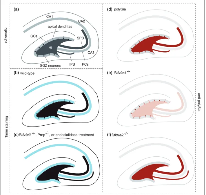
Hippocampal formation. The hippocampus represents in ver-tebrates the primary brain region tasked with memory con-solidation and spatial navigation. Detailed reports have been published that inform on the distribution of PrPC, polySTs, polySia, and NCAM1 within this brain region. Other docu-ments show how the architecture of the hippocampal forma-tion is changed in mice lacking the respective genes (Colling et al., 1997; Nacher et al., 2010). The subsequent summary of this literature will also first deal with localization before moving on to knockout phenotypes.
Both ST8SIA2 and 4 are most prominently observed in dentate granule cells (Angata et al., 1997). As analyzed
in young rats, St8sia2mRNA is localized to cells in the
subgranular zone, the innermost region of the granule cell layer and neurogenic niche of the dentate gyrus (Hildebrandt et al., 1998). Consistent with this, St8sia2
knockouts possess a diminished number of polySia-posi-tive cells in the subgranular zone (Angata et al., 2004).
The frequency of mitotic neural progenitors was not altered (Angata et al., 2004), but many of the immature granule neurons showed aberrant location and altered morphology, suggesting a role of ST8SIA2 in their guid-ance and differentiation (Nacher et al., 2010). In contrast,
St8sia2 mRNA signals are broadly distributed over the entire depth of the granule cell layer (Hildebrandt et al., 1998), which is consistent with the almost complete loss of the prominent polySia immunoreactivity on mossy fibers, the unmyelinated axons projecting from the gran-ule cells to pyramidal cells of the CA3 region of Ammon’s horn (Eckhardt et al., 2000; Nacher et al., 2010).
Likewise, the presence of St8sia4 but not St8sia2
mRNA in the pyramidal cell layer of the CA1 region (Hildebrandt et al., 1998) corresponds to the loss of polySia from the stratum lacunosum moleculare of the CA1 field inSt8sia4/
but notSt8sia2/
mice (Nacher et al., 2010). Resembling the pattern of hippocampal
polyST expression, PrPC is within this brain region
most prominently expressed in dentate granule cells and in CA1 to CA3 pyramidal cells (Tremblay et al., 2007).
Two types of mossy fiber bundles are known, a supra-pyramidal (SPB) main bundle and a smaller infrapyrami-dal bundle (IPB) that innervates dendrites on basal CA3 pyramidal cells and normally crosses the pyramidal cell layer to connect to the SPB within the CA3 field and in proximity to the hilus. As early as 1997, it was reported
that PrPC-deficient mice exhibit abnormal targeting of
the IPB (Colling et al., 1997). More specifically, in affected knockout mouse lines, the IPB—which can be visualized at low resolution by TIMM staining—did not cross over but instead continued to run underneath the pyramidal cell layer to the apex of its curvature (Figure 3). Significantly, since that time, only few other genes were shown to play a role in the biology that governs
proper IPB targeting. Among these areNcam1-knockout
mice (Seki and Rutishauser, 1998) and St8sia2-deficient
mice (Angata et al., 2004) as well as mice lacking both polySTs (Weinhold et al., 2005) and mice treated with endosialidase (Seki and Rutishauser, 1998) but not
St8sia4-deficient mice (Eckhardt et al., 2000). Notably, the increased granulation suggestive of mossy fiber ter-minal sprouting as observed in the extended infrapyrami-dal layer of PrPC-deficient mice (Colling et al., 1997) is highly reminiscent to the extension of the IPB and the formation of ectopic synapses in the far CA3 region of
St8sia2-knockout mice (Angata et al., 2004). The import-ance of balimport-anced polySia synthesis during mossy fiber tract formation has been corroborated in a slice culture model, in which aberrant mossy fiber projections and formation of ectopic synapses were induced by treatment with a modified sialic acid precursor and inhibitor of ST8SIA2 activity (Vogt et al., 2012).
modulation of cell surface interactions. Loss of polySia could either increase fasciculation by increasing
inter-actions between axons or reduce fasciculation
by increasing interactions with the environment.
Alternatively, the mossy fiber phenotype could be explained by alterations of the retraction and pruning of neuritic processes and axons, a major developmental
mechanism known to contribute to neuronal maturation. It has long been known that mossy fibers first send out, then retract filamentous extensions to nearby cells during their transit through the hilar region of the dentate gyrus (Amaral, 1979). This concept was further corroborated when a careful immunohistochemical study revealed
that during postnatal neurodevelopment even
polySia
St8sia4
St8sia2
IPB
wild-type
SPB
CA3 CA2 CA1
Hi
GCs
PCs apical dendrites
schematic
(a)
(b)
(c)
(d)
(e)
(f)
T
imm staining
anti-polySia
SGZ neurons
-/-St8sia2 , Prnp , or endosialidase treatment-/-
hippocampal formations of wild-type mice exhibit extended IPBs which, however, get pruned back around postnatal day 20 (Bagri et al., 2003).
In addition to the aforementioned players, the analysis of mice producing a loss-of-function NRP2 mutant revealed a critical role of this protein in the correct tar-geting of IPBs (Chen et al., 2000). Finally, a phenocopy of this IPB pathfinding defect can also be observed in mice deficient for the coreceptor of NRP2, plexin-A3 (Cheng et al., 2001), and its ligand, semaphorin-3F (Sahay et al., 2003). Thus, a model is emerging whereby pruning of IPBs not only depends on the semaphorin-3 ligand recognizing its NRP2/plexin-A3 heterodimeric
receptor but may also require PrPC and
ST8SIA2-mediated polysialylation. Interestingly, NRP2 is a target for polysialylation but polySia-NRP2 is exclusively pro-duced by ST8SIA4 (Rollenhagen et al., 2013) and so far has only been detected in dendritic cells (DCs), macro-phages, and microglia and not in neurons (Curreli et al., 2007; Stamatos et al., 2014; Werneburg et al., 2015). PolySia, however, may modulate the local concentration of ligands that act as attractants or repellents during mossy fiber guidance or may promote the retraction of temporary axonodendritic contacts formed between mossy fibers and pyramidal cell dendrites.
Taken together, the knockout phenotypes and patterns
of expression of PrPC and polySTs in brain areas
undergoing neurogenesis are consistent with a coordi-nated role of these proteins in neuroblast differentiation. And although the notion that these proteins contribute to both EMT and neuronal differentiation might be coun-terintuitive, it is increasingly being recognized that the morphogenetic reprogramming underlying both pro-cesses are overlapping (Itoh et al., 2013).
HSCs and their derivatives. The precise steps that govern the conversion of totipotent ESCs into multipotent mesoderm-committed cells are still not known. Recent gene expression profiling experiments, however, revealed the underlying shift of the transcriptome to bear yet again striking resem-blances to previously established molecular signatures of cells undergoing EMT (Evseenko et al., 2010). Thus, whereas ESCs rely on E-cadherin-dependent cell-to-cell contacts and express claudins, beta-catenin, and occludin, during mesoderm-commitment, these epithelial marker teins are replaced by the expression of mesenchymal pro-teins, including snail, vimentin, fibronectin, and NCAM1—note that the latter is better known as CD56 in the literature not concerned with the study of the brain. Interestingly, multipotent HSCs, which represent the main mesoderm-committed progenitors within the bone marrow of vertebrates, together with progenitors of endothelial and mesenchymal cells, smooth muscle cells, and cardiomyo-cytes, express ST8SIA4, but not ST8SIA2, and rely on this polyST for the expression of polySia on their cell surface
(Drake et al., 2008). A recent report extended these obser-vations by showing that ST8SIA4 is upregulated during early meso- and endoderm differentiation of human pluripo-tent stem cells, whereas ST8SIA2 is elevated in ectodermal cells (Berger et al., 2016).
Like NCAM1, PrPCis not found on totipotent human
ESCs but is expressed on HSCs expressing the marker
protein CD34 (Dodelet and Cashman, 1998). In PrPC
-deficient mice, self-renewal of HSCs is impaired leading to a gradual reduction in the proportion of multipotent long-term HSCs (Zhang et al., 2006). Because long-term HSCs serve as progenitors of myeloid and lymphoid lin-eage cells, another way to investigate their health is to monitor certain populations of cells derived from them by further differentiation and maturation. This approach
has been taken with St8sia4-deficient mice which have
been reported to exhibit reduced levels of the earliest thymocyte progenitors leading to an overall 30% reduc-tion of total thymocytes. Mechanistic investigareduc-tions, in which depleted T-cell progenitor reservoirs were repopu-lated by engraftment from wild-type orSt8sia4-deficient mice, suggested that the absence of polySia impairs the ability of progenitors to leave the bone marrow (Drake et al., 2009).
For PrPC, relatively detailed characterizations of its expression within the various lineages of HSCs-derived cells are available (Isaacs et al., 2006). According to these, levels of PrPCand trends of its expression are dis-tinct for each cell type as it matures. For example, PrPC expression is relatively high in monocytes and increases further as these mature into DCs but is gradually reduced upon differentiation of HSC progenitors into granulo-cytes. PrPCis further expressed on T- and B-lymphocytes as well as natural killer cells. How polysialylation levels change as HSC progenitors commit to specific myeloid or lymphoid cell lineages is currently less well understood. However, similar to PrP, relatively high levels of polySia-NCAM1 have been observed on the cell surface of mono-cytes and natural killer cells (Husmann et al., 1989; Drake et al., 2008).
Interestingly, as monocytes differentiate, initially, into immature, then mature DCs, the substrate for attachment of polySia shifts from NCAM1 to NRP2 (Curreli et al., 2007). Efforts to determine the polyST responsible for NRP2 polysialylation revealed that, although DCs
express both ST8SIA2 and ST8SIA4, only St8sia4
polySia on non-NCAM1 protein carriers is confined to the Golgi compartment of microglia but released in response to the inflammatory activation by lipopolysac-charides, mimicking an infection by Gram-negative bac-teria. Considering the anti-inflammatory activity of polySia (Shahraz et al., 2015; Werneburg et al., 2015, 2016), it can be hypothesized that this LPS-induced release is part of a negative feedback regulation of micro-glia or macrophage activation.
What is the functional significance of NRP2 polysia-lylation on the surface of DC cells? It is well known that mature DCs migrate toward secondary lymphoid organs under the guidance of specific chemokines. In particular, chemokine C-C motif ligands 19 (CCL19) and 21 (CCL21) are known to be essential in this context. Significantly, polySia on DC cells facilitates this migra-tion in a manner that depends on the presence of a basic motif present at the C-terminus of CCL21 and, presum-ably, involves its interaction with the polySia negative charge cluster (Rey-Gallardo et al., 2010). This chemo-taxis is strictly dependent on ST8SIA4 and not on ST8SIA2 (Rey-Gallardo et al., 2011). Originally thought to depend on polySia-NRP2, a recent study reveals that polysialylation of CCR7, the central chemokine receptor controlling immune cell trafficking to secondary lymph-atic organs, is essential for the recognition of CCL21 by DC cells (Kiermaier et al., 2016). The same study also provides a mechanistic explanation why CCR7 needs polySia. CCL21 adopts an autoinhibited conformation, which is released by polySia interactions with its basic C-terminal extension.
The question arises if the ability of PrPCto influence polysialylation levels extends to ST8SIA4 and non-NCAM1 substrates in certain cellular contexts. If so, it will be attractive to specifically investigate if PrPC influ-ences in immune cells the expression of ST8SIA4, thereby modulating microglia or macrophage activation or CCL21-dependend migration of DCs. Although this question has not yet been directly addressed, it was reported that transgenic overexpression of CCL21 from an insulin promoter causes chronic inflammatory disease
in the pancreas that was characterized by PrPSc
accumu-lation in prion-infected mice in this peripheral organ that
in wild-type mice is spared by PrPSc infection
(Heikenwalder et al., 2005). It appears as if CCL21 served in this paradigm as a chemotactic attractant which stimulated invasion of this organ by PrPC express-ing cells, a scenario consistent with a broader role of PrPC in controlling polysialylation, transcending its more lim-ited role in facilitating polysialylation of NCAM1.
Myelin repair and maintenance. Myelination of peripheral nerves, including the sciatic nerves, depends on a complex molecular dialogue between Schwann cells and neurons that is only partially understood. Schwann cells are one of
several cell lineages derived from neural crest stem cells that are known to express the polysialylated form of NCAM1. In fact, NCAM1 is not merely a marker of cells in this lineage but appears to modulate several aspects of myelination, including axonal outgrowth, the formation of contacts between axons and Schwann cells, and Schwann cell differentiation (Rutishauser, 2008; Irintchev and Schachner, 2012). Early steps of the axon-Schwann cell interaction appear to be governed by a molecular biology of Schwann cells adapted from the EMT program. Consistent with this notion, not only TGFB1 but also fibro-nectin and tenascin, well-established marker proteins of mor-phogenetically active mesenchyme (Ignotz and Massague, 1986), are expressed during these early steps. NCAM1 has been observed on fasciculating axons and on non-myelinat-ing Schwann cells until the onset of myelination when Schwann cell processes had completed up to 1.5 turns of their axon ensheathment (Martini and Schachner, 1986). A downregulation of both TGFB1 and NCAM1 expression is essential for myelination to proceed (Einheber et al., 1995). During this step, the NCAM1 pool is largely replaced by other extracellular matrix proteins, including the myelin-associated glycoprotein (Martini and Schachner, 1988), but appears to persist in the periaxonal region of larger diameter myelinated axons (Martini and Schachner, 1986). Lesioning of peripheral nerves, including nerve transection or crushing, induces TGFB1 and repopulates axon stumps with polysia-lylated NCAM1, facilitating proliferation of non-myelinat-ing Schwann cell (Covault et al., 1986; Daniloff et al., 1986; Martini and Schachner, 1988). Indeed, it has been demon-strated that peripheral nerve regeneration is reduced in the absence of ST8SIA2 (Jungnickel et al., 2012; Koulaxouzidis et al., 2015) and that induced expression of polySTs in the environment promotes peripheral nerve regeneration (Gravvanis et al., 2005; Lavdas et al., 2006; Jungnickel et al., 2012). However, exquisite timing is critical for success-ful peripheral nerve regeneration, as a forced continuous overexpression of polySTs in Schwann cells leads to fewer regenerated myelinated axons (Jungnickel et al., 2012).
One of the most thoroughly investigated phenotypes in
Prnp/
mice is a peripheral neuropathy that most con-spicuously is characterized by a myelin maintenance defect (Bremer et al., 2010). Reminiscent of the
distribu-tion of NCAM1, PrPC was observed on both the axon
and ensheathing Schwann cells during peripheral nerve
development, with Schwann cell expression of PrPC
coming to a halt once the ensheathment process is under-way. Elegant genetic rescue experiments led the authors to conclude that PrPChas to be expressed on the axon to prevent this demyelinating neuropathy. In the absence of PrPC, myelinated axons were seen to be considerably less tightly packed and the number of large diameter myelin-ensheathed axons was reduced. There also were ultra-structural differences, including loosely condensed onion
non-myelinated axons, also known as Remak bundles, which can be seen in wild-type controls. A deficiency in the formation of Remak bundles has also been observed in the aforementioned transgenic mice, which continuously overexpress polySTs in Schwann cells (Jungnickel et al., 2012) and in laminin-deficient Schwann cells (Yu et al., 2009). Notable characteristics in the latter mice are impaired developmental expression and interactions of NCAM1 in the mutant nerves (Yu et al., 2009).
Thus, it is conceivable that the presence of PrPC on
peripheral axons and myelinating Schwann cells might impact myelin maintenance and peripheral nerve regen-eration on capacity of its ability to interact with NCAM1 or by influencing its levels of polysialylation.
Circadian rhythm. Profound sleep disturbances are a hallmark of the disease in individuals afflicted with Fatal Familial Insomnia (FFI). In humans, FFI can be caused by a single amino acid change within the prion protein sequence that replaces an aspartate with an asparagine residue at Position 178 on a background of the common methionine or methio-nine polymorphism at PrPCamino acid Position 129. Using a knock-in strategy, this disease could be modeled in mice (Jackson et al., 2013). While the exact causes for this pheno-type are not known, the neural structures for time keeping are increasingly well understood. In humans and mice, a brain region that sits atop the optic chiasm, the suprachias-matic nucleus (SCN), is known to not only regulate the sleep or wake cycle but also to act as a master conductor that harmonizes the speed of cellular clocks within the body (O’Neill et al., 2013). The insomnia phenotype observed in FFI could be caused by a mere loss of neurons within the SCN because this area, together with surrounding thalamic regions, is strongly affected in the disease. This explanation is unsatisfying, however, on account of evidence that similar sleep or wake cycle disturbances can also be observed in cases of sporadic Creutzfeldt-Jakob disease in the absence of prominent neuronal loss in this brain region (Landolt et al., 2006). A more plausible scenario, therefore, may be that PrPCplays a role in the control of the sleep or wake cycle, and that mutation or its structural conversion causes a loss or perturbation of this role. PolySia-NCAM1 is expressed in adult rodent brain (Glass et al., 1994) and also subject to diurnal fluctuations during a 24-h light or dark cycle in the SCN (Glass et al., 2003), with higher and lower levels observed during the early subjective day and night, respect-ively (Prosser et al., 2003). Investigations of mice deficient for individual NCAM1 isoforms or all of NCAM1, and con-sequently its polySia modification, revealed that polySia-modified NCAM1 is critical for intact free-running circadian rhythmicity under continuous darkness (Shen et al., 1997, 2001). The knockout of all NCAM1 isoforms was shown to cause a yet more severe phenotype that included a defect-ive activity profile during the light or dark entrainment period (Shen et al., 2001). Although the mechanism by
which polySia-NCAM exerts its control of the circadian rhythm is not yet understood, a recent study proposed that an endoproteolytic polySia-carrying NCAM1 fragment might be transported to the nucleus of cells involved in cir-cadian control to regulate the gene expression of clock-related genes (Westphal et al., 2016).
Indicative of the insomnia component observed in a subset of prion diseases reflecting a disturbance of the
physiological role of PrPC, one of the first prominent
phenotypic changes observed in Prnp/
mice was an altered circadian rhythm (Tobler et al., 1996). More spe-cifically, twoPrnp/
mouse lines, generated by different targeting strategies, entrained like wild-type mice to light or dark cycles but showed longer periods of activity than wild-type control mice, when subjected to continuous darkness. A follow-up investigation determined that
mRNA levels of PrPC fluctuate during the day in the
SCN and other forebrain regions (Cagampang et al., 1999), peaking early in the phase of increased locomotor activity, together with the gene products of several other
clock-relatedgenes (Buhr and Takahashi, 2013).
What might be a cause for the phase shift observed in
PrPCand polySia-NCAM-deficient mice? Light is known
to induce retinal ganglion cells to release glutamate onto SCN neurons. The increased glutamate levels, in turn, are understood to activate NMDA receptors (NMDARs).
Both PrPC and polySia-NCAM1 have been shown to
impact NMDAR signaling, possibly, by modulating local copper concentrations (see later). In support of this model, copper has been shown to cause a delay in circadian neural activity rhythms (Yamada and Prosser, 2014). Addition of glutamate to a slice preparation of the SCN increases polySia-NCAM1 levels causing phase delays (Prosser et al., 2003). Pretreatment of this slice preparation of the SCN with endosialidase abolishes these phase delays (Prosser et al., 2003). Thus, an intri-cate interplay between glutamate release, the circadian
control of polySia-NCAM1 and PrPC levels, as well as
their ability to modulate NMDA-mediated glutamate sig-naling might influence the circadian phase.
Glutamatergic signaling and long-term potentiation. Long-term potentiation (LTP) denotes a complex postsynaptic response to extracellular glutamate exposure, widely considered the cellular correlate to memory and learning. Central to LTP are the functional interplay of two types of glutamate recep-tors, AMPA receptors (AMPARs) and NMDARs. Briefly, when glutamate binds to AMPARs, their ion channels open causing Naþ
influx. As the intracellular milieu becomes increasingly depolarized, inhibitory Mg2þ
ions occluding the channels of nearby NMDARs are released, causing them to also contribute to the Naþ
influx and allowing add-itional calcium ions to enter the cell. Ca2þ
membrane, thereby increasing the AMPAR-NMDAR ratio and the ability of the synapse to respond to future glutamate exposure. PolySia-NCAM1 has repeatedly been shown to modulate glutamatergic signaling reliant on AMPARs and NMDARs. For example, St8sia4/
mice were shown to exhibit LTP impairments following stimulation of Schaffer collateral-CA1 synapses (Eckhardt et al., 2000). Indicative of available polySia levels playing a critical role in this pheno-type, these CA1 synapses are known to express polySia-NCAM1 in wild-type mice and the level of impairment appeared to correlate with the gradual loss of polySia in adult mice. Ncam1/
mice are similarly characterized by LTP disturbances that manifest as memory deficits in hippo-campus-dependent contextual fear conditioning and extrahip-pocampal-cued memory paradigms. Interestingly, of these two types of memory deficits, only the contextual fear deficit is shared bySt8sia4/mice, indicating that this adaptation is more dependent on polySia than NCAM1 itself. Consistent with this interpretation, when acute hippocampal slices from NCAM1/
mice were injected with NCAM1, the extracel-lular domain of polySia-NCAM1 or polySia alone, restoration of LTP was only observed with injected material containing polySia (Senkov et al., 2006). The interaction of polySia-NCAM1 appears to be specific with regard to subsets of NMDARs and AMPARs and the directionality of its modu-lating effect is opposite for these receptors, that is, activity promoting for AMPARs (Vaithianathan et al., 2004) and activity restricting for NMDARs (Hammond et al., 2006; Kochlamazashvili et al., 2010).
What is the physiological significance of these effects? The polySia-dependent activity restriction of GluN2B-containing NMDARs serves a useful role. It prevents the neurotoxic chronic low-level activation of these recep-tors and their downstream signaling pathways, possibly on the basis of it acting as a GluN2B antagonist that competes with low levels of glutamate for binding to the receptor (Hammond et al., 2006). Thus, removal of polySia was shown to translate downstream of GluN2B into higher levels of signaling through Ras and GRF1 to p38 MAPK, which, in turn, led to increased cell death. Consistent with this model, LTP and contextual fear
con-ditioning were restored in Ncam1-deficient mice by
inhibiting GluN2B or by scavenging free glutamate with GTP (Kochlamazashvili et al., 2010). Not surprisingly, there also is a literature that ties NMDA activation to the expression of polySia-NCAM (Butler et al., 1999; Singh and Kaur, 2009).
Ever since the first Prnp/
mouse models became available (Bueler et al., 1992), researchers have attempted
to gauge the extent to which the loss of PrPC function
may contribute to synaptic deficits observed in prion dis-eases. Several groups reported in the mid-90s that PrPC -deficiency translates into impairments in LTP (Clinton et al., 1993; Collinge et al., 1994; Manson et al., 1995) and learning and memory. As with polySia-NCAM1, the
presence of PrPChas most often been shown to
desensi-tize NMDARs, making them less responsive to low levels of EtOH, NMDA, kainite, or glutamate (Shyu et al., 2005; Rangel et al., 2007; Carulla et al., 2011; Llorens and Del Rio, 2012; Black et al., 2014). This protective role has, however, not remained unchallenged (Herms et al., 1995) and was, for example, not detected when the influence of PrP on LTP was investigated in
hippo-campal slices dissected from intercrosses ofPrnp
knock-out mice and an AD mouse model, which itself exhibits an LTP defect (Calella et al., 2010). Nonetheless, the pre-vailing trend seems to be that protective roles of PrPCcan be observed in a wide range of experimental settings and extend to PrPCorthologs in species as distant as zebrafish (Fleisch et al., 2013). The mechanism through which PrPC ameliorates NMDAR-dependent excitotoxicity is under intense investigation but has so far not been elucidated. It has been proposed that PrPCin its copper-loaded state binds to the NMDAR complex, thereby reducing the affinity of the receptor to glycine, possibly through an allosteric effect (You et al., 2012). An alternative model
sees cooperative binding of PrPC and copper protect
NMDA receptors by promoting their S-nitrosylation (Gasperini et al., 2015). To date, neuroprotective roles
of polySia-NCAM1 or PrPCon NMDA receptor biology
have only been considered independently. It will be inter-esting to explore to which degree these phenomena mani-fest independently or reflect a closely linked biology of
PrPCand NCAM1.
Evolutionary Context
The prion gene family evolved approximately less than a half billion years ago from an ancestral gene coding for a member of the Zrt-, Irt-like protein (ZIP) family of metal ion transporters (Schmitt-Ulms et al., 2009). The evolution-ary relationship between PrPCand ZIP transporters acted as a catalyst in the research path that precipitated the
dis-covery of PrPC’s involvement in the control of NCAM1
A first indication of a relationship between PrPC and ZIP transporters surfaced when it was established that two of these transporters (ZIP6 and ZIP10) can be crosslinked
to PrPC in Neuro-2a neuroblastoma cells exposed to low
levels of formaldehyde (Watts et al., 2009). During the follow-up analysis of candidate PrPCinteractors, striking
similarities in the molecular organization of PrPC and
these ZIP transporters were noticed, which precipitated an investigation into whether these proteins are evolution-arily related. Subsequent bioinformatic and structural modeling analyses then led to the surprising discovery
that PrPC and the ectodomains of a subset of ZIP
trans-porters indeed share considerable sequence identity and are predicted to acquire homologous folds. Spurred by these observations, additional in-depth bioinformatic ana-lyses sealed the conclusion that PrPCand ZIP transporters are members of one large protein family comprising 17 genes in humans (Schmitt-Ulms et al., 2009; Ehsani, Huo, et al., 2011). Note that the initial observation of a molecular interaction between PrPCand its paralogs ZIP6 and ZIP10 might reflect a reality of ZIP transporters
assembling into dimers in vivo (Bin et al., 2011;
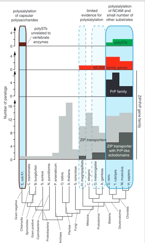
Pocanschi et al., 2013), that is, PrPCmost likely inherited and retained this property from its ancestral ZIP trans-porter. Whereas ZIP transporters are expressed in all branches of life, the subset of these transporters compris-ing a PrP-like ectodomain only evolved in the metazoan lineage (Schmitt-Ulms et al., 2009). On the basis of synteny analyses and detailed gene structure comparisons, it can be inferred that the genomic rearrangement, which led to the creation of the prion founder gene, occurred relatively early in the vertebrate lineage. Mechanistically, this event can be reconstructed to have involved a spliced ancestral ZIP transporter mRNA and most likely was based on the genomic insertion of a reverse transcribed copy of this mRNA by a retrovirus (Ehsani, Tao, et al., 2011).
There is compelling sequence evidence in the genomes of other organisms to conclude that the aforementioned steps, which precipitated the formation of the prion gene family in vertebrates, were not an isolated event but occurred more than once. For example, in the oppossum (Monodelphis domestica) genome, a PrPC-like ZIP6-related
pseudogene could be identified, which is, like the Prnp
gene, devoid of introns within its protein coding sequence and also lacks the sequence elements coding for the trans-membrane domain observed in intact ZIP transporter genes (Ehsani, Tao, et al., 2011). In contrast to the prion founder gene sequence, which upon its genomic insertion became a fully functional retrogene by adapting an upstream promoter for its expression, the oppossum gene has become a non-expressed pseudogene subject to slow sequence decay (Ehsani, Tao, et al., 2011).
Similar to the subset of ZIP transporters carrying
PrPC-like ectodomains, NCAM1 is an ancient molecule
that evolved early in the metazoan lineage but there is no
evidence for its polysialylation in protostomes (arthro-podes and mollusks). Consistent with this assertion, the polySTs responsible for its polysialylation may have evolved around 500 million years ago (Harduin-Lepers et al., 2005; and most likely preceded the evolutionary appearance of the prion founder gene). Available evi-dence suggests that around that time, a gene duplication involving an ancestral sialyltransferase must have given
rise to a primordial ST8SIA2/4 polyST gene
(Harduin-Lepers et al., 2008). The proposed turn of events can be
deduced from the presence of an array of fourST8SIA
-genes, including a coding sequence for anST8IA2/4gene,
that map to a single chromosome in lancelets
(Branchiostoma floridae; Harduin-Lepers et al., 2008). Lancelets are members of the phylogenetic branch of amphioxi, subphylum cephalochordate, which did not participate in the two whole genome duplication events that have been credited with the emergence of separate ST8SIA2 and ST8SIA4 paralogs and were restricted to the vertebrate lineages comprising fish and tetrapods (Harduin-Lepers et al., 2008).
Around the time of chordate speciation, and most dra-matically following the whole genome duplications in early vertebrates, genomes coded for many more cell sur-face receptors than prior to these events. The expanded repertoire of cell surface receptors and their ability to interactin transwith cell surface receptors on neighboring cells allowed cell-cell communication to become more refined but also must have posed an obstacle for the exe-cution of plasticity-related tasks. Thus, for a cell to become motile, a larger number ofin transprotein inter-actions had to be negotiated or broken. It has been pro-posed that the emergence of NCAM1 polysialylation represented an adaptation to this reality (Rutishauser, 2008). Taken together, this crude outline of evolutionary
events pertinent to the formation of the PrPC
-ST8SIA2-NCAM1 signaling loop posits that the appearance of the
Prnpgene served as a timely and meaningful adaptation
to a new challenge.
Naturally, for a cell to become motile, many other elements need to be in place. For instance, (a) stable cell-cell adhesion complexes need to be replaced with dynamic cell-substrate interactions; (b) instead of provid-ing structural integrity, the cytoskeleton need to convey cell movement; and (c) the cell needs to have systems in place that alert it to guidance cues and inform it on when to stop moving and re-establish connections to neighbor-ing cells. Thus, although a defect in the control of NCAM1 polysialylation may turn out to be the single
most profound change observed in PrPC deficiency
In particular, proteins which undergo level changes in EMT in wild-type cells are highly significantly overrepre-sented among the subset of proteins whose levels are altered in stable PrPC-deficient cells.
Consistent with the view that the involvement of the
PrPC-NCAM1 heteromer in cellular plasticity programs
did not evolve abruptly, NCAM orthologs have been linked to these programs even in cells and species whose
S.
c
erevisiae
C.
elegans
A.
gambiae
D
. melanogaster
T
. rubripes
D
. rerio
M.
musculus
H.
sapiens
A.
thaliana
O
. sativ
a
Protostomia Deuterostomia
Bilateria
H.
magnipapillata
Metazoa
P
. arsenaticum
Plantae
Fungi
Gram positive
Chordata
A
rchaea
Cyanobacteria
Proteobacteria
Gram negative
Chlamydia
Spirochetes
Number of par
alogs
0 4 8 12 16 0 4 0 4 0 4
NCAM family genes
ZIP transporter with PrP-like ectodomains
polysialylation of NCAM and small number of other substrates polysialylation
of capsular polysaccharides
limited evidence for polysialylation
polySTs unrelated to
vertebrate enzymes
E.
c
oli K1
S.
aureus
B
. bur
g
dor
feri
N.
punc
tiforme
C.
tr
achomatis
ZIP/PrP
gene family
PrP family
polySTs
ZIP transporters
genomes do not code for polySTs. For example, fasciclin2
(Fas2), a fruit fly (Drosophila melanogaster) NCAM
ortholog, is required for sensory axon guidance
(Kristiansen et al., 2005), motility of glial cells along axons (Silies and Kla¨mbt, 2010), and has been shown to be essential for the coordinated developmental move-ment of a cluster of cells known as border cells (Szafranski and Goode, 2004). In some mammalian cancer paradigms, unpolysialylated NCAM1 might be sufficient and indispensable for a lipid-raft centered sig-naling program that coordinates the concomitant disas-sembly of E-cadherin-based adherence junctions and formation of focal adhesions as a prelude to cellular movement (Frame and Inman, 2008; Lehembre et al., 2008). In contrast to invertebrates, which do not synthe-size polySia-like structures and control the cell surface presentation of NCAM orthologues by internalization and proteolytic degradation (Schuster et al., 1996; Bailey et al., 1997), the possibility to steer NCAM1 inter-actions by means of polysialylation provides vertebrates with an additional level of regulation, and the ratio between unpolysialylated and polysialylated NCAM1 seems to be critical for balancing cell and cell-matrix adhesion as a prerequisite for the morphogenetic events during migration and differentiation (Seidenfaden et al., 2006; Eggers et al., 2011).
A similar connection to cellular plasticity programs has also been established for the subset of ZIP trans-porters carrying PrPC-like ectodomains. For instance, in addition to the aforementioned gastrulation arrest pheno-type observed in ZIP6-deficient zebrafish (Yamashita et al., 2004), the fruit fly gene fear-of-intimacy, a ZIP5/6/10 ortholog (Mathews et al., 2005), was shown to be essen-tial for the execution of a cell migration and coalescence program during gonad formation (Van Doren et al., 2003; Mathews et al., 2006) and tracheal development (Van Doren et al., 2003). Moreover, mice deficient in the ZIP13 transporter exhibit defects in bone, teeth, and connective tissue development that implicated this porter in impaired bone morphogenetic protein or trans-forming growth factor signaling (Fukada et al., 2008). Similarly, ZIP14 knockout mice are characterized by stunted growth and defects in bone morphology, possibly by affecting CREB-dependent developmental signaling in chondrocytes (Hojyo et al., 2011).
Although at least theoretically the participation of
orthologs of NCAM and ZIP transporters with PrPC
-like ectodomains in these cellular plasticity programs could be realized independent of each other, this scenario
seems unlikely at this time. Existing PrPC interactome
data already established close proximity of PrPC to
NCAM1, ZIP6, and ZIP10. These data have since been corroborated by reciprocal analysis of ZIP6 interactors, which revealed NCAM1 to represent the main binding
partner of ZIP6 (unpublished observation). Thus,
consistent with their coinciding evolutionary emergence,
NCAM1 and ZIP transporters harboring PrPC-like
ecto-domains may have evolved to collaborate during the exe-cution of cell plasticity programs. A need for these programs materialized during metazoan development to promote a motile lifestyle. Later in evolution, with the emergence of complex body plans, elements of these pro-grams could be adapted for developmental propro-grams that required the migration of defined cell populations. Components required for NCAM1 polysialylation only emerged much later. These may have served the purpose to further enhance the ability of preexisting plasticity pro-grams to cope with both an unprecedented complexity of cell surface receptor interactions and an increasing com-plexity of body plans. In particular, the migration of spe-cialized cell populations in different parts of the developing body would be expected to require a more specialized toolkit to choreograph this movement in time and space, possibly requiring further differentiation of players involved. Perhaps adapting to this need, the subbranch of the ZIP transporter family comprising
PrPC-like ectodomains expanded and diversified in the
vertebrate lineage (Figure 4). It will be interesting to
learn if PrPC-like ectodomains in ZIP transporters are
able to modulate the transcription of polySTs during the execution of specific mammalian morphogenesis pro-grams in the same way as PrPC.
Ironically, although this line of research was precipi-tated by the striking resemblance of gastrulation arrest
phenotypes independently observed following PrPC or
ZIP6 knockdown in zebrafish embryos (Yamashita et al., 2004; Malaga-Trillo et al., 2009), it can currently not be concluded if these zebrafish defects rely on per-turbed NCAM polysialylation or rely on defects in other,
yet to be identified, signals that emanate from PrPC or
ZIP6. However, consistent with the possibility of an
involvement of the PrPC-ST8SIA2-NCAM signaling
loop in this program, the expression of ST8SIA2 (Marx et al., 2007) has been observed just prior to gastrulation during zebrafish development.
Conclusions
This review was written with a view to provide context and perspective to a recent report, which proposed a
novel role for PrPC as a key player within a signaling
loop that controls the polysialylation of NCAM1 during the execution of cellular plasticity programs. The
role proposed for PrPCreconciles within a single model