Vojnosanit Pregl 2015; 72(3): 233–240 VOJNOSANITETSKI PREGLED Page 233
O R I G I N A L A R T I C L E UDC: 615.849.19::616.314-089.843
DOI: 10.2298/VSP131202075M
Influence of postoperative low-level laser therapy on the
osseointegration of self-tapping implants in the posterior maxilla:
A 6-week split-mouth clinical study
Uticaj postoperativne terapije laserom male snage na oseointegraciju
samourezuju
ć
ih implantata u bo
č
noj regiji gornje vilice:
šestonedeljna split-mouth klini
č
ka studija
Borka Mandić*, Zoran Lazi憇, Aleksa Marković*, Bojan Mandić§, Miška Mandić¶, Ana Djinić*, Biljana Miličić||
*
Clinic of Oral Surgery, §Clinic of Maxillofacial Surgery, ||Department for Medical Statistics and Informatics, ¶Department of Orthodontics, Faculty of Dental Medicine, University of Belgrade, Belgrade, Serbia; †Clinic of Dental Medicine, Military Medical
Academy, Belgrade, Serbia; ‡Faculty of Medicine of the Military Medical Academy, University of Defence, Belgrade, Serbia
Abstract
Background/Aim. Low-level laser therapy (LLLT) has been proven to stimulate bone repair, affecting cellular prolifera-tion, differentiation and adhesion, and has shown a potential to reduce the healing time following implant placement. The aim of this clinical study was to investigate the influence of postoperative LLLT osseointegration and early success of self-tapping implants placed into low-density bone. Meth-ods. Following the split-mouth design, self-tapping implants (n = 44) were inserted in the posterior maxilla of 12 patients. One jaw side randomly received LLLT (test group), while the other side was placebo (control group). For LLLT, a 637 nm gallium-aluminum-arsenide (GaAlAs) laser (Medicolaser 637, Technoline, Belgrade, Serbia) with an output power of 40 mW and continuous wave was used. Low-level laser treat-ment was performed immediately after the surgery and then repeated every day in the following 7 days. The total irradia-tion dose per treatment was 6.26 J/cm² per implant. The study outcomes were: implant stability, alkaline-phosphatase (ALP)
activity and early implant success rate. The follow-up took 6 weeks. Results. Irradiated implants achieved a higher stability compared with controls during the entire follow-up and the difference reached significance in the 5th postoperative week (paired t-test, p = 0.030). The difference in ALP activ-ity between the groups was insignificant in any observation point (paired t-test, p > 0.05). The early implant success rate
was 100%, regardless of LLLT usage. Conclusion. LLLT
applied daily during the first postoperative week expressed no significant influence on the osseointegration of self-tapping implants placed into low density bone of the poste-rior maxilla. Placement of self-tapping macro-designed im-plants into low density bone could be a predictable thera-peutic procedure with a high early success rate regardless of LLLT usage.
Key words:
dental implants; oral surgical procedures; laser therapy, low-level; bone regeneration; alkaline phosphatase; treatment outcome.
Apstrakt
Uvod/Cilj. Terapija laserom male snage (TLMS) stimuliše reparatorne sposobnosti kosti utičući na ćelijsku proliferaci-ju, diferencijaciju i adheziproliferaci-ju, i ima potencijal da skrati vreme zarastanja kosti nakon ugradnje implantata. Cilj ove kliničke studije bio je da se ispita uticaj postoperativne primene TLMS na oseointegraciju i rani uspeh ugradnje samourezu-jućih implantata u kost male gustine. Metode. Prateći split-mouth dizajn, samourezujući implantati (n = 44) ugrađeni su u posteriorne regije gornje vilice 12 pacijenata. Slučajnim
Zra-čeni implantati imali su veću stabilnost u odnosu na kon-trolne tokom celog perioda praćenja, a statistički značajno veća stabilnost bila je u petoj postoperativnoj nedelji (t-test za vezane uzorke, p = 0.030). Razlika u aktivnosti ALP iz-među grupa nije bila statistički značajna ni u jednoj tački po-smatranja (t-test za vezane uzorke, p > 0.05). Procenat rane uspešnosti terapije implantatima bio je 100%, bez obzira na
primenjenu TLMS. Zaključak. Svakodnevna primena
TLMS u prvoj postoperativnoj nedelji nije pokazala znač a-jan uticaj na oseointegraciju samourezujućih implantata u
kost male gustine bočne regije gornje vilice. Primena im-plantata samourezujućeg makrodizajna u kosti male gustine mogla bi predstavljati predvidljivu terapijsku proceduru sa visokim procentom rane uspešnosti, bez obzira na prime-njenu TLMS.
Ključne reči:
implantati, stomatološki; hirurgija, oralna, procedure; lečenje laserom male snage; kost, regeneracija; alkalna fosfataza; lečenje, ishod.
Introduction
Low-level laser therapy (LLLT) has been used for more than 30 years in the medical field and no adverse effects have been reported 1. It is defined as red beam or near-infrared laser therapies of low energy density and output power, with wavelengths between 500 and 1,200 nm, that do not increase normal tissue and body temperature 1. Its effects are therefore nonthermal and biostimulative.
As LLLT affects various tissue responses such as blood flow, inflammation, cellular proliferation and/or differentia-tion 2, stimulation with LLLT creates a number of environ-mental conditions that appeared to have accelerated healing of bone defects in animal models and clinical investiga-tions 2–5.
Though the exact mechanism of these effects is not elu-cidated yet, they are considered to be results of laser irradia-tion on the cell membrane, mitochondria, DNA and RNA synthesis, collagen synthesis, neovascularization, cell prolif-eration, and the production of ATP 6.
In oral implantology, research has been focused on the potential of LLLT to reduce the healing time following im-plant placement and to improve the potential for bone regen-eration 2.
Previous experimental studies reported that low-level laser treatment stimulated proliferation and differentiation of osteoblasts 7–11 as well as their bonding to titanium implant 7. It significantly increased alkaline phosphatase (ALP) activ-ity, which is considered to be a marker of differentiated os-teoblasts, in culture 8, 9, 11 and animal models 10. When ap-plied in the early postoperative period, LLLT lead to an en-hancement of the mechanical strenght of bone-implant inter-face 12–14 and stimulation of bone matrix production and bone nodule formation 9.
There are a number of studies suggesting that low-level laser treatment in the early postoperative period after implant placement may lead to a positive clinical effect 2.
As low-density bone (D3 and D4 class of bone, Leck-holm & Zarb classification 15) is usually present in the molar region of the upper jaw, this has proven to be the region of lower success rates of dental implant therapy due to lack of primary stability that can be obtained 16. Postoperative LLLT might have potential beneficial influence on dental implant treatment in this area, making it more predictable.
The aim of our study was to investigate the influence of postoperative LLLT on osseointegration of self-tapping
im-plants placed into low density bone, by investigating and comparing clinical status – implant stability with the ap-pearance of the marker of alkaline phosphatase in the periimplant crevicular fluid. The second aim was to evaluate early success rate of implants placed into the premolar/molar maxillary region, regarding LLLT.
Methods
The study was conducted in accordance with the 1975 Declaration of Helsinki, as revised in 2002. The protocol was approved by the Ethics Commitee of the Faculty of Den-tistry, University of Belgrade (No.36/22), and the patients gave their written informed consent. Written patient’s con-sent was also obtained to publish clinical photographs.
A total of 12 patients (6 males and 6 females) seeking implant therapy for bilateral reconstruction in the posterior maxilla were recruited for this study. All the patients were healthy adults, age 18 or older. The patients were selected in accordance with the following inclusion criteria: sufficient bone volume to receive implants without requiring bone augmentation (reconstruction) procedures and no history of previous tooth extraction in the last six months in the se-lected area. Exclusion criteria were: 1) systemic: pregnancy or lactation, systemic disease that affects osseointegration, anticoagulant therapy, systemic glucocorticoid therapy, his-tory of radiotherapy in the craniofacial region within last 12 months, smoking habit of more than 10 cigarettes per day and 2) local: acute infection in the mouth, uncontrolled or untreated periodontal disease.
For patients' selection and treatment planning, pano-ramic radiographs and 3D computed tomography scans were required, followed by clinical intraoral examination.
Following split mouth design, a total of 44 self-tapping BlueSky® (Bredent, Germany) implants with diameter of 4 mm and length of 10 mm were inserted bilaterally and simet-rically in the posterior maxilla of the selected patients.
Local anesthesia was induced by infiltration with 2% lidocaine hydrochloride and 1: 80 000 adrenaline. After cre-stal incision and mucoperiosteal flap elevation, preparations of implant recipient sites were performed under cooling with physiological solution, according to the protocol following the manufacturer’s instructions (Bredent, Germany). The speed of 15 rpm with a torque of 35 Ncm was set for inser-tion of all implants. The implants were allowed to heal transmucosally and sutures were removed after 7 days.
Vol. 72, No. 3 VOJNOSANITETSKI PREGLED Page 235
Postoperatively all the patients were prescribed amox-icillin (1.5 g) or clindamycin (1.8 g) daily, for three days as well as nonsteroidal anti-inflammatory drugs for pain relief. The patients were also given detailed instructions with regard to oral hygiene. No temporary prosthesis was placed during the entire 6-week observation period.
After the surgery, one of the sides of the upper jaw of the patients was randomly (computer-generated random numbers) chosen to receive low-level laser treatment (test group). The other side of the jaw was placebo, without any treatment performed and served as a control (control group).
A 637 nm gallium-aluminum-arsenide (GaAlAs) laser (Medicolaser 637, Technoline, Belgrade, Serbia) with an out-put power of 40 mW and continuous wave was used. The im-plant on the chosen side was irradiated intraorally, orthora-dially to the implant's longitudinal axis (Figure 1). Low-level laser treatment was performed immediately after the surgery and then repeated every day in the following 7 days. The total irradiation dose per treatment was 6.26 J/cm² per implant.
Fig. 1 – Postoperative low-level laser therapy. The operational field was irradiated by laser probe positioned
intraorally, at a distance of 1 cm and orthoradially to the implant's longitudinal axis.
Evaluation of osseointegration of implants
All assessments of the study outcomes were preformed in a double blind manner, since neither patients (due to pla-cebo) or assessors (not involved in LLLT) were aware of treatment allocation.
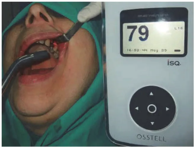
Resonance frequency analysis (RFA) was performed us-ing the Osstell™ Mentor instrument (Integration Diagnostics, Göteborg, Sweden) by a trained calibrated operator who was unaware of which side would be irradiated. Measurements were recorded immediately after implant insertion and then postoperatively in a weekly manner during the following 6 weeks. A standardized abutment of fixed length (Smartpeg™ Integration Diagnostics, Göteborg, Sweden) was inserted and hand-tightened into each implant. The transducer probe (Osstell™ Mentor Probe) was held so that the probe tip was aimed at the small magnet on top of the Smartpeg™ at a dis-tance of 2–3 mm (Figure 2). It was held still until the instru-ment beeped and displayed the implant stability quotient (ISQ) value. Each measurement was repeated until the same value
was recorded twice, which was accepted as the authentic val-ue. For the post-surgical stability measurements, abutments were removed from the implants.
Fig. 2 – Implant stability measurement by means of resonance frequency analysis. The hand-held probe stimulates magnetically the transducer attached to the implant. The degree of implant stability is shown on the
display as implant stability quotient value.
Evaluation of bone remodelation intensity and osteoblast differentiation
Peri-implant crevicular fluid (PICF) sampling was per-formed on the postoperative day 7, 14, 21 and 28.
To avoid mechanical irritation, blood contamination or stimulation of the PICF, PICF samples were collected before the clinical measurements. Briefly, following the isolation of the sampling area with sterile cotton rolls, supragingival plaque was removed and the sampling site was gently air dried to reduce any contamination with plaque and saliva. Extreme care was taken to minimize the level of mechanical irritation during PICF sampling as this is known to affect the actual flu-id volume in a given site. Standardized sterile paper strip
(Pe-riopaper® N° 593525, Oraflow Inc, Amityville NY) was
placed at the entrance of peri-implant sulcus and pushed until minimal resistance was felt (Figure 3). Sampling time was
Fig. 3 – Peri-implant crevicular fluid collection. After the isolation of implant sites with cotton rolls, standardized
standardized as 60 s. Samples with visible blood contamina-tions were discarded. Paperstrips with PICF from single im-plants were immediately used for ALP activity determination. A quantity of 20 µl of distilled water was added to each sample. The tubes were vigorously shaken for 1 min and then centrifuged at 2,000 g for 5 min with the strips kept at the collar of the tube in order to completely elute PICF com-ponents.
ALP activity was assayed spectrophotometrically with spectrophotometer at 405 nm (Secomam Basic, France). The principle of method is coloured reaction in which ALP hy-drolyses p-nitrophenyl phosphate in the presence of magne-sium ions to yellow product p-nitrophenol and inorganic phosphate. The reaction of 10 µl of the sample with 500 µl of the working reagent is at 37 °C, and the rate of increase in absorbance is read after 1 min, then in 1 min intervals and fi-nally recorded after 4 minutes at 405 nm. ALP activity is ex-pressed in U, where U (international unit) represents the amount of enzyme that catalyses release of 1 µmol of p-nitrophenol per min at 37 °C. The final results were reported as total ALP activity (U/sample).
Evaluation of early implant success
Early implant success was evaluated after the sixth postoperative week using the following criteria proposed by Buser et al. 17: 1) the absence of recurring peri-implant infec-tion with suppurainfec-tion; 2) the absence of persistent subjective complaints such as pain, foreign body sensation, and/or dysesthesia, 3) the absence of a continuous radiolucency around the implant and 4) the absence of any detectable im-plant mobility.
Possible adverse events related to LLLT were also re-corded during a 6-week follow-up.
Statistical analysis was performed using the SPSS® 17.0 software (SPSS Inc., Chicago, IL, USA). Implants were used as units of analysis. ISQ and ALP activity data were reported using measures of central tendency (mean, median) and varia-tion (standard deviavaria-tion, min, max, 95% confidence interval
(CI). One-sample Kolmogorov–Smirnov test was used to as-sess the normality of data distribution. Repeated measures analysis of variance was performed to analyze changes of ISQ, as well as ALP activity data, during the observation period and was followed by post hoc least significant difference test to de-termine differences within groups between particular observa-tion points. The statistical significance of differences in the observed parameters (ISQ and ALP activity) between the groups in each observation point was analyzed using paired samples t-test since data from strictly symmetrical positions of the implants were compared (split-mouth design). The statisti-cal significance of all tests was defined as p < 0.05.
Results
Twelve eligible patients were enrolled in the study. They received a total of 44 implants. Since all 4 implants of one male patient aged 68 inserted bilaterally into the regions of the first and the second maxillary molars failed to achieve primary stability sufficient for the one- stage surgery approach, they were covered, not irradiated and excluded from the study. Eleven remaining patients of both genders (5 females and 6 males), mean age 61.28 years (55 to 75) enrolled in this study completed the study protocol. They received a total of 40 im-plants bilaterally inserted into premolar and/or molar maxillary regions, with 20 implants randomly and symmetrically attrib-uted to each of the two groups, irradiated (test) or non-irradiated (control) group that were included in the analyses. A total follow-up period per patient was 6 weeks.
Resonance frequency analysis
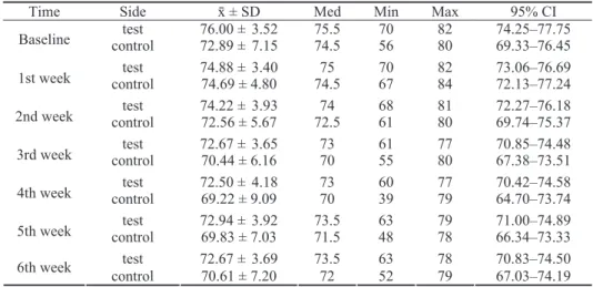
Within the test group significant changes were recorded during a 6-week follow-up (p = 0.016) (Table 1, Figure 4).
The maximum stability was achieved at baseline and after-wards significantly declined in the 2nd, 3rd and 4th week (p
= 0.029; p = 0.007; p = 0.008; respectively) with the minimal recorded value in the 4th week. In the 5th week it started to rise insignificantly, but fell again in the 6th week, in both
ob-Table 1 Descriptive statistics for implant stability measurements by means of resonance frequency analysis in test (irradiated) and
control (non-irradiated) implants at baseline and during six postoperative weeks
Time Side ґ ± SD Med Min Max 95% CI
test 76.00 ±3.52 75.5 70 82 74.25–77.75
Baseline control 72.89 ±7.15 74.5 56 80 69.33–76.45
test 74.88 ±3.40 75 70 82 73.06–76.69
1st week control 74.69 ± 4.80 74.5 67 84 72.13–77.24
test 74.22 ±3.93 74 68 81 72.27–76.18
2nd week control 72.56 ± 5.67 72.5 61 80 69.74–75.37
test 72.67 ±3.65 73 61 77 70.85–74.48
3rd week control 70.44 ± 6.16 70 55 80 67.38–73.51
test 72.50 ±4.18 73 60 77 70.42–74.58
4th week control 69.22 ± 9.09 70 39 79 64.70–73.74
test 72.94 ±3.92 73.5 63 79 71.00–74.89
5th week control 69.83 ± 7.03 71.5 48 78 66.34–73.33
test 72.67 ±3.69 73.5 63 78 70.83–74.50
6th week
control 70.61 ± 7.20 72 52 79 67.03–74.19 The results are presented as implant stability quotient values.
CI – confindence interval.
Vol. 72, No. 3 VOJNOSANITETSKI PREGLED Page 237
servation points still being significantly lower than the base-line stability (p = 0.017; p = 0.005; respectively). The differ-ences in ISQ values between both consecutive weeks within the test group were not significant (p > 0.05).
Fig. 4 – Effect of low-level laser therapy on implant stability measured by resonance frequency analysis.
In the control group significant changes in implant sta-bility over time were revealed (p = 0.023) (Table 1, Figure 4). The maximum implant stability was achieved in the 1st week, and afterwards significantly decreased in the consecu-tive 2nd and 3rd week (p = 0.047; p = 0.044; respectively).
An insignificant decrease continued in the 4th week (p =
0.234), when the minimum value was recorded and was sig-nificantly lower than baseline stability (p = 0.039). After-wards it started to rise insignificantly during the 5th and 6th consecutive weeks (p = 0.401; p = 0.110; respectively) with ISQ values recorded in the 5th week being significantly low-er compared to baseline stability (p = 0.029) whereas stabil-ity recorded in the 6th week was insignificantly different compared to baseline (p = 0.074).
Between group comparative analysis revealed higher ISQ values in the test group compared to the controls
dur-ing the entire 6-week observation period with the differ-ence being statistically significant in the 5th week (p = 0.030) (Table 2). The highest implant stability was re-corded at baseline, in the test group. Both groups showed the "stability dip" (with the lowest ISQ values) in the 4th week, with the minimal recorded ISQ value in the control group (Figure 4).
Alkaline-phosphatase activity
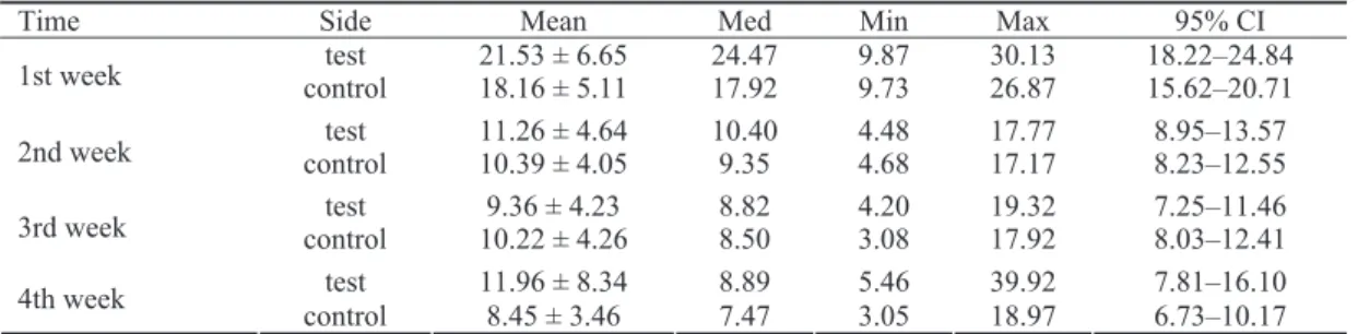
Within the test group, statistically significant changes of ALP activity were observed during the 4-week observa-tion period (p < 0.0005) (Table 3, Figure 5). The highest ALP activity was recorded in the 1st week and afterwards significantly decreased in the 2nd week (p≤ 0.005). An in-significant decrease continued from the 2nd week till the 3rd week (p = 0.175) followed by an insignificant increase re-corded in the 4th week (p = 1.000). The ALP activity value in each observation point (2nd, 3rd and 4th week) was
sig-nificantly lower than in the 1st postoperative week (p ≤
0.0005; p≤ 0.0005; p = 0.010; respectively).
Fig. 5 – Effect of low-level laser therapy on alkaline phosphatase (ALP) activity in peri-implant crevicular fluid,
measured spectrophotometrically during a 4-week observation period.
Table 2 Differences in implant stability between irradiated (test ) and non-irradiated (control) implants
Implant stability quotient (ґ ± SD) Time
test control 95% CI for MD
p
Baseline 76.00 ± 3.52 72.89 ± 7.15 -0.78177 to 7.00399 0.110 1st week 74.88 ± 3.40 74.69 ± 4.80 -3.45378 to 3.82878 0.914 2nd week 74.22 ± 3.93 72.56 ± 5.67 -1.73616 to 5.06950 0.316 3rd week 72.67 ± 3.65 70.44 ± 6.16 -0.88360 to 5.32805 0.150 4th week 72.50 ± 4.18 69.22 ± 9.09 -0.72534 to 7.28089 0.102 5th week 72.94 ± 3.92 69.83 ± 7.03 0.34554 to 5.87668 0.030* 6th week 72.67 ± 3.69 70.61 ± 7.20 -0.60045 to 4.71157 0.121 MD– mean difference; *p values (paired samples t-test) – statistically significant;
CI – confidence interval.
Table 3 Descriptive statistics for alkaline phosphatase activity assayed spectrophotometrically in test (irradiated)
and control (non-irradiated) implants during four week observation period.
Time Side Mean Med Min Max 95% CI
test 21.53 ± 6.65 24.47 9.87 30.13 18.22–24.84
1st week control 18.16 ± 5.11 17.92 9.73 26.87 15.62–20.71
test 11.26 ± 4.64 10.40 4.48 17.77 8.95–13.57
2nd week control 10.39 ± 4.05 9.35 4.68 17.17 8.23–12.55
test 9.36 ± 4.23 8.82 4.20 19.32 7.25–11.46
3rd week control 10.22 ± 4.26 8.50 3.08 17.92 8.03–12.41
test 11.96 ± 8.34 8.89 5.46 39.92 7.81–16.10
4th week
In the control group ALP activity values significantly changed during the 4-week follow-up (p < 0.0005) (Table 3, Figure 5). The maximum ALP activity was recorded in the 1st postoperative week and then continuously declined until the end of the 4th week. This decline was significant in the 2nd, 3rd and 4th week (p = 0.006; p = 0.003; p < 0.0005; re-spectively) in comparison with the 1st one. The decrease in ALP activity between the 1st and the 2nd week was statisti-cally significant (p = 0.006) whereas no significant differ-ence in ALP activity was observed between the 2nd and 3rd week (p = 1.000), neither between the 3rd and 4th postopera-tive week (p = 0.743).
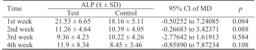
The mean ALP values were higher in the test group during a 4-week follow-up, except in the 3rd postoperative week, but the difference between the groups was not statisti-cally significant at any time of observation (Table 4). The
pattern of ALP activity changes over time was different in the test and control groups (Figure 5). After the initial de-cline of ALP activity in the test group an increase in the 4th week was observed reaching values similar to those of the 2nd week (p = 1.000), whereas in the control group a con-tinuous decrease was recorded.
Early implant success
The early implant success rate after the first six weeks (prior to implant placement) was 100%, regardless of LLLT usage. No adverse event was recorded during the follow-up.
Discussion
Osseointegration is an essential prerequisite for the dental implants' long-term prognosis. Therefore, chemical, biological and biophysical adjunctive therapies to improve and accelarate healing at bone-implant interface have been
widely investigated 18. This randomized, double blind,
split-mouth clinical study was focused on the effect of postoperative LLLT using a 637 nm GaAlAs laser with an
output power of 40 mW and total irradiation dose per
treatment of 6.26 J/cm² per implant, on osseointegration of self-tapping implants placed into posterior maxilla. Our in-tention was to explore this effect on bone healing after den-tal implant placement in the maxillary premolar and/or mo-lar region, being the area of the least predictible success of implant therapy 16, where the use of LLLT might be of ma-jor clinical relevance. The results of our study suggest that LLLT did not significantly affect the osseointegration of
self-tapping implants placed into low density bone of pos-terior maxilla.
A 637 nm GaAlAs laser has been chosen due to its ben-eficial effects on bone regeneration reported in animal 3 and
clinical studies 4. LLLT has been found to increase
os-teoblastic proliferation, collagen deposition, and bone neo-formation in the irradiated comparing to non-irradiated bone 3, 9. Studies using animal models and human osteoblast-like cells cultures, demonstrated that the use of low-level la-ser after titanium implant inla-sertion promoted osseointegra-tion due to rapid bone turnover 7, 12 and seemed to accelerate active bone replacement without causing tissue or implant
damage 7. Histomorphometric evaluation in animal models
revealed more bone-implant contact in the irradiated groups as compared to the controls at 3 and 6 19 and 16 weeks post-operatively 20. These results suggest that LLLT may
stimu-late bone repair, affecting cellular proliferation, differentia-tion and adhesion 7–14, 19, 20.
Table 4 Differences in alkaline phosphatase (ALP) activity between irradiated (test)
and non-irradiated (control) implants
ALP (ґ ± SD) Time
Test Control 95% CI of MD p
1st week 21.53 ± 6.65 18.16 ± 5.11 -0.50252 to 7.24085 0.084 2nd week 11.26 ± 4.64 10.39 ± 4.05 -0.26683 to 3.42371 0.088 3rd week 9.36 ± 4.23 10.22 ± 4.26 -2.77642 to 1.61913 0.584 4th week 11.9 ± 8.34 8.45 ± 3.46 -0.85890 to 7.87234 0.108 ALP activity is presented in U/L; MD – mean difference; p- values (paired samples t-test)
CI – confidence interval.
In this study osseointegration was evaluated through its two indicators – secondary implant stability measured by means of RFA and ALP activity assayed spectrophotometri-cally. Secondary implant stability is a clinical reflection of cellular events in peri-implant healing department and there-fore indicates the rate and extent of osseointegration 21. We used RFA as a non-invasive method that has proved to be a reliable tool to assess implant stability, determine different healing phases of dental implants and predict success of im-plant treatment 21. Longitudinal ISQ values in both groups followed the usual pattern of changes with "stability dip" in the 4th postoperative week that reflected bone remodeling process when primary spongiosa was being replaced with lamellar and/or parallel-fibered bone 16, 22. The trend of higher ISQ values recorded in the test group compared to controls during the entire 6-week period of observation, reached a significant difference in the 5th postoperative week. This result might suggest biomodulatory effect of LLLT that increases cellular activity and bone apposition but still not clinically significant to provide an earlier and better anchorage of implants. Statistically significant regeneration of bone tissue around irradiated implants was recorded in an intermediate period, which was in agreement with literature data 13, 23. It has been shown that although LLLT is capable to increase the number of osteogenic cells in the very initial stage of healing, its effect on implant stabilization in this stage is still insignificant 13, 23. Conversely, previous reports of animal studies reported that postoperative LLLT improved
Vol. 72, No. 3 VOJNOSANITETSKI PREGLED Page 239
biomechanical characteristics of bone-implant interface 12–14. The authors agreed that single 14 or multisession 12, 13 LLLT was beneficial to improve bone-implant interface strength, resulting in higher values of removal torque required to de-tach bone and implant in sites previously submitted to irra-diation in comparison to non-irradiated sites 13, 14.
The only clinical study that investigated the stability of oral implants after LLLT was the study of Garcià-Morales et al. 24. Under the conditions of their study, no evidence was found of any effect of LLLT on the stability of implants when measured by RFA. The authors remarked that potential beneficial effect of LLLT was perhaps masked by high initial stability attained in the posterior mandible region 24. With regard to different irradiation protocol used in a Garcià-Morales study 24 (infrared laser with seven irradiations re-peated every 48 h for the first 14 days), as well as different implantation sites, comparison with our results is difficult.
In our study, during the whole 6-week observation pe-riod in both irradiated and non-irradiated implants, implant stability rates were high (≥ 69 ISQ), which is interesting, since the implantation site was the posterior maxilla. These results could probably be explained by the self-tapping im-plant design as has been previously demonstrated by a recent randomized clinical trial 25. Exceptionally, four implants of one male patient aged 68 inserted bilaterally into the regions of the first and the second maxillary molars failed to achieve primary stability sufficient for one stage surgery approach. Although the cause of poor implant stability remains unclear, the fact that all the implants were placed to the same patient indicates the probable systemic factor despite the incon-spicuous medical history. Regardless of the possibility of LLLT to promote the osseointegration of implants with poor primary stability demonstrated in animal model 26 we decided to cover them and exclude from the study due to concerns that weekly RFA measurements during early healing might damage weak bone-implant interface resulting in implant failure.
We compared clinical status of the implant – its stability, with the appearance of the marker of ALP in the peri-implant crevicular fluid. ALP is considered to be a marker of differen-tiated osteoblasts and their activity, as early progenitor cells do not express ALP activity but differentiate through a defined number of cell divisions to express ultimately a mature os-teoblast phenotype that is capable of bone formation 27. Our results revealed significant changes in ALP activity longitudi-nally in time, i.e. during the 4-week observation period, within both groups. The significantly enhanced ALP activity in the early stage of bone tissue healing (first postoperative week) was found in both irradiated and non-irradiated implants. As new bone formation starts as early as 1 week after implant placement when the primary bone contacts are supplemented by newly formed secondary bone contacts 28, this result may indicate an intensive osteoblastic activity around implants, i.e. bone formation. On the other hand, a subsequent decrease of ALP activity from the second week and onwards, would there-fore be the result of greater presence of differentiated cells (os-teocytes) at the implant-bone interface. However, this is
un-likely the case, as this is too early for the bone deposition pro-cess to decline. Apart from that released from osteoblasts dur-ing bone remodeldur-ing, ALP found in PICF can also derive from polymorphonuclear cells during inflamation 29 and periodontal fibroblasts during periodontal regeneration 30. Increased ALP activity in the first postoperative week is therefore more likely the result of inflammation that occurs as a physiological re-sponse to operation trauma, and which presents the first phase of osseointegrating process.
Although our results showed no statisticaly significant difference in ALP activity between the test and control group in all observation points, the pattern of ALP activity changes over time was different. In contrast to the control group where continuous decrease of ALP activity was recorded, in the test group after the initial decline, an increase was ob-served in the 4th week. The increase in ALP activity in the laser group might be interpreted as an indication of enhanced osteoblast activity and therefore, improved bone neoforma-tion and mineralizaneoforma-tion. This biochemical result was sup-ported by our clinical finding from the 5th observation week when a significantly higher stability was recorded for irradi-ated implants compared to controls, suggesting beneficial ef-fect of LLLT on osseointegration.
Previous in vitro8, 9, 11 and animal 10 studies reported on enhancements in the ALP activity as well as matrix forma-tion after LLLT, which the authors considered as an indica-tion of increased osteoblastic activity after LLLT.
Generalisation of our results might be affected by bone density, implant macro design, as well as irradiation protocol we used. In the literature, there is no consensus regarding LLLT protocol. The ideal wave length, energy density and irradiation protocol are perhaps yet to be determined. Fur-thermore, we have used self-tapping implant macro design since it has been recommended for low density bone of pos-terior maxilla in order to achieve sufficient implant stabil-ity 25. However, non self-tapping implants are not so effec-tive in providing good primary stability into spongy bone and more pronounced effect of LLLT on the healing of such implants could be expected since the effect of LLLT in our study might be masked by self-tapping design.
Conclusion
Low-level-laser therapy applied daily during the first postoperative week using a 637 nm gallium-aluminum-arsenide (GaAlAs) laser with an output power of 40 mW and total irradiation dose per treatment of 6.26 J/cm² per implant expressed no significant influence on the osseointegration of self-tapping implants placed into low density bone of poste-rior maxilla. Placement of self-tapping macro-designed im-plants into low density bone could be predictable therapeutic procedure with a high early success rate regardless the low-level laser therapy use.
Acknowledgements
R E F E R E N C E S
1. Harris DM. Biomolecular mechanism of laser biostimulation. J Clin Laser Med Surg 1991; 9(4): 277−80.
2. Stanford OT, Beirne R, Ellingsen JE. Effects of Low-Level Laser Treatment on Bone Regeneration and Osseointegration of Dental Implants. Int J Oral Maxillofac Implants 2007; 22(5): 691−5.
3. Markovic A, Kokovic V, Todorovic L. The influence of low-power laser on healing of bone defects: An experimental study. J Oral Laser Applic 2005; 5: 169−72.
4. Marković A, Todorović L. The Influence of Low-power Laser on Healing of Bone Defects after Periapical Surgery: A Clinical Study. J Oral Laser Applic 2006; 6: 163−8.
5. Pinheiro AL, Gerbi ME. Photoengineering of bone repair proc-esses. Photomed Laser Surg 2006; 24(2): 169−78.
6. Karu T. Photobiology of low-power laser effects. Health Phys 1989; 56(5): 691−704.
7. Khadra M, Lyngstadaas SP, Haanaes HR, Mustafa K. Effect of la-ser therapy on attachment, proliferation and differentiation of human osteoblast-like cells cultured on titanium implant mate-rial. Biomaterials 2005; 26(17): 3503−9.
8. Stein E, Koehn J, Sutter W, Wendtlandt G, Wanschitz F, Thurnher D, et al. Initial effects of low-level laser therapy on growth and differentiation of human osteoblast-like cells. Wien Klin Wochenschr 2008; 120(3−4): 112−7.
9. Ozawa Y, Shimizu N, Kariya G, Abiko Y. Low-energy laser irra-diation stimulates bone nodule formation at early stages of cell culture in rat calvarial cells. Bone 1998; 22(4): 347−54. 10.da Silva AP, Petri AD, Crippa GE, Stuani AS, Stuani AS, Rosa
AL, et al. Effect of low-level laser therapy after rapid maxillary expansion on proliferation and differentiation of osteoblastic cells. Lasers Med Sci 2012; 27(4): 777−83.
11.Abramovitch-Gottlib L, Gross T, Naveh D, Geresh S, Rosenwaks S, Bar I, et al. Low level laser irradiation stimulates osteogenic phenotype of mesenchymal stem cells seeded on a three-dimensional biomatrix. Lasers Med Sci 2005; 20(3−4): 138−46. 12.Khadra M, Rønold HJ, Lyngstadaas SP, Ellingsen JE, Haanaes HR.
Low-level laser therapy stimulates bone-implant interaction: an experimental study in rabbits. Clin Oral Implants Res 2004; 15(3): 325−32.
13.Maluf AP, Maluf RP, da Brito CR, França FM, de Brito RB. Me-chanical evaluation of the influence of low-level laser therapy in secondary stability of implants in mice shinbones. Lasers Med Sci 2010; 25(5): 693−8.
14.Boldrini C, de Almeida JM, Fernandes LA, Ribeiro FS, Garcia VG, Theodoro LH, et al. Biomechanical effect of one session of low-level laser on the bone-titanium implant interface. Lasers Med Sci 2013; 28(1): 349−52.
15.Lekholm U, Zarb GA. Patient selection and preparation. In:
Branemark PI, Zarb GA, Albrektsson T, editors. Tissue-Integrated Prostheses: Osseointegration in clinical dentistry. 1st ed. Chicago: Quintessence; 1985. p. 199−210.
16.BischofM, Nedir R, Szmukler-Moncler S, Bernard J, Samson J. Im-plant stability measurement of delayed and immediately loaded implants during healing. Clin Oral Implants Res 2004; 15(5): 529−39.
17.Buser D, Weber HP, Lang NP. Tissue integration of non-submerged implants. 1-year results of a prospective study with
100 ITI hollow-cylinder and hollow-screw implants. Clin Oral Implants Res 1990; 1(1): 33−40.
18.Mavrogenis AF, Dimitriou R, Parvizi J, Babis GC. Biology of im-plant osseointegration. J Musculoskelet Neuronal Interact 2009; 9(2): 61−71.
19.Pereira CL, Sallum EA, Nociti FH, Moreira RW. The effect of low-intensity laser therapy on bone healing around titanium implants: a histometric study in rabbits. Int J Oral Maxillofac Implants 2009; 24(1): 47−51.
20.Jakse N, Payer M, Tangl S, Berghold A, Kirmeier R, Lorenzoni M. Influence of low-level laser treatment on bone regeneration and osseointegration of dental implants following sinus aug-mentation. An experimental study on sheep. Clin Oral Im-plants Res 2007; 18(4): 517−24.
21.Meredith N. Assessment of implant stability as a prognostic de-terminant. Int J Prosthodont 1998; 11(5): 491−501.
22.Huwiler MA, Pjetursson BE, Bosshardt DD, Salvi GE, Lang NP. Resonance frequency analysis in relation to jawbone character-istics and during early healing of implant installation. Clin Oral Implants Res 2007; 18(3): 275−80.
23.Lopes CB, Pinheiro AL, Sathaiah S, Duarte J, Cristinamartins M. Infrared laser light reduces loading time of dental implants: a Raman spectroscopic study. Photomed Laser Surg 2005; 23(1): 27−31.
24.Garcia-Morales JM, Tortamano-Neto P, Todescan FF, de Andrade JC, Marotti J, Zezell DM. Stability of dental implants after irradia-tion with an 830-nm low-level laser: a double-blind random-ized clinical study. Lasers Med Sci 2012; 27(4): 703−11. 25.Marković A, Calvo-Guirado JL, Lazić Z, Gómez-Moreno G, Ćalasan
D, Guardia J, et al. Evaluation of primary stability of self-tapping and non-self-self-tapping dental implants. A 12-week clini-cal study. Clin Implant Dent Relat Res 2013; 15(3): 341−9. 26.Campanha BP, Gallina C, Geremia T, Loro RC, Valiati R, Hubler
R, et al. Low-level laser therapy for implants without initial stability. Photomed Laser Surg 2010; 28(3): 365−9.
27.Owen TA, Aronow M, Shalhoub V, Barone LM, Wilming L, Tassi-nari MS, et al. Progressive development of the rat osteoblast phenotype in vitro: reciprocal relationships in expression of genes associated with osteoblast proliferation and differentia-tion during formadifferentia-tion of the bone extracellular matrix. J Cell Physiol 1990; 143(3): 420−30.
28.Berglundh T, Abrahamsson I, Lang NP, Lindhe J. De novo alveolar bone formation adjacent to endosseous implants. Clin Oral Implants Res 2003; 14(3): 251−62.
29.Plagnat D, Giannopoulou C, Carrel A, Bernard J, Mombelli A, Belser UC. Elastase, alpha2-macroglobulin and alkaline phosphatase in crevicular fluid from implants with and without periimplan-titis. Clin Oral Implants Res 2002; 13(3): 227−33.
30.Groeneveld MC, van den Bos T, Everts V, Beertsen W. Cell-bound and extracellular matrix-associated alkaline phosphatase activ-ity in rat periodontal ligament. Experimental Oral Biology Group. J Periodont Res 1996; 31(1): 73−9.
Received on December 2, 2013. Accepted on February 6, 2014. OnLine-First November, 2014.