Tailor-Made Ezrin Actin Binding Domain to
Probe Its Interaction with Actin
In-Vitro
Rohini Shrivastava1, Darius Köster2, Sheetal Kalme1, Satyajit Mayor2, Muniasamy Neerathilingam1*
1Protein Technology Core, Centre for Cellular and Molecular Platforms NCBS-TIFR, GKVK Post, Bangalore, India,2NationalCentre for Biological Sciences Tata Institute of Fundamental Research GKVK, Bangalore, India
Abstract
Ezrin, a member of the ERM (Ezrin/Radixin/Moesin) protein family, is an Actin-plasma membrane linker protein mediating cellular integrity and function.In-vivostudy of such
inter-actions is a complex task due to the presence of a large number of endogenous binding partners for both Ezrin and Actin. Further, C-terminal actin binding capacity of the full length Ezrin is naturally shielded by its N-terminal, and only rendered active in the presence of Phosphatidylinositol bisphosphate (PIP2) or phosphorylation at the C-terminal threonine. Here, we demonstrate a strategy for the design, expression and purification of constructs, combining the Ezrin C-terminal actin binding domain, with functional elements such as fu-sion tags and fluorescence tags to facilitate purification and fluorescence microscopy based studies. For the first time, internal His tag was employed for purification of Ezrin actin bind-ing domain based onin-silicomodeling. The functionality (Ezrin-actin interaction) of these
constructs was successfully demonstrated by using Total Internal Reflection Fluorescence Microscopy. This design can be extended to other members of the ERM family as well.
Introduction
Cytoskeleton proteins are involved in several cellular processes such as maintaining cell shape, motility or cell division and provide structural support to the plasma membrane [1,2]. Actin, one such cytoskeleton protein, is highly conserved and abundantly available in eukaryotic cells [3]. Particularly, cortical actin, which underlies the inner surface of the plasma membrane, is important for various membrane related processes like endocytosis, migration and signal trans-duction from the surface to the interior of the cell. Cortical actin is connected to the plasma membrane through linker proteins such as, Ezrin, Radixin and Moesin (ERM). The common structure of these linker proteins comprise a) an N-terminus membrane binding FERM do-main {band 4.1 protein(F), Ezrin(E), Radixin(R) and Moesin(M)}, b) an intermediary dodo-main and c) a C-terminus F-Actin binding domain [4]. Here, the FERM domain binds to the plasma membrane and the C-terminal F-actin binding domain interacts with actin to form ternary complexes, which are involved in signal transduction. However, studying these interactions
OPEN ACCESS
Citation:Shrivastava R, Köster D, Kalme S, Mayor S, Neerathilingam M (2015) Tailor-Made Ezrin Actin Binding Domain to Probe Its Interaction with Actin In-Vitro. PLoS ONE 10(4): e0123428. doi:10.1371/ journal.pone.0123428
Academic Editor:Vladimir N. Uversky, University of South Florida College of Medicine, UNITED STATES
Received:January 28, 2015
Accepted:March 3, 2015
Published:April 10, 2015
Copyright:© 2015 Shrivastava et al. This is an open access article distributed under the terms of the Creative Commons Attribution License, which permits unrestricted use, distribution, and reproduction in any medium, provided the original author and source are credited.
Data Availability Statement:All relevant data are within the paper and its Supporting Information files.
Funding:This study was supported by Indo Finland and TIFR, Department of Biotechnology, India.
in-vivois difficult due to situational association of ERM proteins like Ezrin to multiple proteins in the cell [1]. To overcome this, interactions between plasma membrane-Ezrin-actin ternary complexes are analyzedin-vitrousing supported lipid bilayers (SLB) [5,6]. One of the primary advantages of using SLBs is the two-dimensional platform of this model membrane system that readily allows protein-membrane interactions to be imaged and characterized with a small amount of protein [7,8].
Full length Ezrin is normally in a dormant confirmation masking the C-terminal actin bind-ing domain with its own N-terminal FERM domain [9,10] resulting in the absence of interac-tion with Actin [11]. Conformational change in full length Ezrin occurs after addition of Phosphatidylinositol bisphosphate (PIP2) [4,5] which binds to the N terminal of the full length Ezrin and thus promotes binding to actin. In an alternative approach, we chose to employ the Ezrin C-terminal domain alone in combination with a lipid anchor 10xHis and a fluorescent tag in order to facilitatein vitrostudies with Total Internal Reflection Fluorescence (TIRF) mi-croscopy. In particular, we present a design comprising a combination of affinity tags that allow high level expression and affinity purification of the Ezrin actin binding domain (ABD) without losing its functionality. We tested two constructs, viz., 1) 10xHis-Yellow Fluorescent Protein (YFP)-ezrinABD with Glutathione S-transferase (GST) and 2) 10xHis-Lysine-Cystine-Lysine (KCK)-ezrinABD with Maltose Binding Protein (MBP) for expression, solubility, purity and subsequent functionality test by TIRF microscopy (S1andS2Figs). N-terminus membrane binding FERM domain was replaced by 10xHis in above mentioned constructs. While con-struct1 was fluorescently labeled with YFP and called bright ezrinABD, construct 2 has a non-fluorescent KCK sequence instead, which we refer to as dark ezrinABD (Fig 1). By demonstrat-ing this successfully, we show how a strategic combination of affinity tags can be instrumental
Fig 1. Schematic representation for generation of bright ezrinABD and dark ezrnABD.(a) Step 1. Amplification of templates for splicing overlap PCR, Step 2. Product of splicing overlap PCR (S3 Fig YFP-ezrinABD), Step 3. Addition of 6xHis at the N-terminal of YFP-ezrinABD (S4A Fig), Step 4. Addition of 10xHis at the N-terminal of YFP-ezrinABD (S4B Fig), (b) Step 1. Amplification of KCK-ezrinABD, Step 2. Addition of 6xHis at the N-terminal of ezrinABD (S4C Fig), Step 3. Addition of 10xHis at the N-terminal of KCK-ezrinABD (S4D Fig).
for large scale purification of complex, multifunctional protein constructs without addition of PIP2 or phosphorylation.
Materials and Methods
Generation of constructs with fusion tags
YFP was amplified using forward primer YFP I, reverse primer YFP I, and spliced with Ezrin, which was amplified using forward primer Ezrin I, reverse primer Ezrin I (S1 Table). The re-sulting PCR product (YFP-ezrinABD) (S3 Fig) was gel purified and digested withNdeIand
BamHI. This insert was cloned into pET-15b vector containing 6xHis at the N-terminal, using
NdeIandBamHIsites. 6xHis-YFP-ezrinABD was confirmed by restriction digestion and se-quencing. To create constructs bearing 10xHis tag, YFP-ezrinABD with 6xHis was amplified with forward and reverse primers containing additional 4xHis residues. The resulting product (bright ezrinABD) was purified and ligated into pGEX-4T-1 (Fig 1A) and pMAL-c5X. Clones were confirmed by restriction digestion and sequencing. Similarly, KCK-ezrinABD (dark ezri-nABD) construct was PCR amplified, 10xHis was added at N-terminal and cloned into vector pMAL-c5X (Fig 1B) and pGEX-4T-1.
Expression and purification of recombinant constructs
E coliBL21 (DE3) transformants containing GST tagged bright ezrinABD were grown in 2xYT media containing ampicillin (100μg/ml) to an OD600of 0.4–0.6 at 37°C. Further, cultures were
induced with 0.3 mM IPTG and grown at 18°C overnight. Post-induction, the cells were har-vested by centrifugation and resuspended in 50 mM NaH2PO4,150 mM NaCl,10 mM
imidaz-ole,1 mMβ-Mercaptoethanol pH 7.5 and lysed by sonication (15 seconds on, 30 second off; 10 Cycles). This was followed by centrifugation at 13,000 ×gfor 20 min at 4°C twice. The superna-tant was loaded onto a Ni2+-nitrilotriacetic acid column(Ni-NTA) (GE healthcare), which was washed with a gradient of 50 mM NaH2PO4, 150 mM NaCl, 10 mM imidazole, 1mMβ
-Mercap-toethanol and 50 mM NaH2PO4, 300 mM NaCl, 50 mM imidazole, 1 mMβ-Mercaptoethanol,
pH 7.5 spanning 20 Column Volume (20 CV). The GST tagged bright ezrinABD protein was eluted using linear gradient of buffer containing 50 mM NaH2PO4, 300 mM NaCl,10 mM
imid-azole, 1 mMβ-Mercaptoethanol and 50 mM NaH2PO4, 150 mM NaCl, 500 mM imidazole, 1
mMβ-Mercaptoethanol pH 7.5 spanning 10 CV. Further, the protein was concentrated and buffer exchanged using 30 kDa Amicon (Millipore) and quantified using Nanovue plus (GE healthcare). GST tag was cleaved using 10 U/mg of thrombin for 8–10hr at 4°C.
Similarly, MBP-tagged dark ezrinABD was transformed intoE.coliBL21 (DE3) star strain and expression was induced using 0.4 mM IPTG at 30°C. Post induction, the harvested cells were resuspended in 50 mM NaH2PO4, 150 mM NaCl, 10 mM imidazole, and 1 mMβ
-mer-captoethanol pH 7.5 and sonicated. Further, purification protocol was performed similar to GST tagged bright ezrinABD and MBP-tag was cleaved using Factor Xa.
Size exclusion chromatography (SEC) analysis
Purification of GST tagged and MBP-tagged proteins
GST tagged bright ezrinABD was purified using GSTrap HP prepacked column (5 ml), which was equilibrated with 50 mM NaH2PO4, 150 mM NaCl, 1 mM DTT pH 7.4 and washed in
buffer containing 50 mM NaH2PO4, 300 mM NaCl, 1 mM DTT pH 7.4. The elution of GST
tagged bright ezrinABD was done with 50 mM Tris-Cl, 10mM Glutathione, and 1 mM DTT pH 7.4.
For MBP-tagged dark ezrinABD, dextrin sepharose beads (2 ml) were used. The beads were equilibrated with equilibration buffer (20 mM Tris-Cl, 200 mM NaCl, 1 mM EDTA and 1mM DTT pH 7.4) and the lysate was loaded on to the column. This was followed by washing with equilibration buffer and eluted with 10 mM Maltose.
Supported lipid bilayers formation
Supported lipid bilayers (SLBs) were formed by fusion of Small Unilamelar Vesicles (SUV) with a lipid composition of 98%PC (phosphatidylcholine (99%) (Egg, Chicken), Avanti Polar Lipids, Alabaster, AL, USA) and 2% DGS-Ni-NTA (18:1 DGS-NTA (Ni2+1, 2-dioleoyl-sn-gly-cero-3-(N-(5-amino-1-carboxypentyl) iminodiacetic acid) succinyl) (nickel salt), Avanti Polar Lipids, Alabaster, AL, USA) on freshly cleaned silica glass slides (Lab-Tek II#1.5 chambered coverglass, 8 well (Thermo Fisher Scientific, USA)). First, lipids were mixed in chloroform, dried with nitrogen gas followed by 3 hours in a vacuum desiccator, and re-hydrated in SUV buffer (20 mM HEPES, 150 mM NaCl, 5%sucrose, pH 7.2) to a final lipid concentration of 4 mM. After the lipid mix underwent 10 cycles of freeze-thaw, SUVs were formed by the extru-sion method using a membrane with 80 nm diameter holes (Mini-Extruder, Avanti Polar Lip-ids, Alabaster, AL, USA). Glass slides were immersed in 2% Hellmanex III solution for 30 min at 50°C and rinsed thoroughly in MilliQ water. Further, it was immersed in 3M NaOH for 15 min, rinsed thoroughly in MilliQ water and blow dried with nitrogen gas. For SLB forma-tion, 15μl SUV mix was added to 100μl SLB formation buffer (150 mM NaCl, 20 mM HEPES,
pH 5.5) in each well and incubated for 20 min followed by thorough washing with SLB buffer (20 mM HEPES, 150 mM NaCl, pH 7.2). For F-actin binding experiments, SLB buffer was ex-changed with KMEH (50 mM KCl, 1 mM MgCl2, 1 mM EGTA, 20 mM HEPES, pH 7.2).
Actin polymerization
Actin was purified from chicken breast following the protocol from Spudich and Watt [12] and labeled with NHS-Cy3 as described by Kellogg et.al [13]. The chicken breast was purchased in a chicken shop close to our laboratory (Venky Chicken Center, D Rajgopal Road, Kodigehali, Bangalore, India (geographic coordinates: 13.061640 latitude, 77.574763 longitude)) which freshly and professionally butchers chicken originating from a local farmer, and was directly used for actin and myosin extraction. 10μM G-actin (10% Cy3 labeled) was polymerized in
20μl of polymerization buffer (50 mM KCl, 1 mM MgCl2, 1 mM EGTA, 20 mMβ
-mercapto-ethanol, 1 mg/ml BSA, 1 mM ATP, 20 mM HEPES, pH 7.2) for 30 min at RT, then added to the SLB containing well filled with 100μl KMEH. Final F-actin concentration on the SLB was
400 nM. 0.1–1.2 nM of bright ezrinABD or dark ezrinABD was added to the SLB, and binding
of F-actin was checked with TIRF microscopy.
Imaging and data analysis
measurements were made at 20°C. Images were collected at a fixed magnification using 100 × 1.49NA objective lens, yielding a pixel size corresponding to 154 nm. F-actin and bright ezrinABD images were acquired with 200 ms exposure time at minimal laser intensities to re-duce photo bleaching.
To estimate the ratio of F-actin bound to SLBs, we analyzed the actin images with a self-de-veloped code in Matlab (MathWorks, Natick, MA, USA). Briefly, the laplacian (divergence of the gradient) of the actin intensity was computed and subjected to a threshold selecting for high negative divergence (intensity 'sinks') and isolated pixels were discarded as noise. This procedure allowed us to reliably segment pixels of the image occupied by actin filaments. F-actin occupation was then computed as the area fraction of these pixels.
In-silico studies
The structure prediction of the constructs was performed using the HMM (Hidden Markov Model) based software T-SAM 08. The structures with the highest E values were selected and were visualized using PyMOL.
Results
Large scale expression and purification of GST-tagged bright ezrinABD
and MBP-tagged dark ezrinABD
Constructs of YFP ezrinABD and KCK ezrinABD were cloned in pET-15b. These constructs were expressed in different strains ofE.colias listed inS3 Table. However, no yield was ob-served with pET-15b during the initial optimization with different IPTG concentrations and various temperatures. Bright and dark ezrinABD were cloned in both pGEX-4T-1 (containing GST) and pMAL-c5X (containing MBP) to check the influence of GST and MBP fusion tags on solubility and to improve expression. Expression of bright ezrinABD in pGEX-4T-1 resulted in soluble protein and relatively better yield than pMAL-c5X in BL21 (DE3). Although dark ezrinABD in pGEX-4T-1 expressed inE.coliBL21 (DE3) gave higher yield, it was found to be insoluble. In contrast, dark ezrinABD cloned in pMAL-c5X gave soluble protein when express-ed inE.coliBL21 (DE3) star whereas yield was lesser than that of pGEX-4T-1.
The optimum temperature and IPTG concentration for expression of GST tagged bright ezrinABD YFP-ezrinABD-construct1) and MBP-tagged dark ezrinABD (10xHis-KCK-ezrinABD-construct2) were different. For expression of GST tagged bright ezrinABD in
E.coliBL21 (DE3), 0.3 mM IPTG concentration at 18°C was employed, (Fig 2(A)). Whereas 0.4 mM IPTG concentration at 30°C was optimum for expression of MBP-tagged dark ezri-nABD inE.coliBL 21(DE3) star (Fig 3(A)).
We purified GST tagged bright ezrinABD and MBP-tagged dark ezrinABD using Glutathi-one 4B and dextrin sepharose beads, respectively. However, purification of GST tagged bright ezrinABD using Glutathione 4B beads rendered low yield (0.25 mg/ml) after purification (Fig 2 (B)). Therefore, the non-binding of GST tagged bright ezrinABD to GST beads was further analysed usingin-silicostudies. The structure of the protein was predicted and the model with the best E-value was chosen for analysis. As indicated inFig 4B, it was observed that the GST binding site (residues 33–37) was masked by the residues in both ezrinABD domain (residues
480–600) and YFP domain (residues 237–479). The residue E34 was found to be directly
ezrinABD was purified using Histidine affinity chromatography (Fig 2(C)). Further, the puri-fied protein was then subjected to thrombin cleavage with 10 U/mg of thrombin for 8–10 hr at
4°C and purified using size exclusion chromatography (Fig 2(D)). GST tag and cleaved 10xHis-YFP-Ezrin (bright ezrinABD) was separated by size exclusion chromatography as
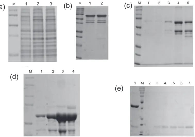
Fig 2. SDS-PAGE analysis of GST tagged bright ezrinABD.(a)12% SDS-PAGE gel showing the expression of 69 kDa protein, lane M; Protein marker, lane 1; Uninduced, lane 2; Total, lane 3; Supernatant (soluble), lane 4; Flow through, (b) GST-tagged bright ezrinABD(69 kDa) purified using GSTrap HP column, lane M; Protein marker, lane 1–6; eluted fractions, (c) GST-tagged bright ezrin purified using His Trap HP column, lane M; Protein marker, lane 1–6; eluted fractions, (d) Gel permeation of 42 kDa bright ezrinABD, lane M; Protein marker, lane 1; thrombin cleaved GST-tagged bright ezrinABD, lane 2–7; purified fractions, (e) AKTA chromatogram of Gel Permeation of bright ezrinABD after cleavage of GST-tag Peak (1) GST-tagged bright ezrinABD, Peak (2) bright ezrinABD, Peak (3) GST.
shown inFig 2(E). Bright ezrinABD was purified as monomer which indicated no intramolecu-lar interaction. The concentration of purified bright ezrinABD was 1 mg/ml after quantifica-tion with Nano plus (GE healthcare). Purificaquantifica-tion of MBP-tagged dark ezrinABD using dextrin sepharose beads showed loss of protein during wash, suggesting very less binding of MBP-tag to the sepharose. As the total protein concentration in the load (10ml) was about1mg/ml, while wash through showed concentration of 0.4mg/ml for the concentrated sample (5ml was con-centrated to 1ml) and 0.2mg/ml for the eluted sample, yielding 0.1 mg/ml of dark ezrinABD after purification (Fig 3(B and 3C)). Similar to YFP-Ezrin, 10xHis internal tag was used to puri-fy MBP-tagged dark ezrinABD (Fig 3(D)). Further, factor Xa was used to cleave MBP-tag from dark ezrinABD with 6.25 U/mg for 8 hr at 4°C and purified using gel permeation. After gel per-meation MBP was not completely separated from dark ezrinABD, consequently MBP was trapped by dextrin sepharose beads and final concentration of dark ezrinABD after purification was 1 mg/ml (Fig 3(E)). After cleavage of MBP from dark ezrinABD, interaction of free MBP with dextrin sepharose beads confirmed masking of MBP binding site in MBP-dark ezrinABD.
Fig 3. SDS-PAGE analysis of MBP-tagged dark ezrinABD.(a)12% SDS-PAGE gel showing the expression of 57 kDa protein, lane M; Protein marker, lane 1; Uninduced, lane 2; load, lane 3; Flowthrough, (b) MBP-tagged dark ezrinABD purified using Dextrin Sapharose,lane M; Protein marker, lane 1&2; Washthrough, (c) lane M; Protein marker, lane 1–5; eluted fractions, (d) MBP-tagged dark ezrinABD purified using His Trap HP column, lane M; Protein marker, lane 1–4; eluted fractions, (e) The purification of 15 kDa dark ezrinABD protein using Dextrin Sepharose, lane M; Protein marker, lane 1; Load, lane 2–7; eluted fractions.
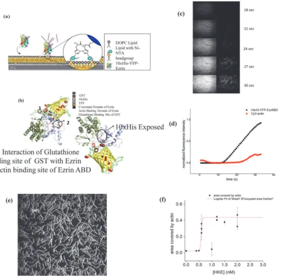
Confirmation of Ezrin functionality by TIRF
The functionality of both the ezrinABD constructs was confirmed by an actin binding assay. Here, Cy3 labeled F-actin was allowed to settle on SLBs doped with 2 mol% DGS-NiNTA lipids at different concentrations of the ezrinABD constructs, and the amount of membrane bound F-actin was estimated from the images obtained by TIRF microscopy (seeMaterials and Meth-odssection). In the absence of bright and dark ezrinABD, no F-Actin signal was observed in the TIRF field indicating that F-Actin does not bind to bare SLBs. In contrast, after addition and binding of bright and dark ezrinABD to SLBs, F-actin was rapidly detected in the TIRF field indicating ezrinABD mediated binding of F-actin to SLBs (Fig 4). This confirms the purity and functionality of bright and dark ezrinABD.
Discussion
Ezrin has three domains, out of which, two domains participate in signal transduction namely N-terminal FERM domain and C-terminal actin binding domain. Dormant state of Ezrin
Fig 4. GST. (a-d) ezrinABD interaction: (a) binding of bright ezrinABD to SLB, (b) Predicted structure of GST-tagged bright ezrinABD, (c) images from TIRF microscopy, (d) graphical presentation of cy3 Actin binding to Ezrin in different time frame;(e, f) Saturation of ezrinABD and actin binding: (e) TIRF microscopy image of f-actin binding to SLB via HKE (dark ezrinABD). (f) Plot of actin binding (area fraction covered by actin) at different concentrations of HKE; red line is a fit of a logistic function, to visualize the sharp onset of actin binding to the SLB at CHKE>0.7 nM, data points represent mean and standard
deviation of individual experiments.
involves masking of C-terminal actin binding domain by its N-terminal FERM domain, which renders inability to bind with actin. Furthermore, Ezrin interacts with other endogenous pro-teinsin-vivo, thus making analysis difficult [1] i.e. N-terminal and C-terminal are engage in in-teractions with membrane proteins and F-actin, as well as intramolecular and intermolecular interactions. Ezrin gets activated by the binding of PIP2 or by phosphorylation at the C-termi-nal threonine. When Ezrin is activated, it binds directly to the cortical actin cytoskeleton. If there is dephosphorylation of the C-terminal threonine and reduction in PIP2levels, it leads to inactivation of ERM and their release from the cell membrane [14].
To reproduce plasma membrane-Ezrin-actin interactionsin-vitro, a system that consists of a minimal set of defined components is used. The preparation of cytoskeleton-membrane link-er proteins like Ezrin forin-vitrostudies is challenging with respect to protein solubility and high yield. Forin-vitrostudy, use of microtiter plate and surface plasmon resonance technology (SPR) encountered less binding and lower solubility of recombinant Ezrin protein [15]. In our study, various fusion tags GST, MBP and 10xHis were employed to enhance the expression and solubility as shown inS2 Fig.
The supported lipid bilayer (SLB) is an experimental research design particularly useful for studies associated with plasma membrane and its related proteinsin vitro. SLB indicates struc-tural and dynamic properties like cell membrane but former is better with respect to this study as one can perform restricted set of experiments without involvement of accessory components of native membranes, still proving functionality comparable to latter. SLB can overcome the problems that come into picture because of surface sensitivity of cell membranes during mi-croscopy and surface sensitive spectroscopies [16]. Due to less understanding of suitable bind-ing partners between recombinant proteins and SLB these studies are restricted, but Nickel-Chelating lipids have been observed to bind efficiently with fusion proteins containing histi-dine tag [17]. These artificial chelating lipid like nickel also reduce the trouble in fixing the bio-logical samples. We facilitate this study by using 10xHis in both bright and dark Ezrin.
expected, the supernatant and flow through contained yellowish green fluorescence indicating properly folded glutathione binding site and YFP, and hence a properly folded protein. Howev-er, as the binding to glutathione beads was not successful, GST could not be used for purifica-tion of the bright ezrinABD. ERM has the property of dimers or oligomers formapurifica-tion with which, Ezrin can form heterotypic associations [15]. Our findings indicated that the non-bind-ing of GST could be due to masknon-bind-ing of glutathione bindnon-bind-ing site by the YFP ezrinABD region of the construct by intramolecular interaction. This was predicted by the homology modeling, where the interaction between ezrinABD and GST was elucidated (Fig 4B). In the same model, we observed that 10xHis tag is a random coil and suspected that it could be exploited for purifi-cation. Indeed, GST tagged bright ezrinABD internal 10xHis was used to purify the protein successfully by affinity purification using affinity (Ni+2column) and size exclusion chromatog-raphy (Fig 2C). The GST tag ensured the solubility, and was cleaved later with thrombin to get 10xHis-YFP-ezrinABD (bright ezrinABD). Using the internal 10xHis we also purified MBP-tagged dark ezrinABD along with MBP because of improper binding of MBP-tag with dextrin sepharose beads. MBP-tag has not been used for ezrinABD purification till now. However, in the present study MBP helped for solubility but it failed in affinity purification.
The SDS-PAGE analysis of His-affinity purified protein revealed YFP-ezrinABD, a protein of 69 kDa, to run at 75 kDa along with other impurities. A similar shift in gel mobility of GST tagged Ezrin was observed by Algrainet al. as well as Gary and Bretschert during purification of GST tagged Ezrin, which was suggested due to the presence of acidic amino acid [10,21]. As impurities were observed after affinity purification of YFP-ezrinABD, ion exchange chroma-tography was performed, however, this resulted in less yield and the impurities still persisted. It has been observed that Ezrin is susceptible for proteolysis [18] which correlates to our observa-tion during purificaobserva-tion of GST and MBP-tagged YFP-ezrinABD and KCK-ezrinABD respec-tively (data not shown).
To study the functionality of bright ezrinABD the protein was bound to SLB (doped with 2 mol% DGS-Ni-NTA), and then Cy3 labeled F-actin was added. The observed increase in fluo-rescence in the Cy3 and YFP-channels illustrate binding of F-actin to the SLB mediated by bright ezrinABD. A similar study was done by Jankeet al., where full length Ezrin protein bound onto DGS-Ni-NTA but F-actin-Ezrin binding failed as C-terminus was not free due to dormant confirmation [5]. Our study employs ezrinABD domain alone thus circumventing the problem caused by full length Ezrin. Further, TIRF microscopy confirmed ezrinABD and F-Actin interaction thus validating the availability of the F-Actin binding site as seenin-silico. The threshold concentration of dark ezrinABD for actin binding to the SLB was 0.5 nM and actin binding saturated at higher concentrations of HEK (dark ezrinABD) (Fig 4E and 4F). Use of the combination of GST and His domain to protect delicate protein domains during purifi-cation could be a useful strategy to express a variety of proteins that would otherwise lose their activity using conventional purification strategies. The present strategy is applicable for teins of the ERM family that allows the purification of their C-terminus alone and similar pro-teins containing a charged C-terminal domain.
Conclusion
of functional 10xHis-YFP-ezrinABD at high yields was successful. This design can be used for members of ERM family proteins and other proteins as well.
Supporting Information
S1 Fig. Diagram showing arrangement of the ezrinABD protein with 10xHis, GST and MBP fusion tags.
(TIF)
S2 Fig. Diagram showing arrangement of engineered protein for microscopic studies.
(TIF)
S3 Fig. Splicing overlap PCR for YFP-ezrinABD fusion.Lane M; Marker (Gene Ruler 1kb DNA Ladder), Lane 1and 2; YFP and ezrinABD respectively, Lane 3; YFP-ezrinABD (1.08kb) fusion PCR product.
(TIF)
S4 Fig. Addition of 10xHis at N-terminus of YFP-ezrinABD and KCK-ezrinABD: Agarose gel showing amplification of (a) 6xHis YFP-ezrinABD, (b) 10xHis YFP-ezrinABD, (c) 6xHis KCK-ezrinABD, (d) 10xHis KCK-ezrinABD.
(TIF)
S1 Table. List of primers used for construction of bright and dark ezrinABD.
(DOCX)
S2 Table. Plasmids used for bright and dark ezrinABD preparation.
(DOCX)
S3 Table. Constructs tried for expression in different strains ofE.coli.
(DOCX)
S1 Text. Reagents.
(DOCX)
Acknowledgments
We thank to members of Protein Technology Core, Center for Cellular And Molecular Plat-forms (C-CAMP) and National Center for Biological Sciences for supporting this research work. Special thanks to Mrs Sneha Bairy for helping the structure prediction modeling and Mr. Sumukh Mysore for figure preparation. We also like to thank to Prof. Ramaswamy.S, The CEO and Dr.Taslimarif Saiyed, COO, C-CAMP for their support.
Author Contributions
Conceived and designed the experiments: MN SM. Performed the experiments: SK RS DK. An-alyzed the data: SK RS SM DK MN. Contributed reagents/materials/analysis tools: MN SM. Wrote the paper: SK RS SM DK MN.
References
1. Bretscher A, Reczek D, Berryman M. Ezrin: a protein requiring conformational activation to link microfil-aments to the plasma membrane in the assembly of cell surface structures. J. Cell Sci. 1997 Dec; 110: 3011–8. PMID:9365271
3. Ballestrem C, Wehrle-Haller B, Imhof BA. Actin dynamics in living mammalian cells. J. Cell Sci. 1998 Jun; 111: 1649–58. PMID:9601095
4. Maniti O, Carvalho K, Picart C. Model membranes to shed light on the biochemical and physical proper-ties of ezrin / radixin / moesin. Biochime 2013 Jan; 95: 3–11.
5. Janke M, Herrig A, Austermann J, Gerke V, Steinem C, Janshoff A. Actin binding of ezrin is activated by specific recognition of PIP2-functionalized lipid bilayers. Biochemistry 2008 Mar 25; 47: 3762–9. doi: 10.1021/bi702542sPMID:18302339
6. Herrig A, Janke M, Austermann J, Gerke V, Janshoff A, Steinem C. Cooperative adsorption of ezrin on PIP2-containing membranes. Biochemistry 2006 Oct 31; 45: 13025–34. PMID:17059219
7. Galush WJ, Nye JA, Groves JT. Quantitative fluorescence microscopy using supported lipid bilayer standards. Biophys. J. 2008 Sep; 95: 2512–9. doi:10.1529/biophysj.108.131540PMID:18515392
8. Mingeot-Leclercq MP, Deleu M, Brasseur R, Dufrêne YF. Atomic force microscopy of supported lipid
bi-layers. Nat. Protoc. 2008 Oct 2; 3: 1654–9. doi:10.1038/nprot.2008.149PMID:18833202
9. Garbett D, Bretscher A. PDZ interactions regulate rapid turnover of the scaffolding protein EBP50 in mi-crovilli. J. Cell Biol. 2012 Jul 23; 198: 195–203. doi:10.1083/jcb.201204008PMID:22801783
10. Gary R, Bretscher A. Ezrin self-association involves binding of an N-terminal domain to a normally masked C-terminal domain that includes the F-actin binding site. Mol Biol Cell. 1995 Aug; 6: 1061–75. PMID:7579708
11. Fehon RG, McClatchey AI, Bretscher A. Organizing the cell cortex: the role of ERM proteins. Nat Rev Mol Cell Biol. 2010 Apr; 11: 276–87. doi:10.1038/nrm2866PMID:20308985
12. Spudich JA, Watt S. The regulation of rabbit skeletal muscle contraction. I. Biochemical studies of the interaction of the tropomyosin-troponin complex with actin and the proteolytic fragments of myosin. J. Biol. Chem. 246, 4866–71 (1971). PMID:4254541
13. Kellogg DR, Mitchison TJ, Alberts BM. Behaviour of microtubules and actin filaments in living Drosophi-la embryos. Development 103, 675–86 (1988).
14. Hao JJ, Liu Y, Kruhlak M, Debell KE, Rellahan BL, Shaw S. Phospholipase C-mediated hydrolysis of PIP2 releases ERM proteins from lymphocyte membrane. J. Cell Biol. 2009 Feb 9; 184:451–62. doi: 10.1083/jcb.200807047PMID:19204146
15. Roy C, Martin M, Mangeat P. A Dual Involvement of the Amino-terminal Domain of Ezrin in F-and G-actin Binding. J Biol Chem. 1997 Aug 8; 272: 20088–20095. PMID:9242682
16. Nye JA, Groves JT. Kinetic control of histidine-tagged protein surface density on supported lipid bilay-ers. Langmuir 2008 Apr 15; 24: 4145–9. doi:10.1021/la703788hPMID:18303929
17. Kubalek EW, Le Grice SF, Brown PO. Two-dimensional crystallization of histidine-tagged, HIV-1 re-verse transcriptase promoted by a novel nickel-chelating lipid. J Struct Biol. 1994 Sep-Oct; 113:117–23 PMID:7536435
18. Andréoli C, Martin M, Le Borgne R, Reggio H, Mangeat P. Ezrin has properties to self-associate at the plasma membrane. J. Cell Sci. 1994 Sep; 107 9: 2509–21. PMID:7844168
19. Elliott BE, Meens J A, SenGupta SK, Louvard D, Arpin M. The membrane cytoskeletal crosslinker ezrin is required for metastasis of breast carcinoma cells. Breast Cancer Res. 2005 March 21; 7: R365–73. PMID:15987432
20. Hewitt SN, Choi R, Kelley A, Crowther GJ, Napuli AJ, Van Voorish WC. Expression of proteins in Escherichia coli as fusions with maltose-binding protein to rescue non-expressed targets in a high-throughput protein-expression and purification pipeline.Acta Crystallogr. Sect. F. Struct. Biol. Cryst. Commun. 2011 Sept 1; 67: 1006–9. doi:10.1107/S1744309111022159PMID:21904041