Affects Actin Structure
Asaf Sol1, Edna Blotnick2, Gilad Bachrach1*., Andras Muhlrad1.
1Institute of Dental Sciences, The Hebrew University-Hadassah School of Dental Medicine, The Hebrew University of Jerusalem, Jerusalem, Israel,2Department of Medical Neurobiology, The Institute for Medical Research-Israel-Canada, The Hebrew University of Jerusalem, Jerusalem, Israel
Abstract
Actin exists as a monomer (G-actin) which can be polymerized to filaments) F-actin) that under the influence of actin-binding proteins and polycations bundle and contribute to the formation of the cytoskeleton. Bundled actin from lysed cells increases the viscosity of sputum in lungs of cystic fibrosis patients. The human host defense peptide LL-37 was previously shown to induce actin bundling and was thus hypothesized to contribute to the pathogenicity of this disease. In this work, interactions between actin and the cationic LL-37 were studied by optical, proteolytic and surface plasmon resonance methods and compared to those obtained with scrambled 37 and with the cationic protein lysozyme. We show that LL-37 binds strongly to CaATP-G-actin while scrambled LL-LL-37 does not. While LL-LL-37, at superstoichiometric LL-LL-37/actin concentrations polymerizes MgATP-G-actin, at lower non-polymerizing concentrations LL-37 inhibits actin polymerization by MgCl2or NaCl. LL-37 bundles Mg-F-actin filaments both at low and physiological ionic strength when in equimolar or
higher concentrations than those of actin. The LL-37 induced bundles are significantly less sensitive to increase in ionic strength than those induced by scrambled LL-37 and lysozyme. LL-37 in concentrations lower than those needed for actin polymerization or bundling, accelerates cleavage of both monomer and polymer actin by subtilisin. Our results indicate that the LL-37-actin interaction is partially electrostatic and partially hydrophobic and that a specific actin binding sequence in the peptide is responsible for the hydrophobic interaction. LL-37-induced bundles, which may contribute to the accumulation of sputum in cystic fibrosis, are dissociated very efficiently by DNase-1 and also by cofilin.
Citation:Sol A, Blotnick E, Bachrach G, Muhlrad A (2012) LL-37 Induces Polymerization and Bundling of Actin and Affects Actin Structure. PLoS ONE 7(11): e50078. doi:10.1371/journal.pone.0050078
Editor:Theresia E.B. Stradal, University of Muenster, Germany
ReceivedJuly 16, 2012;AcceptedOctober 16, 2012;PublishedNovember 26, 2012
Copyright:ß2012 Sol et al. This is an open-access article distributed under the terms of the Creative Commons Attribution License, which permits unrestricted use, distribution, and reproduction in any medium, provided the original author and source are credited.
Funding:This work was supported by the Israel Science Foundation (grant No. 208/10). The funders had no role in study design, data collection and analysis, decision to publish, or preparation of the manuscript.
Competing Interests:The authors have declared that no competing interests exist. * E-mail: [email protected]
.These authors contributed equally to this work.
Introduction
Actin is the most abundant protein in the eukaryotic cell. It is a highly dynamic structural protein with multiple functions in cell physiology which include cytoskeleton formation, cell division, motility, adhesion, signaling and more [1]. Actin exists in either monomer- globular (G) or polymer- filament (F) form. These forms can interconvert into each other by a plethora of different factors. G-actin, which is negatively charged, is polymerized into F-actin by an increase in monovalent or divalent cation concentration and by positively charged proteins and peptides.
F-actin filaments may form bundles via specific actin bundling proteins [2], which cross-link two filaments when their two discrete actin-binding sites attach to separate filaments. These proteins, including a-actinin, filamin, fimbrin, spectrin and others, form tightly packed, well organized bundles containing parallel ar-ranged filaments [3], [4]. The other group of actin bundling factors are the polycations, including polycationic proteins or peptides and polyamines. This group induces bundle formation via non-specific electrostatic interactions by eliminating repulsion between negatively charged actin filaments [5]. LL-37 [6], lysozyme [7] MARCKS [8], [9], ENA/VASP [10], fesselin [11], [12] and calponin [13], [14] all belong to this group. Polycationic polyamines such as spermine and spermidine [15], [16] and
polylysine [17] also bundle F-actin. Site specific hydrophobic interactions between actin and polycation might contribute to the bundling especially at high ionic strength, where the bundling effect of polycations decreases because the relatively high concentration of monovalent cations masks the electrostatic interactions between polycations and actin [18]. The bundling process of F-actin by lysozyme, spermine and polylysine was recently studied in some detail [19], [20].
of LL-37 attached to actin bundles was reported to be significantly reduced [6]. Gelsolin and polyanions dissolve actin bundles [30] and restore LL-379s antibacterial activity [6]. Despite these significant advances, important processes, such as polymerization of G-actin by LL-37, kinetics of bundle formation, characteriza-tion of actin-LL-37 interaccharacteriza-tion at molecular level and the reaccharacteriza-tion of LL-37-induced bundles with actin binding and severing proteins remain uncharacterized and poorly understood.
In this study, we examined the LL-37-induced polymerization of MgATP-G-actin and the bundling of Mg-F-actin, and compared them to the well characterized polymerization and bundling of actin by the antibacterial polycationic protein lysozyme [20]. We show that LL-37 polymerizes actin only at concentrations greater than twice that of actin. Low concentra-tions of LL-37, which are not sufficient to polymerize actin, inhibit actin polymerization induced by MgCl2or NaCl. LL-37-induced F-actin bundles are less sensitive to ionic strength when compared to sLL-37 (scrambled LL-37) or lysozyme induced bundles. Substoichiometric LL-37 concentrations relative to actin acceler-ate the subtilisin digestion of the protein. Our results indicacceler-ate that in addition to electrostatic interactions there are specific hydro-phobic interactions between LL-37 and actin. These interactions may involve the DNase1 binding (D) loop since DNase1 dissociates LL-37 induced F-actin bundles very efficiently.
Materials and Methods
Materials
N-(1-pyrene) maleimide was obtained from Molecular Probes (Eugene, OR). Hen lysozyme, DNase1, ATP, ADP, dithiotreitol (DTT), and EGTA were purchased from Sigma Chemical Co. (St Louis, MO). LL-37 (LLGDFFRKSKEKIGKEFKRIVQ-RIKDFLRNLVPRTES) peptide and scrambled LL-37 (sLL-37) peptide (GLKLRFEFSKIKGEFLKTPEVRFRDIKLKDN-RISVQR) were purchased from (Genemed Synthesis Inc., San Antonio,TX ). The peptides were purified by HPLC and purity (greater than 90%) was determined by Mass Spectrometry. Yeast cofilin was a generous gift from Prof. Emil Reisler Dept. of Chemistry and Biochemistry, Univ. of California Los Angeles CA.
Preparation of Actin
CaATP-G-actin was prepared from acetone dried powder derived from the back and leg muscles of rabbit by the method of Spudich and Watt [31] that even without gel filtration yields highly homogeneous actin in purity greater than 90%. CaATP-G-actin was stored in a buffer containing 5 mM TrisHCl, 0.2 mM CaCl2, 0.2 mM ATP, 0.5 mM b-mercaptoethanol, pH 8.0 (CaATP-G-buffer). MgATP-G-actin was obtained by incubating CaATP-G-actin with 0.2 mM EGTA and 0.1 mM MgCl2 at room temperature for 5 min. MgATP-G-actin was diluted for further treatments in MgATP-G-buffer containing 5 mM MOPS, 0.1 mM MgCl2, 0.2 mM EGTA, 0.2 mM ATP and 0.5 mM DTT, pH 7.4. MgF-actin was polymerized from MgATP-G-actin or CaATP-G-actin by 30 min incubation with 2 mM MgCl2 at room temperature. The concentration of un-labeled rabbit skeletal musclea-G-actin was determined spectro-photometrically using the extinction coefficients E1%290= 11.5 cm21. (The optical density of actin was measured in the presence of 0.5 M NaOH, which shifts the maximum of absorbance from 280 nm to 290 nm). Molecular masses of skeletal actin, yeast cofilin, hen-lysozyme and LL-37 were assumed to be 42 kDa, 15.9 kDa, 14.3 kDa and 4.5 kDa respectively.
Pyrene Labeling
Labeling of Mg-F-actin at Cys-374 with pyrene maleimide was carried out according to Kouyama and Mihashi [32] with some modifications. CaATP-G-actin was filtered through a PD-10 column (GE Healthcare) equilibrated withb-mercaptoethanol-free CaATP-G-buffer. After filtration, actin (1 mg/ml) was polymer-ized by 2 mM MgCl2and 100 mM KCl at room temperature for 30 min, and reacted with pyrene maleimide (16mg/ml) on ice, for one hour. The reaction was terminated with 1 mM DTT. Labeled F-actin was centrifuged at 129,1516gfor two hours, the pellet was resuspended in Ca-ATP-G-buffer and depolymerized by dialyzing in this buffer for over 36 hours at 4uC. Finally, actin was centrifuged again at 129,1516gfor two hours. The supernatant contained the purified pyrene-labeled CaATP-G-actin. The concentration of modified actin was determined by the procedure of Bradford [33] using unmodified actin as a standard. The extent of labeling, which was measured by using pyrene extinction coefficient E344 nm= 22000 cm21M21, was,100%.
Fluorescence and Light Scattering Measurements Actin polymerization was followed as an increase in fluores-cence of pyrene-labeled G-actin [32], which was added to unlabeled G-actin in 10–15%. The time course of pyrene-labeled actin polymerization was monitored by measuring fluorescence increase (with 365 nm excitation and 386 nm emission wave-lengths) in a PTI spectrofluorometer (Photon Technology In-dustries, South Brunswick, NJ). Bundling of MgF-actin was followed by increase in light scattering [20]. The time course of light scattering changes was also measured in a PTI spectroflu-orometer, with both excitation and emission wavelengths adjusted to 450 nm. All fluorescence and light scattering measurements were carried out at 22uC.
Monitoring Bundling by Low Speed Sedimentation After addition of polycations, actin samples were centrifuged at 20,8006g at 4uC for 8 min (in an Eppendorf centrifuge). The supernatants were run using SDS-PAGE and analyzed by densitometry using the TINA 2.07d software.
Binding Measurement of LL-37 or Scrambled LL-37 to G-actin by Surface Plasmon Resonance
Results
Polymerization of MgATP-G-actin by LL-37
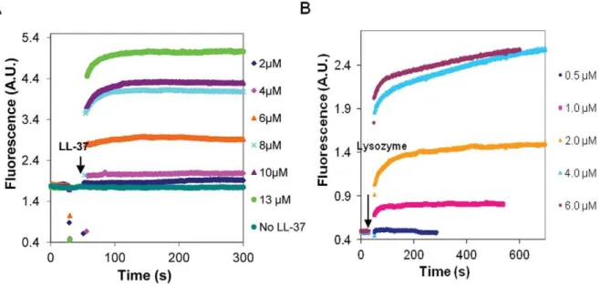
In low ionic strength MgATP-G-buffer, LL-37 was found to polymerize MgATP-G-actin (Figure 1A). The onset of polymer-ization was fast and its extent was dependent on the concentration of LL-37. Significant polymerization was observed when the concentration of LL-37 was three times higher than that of MgATP-G-actin (6mM LL-37, and 2mM actin) (Figure 1A). Lysozyme polymerized MgATP-G-actin at half of the concentra-tion of MgATP-G-actin (1mM lysozyme and 2mM actin) (Figure 1B). One may suppose that lysozyme polymerizes G-actin at lower concentrations than LL-37; because its net positive charge [34] is higher than that of LL-37 [6]. However, LL-37’s affinity to MgATP-G-actin also seems to be high as implied by the finding that LL-37 affects G-actin even when its concentration is too low to induce polymerization. Under these conditions, LL-37 inhibits polymerization induced by 2 mM MgCl2 or 100 mM NaCl (Figure 2A) while similar concentrations of lysozyme have no inhibitory effect (Figure 2B). These results may indicate that monomeric actin forms complexes with LL-37 that at low LL-37 concentrations inhibit salt-induced actin polymerization.
Bundling of MgF-actin by LL-37
Bundling kinetics of MgF-actin by LL-37, lysozyme and scrambled LL-37 were studied by increase in light scattering and the extent of bundling was determined using low speed centrifu-gation. The bundling of MgF-actin by LL-37 is also shown by electron microscopy (Figure S1). Mg-F-actin bundle formation was very fast both with LL-37 (Figure 3A) and lysozyme [20]. The extent of bundling was dependent on the concentration and the nature of the polycation used (Figure 3B). At low ionic strength more than 50% of Mg-F-actin was bundled upon addition of lysozyme (1mM) to Mg-F-actin (4mM). A similar degree of bundling was observed with LL-37 when its concentration was equal to that of actin (4mM) (Figure 3B). At low ionic strength scrambled LL-37 induced significant bundling when its concen-tration was 2.5 fold higher than that of MgF-actin (Figure 3C).
These results are similar to those obtained for MgATP-G-actin polymerization by lysozyme and LL-37. Bundling of F-actin by polycations is ionic-strength-sensitive and bundles induced by polycations dissociate (unbundle) at physiological ionic strength [20]. This is also observed with lysozyme where a six fold higher concentration of lysozyme (6mM ) was needed for half maximal bundling of 4mM Mg-F-actin at physiological (100 mM NaCl) than at low ionic strength (1mM lysozyme) (Figure 3B). With scrambled LL-37, whose net number of positive charges are identical to those of LL-37, even 14mM were insufficient to cause significant bundling in the presence of 100 mM NaCl, while at low ionic strength more than 60 percent of MgF-actin bundled at 10mM concentration of sLL-37 (Figure 3C). The results show the competing effect of ionic strength and polycation concentration on bundling. Unlike with lysozyme and sLL-37, the kinetics (Figure 3A) and the extent (Figure 3B and C) of bundling by LL-37 are practically identical at low and at physiological ionic strength. The decreased salt sensitivity of LL-37-induced bundles may indicate that the interactions between LL-37 and actin are partially electrostatic and partially hydrophobic.
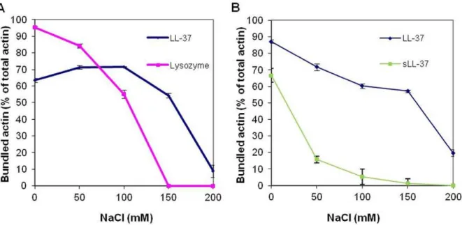
We studied the effect of NaCl concentration on the stability of bundles formed from 4mM MgF-actin, that were induced by 6mM LL-37 or lysozyme (Figure 4a), or by 10mM LL-37 or sLL-37 (Figure 4B). The LL-sLL-37-induced bundles were reasonably stable in the 0–150 mM NaCl range and dissociated only at 200 mM NaCl. In contrast, the stability of lysozyme and sLL-37 induced bundles was decreased already at 50 mM and the bundles completely dissociated in 150 mM NaCl. These results support our assumption that reduced salt sensitivity of the LL-37-induced bundles is due to hydrophobic interactions between LL-37 and actin.
Effect of LL-37 on Subtilisin Digestion of CaATP-G-, MgATP-G-actin and Mg-F-actin
Subtilisin at low concentrations cleaves G-actin between Met-47 and Gly-48 [35]. In order to cleave F-actin, about a magnitude higher concentration of subtilisin is needed. The subtilisin digestion of actin is dependent on the structure of the protein.
Figure 1. Polymerization of MgATP-G-actin by LL-37 and lysozyme at low ionic strength MgATP-G-buffer.LL-37 (A), or lysozyme (B) were added to pyrene labeled (10% labeling ratio) 2mM MgATP-G-actin and the polymerization was followed by increase in pyrene fluorescence. Presented data are representative of three independent experiments.
For example, the exchange of tightly bound ATP to ADP in the nucleotide binding cleft, significantly decreases the rate of actin cleavage by subtilisin [36]. Addition of cofilin affects MgF-actin’s structure, and strongly accelerates the digestion of actin filaments by subtilisin [37]. In light of our assumption that the binding of LL-37 to actin is partially hydrophobic, and may affect actin structure, we examined the effect of LL-37 on actin digestion by subtilisin (Figure 5). CaATP- and MgATP-G-actin (8mM) were digested by subtilisin (4mg/ml) for 2 minutes. Mg-F-actin (8mM) was digested in the presence and absence of 200 mM NaCl, by 20mg/ml subtilisin for 30 min. Digestions were carried out in the presence or absence of 6mM LL-37, which in all cases enhanced the subtilisin cleavage in a statistically significant manner (Figure 5). These results indicate that LL-37 binds both the monomer and polymer forms of actin. It also binds F-actin in the presence of 200 mM NaCl, which abolish electrostatic interactions between F-actin and LL-37 (Figure 4). Thus, the binding of LL-37 to actin is at least partially hydrophobic and induces changes in actin structure, which is manifested in increased sensitivity toward subtilisin.
Surface Plasmon Resonance Measurements of Binding of LL-37 to Immobilized G-actin
In light of the subtilisin digestion results, which showed that LL-37 binds G-actin and affects its structure, we measured the dissociation constants of LL-37 and of scrambled LL-37 from CaATP-G-actin using the SPR BIACORE approach (Figure 6). CaATP-G-actin was bound to the sensor chip at low ionic strength and washed. LL-37 or sLL-37 were applied to the surface-bound G-actin followed by washing with the 150 mM NaCl supplemen-ted CaATP-G-buffer (G-actin was not polymerized by this high ionic strength buffer, because it was bound to the chip surface). The results presented in Figure 6A show that LL-37 binds strongly to G-actin (Table 1) in the presence of 150 mM NaCl, which masks most of the electrostatic interactions between actin and LL-37. The KD value of LL-37 binding obtained in this measurement is slightly different when compared with the results of the bundling experiments (Figure 3C). This might result from the fact that the binding of LL-37 was measured to monomeric CaATP-G-actin,
while the bundling experiments were carried out with polymeric MgF-actin. sLL-37 binds very weakly to G-actin under the same conditions (Figure 6B, Table 1). These results indicate a strong, sequence dependent hydrophobic interaction between LL-37 and actin. It appears that LL-37 contains an actin binding sequence, which is perturbed by scrambling, that abolished the hydrophobic interaction.
Dissociation of LL-37-induced Mg-F-actin Bundles by DNase1 and Cofilin
Bundles of actin filaments are major contributors to the viscosity of sputum in the airways of cystic fibrosis patients. LL-37, which also has been found in the sputum, may contribute to actin bundle formation [38]. Gelsolin, an actin filament severing protein was found to significantly reduce the viscosity of sputum [30] and dissociate LL-37-induced actin filament bundles [38]. Here, we studied the effect of cofilin [39] and DNase1 on the stability of LL-37-induced bundles.
Cofilin, like gelsolin, is an actin-filament severing protein [39]. DNase1, depolymerizes F-actin and forms a tight complex with the actin monomer by binding to its DNase1 binding loop (D-loop) [40]. The effect of 5mM DNase1 (Figure 7A) or cofilin (Figure 7B) on 4mM Mg-F-actin bundled with 4–9mM LL-37 in the presence or absence of 100 mM NaCl was examined. Both DNase1 and cofilin were found to dissociate actin bundles and their effect decreased with the increase in the LL-37 concentration. This indicates a competition for a binding site on actin between LL-37 and DNase1 or cofilin. Dissociation was more efficient in the presence of 100 mM NaCl than in low ionic strength, indicating that the increased ionic strength contributed to the disassembly of the bundles. DNase1 dissociated F-actin bundles far more efficiently than cofilin. This was especially conspicuous at low ionic strength, where cofilin had a poor dissociating effect and almost did not affect bundling at high (9mM) LL-37 concentra-tions.
The kinetics of dissociation of LL-37-induced Mg-F-actin bundles was also studied (Figure 8). In this experiment, LL-37 (9mM) was added to MgF-actin (4mM), which induced immediate bundling. This was followed by the addition of Figure. 2. Effect of LL-37and lysozyme, on the polymerization of MgATP-G-actin induced by 100 mM NaCl or 2 mM MgCl2.4mM
LL-37 (A) or lysozyme (B) were added (simultaneously with NaCl or MgCl2) to pyrene labeled (10% labeling ratio) 2mM MgATP-G-actin and the
6mM DNase1, cofilin, or 200 mM NaCl. Bundling and dissociation was monitored as a change in light scattering. Dissociation was complete and very fast with 200 mM NaCl, fast but only partial with 6mM cofilin and relatively slow but led to near complete unbundling with 6mM DNase1. The very fast dissociation by NaCl may indicate the abolishment the interactions between the filaments. The dissociation by cofilin may be caused by severing while the slow scattering decrease induced by DNase1 can be due to depolymerization of actin filaments. These assumptions were further examined by
in-vestigating the mechanism of dissociation of LL-37-induced Mg-F-actin bundles by DNase1 and cofilin using low and high speed sedimentation (Figure 9). G-actin, which does not sediment even by high speed centrifugation, was separated from F-actin, which was sedimented by one hour centrifugation at 352,2716g. The bundled F-actin, unlike the unbundled one, is sedimented at low speed centrifugation (20,8006g, 8 min). Thus, the three actin forms (G-, unbundled and bundled F-actin) were separated from each other by low and high speed centrifugations. Addition of 6mM DNase1 and 9mM cofilin to Figure 3. LL-37, scrambled LL-37 and lysozyme induced bundle formation of Mg-F-actin at low and physiological (100 mM NaCl) ionic strength.(A) Kinetics of LL-37-induced bundle formation: LL-37 (2–8mM) was added to MgF-actin (2mM) and bundle formation was followed as an increase in light scattering at 350 nm. Presented data are representative of three independent experiments. (B) Extent of LL-37 and lysozyme induced bundle formation: LL-37 or lysozyme (0.5–9mM) were added to Mg-F-actin (4mM). Samples were centrifuged at 20,8006g, for 8 min and the supernatants were analyzed by SDS-PAGE and densitometry. (C) Extent of LL-37 and scrambled LL-37 induced bundle formation: LL-37 or scrambled LL-37 (2–14mM) were added to MgF-actin (4mM) and bundle formation was measured as in (B). The presented data are mean and standard deviation
4mM Mg-F-actin (bundled by 9mM LL-37) induced bundle disappearance at 100% and 73% respectively (Figure 9). However, as indicated by the high speed centrifugation, the cofilin treated F-actin bundles remained polymerized, while the DNase1 treated one completely depolymerized (as shown by the lack of sedimentation). These results are in accordance with the well known depolymerizing effect of DNase1 through the formation of G-actin-DNase1 complex [40] and cofilin severing [41] of the actin filament. Finally, we compared the dissociation of LL-37- (Figure 10A) or lysozyme-induced (Figure 10B) actin bundles by DNase1 and cofilin. In this experiment MgF-actin (4mM) was bundled by 9mM LL-37 or lysozyme in the presence of 100 mM NaCl. DNase1 or cofilin were subsequent-ly added in increasing concentrations to the bundled actin, which after 10 min incubation was separated from the unbundled actin by low speed sedimentation. The dissociation of LL-37-induced actin bundles was found to be more extensive with DNase1 than with cofilin and the unbundling effect of the two proteins was additive (Figure 10A). Contrary to the LL-37-induced bundles, the dissociation of the lysozyme-LL-37-induced bundles is more extensive with cofilin than with DNase1. These findings indicate that there are structural differences between the two types of bundles. It seems that the LL-37-induced bundles are more easily depolymerized by DNase1 while for the lysozyme-induced ones the filament severing is the preferred unbundling process.
Figure 4. NaCl concentration dependent bundling of MgF-actin by LL-37, lysozyme and sLL-37.(A) 6mM lysozyme or LL-37 were added to Mg-F-actin (4mM). (B) 10mM LL-37 or sLL-37 were added to 4mM MgF-actin After 10 min incubation at room temperature samples were
centrifuged at 20,8006g for 8 min. The supernatants were analyzed by SDS-PAGE and densitometry. The presented data are mean and standard deviation of three independent experiments.
doi:10.1371/journal.pone.0050078.g004
Figure 5. Effect of 6mM LL-37 on subtilisin digestion of CaATP-G-actin, MgATP-G-actin and Mg-F-actin.8mM CaATP-G-actin or 8mM MgATP-G-actin were digested by 4mg/ml subtilisin for 2 min,
MgF-actin (8mM) was digested by 20mg/ml subtilisin for 30 min in the presence or absence of 6mM LL-37. Digestions were carried out at 22uC and quenched by 1 mM PMSF. Samples were analyzed by 12% SDS-PAGE and densitometry. Gel insert shows representative actin bands: (A), actin only; (B), actin, subtilisin, no LL-37; (C), CaATP-G-actin,subtilisin and LL-37, (D) MgATP-G-actin, subtilisin, no LL-37, (E) MgATP-G-actin, subtilisin and LL-37, (F) Mg-F-actin, subtilisin, no LL-37, (G) Mg-F-actin, subtilisin and LL-37, (H) Mg-F-actin, 200 mM NaCl, subtilisin, no LL-37, (I) Mg-F-actin, 200 mM NaCl, subtilisin and LL-37. The presented quantitation data are mean and standard deviation of three independent experiments.
doi:10.1371/journal.pone.0050078.g005
Table 1.Dissociation constants of LL-37 bound to immobilized G-actin using the BIACORE 3000 system.
KD (M)
LL-37 1.8661027
Scrambled LL-37 1.3861024
Discussion
LL-37 is a polycationic 37-mer human host defense peptide with multiple functions that include antimicrobial, immunoregulatory and tissue repair activities [21], [42]. It polymerizes G-actin and bundles F-actin if its concentration is higher than that of actin and reacts with actin monomers or protomers also at low concentra-tions, which do not polymerize actin. The LL-37-induced bundles could play a role in cellular actin dynamics and may participate in the formation of cytoskeletal structures. LL-37 has been shown to have many cellular functions, including chemoattraction, cytokine release, tissue regeneration, inhibition of immunostimulation and
apoptosis [21,43]. It is assumed that the direct or indirect effects of LL-37 on actin dynamics have a significant role on antibacterial defense mechanism of lung epithelial cells [27].
LL-37 polymerizes G-actin at concentrations greater than twice than that of actin. At 4mM LL-37, a concentration not sufficient to cause polymerization (Figure 1A), LL-37 still reacts with actin monomers, as shown by the LL-37 inhibition of MgCl2- and NaCl-induced actin polymerization (Figure 2A). This finding may indicate the existence of a stable LL-37-actin- monomer complex. This assumption is further supported by the acceleration of the subtilisin digestion of both CaATP-G-actin, MgATP-G-actin and Figure 6. Binding measurements of LL-37 and of scrambled LL-37 to G-actin at high ionic strength.Biacore sensorgram [fit (solid) and experimental (dashed)] showing the interactions between LL-37 (A) or scrambled LL-37 (B) and actin, using series of concentrations of 0–500 nM (A), 0–2500 nM (B). Calculation for sLL-37 (B) were performed using a steady state affinity model based on the sensorgram shown as an insert in B. Presented data are representative of three independent experiments.
doi:10.1371/journal.pone.0050078.g006
Figure 7. Dissociation of LL-37-induced Mg-F-actin bundles by DNase1 and cofilin at low and physiological ionic strength.5mM DNase1 or cofilin were added to 4mM Mg-F-actin bundles (bundled by 4–9mM LL-37) in the presence or absence of 100 mM NaCl. Following 30 min
incubation the samples were centrifuged at 20,8006g for 8 min and the supernatants were analyzed by SDS-PAGE and densitometry. (A) Dissociation by DNase1. (B) Dissociation by cofilin. The presented data are mean and standard deviation of three independent experiments.
MgF-actin by substoichiometric LL-37 concentrations relative to actin. Subtilisin digestion of actin is highly sensitive to subtle changes in actin structure [37]; therefore, the increased subtilisin sensitivity of actin in the presence of LL-37 indicates LL-37-induced changes in actin’s conformation. LL-37 enhanced sub-tilisin digestion of Mg-F-actin also in the presence of 200 mM NaCl, which dissolves LL-37-induced bundles by mask-ing the electrostatic interactions between F-actin and LL-37. The observation that LL-37 affects actin structure even in the absence of electrostatic interactions indicates the existence of specific hydrophobic interactions between the protein and the peptide. The present investigation, which showed that F-actin bundles induced by LL-37 are significantly more resistant to increasing ionic strength compared to those formed by lysozyme and other polycations (Figure 4 and [38]), supports the existence of hydrophobic interactions between F-actin and LL-37. This was further confirmed by measuring the binding of LL-37 and scrambled LL-37 to actin by surface plasmon resonance, which showed that LL-37 strongly binds G-actin, while sLL-37 does not (Figure 6). This indicates the existence of a special actin binding sequence in LL-37, which is responsible for the hydrophobic interactions between the peptide and actin. These interactions help to stabilize the bundles formed by the polycationic peptide via neutralization of the repulsive negative charges of actin filaments. DNase1, a G-actin binding and F-actin depolymerizing protein [40] and cofilin, an actin filament severing and depolymerization promoting protein [41], [44] and [45] were shown in the present study to disassemble LL-37- and lysozyme-induced actin bundles. DNase1 unbundles polycation-induced bundles by depolymerizing actin filaments attached to each other. This is indicated by the finding that essentially no F-actin remained in the solution after DNase1 caused disassembly of the bundles. Cofilin dissociates
LL-37-induced bundles by severing filaments, as actin remained polymerized after cofilin addition (Figure 10). The DNase1-caused dissociation of LL-37-induced bundles is slower but much more efficient than the disassembly induced by cofilin. On the other hand, cofilin is more efficient than DNAse1 at dissociating lysozyme-induced bundles at physiological ionic strength. This implies possible structural differences between LL-37- and lysozyme-induced actin filament bundles. The strong dissociating effect of DNase1 on LL-37-induced bundles may indicate that DNase1 and LL-37 compete for the same site of the actin protein, specifically for the DNase1 binding (D) loop.
In cystic fibrosis, which is the most common fatal inherited disease in the western world [29], large quantities of F-actin and DNA released from lysed inflammatory cells are found in the surface airway liquid, together with polycationic antimicrobial polypeptides, such as LL-37, lysozyme, ß-defensin and lactoferrin [38]. These polycations promote the formation of actin and DNA bundles by neutralizing the repulsive negative charges of the F-Figure 8. Kinetics of dissociation of MgF-actin bundles by
DNase 1, cofilin and NaCl.LL-37 (9mM) was added to 4mM
MgF-actin (thin arrow), inducing immediate bundling. This was followed by the addition of 6mM DNase1, 6mM cofilin, or 200 mM NaCl (thick
arrow). Bundling and dissociation were followed as an increase and decrease in light scattering, respectively, measured at 450 nm. Presented data are representative of three independent experiments. doi:10.1371/journal.pone.0050078.g008
Figure 9. Mechanism of dissociation of LL-37-induced Mg-F-actin bundles by DNase1 and cofilin.LL-37 (9mM) was added to
2mM Mg-F-actin. Following a 10 minute incubation, 6mM DNase1 or
9mM cofilin were added. After 30 min incubation the samples were
divided into two pools and centrifuged at 20,8006g or 352,2716gfor 8
and 60 min, respectively. The supernatants were analyzed by SDS-PAGE and densitometry. Monomer (G-actin) separated from actin filaments (F-actin) by one hour 352,2716g centrifugation. Bundled F-actin was
separated from unbundled F-actin by low speed (20,8006gfor 8 min)
centrifugation. Thus, the three actin forms (G-, unbundled F- and bundled F-actin) were separated from each other by low and high speed centrifugations. Addition of 6mM DNase1 and 9mM cofilin to
4mM Mg-F-actin (bundled by 9mM LL-37) promoted disappearance of
actin and DNA polyanions [18], [20]. The bundles have a significant role in symptom aggravation in cystic fibrosis patients. LL-37 was previously shown to bundle actin in the sputum of cystic fibrosis patients [6]. However, in sputum of cystic fibrosis patients LL-37 concentration was found to reach ,15mg/ml
(,3mM) [46] while extracellular F-actin ranged between 0.1–
5 mg/ml (,2–120mM) [30]. This LL-37/actin ratio is seemingly
lower than the superstoichiometric one found here necessary for actin bundling. It is possible that in inflamed site, temporary local LL-37 concentrations together with those of other cationic actin bundling proteins (such as lysozyme) reach sufficient concentra-tions to induce bundling of actin released from lysed cells.
LL-37-induced F-actin bundles have a special impact on the disease because they form at relatively low LL-37 concentrations and are stable at physiological ionic strength, unlike those induced by low micromolar concentrations of lysozyme, which are dissociated at physiological ionic strength. Another important characteristic of LL-37-induced bundles is their very efficient DNase1 induced disassembly. DNase1 dissociates DNA bundles by cleaving the DNA double helix. This activity is inhibited by actin, which is tightly bound to DNase1. Therefore, a specially constructed human recombinant DNase1, whose actin binding site is missing was developed to dissociate DNA bundles in the airways of cystic fibrosis patients [47]; however, this DNase1 is not suitable to disassemble actin bundles due to the fact that it does not react
with actin. In light of the efficient dissociation of LL-37-induced actin bundles by native, non-recombinant DNase1 (used in this study), it would be worth to study the use of a mixture of non-recombinant and non-recombinant DNase1, the former to dissociate actin and the latter to sever DNA bundles, for cystic fibrosis treatment.
Supporting Information
Figure S1 LL-37 induces bundling of Mg-F-actin. Scan-ning electron microscopy showing 2mM of Mg-F-actin before (A) and after (B) incubation with 4mM LL-37.
(TIF)
Acknowledgments
We are very grateful to Dr. Hagit Zer for her assistance in the BIAcore studies.
Author Contributions
Conceived and designed the experiments: AM GB AS EB. Performed the experiments: AM AS, EB. Analyzed the data: AM GB AS EB. Contributed reagents/materials/analysis tools: AM GB AS EB. Wrote the paper: AM GB AS.
References
1. Rottner K, Stradal TE (2011) Actin dynamics and turnover in cell motility. Curr Opin Cell Biol 23: 569–578.
2. Puius YA, Mahoney NM, Almo SC (1998) The modular structure of actin-regulatory proteins. Curr Opin Cell Biol 10: 23–34.
3. Furukawa R, Fechheimer M (1997) The structure, function, and assembly of actin filament bundles. Int Rev Cytol 175: 29–90.
4. Bartles JR (2000) Parallel actin bundles and their multiple actin-bundling proteins. Curr Opin Cell Biol 12: 72–78.
5. Tang JX, Janmey PA (1996) The polyelectrolyte nature of F-actin and the mechanism of actin bundle formation. J Biol Chem 271: 8556–8563. 6. Weiner DJ, Bucki R, Janmey PA (2003) The antimicrobial activity of the
cathelicidin LL37 is inhibited by F-actin bundles and restored by gelsolin. Am J Respir Cell Mol Biol 28: 738–745.
7. Sanders LK, Xian W, Guaqueta C, Strohman MJ, Vrasich CR, et al. (2007) Control of electrostatic interactions between F-actin and genetically modified lysozyme in aqueous media. Proc Natl Acad Sci U S A 104: 15994–15999.
Figure 10. Comparison of DNase1 and cofilin dissociation of LL-37- and lysozyme- induced Mg-F-actin bundles.Mg-F-actin (4mM) was
bundled by 9mM LL-37 or lysozyme in the presence of 100 mM NaCl. After 10 min of incubation, DNase1 or cofilin were added in increasing
concentrations to bundled actin. After 30 min of incubation, the samples were centrifuged at 20,8006gfor 8 min and the supernatants were
analyzed by SDS-PAGE and densitometry. (A) Unbundling of LL-37-induced Mg-F-actin bundles. (B) Unbundling of lysozyme-induced Mg-F-actin bundles. The presented data are mean and standard deviation of three independent experiments.
8. Bubb MR, Lenox RH, Edison AS (1999) Phosphorylation-dependent confor-mational changes induce a switch in the actin-binding function of MARCKS. J Biol Chem 274: 36472–36478.
9. Yarmola EG, Edison AS, Lenox RH, Bubb MR (2001) Actin filament cross-linking by MARCKS: characterization of two actin-binding sites within the phosphorylation site domain. J Biol Chem 276: 22351–22358.
10. Harbeck B, Huttelmaier S, Schluter K, Jockusch BM, Illenberger S (2000) Phosphorylation of the vasodilator-stimulated phosphoprotein regulates its interaction with actin. J Biol Chem 275: 30817–30825.
11. Beall B, Chalovich JM (2001) Fesselin, a synaptopodin-like protein, stimulates actin nucleation and polymerization. Biochemistry 40: 14252–14259. 12. Schroeter M, Chalovich JM (2004) Ca2+-calmodulin regulates fesselin-induced
actin polymerization. Biochemistry 43: 13875–13882.
13. Tang JX, Szymanski PT, Janmey PA, Tao T (1997) Electrostatic effects of smooth muscle calponin on actin assembly. Eur J Biochem 247: 432–440. 14. Winder SJ, Walsh MP (1993) Calponin: thin filament-linked regulation of
smooth muscle contraction. Cell Signal 5: 677–686.
15. Oriol-Audit C (1978) Polyamine-induced actin polymerization. Eur J Biochem 87: 371–376.
16. Sowa GZ, Cannell DS, Liu AJ, Reisler E (2006) Polyamine-induced bundling of F-actin. J Phys Chem B 110: 22279–22284.
17. Brown SS, Spudich JA (1979) Nucleation of polar actin filament assembly by a positively charged surface. J Cell Biol 80: 499–504.
18. Tang JX, Ito T, Tao T, Traub P, Janmey PA (1997) Opposite effects of electrostatics and steric exclusion on bundle formation by F-actin and other filamentous polyelectrolytes. Biochemistry 36: 12600–12607.
19. Grintsevich EE, Phillips M, Pavlov D, Phan M, Reisler E, et al. (2010) Antiparallel dimer and actin assembly. Biochemistry 49: 3919–3927. 20. Muhlrad A, Grintsevich EE, Reisler E (2011) Polycation induced actin bundles.
Biophys Chem 155: 45–51.
21. Bucki R, Leszczynska K, Namiot A, Sokolowski W (2010) Cathelicidin LL-37: a multitask antimicrobial peptide. Arch Immunol Ther Exp (Warsz) 58: 15–25. 22. De Y, Chen Q, Schmidt AP, Anderson GM, Wang JM, et al. (2000) LL-37, the neutrophil granule- and epithelial cell-derived cathelicidin, utilizes formyl peptide receptor-like 1 (FPRL1) as a receptor to chemoattract human peripheral blood neutrophils, monocytes, and T cells. J Exp Med 192: 1069–1074. 23. Elssner A, Duncan M, Gavrilin M, Wewers MD (2004) A novel P2X7 receptor
activator, the human cathelicidin-derived peptide LL37, induces IL-1 beta processing and release. J Immunol 172: 4987–4994.
24. Carretero M, Escamez MJ, Garcia M, Duarte B, Holguin A, et al. (2008) In vitro and in vivo wound healing-promoting activities of human cathelicidin LL-37. J Invest Dermatol 128: 223–236.
25. Rosenfeld Y, Papo N, Shai Y (2006) Endotoxin (lipopolysaccharide) neutrali-zation by innate immunity host-defense peptides. Peptide properties and plausible modes of action. J Biol Chem 281: 1636–1643.
26. Nagaoka I, Tamura H, Hirata M (2006) An antimicrobial cathelicidin peptide, human CAP18/LL-37, suppresses neutrophil apoptosis via the activation of formyl-peptide receptor-like 1 and P2X7. J Immunol 176: 3044–3052. 27. Lau YE, Rozek A, Scott MG, Goosney DL, Davidson DJ, et al. (2005)
Interaction and cellular localization of the human host defense peptide LL-37 with lung epithelial cells. Infect Immun 73: 583–591.
28. Byfield FJ, Kowalski M, Cruz K, Leszczynska K, Namiot A, et al. (2011) Cathelicidin LL-37 increases lung epithelial cell stiffness, decreases
transepithe-lial permeability, and prevents epithetransepithe-lial invasion by Pseudomonas aeruginosa. J Immunol 187: 6402–6409.
29. Welsh MJ, Smith AE (1995) Cystic fibrosis. Sci Am 273: 52–59.
30. Vasconcellos CA, Allen PG, Wohl ME, Drazen JM, Janmey PA, et al. (1994) Reduction in viscosity of cystic fibrosis sputum in vitro by gelsolin. Science 263: 969–971.
31. Spudich JA, Watt S (1971) The regulation of rabbit skeletal muscle contraction. I. Biochemical studies of the interaction of the tropomyosin-troponin complex with actin and the proteolytic fragments of myosin. J Biol Chem 246: 4866– 4871.
32. Kouyama T, Mihashi K (1981) Fluorimetry study of N-(1-pyrenyl)iodoaceta-mide-labelled F-actin. Local structural change of actin protomer both on polymerization and on binding of heavy meromyosin. Eur J Biochem 114: 33– 38.
33. Bradford MM (1976) A rapid and sensitive method for the quantitation of microgram quantities of protein utilizing the principle of protein-dye binding. Anal Biochem 72: 248–254.
34. Canfield RE (1963) The Amino Acid Sequence of Egg White Lysozyme. J Biol Chem 238: 2698–2707.
35. Schwyter D, Phillips M, Reisler E (1989) Subtilisin-cleaved actin: polymerization and interaction with myosin subfragment 1. Biochemistry 28: 5889–5895. 36. Strzelecka-Golaszewska H, Moraczewska J, Khaitlina SY, Mossakowska M
(1993) Localization of the tightly bound divalent-cation-dependent and nucleotide-dependent conformation changes in G-actin using limited proteolytic digestion. Eur J Biochem 211: 731–742.
37. Muhlrad A, Kudryashov D, Michael Peyser Y, Bobkov AA, Almo SC, et al. (2004) Cofilin induced conformational changes in F-actin expose subdomain 2 to proteolysis. J Mol Biol 342: 1559–1567.
38. Bucki R, Byfield FJ, Janmey PA (2007) Release of the antimicrobial peptide LL-37 from DNA/F-actin bundles in cystic fibrosis sputum. Eur Respir J 29: 624– 632.
39. Bamburg JR (1999) Proteins of the ADF/cofilin family: essential regulators of actin dynamics. Annu Rev Cell Dev Biol 15: 185–230.
40. Kabsch W, Mannherz HG, Suck D, Pai EF, Holmes KC (1990) Atomic structure of the actin:DNase I complex. Nature 347: 37–44.
41. Pavlov D, Muhlrad A, Cooper J, Wear M, Reisler E (2007) Actin filament severing by cofilin. J Mol Biol 365: 1350–1358.
42. Nijnik A, Hancock RE (2009) The roles of cathelicidin LL-37 in immune defences and novel clinical applications. Curr Opin Hematol 16: 41–47. 43. Bowdish DM, Davidson DJ, Hancock RE (2006) Immunomodulatory properties
of defensins and cathelicidins. Curr Top Microbiol Immunol 306: 27–66. 44. Andrianantoandro E, Pollard TD (2006) Mechanism of actin filament turnover
by severing and nucleation at different concentrations of ADF/cofilin. Mol Cell 24: 13–23.
45. Pavlov D, Muhlrad A, Cooper J, Wear M, Reisler E (2006) Severing of F-actin by yeast cofilin is pH-independent. Cell Motil Cytoskeleton 63: 533–542. 46. Chen CI, Schaller-Bals S, Paul KP, Wahn U, Bals R (2004) Beta-defensins and
LL-37 in bronchoalveolar lavage fluid of patients with cystic fibrosis. J Cyst Fibros 3: 45–50.