against House Dust Mite Allergy in a BALB/c Mouse
Model
Chunqing Ai1, Qiuxiang Zhang1*, Chengcheng Ren1, Gang Wang1, Xiaoming Liu1, Fengwei Tian1, Jianxin Zhao1, Hao Zhang1, Yong Q. Chen1,2, Wei Chen1,2*
1State Key Laboratory of Food Science and Technology, School of Food Science and Technology, Jiangnan University, Wuxi, Jiangsu, P. R. China,2Synergistic Innovation Center for Food Safety and Nutrition, Jiangnan University, Wuxi, Jiangsu, P. R. China
Abstract
Background:Mucosal vaccine based on lactic acid bacteria is an attractive concept for the prevention and treatment of allergic diseases, but their mechanisms of actionin vivoare poorly understood. Therefore, we sought to investigate how recombinant major dust mite allergen Der p2-expressing Lactococcus lactisas a mucosal vaccine induced the immune tolerance against house dust mite allergy in a mouse model.
Methods:Three strains of recombinantL. lactisproducing Der p2 in different cell components (extracellular, intracellular and cell wall) were firstly constructed. Their prophylactic potential was evaluated in a Der p2-sensitised mouse model, and immunomodulation properties at the cellular level were determined by measuring cytokine productionin vitro.
Results:Der p2 expressed in the different recombinantL. lactisstrains was recognized by a polyclonal anti-Der p2 antibody. Oral treatment with the recombinant L. lactis prior sensitization significantly prevented the development of airway inflammation in the Der p2-sensitized mice, as determined by the attenuation of inflammatory cells infiltration in the lung tissues and decrease of Th2 cytokines IL-4 and IL-5 levels in bronchoalveolar lavage. In addition, the serum allergen-specific IgE levels were significantly reduced, and the levels of IL-4 in the spleen and mesenteric lymph nodes cell cultures were also markedly decreased upon allergen stimulation in the mice fed with the recombinantL. lactisstrains. These protective effects correlated with a significant up-regulation of regulatory T cells in the mesenteric lymph nodes.
Conclusion: Oral pretreatment with live recombinant L. lactis prevented the development of allergen-induced airway inflammation primarily by the induction of specific mucosal immune tolerance.
Citation:Ai C, Zhang Q, Ren C, Wang G, Liu X, et al. (2014) Genetically EngineeredLactococcus lactisProtect against House Dust Mite Allergy in a BALB/c Mouse Model. PLoS ONE 9(10): e109461. doi:10.1371/journal.pone.0109461
Editor:Guido Favia, University of Camerino, Italy
ReceivedApril 29, 2014;AcceptedSeptember 10, 2014;PublishedOctober 7, 2014
Copyright:ß2014 Ai et al. This is an open-access article distributed under the terms of the Creative Commons Attribution License, which permits unrestricted use, distribution, and reproduction in any medium, provided the original author and source are credited.
Data Availability:The authors confirm that all data underlying the findings are fully available without restriction. All relevant data are within the paper. Funding:This work was supported by the National Natural Science Foundation of China (No. 31200691), the National Science Fund for Distinguished Young Scholars (No. 31125021), the 111 project B07029, IRT124, and the Priority Academic Program Development of Jiangsu Higher Education Institutions. The funders had no role in study design, data collection and analysis, decision to publish, or preparation of the manuscript.
Competing Interests:The authors have declared that no competing interests exist. * Email: [email protected] (QZ); [email protected] (WC)
Introduction
Asthma is a chronic inflammatory disease of the airway affecting 300 million people worldwide, which is also the most common chronic disease among children [1]. Epidemiological studies have shown that despite there are geographical differences, roughly 70% to 80% of asthmatics are allergic to house dust mite (HDM) [2]. The common symptoms of asthma, including wheezing, coughing, chest tightness and shortness of breath, generally make patients uncomfortable, whereas an acute asthma exacerbation may pose a life threat to asthma patients [3]. In addition, recurrent attacks, a major characteristic of allergic asthma, have a serious effect on the quality of life in asthmatic patients. At present, 10-15% of individuals in western population have asthma; about 25% of whom experience weekly symptoms and 15% daily symptoms [4]. Attempts at HDM reduction in the management of HDM-sensitive
patients are logical, but there is considerable uncertainty regarding the efficacy and effectiveness of interventions as a result of wide-spread existence of HDM in the environment. Therefore, more convenient and efficacious prophylactic or therapeutic strategies for HDM allergic asthma are now required.
v1 was as effective as a natural Bet v1 in the treatment of respiratory allergy [7]. At present, recombinant allergens are mainly produced in large amounts in Escherichia coli, yeasts or insect cell at low cost. However, the complicated purification process may limit its application in clinical treatment.
Lactic acid bacteria (LAB), widely used in the food industry for a long time, are present in the intestine of most animals including humans. Due to issues of safety and intrinsic immunomodulation properties, there has been increasing interest in the application of LAB as effective vehicles to deliver antigens or biologically active proteins in the mucosal tissues [8,9]. The protective effects of these genetically engineeredLactococcus lactisstrains for a wide range of diseases have been verified in a number of animal experiments and clinical trials [10-12]. Lee and colleagues demonstrated that recombinant Giardia lamblia cyst wall protein-2-expressing L. lactis significantly increased the local immune responses in the mesenteric lymph nodes and Peyer’s patches, and reduced cyst output in a mouse model [10]. Furthermore, the results of clinical trials indicated that transgenic L. lactis expressing IL-10 can significantly reduce Crohn’s disease in patients [12]. Therefore, mucosal delivery of recombinant HDM allergen-expressing L. lactis vaccine would be a promising approach for the immuno-therapy of HDM allergic diseases. Among more than 30 HDM allergens, up to 80% of HDM-allergy patients exert positive reaction to Der p2 [13], and thus recombinant Der p2-expressing L. lactiscould be fully efficient in the prevention and treatment of major HDM allergic diseases.
So far, several expression systems have been designed to specifically target protein or antigen to different locations (i.e., intracellular part, the cell wall or the extracellular medium) inL. lactis, and the final localization of heterogenous protein in recombinant LAB vaccine may influence its final immunogenicity in vivo[14]. There were a few researches briefly describing the influence of one or two expression patterns on the final immunogenicity of antigen-expressing mucosal vaccine in vivo. But the protective effect of recombinantL. lactisstrains expressing HDM allergen via three different expression systems has not been
explored. Hence, three recombinant L. lactis strains expressing Der p2 in the intracellular, extracellular and cell wall parts were constructed, and then the immune mechanisms involved and their prophylactic potential in mouse models were evaluated.
Materials and Methods
Bacterial strains, plasmids and growth condition
The bacterial strains and plasmids used in this study are listed in Table 1.Escherichia coliwas cultured aerobically at 37uC in Luria Broth.L. lactiswas grown at 30uC in M17 medium supplemented with glucose (0.5%). Antibiotics (Sigma, USA) were used at the following concentrations: forE. coli, ampicillin (100mg/ml) and kanamycin (50mg/ml); forL. lactis, chloramphenicol (10mg/ml).
Transformation, DNA manipulation and construction of the plasmids
To construct theE. colivector, thederp2coding sequence from the pUC57-Derp2 plasmid (Sangon, China) was amplified using the primers 28a-DF and 28a-DR (Table 2). The resulting fragment digested byNheI andSacI was cloned into aNheI-SacI digested pET28a expression plasmid (named pET28a-Derp2), verified by DNA sequencing and subsequently transformed intoE. coli, yielding E. coliBL21D.
For theL. lactis vectors, thederp2fragment amplified by the primers 8N-F and 8N-R (Table 2) was digested byNcoI andXbaI, cloned into aNcoI-XbaI digested pNZ8148 expression plasmid, and introduced into L. lactis NZ9000 by electroporation. The resulting plasmid verified by DNA sequencing was designated pNZ8148-ID, and the obtainedL. lactisstrain was named LL-I. To construct the other two plasmids, ED and pNZ8148-WD, the signal peptide of Usp45and the anchor gene fragment of N-acetylmuramidase (acmA) on the L. lactis MG1363 genome were amplified using the respective primers [15]. Both plasmids were manipulated with a similar protocol in the correct order to obtain recombinantL. lactisstrains LL-E and LL-W, respectively.
Table 1.Bacterial strains and plasmids used in this study.
Strains and plasmids Characteristics source
L. lactisNZ9000 L. lactisstrain derived fromL. lactisMG1363 In our lab
LL-E L. lactisNZ9000 containing pNZ8148-ED plasmid this work
LL-I L. lactisNZ9000 containing pNZ8148-ID plasmid this work
LL-W L. lactisNZ9000 containing pNZ8148-WD plasmid this work
L. lactis8148 L. lactisNZ9000 containing pNZ8148 plasmid In our lab
E. coliBL21(DE3) Expression strain In our lab
E. coliBL21D E. coliBL21 (DE3) containing pET28a-Derp2 plasmid this work
E. coliTop10 Subclone strain In our lab
E. coliTop10D E. coliTop10 containing pET28a-Derp2 plasmid In our lab
Plasmids
pET28a Kanr, commercial expression plasmid In our lab
pNZ8148 Cmr, pNZ8048 derivative; expression vector with nisA promoter In our lab
pUC57-Derp2 Ampr, pUC57 plasmid carrying Derp2 gene (codon optimization) Sangon
pET28a-Derp2 pET28a plasmid carrying Derp2 gene this work
pNZ8148-ED pNZ8148 carrying signal peptide of Usp45 and Der p2 gene fused tonisA promoter this work
pNZ8148-ID pNZ8148 carrying Der p2 gene fused tonisA promoter this work
pNZ8148-WD pNZ8148 carrying signal peptide of Usp45, Der p2 and fragment of CA fused tonisA promoter this work
Preparation of allergen Der p2 and anti-Der p2 polyclonal antibody
The expression and purification of Der p2 in the recombinant E. coli BL21D was performed as previously detailed [16]. The concentration of purified Der p2 was measured with a BCA protein assay kit (Pierce, USA). The purified Der p2 was used as an allergen in the mouse model and to prepare the polyclonal anti-Der p2 antibody (AbMax Biotechnology Co., Ltd., China) for Western blot analysis.
Expression and detection of Der p2 production
For the induction of Der p2, three recombinantL. lactisstrains (LL-I, LL-W, LL-E) were cultured to an OD600 of 0.5, to which 10 ng/ml of nisin was added. After 6 h of induction, the culture supernatants and bacterial cells were separated and treated as previously described [17,18]. For the Western blot analysis, the protein samples were separated by SDS-PAGE, transferred to a poiyvinylidene fluoride membrane by electroblotting and detected with the rabbit polyclone anti-Der p 2 antibody (1:1000) followed by a horseradish peroxidase (HRP)-conjugated goat anti-rabbit antibody (1:10 000).
Preparation of fresh bacterial suspensions
All bacterial suspensions we used in mice immunization protocol were freshly prepared. Before the animal experiment, the linear relation between the optical density (OD600) and bacterial count has been determined. Recombinant strains LL-E, LL-I, LL-W and control strain L. lactis 8148 were induced as described above and the bacterial cells were harvested and washed twice with sterile PBS. Then the bacterial cells were resuspended in sterile PBS to an OD600of 0.7,0.8, and total bacterial count
was determined based on the linear relation. At last, all bacterial suspensions were adjusted to achieve a final concentration of 16
1010CFU/ml in sterile PBS.
Animal
Female BALB/c mice (SPF, 4 weeks) were purchased from Shanghai SLAC Laboratory Animals Co., Ltd. (China) and housed at the Laboratory Animals Center of Jiangnan University in a barrier environment. Mice were kept at a constant temperature of 23 6 1uC, relative humidity of 55 6 5% and under a regular cycle (light: dark = 12 h : 12 h). The food and sterile water were given ad libitum. Ninety mice were divided
randomly into six groups, and mice in each group were assigned to three cages (5 mice per cage). All mice were housed in the standard cages for one week before the experiments began.
Ethics statement
This study was carried out in strict accordance with the European Community guidelines (Directive 2010/63/EU) for the care and use of experimental animals. The protocol was approved by the Animal Ethics Committee of Jiangnan University, China (JN No. 20121203-0120[29]). All mice were sacrificed by cervical dislocation with sodium pentobarbital anesthesia, and all efforts were made to minimize suffering.
Immunization protocol
The mouse models were established as described by Rigaux et al. [19]. Groups of mice (n = 15) were orally administered on days 0–4 and 7–11 with 2 6 109CFU (200ml) of wild-type or recombinant L. lactis, and PBS. Seven days after the last vaccination, the mice underwent intraperitoneal injection sensiti-zation for 3 weeks at weekly intervals with 10mg of the purified Der p2 formulated with 2 mg of Alum (Pierce, USA). To induce airway inflammation, the sensitized mice were inhalation chal-lenged on days 39–43 with aerosolized HDM allergen (10 mg/ 100 ml PBS) over a 45-min period (Fig. 1). Mice in the positive group were sensitized to Der p2, expressing significant allergic responses, and the naive mice with PBS treatment were used as a negative control.
Measurement of specific antibodies and total IgE in serum
The levels of Der p2-specific IgE, IgG1 and IgG2a in serum were measured as previously detailed by Lee et al. [20]. In addition, the concentration of total IgE in serum was also measured by ELISA in accordance with the manufacturer’s instructions (Rapidbio Lab, Langka Trade Co., Ltd., Shanghai, China).
In vitrostimulation of spleen and mesenteric lymph
nodes cells with Der p2
The cytokine production on thein vitroDer p2-stimulation of pooled spleen or mesenteric lymph nodes (MLN) cells cultures was assayed following the method detailed elsewhere [20]. Briefly, single-cell suspensions were re-suspended in RPMI1640 medium Table 2.Primers used in this study.
Primers Sequence Restriction site
28a-DF CGGCTAGCGATCAAGTTGATGTTAAAGATTGTGC NheI
28a-DR CGAGCTCTTAATCACGAATTTTAGCATGAGTAG SacI
SPusp45-F CGAATTCCATGGTGAAAAAAAAGATTATCTCAGC NcoI
SPusp45-R GGGGTACCCAGCGTAAACACCTGACAAC KpnI
8N-F CACCATGGATCAAGTTGATGTTAAAGATTGTGC NcoI
8N-R GCTCTAGATTAATCACGAATTTTAGCATGAGTAG XbaI
8N-F’ GGGGTACCGATCAAGTTGATGTTAAAGATTGTGC KpnI
8N-R’ GCTCTAGAATCACGAATTTTAGCATGAGTAG XbaI
CA-F GCTCTAGAGACGGAGCTTCTTCAGCTGG XbaI
CA-R GGAGCTCAATAAAATAAGCATCTATG SacI
containing 10% FBS supplemented with 1% penicillin/strepto-mycin and 1% glutamine (Hyclone, USA). After that, 26106cells
(200ml) were added and incubated in 96-well plates (Corning, USA) with/without Der p2 (10mg/well) for 72 h at 37uC. The levels of IL-4, IL-5, IL-10, IL-12 and IFN-cin the cell culture
supernatants were measured by ELISA according to the manu-facturer’s instructions (Rapidbio Lab, Langka Trade Co., Ltd., Shanghai, China).
Bronchoalveolar lavage
The analysis of bronchoalveolar lavage was performed as an earlier description [20]. Briefly, bronchoalveolar lavage was performed using 0.8 ml HBSS instilled bilaterally with a syringe. The bronchoalveolar lavage fluid (BALF) was collected three times by gentle aspiration and then centrifuged. The levels of IL-4, IL-5 and IL-10 in BALF were measured by ELISA in accordance with the manufacturer’s instructions (Abcam, USA).
Lung histology
The lungs were fixed with 10% PBS-buffered formalin overnight and embedded in paraffin. The fixed and embedded tissues were stained with hematoxylin and eosin (H&E) for histologic assessment using a light microscope (Leica, Germany).
Flow cytometry analysis
Fresh spleen and MLN from different groups of mice were harvested, pooled and treated as previously described [20]. Regulatory T cells were stained with a Mouse Regulatory T cell Staining Kit in accordance with the manufacturer’s instructions (eBioscience, USA). Finally, staining T cells was analyzed by FACSCaliber (BD Bioscience, USA).
Statistical analysis
The data were expressed as means 6 standard error of the mean (SEM). Statistical analysis of the results was performed using SPSS 19.0 for Windows software (SPSS Inc., USA). Statistical significances between groups were determined by one-way analysis of variance (ANOVA) followed by Duncan’s test. P ,0.05 was considered to be statistically significant.
Results
Purification of recombinant Der p2 inE. coliand preparation of anti-Der p2 polyclonal antibody
To prepare the Der p2-specific polyclonal antibody, recombi-nant Der p2 inE. coliBL21D was purified by passing a cellular extract over a nickel affinity column in accordance with the protocols from the Novagen pET system manual. The protein obtained was analyzed by SDS-PAGE (Fig. 2A). The Der p2-specific polyclonal antibody was obtained by immunizing rabbits with purified Der p2, and the final antibody titer was about 1: 16
105.
Production and identification of recombinant Der p2 inL. lactis
As the localization of an antigen in recombinant LAB strain is anticipated to influence its final immunogenicity in vivo, three recombinantL. lactisstrains were engineered to express Der p2 in the extracellular environment (LL-E), intracellular (LL-I) and cell wall parts (LL-W). The production of Der p2 in the recombinant L. lactis was evaluated by western blot using the anti-Der p2 polyclonal antibody. After nisin induction, a band of,17 kDa
similar to the size of purified Der p2 was detected in the culture supernatant of strain LL-E and the intracellular fraction of strain LL-I, respectively (Fig. 2B). In addition, a band of,40 kDa was
observed in the cell wall fraction of strain LL-W corresponding to a fusion protein of Der p2 and a fragment of acmA. No signal was detected in the culture supernatant and cell extract of wild-typeL. lactis8148 (Fig. 2B). These results indicated that three recombi-nant strains successfully produced Der p2 in different cell components (extracellular environment, intracellular part, cell wall), respectively.
RecombinantL. lactisstrains modulated Der p2-specific antibody responses
Aiming at evaluating whether the treatment with the recombi-nantL. lactisinfluenced systemic antibody responses, the levels of Der p2-specific antibody (IgE, IgG1, IgG2a) and total IgE in serum were measured. The Der p2-sensitized mice developed
Figure 1. Experimental design.On days 0–4 and 7–11, BALB/c mice were daily received PBS, recombinant or wild-typeL. lactisvia the oral route. On days 17, 24 and 31, mice underwent intraperitoneal (i.p.) injection sensitization with the mixture of Der p2/Alum or sterile PBS. From day 39 to 43, all groups were inhalation challenged with aerosolized HDM allergen over a 45-min period. Positive and Negative group served as positive and negative control in this study, respectively.
apparent Th2-biased specific antibody responses characterized by high levels of specific IgE and IgG1, whereas the treatment with any recombinant L. lactis significantly reduced the specific IgE responses and increased the specific IgG2a levels in serum compared with the positive group (Fig. 3A and B). It seemed that there was more specific IgG2a production in the mice fed with strain LL-E and LL-W than in those fed with strain LL-I. However, this immunomodulation was not observed in those mice fed with wild-typeL. lactis8148 (Fig. 3A and B). Moreover, the levels of specific IgG1 and total IgE were not modulated by the recombinant or wild-type L. lactis (Fig. 3C and D), and no significant signal was detected in the negative group.
RecombinantL. lactisstrains suppressed thein vitro
cellular responses
To further evaluate the effect of the recombinantL. lactison the systemic and mucosal immune response to allergen Der p2, the levels of cytokines in the Der p2-stimulated spleen and MLN cell cultures were measured. In the spleen cell cultures, the treatment with both recombinant and wild-typeL. lactisresulted in a marked reduction in IL-4 and IL-10 levels relative to the positive group, whereas the reduction was most pronounced in the mice immunized with three recombinant strains (Fig. 4A and B). No significant effects on IFN-cand IL-12 levels were observed in all
pretreated groups (Fig. 4C and D). In addition, IL-5 production was not detectable in the spleen cell cultures (data not shown). In the MLN cell cultures, IL-4 and IL-10 production were also significantly suppressed in the mice fed with any recombinantL. lactiscompared with the positive group or group treated with wild-typeL. lactis. Levels of cytokines IFN-c, IL-12 and IL-5 were not
modulated by the recombinant or wild-typeL. lactisin the Der p2-sensitized mice (Table 3).
RecombinantL. lactisstrains alleviated pulmonary inflammation
To investigate whether the recombinant L. lactis suppressed Der p2-induced airway inflammation, histological analysis of the lung tissues and cytokine production in bronchoalveolar lavage fluid (BALF) were investigated. After the inhalation challenge, the
examination of the lung tissues from the positive group revealed numerous inflammatory cells surrounding the airways, whereas the treatment with any recombinantL. lactisproduced a marked decrease in both cellular infiltration and inflammatory changes similar to the negative group, as determined by histopathology (Fig. 5). Oral treatment with both recombinant and wild-typeL. lactissignificantly decreased Th2 cytokines IL-4 and IL-5 content in BALF compared with the positive group, whereas the reduction was most pronounced in the mice fed with three recombinant strains. Surprisingly, IL-10 levels were not affected by any of theL. lactisstrains (Table 4).
RecombinantL. lactisstrains induced the production of Tregs in the MLN
To investigate how the recombinant L. lactis modulated the intestinal mucosal immune responses to inhibit allergic responses, the percentage of CD4+Foxp3+T cells in the MLN was analyzed. After oral treatment with the recombinant or wild-typeL. lactisfor a short time, the levels of Tregs were significantly increased relative to the positive group, whereas three recombinant strains had more significant effect on the Tregs levels than wild-typeL. lactis(Fig. 6, day 12). When the mice were sensitized to Der p2, the treatment with any recombinant L. lactis seemed to be superior to those with wild-type L. lactis, inducing a significant increase in the Tregs level (Fig. 6, day 32). After the inhalation challenge, the Tregs level in all the sensitized mice returned to a normal state similar to those in the non-sensitized mice, and there were no significant differences between the different treatment groups (Fig. 6, day 45).
RecombinantL. lactishad no significant effect on the Tregs level in the spleen
To investigate whether the recombinant L. lactis modulated systemic Tregs responses to inhibit allergic responses, the levels of Tregs in the spleen were assessed. This result showed that wild-typeL. lactishad no significant effect on the proportion of Tregs in the spleen compared with the positive group, and the same result was also observed in the mice immunized with any recombinantL. lactis(Fig. 7).
Discussion
Based on the safety and immunomodulation properties, LAB have been demonstrated to act as potent mucosal adjuvants and/ or antigen-delivery systems [21]. In light of this, recombinant LAB vaccine, genetically engineered to produce and deliver an allergen to the mucosal surface with the aim of inducing allergen-specific immune tolerance, has emerged as a promising concept for mucosal intervention against Type I allergy [22,23]. Therefore, we constructed three strains of recombinantL. lactisproducing Der p2 in different cell components, and evaluated their prophylactic potential in the mouse models.
It was assumed that the localization of an antigen in recombinant LAB vaccine is anticipated to influence its final immunogenicityin vivo [24]. In this study, there was no statistically significant difference among different recombinant strains in the suppression of allergic responses as a whole. These results were different from that of a previous study, demonstrating that mice immunized with cell-wall-anchored E7 antigen exerted a better protective effect than those immunized with a strain expressing intracellular E7 antigen [25]. Although it may indicate that the cell-wall-anchored form of an antigen is more immunogenic, it could also be due to the production of increased total amount of antigen by recombinant LAB strain. This can be explained by the concept that cytoplasmic
proteolysis of expressed proteins or the toxicity of heterologous proteins to the host strains can be avoided if the synthesized proteins are transported extracellular environment [26]. However, not all of recombinant antigen-expressing LAB vaccines exerted the same result. Norton and colleagues showed that tetanus toxin fragment C located on the cytoplasmic was more immunogenic than the cell surface antigen [27]. This may attribute to the fact that intracellular protein is avoided from proteolysis in the gastrointestinal tract and generally secreted as soluble molecules once the host strains lysed. These results showed that the best location of expressed protein for optimal mucosal immunization may be closely associated with specific properties of expressed protein, and thus cannot yet be conclusively identified. In this study, the localization of allergen Der p2 in the recombinantL. lactishas no significant influence on its final immunogenicityin vivo.
Allergen-specific IgE is usually known as a surrogate marker for the clinical diagnosis of allergic diseases [28]. The present study showed that oral prophylactic vaccination with the recombinant L. lactis inhibited the development of Der p2-specific allergic responses as evidenced by reduced specific IgE levels, which were closely associated with increased specific IgG2a levels. Rigaux and colleagues showed the same results that recombinant allergen-producingL. plantarumimpaired allergic responses by increased
Figure 3. Der p2-specific antibodies and total IgE in serum were detected after inhalation challenge.Der p2-specific IgE (A), IgG2a (B) and IgG1 (C) were measured as optical density (OD) units, and total IgE (D) was reported as ng/ml. Results are means6SEM.*P,0.05 vs the positive group.
specific IgG2a production [19]. The Th2-biased allergic immune response is indeed prevented by the induction of a specific Th1 profile characterized by a high specific IgG2a concentration. Despite the fact that the blocking activity of specific IgG2a remains
to be elucidated, it is tempting to speculate that specific IgG2a could compete with specific IgE binding with Der p2 to alleviate allergic inflammation [29]. The antibody isotype switch bias to IgG2a may confer a protection against allergic symptoms in
Figure 4. Effect of oral administration with the recombinantL. lactison cytokine production.Afterin vitrostimulation with Der p2, the levels of IL-4 (A), IL-10 (B), IL-12 (C) and IFN-c(D) in the supernatants of spleen cells from the respective groups were measured. Results are expressed
as means6SEM.*P,0.05 vs the positive group. doi:10.1371/journal.pone.0109461.g004
Table 3.Cytokine production in the MLN cell culture supernatantsin vitro.
Groups Cytokine (pg/ml)
IFN-c IL-10 IL-4 IL-12 IL-5
Positive 13276170 655631 236616 3866 3.660.2
8148 12866192 508641* 214
627 3761 2.660.2
LL-I 13006180 505628* 166
612* 33
66 2.660.2
LL-W 12706178 496618* 178
619* 31
65 3.160.4
LL-E 1317695 531640* 157
636* 36
63 3.660.3
Negative 1298657 584617 216614 3764 3.360.3
specific immunotherapy, as confirmed by Rupa and Mine [30], and thus recombinant LAB vaccine would be an effective strategy in the treatment and prevention of allergic diseases.
It has been well established that the internal profile of Th1/Th2 responses decides the orientation of antibody response [31,32]. At the cellular level, the treatment with the recombinant L. lactis seemed to be superior to that with wild-typeL. lactis, inducing a significant suppression of Th2 cytokine IL-4 in the spleen and MLN cell cultures. IL-4 has been shown to play an important role in promoting B cell proliferation and provoking IgE secretion from B cells [33]. Previous research demonstrated that IL-10, an important regulatory cytokine, is thought to be involved in several regulatory mechanisms including suppression of IgE and monitor the balance of Th1/Th2 responses [34]. Surprisingly, IL-10 levels were significantly reduced in all the pretreated groups. These results were consistent with Schwarzer et al. [9] who found that treatment with recombinant Bet v1-producing L. plantarum significantly reduced the levels of IL-4 and IL-10 in vitro. It is postulated that IL-10 could be produced as a part of a compensatory mechanism to control Th2-associated allergic responses, and may be reduced following recombinant L. lactis treatment if there is associated with the attenuation of allergic responses.
Allergic asthma is characterized as a chronic airway inflamma-tion disease, and it is well recognized that a variety of inflammatory cells play a key role in this process [35]. After the inhalation challenge, inflammatory cells migrate from the periph-eral blood to the inflamed sites in the airway, and Th2 cytokines are dominantly detected in BALF [36]. Under microscope, it was shown that the recombinantL. lactisexerted a beneficial effect on suppressing inflammatory cells infiltration in the lung tissues. Such suppressive effect was associated with the reduction in Th2 cytokines IL-4 and IL-5 in BALF, which were considered to play an important role in eosinophilic infiltration [37]. IL-10 has been reported to reduce Th2 cytokines production and eosinophilic infiltration in inflamed tissues [38], whereas no obvious effect on IL-10 production was observed in the mice fed with recombinant or wild-type L. lactis. Karimi and colleagues also demonstrated thatL. reuteri-induced attenuation of the allergic airway response was closely correlated with decreased IL-5 level in BALF, not increased IL-10 secretion [39]. Accordingly, it can be postulated that the protective effect of recombinant L. lactis in inflamed tissues are possibly dependent primarily on the induction of immune tolerance, which can effectively inhibit allergen-induced Th2 responses.
Exquisitely balanced control mechanisms operating at mucosal sites are able to accommodate potent immune defense to prevent
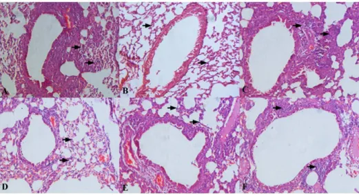
Figure 5. Histological analysis.Representative images (200x) showed the extensive infiltration of inflammatory cells in the lung tissues from the respective groups: Positive group (A), Negative group (B),L. lactis8148 (C), LL-E (D), LL-I (E), LL-W (F). Inflammatory cells were indicated with black arrows.
doi:10.1371/journal.pone.0109461.g005
Table 4.Cytokine production in bronchoalveolar lavage fluid.
Group IL-4 (pg/ml) IL-5 (pg/ml) IL-10 (pg/ml)
Positive 98613 174620 375627
8148 7366* 13466* 340614
LL-I 56613* 9765*# 348
618
LL-W 6066* 93611*#
313612
LL-E 6367* 8867*#
302614
Negative 2764 3367 304647
Results were reported as means6SEM. *P,0.05 compared with the positive group;#P
inflammatory responses caused by environmental allergens [40]. Prominent among multiple regulatory factors operating at the mucosal surface are diverse populations of Tregs. Josefowicz and colleagues have demonstrated that Tregs in the MLN are essential for mucosal tolerance, which is reported to play an important role in maintaining functional tolerance and regulating Th2 responses to allergen [41-43]. Here, we showed that the treatment with any recombinant L. lactis significantly increased the proportion of CD4+Foxp3+Tregs in the MLN of non-sensitized mice (day 12). Such increase is thought to play a critical role in the intervention of the early development of allergic diseases, as demonstrated by Earle and colleagues [44] who found that Tregs could suppress the proliferation and cytokine secretion of effector T cells. Moreover, a population of Tregs could create a regulatory milieu that promotes the outgrowth of a new population of Tregs with antigen specificities distinct from those of the original Tregs population [45]. Therefore, we postulated that the treatment with any recombinant L. lactis would be superior to that with wild-type
L. lactis, inducing a significant increase in the proportion of Tregs in the MLN of the Der p2-sensitized mice, and subsequent results confirmed our assumption (Fig. 6, day 32). However, it was remarkable that after the inhalation challenge, the levels of Tregs in the MLN of the Der p2-sensitized mice were returned to a normal state similar to those of the non-sensitized mice (Fig. 6, day 45). These results can be due to that Tregs in other immune organs could migrate to and remain in inflamed tissues, which play an essential role for their functionin vivo [46]. It can also be explained by postulating that the maintenance of mucosal Tregs is linked to continuing antigen exposure, and antigen withdrawal results in rapid return to baseline Tregs numbers in the mucosa [47]. Changes of Tregs level in the MLN indicated that oral administration with probiotic or recombinant LAB vaccine can strengthen immune tolerance to suppress the early development of allergic diseases, but these protective effect is not durable and to be weakened or disappear without continuous application.
Eyles and colleagues have well described that the modulation of systemic immune responses by mucosal vaccination is developed through antigen transportation to systemic lymphoid organs (spleen) preferentially via the mucosal lymphoid tissues [48]. However, treatment with recombinant or wild-typeL. lactis did not significantly influence the proportion of Tregs in the spleen. This result differed from Karimi et al. [39] who found that oral treatment withL. reuteri increased the Tregs population in the spleen, which is correlated with the attenuation of allergen-induced airway inflammation. The implication of these findings is that there are clear strain-specific immune-regulatory properties of LAB species in vivo [49] and the intrinsic immunogenicity of bacterial vehicle for vaccine delivery would influence the final immune response to recombinant LAB vaccinein vivo.
On the whole, oral treatment with the recombinant L. lactis prior sensitization exerted significant beneficial effects on the suppression of allergic responses, suggesting that recombinant LAB vaccine would be a desirable competitor for the prevention and treatment of allergic diseases in the future. The protective effect is mainly attributed to the enhancement of mucosal immune tolerance, which plays a critical role in the reduction of inflammatory factors (Th2 cytokines, specific IgE and inflamma-tory cells infiltration) both at the systemic levels and the local sites. In the current study, the localization of Der p2 in the recombinant
Figure 6. Effect of the recombinantL. lactistreatment on the proportion of CD4+Foxp3+T cells in the MLN.
On days 12, 32 and 45, the levels of CD4+Foxp3+T cells in the MLN from the respective groups were analyzed by FACS. Results were reported as means
6SEM.*P
,0.05 vs the positive group (day 12).#
P,0.05 vs the positive group (day 32). doi:10.1371/journal.pone.0109461.g006
Figure 7. Measurement of CD4+Foxp3+T cells in the spleen.
On day 12, the percentage of CD4+Foxp3+T cells in the spleen from the
respective groups were evaluated. Results were expressed as means6
SEM.
L. lactisdid not affect its final immunogenicityin vivo, whereas for its application in clinical treatment, it is necessary to further confirm the effect of other factors, i.e., inoculation route and bacterial vehicle, on the suppression of HDM allergic diseases.
Author Contributions
Conceived and designed the experiments: CQA QXZ HZ YQC WC. Performed the experiments: CQA CCR GW. Analyzed the data: CQA FWT JXZ. Contributed reagents/materials/analysis tools: HZ WC. Wrote the paper: CQA QXZ XML.
References
1. Gregory LG, Lloyd CM (2011) Orchestrating house dust mite-associated allergy in the lung. Trends Immunol 32: 402–411.
2. Nelson RP Jr, DiNicolo R, Ferna´ndez-Caldas E, Seleznick MJ, Lockey RF, et al. (1996) Allergen-specific IgE levels and mite allergen exposure in children with acute asthma first seen in an emergency department and in nonasthmatic control subjects. Journal of allergy and clinical immunology 98: 258–263.
3. Martı´nez HQ, Ferguson N (2009) Life-threatening Asthma: Focus on Lung Protection. Intensive Care Medicine: Springer. pp372–382.
4. Kandane-Rathnayake RK, Matheson MC, Simpson JA, Tang ML, Johns DP, et al. (2009) Adherence to asthma management guidelines by middle-aged adults with current asthma. Thorax 64: 1025–1031.
5. Hage-Hamsten V, Valenta R (2002) Specific immunotherapy-the induction of new IgE-specificities? Allergy 57: 375–378.
6. Bousquet J, Lockey R, Malling HJ (1998) Allergen immunotherapy: therapeutic vaccines for allergic diseases. A WHO position paper. J Allergy Clin Immunol 102: 558–562.
7. Pauli G, Malling H-J, Rak S, Horak F, Pastorello E, et al. (2006) Clinical efficacy of subcutaneous immunotherapy in birch pollen allergic patients: a randomized, double-blind, placebo-controlled study with recombinant Bet v 1 versus natural Bet v 1 or standardized birch extract. Blackwell Publishing 9600 Garsington Rd,. Oxford OX4 2DQ, Oxon, England. pp1206–1206.
8. Huibregtse IL, Snoeck V, de Creus A, Braat H, De Jong EC, et al. (2007) Induction of ovalbumin-specific tolerance by oral administration of Lactococcus lactis secreting ovalbumin. Gastroenterology 133: 517–528.
9. Schwarzer M, Repa A, Daniel C, Schabussova I, Hrncir T, et al. (2011) Neonatal colonization of mice with Lactobacillus plantarum producing the aeroallergen Bet v 1 biases towards Th1 and T-regulatory responses upon systemic sensitization. Allergy 66: 368–375.
10. Lee P, Abdul-Wahid A, Faubert GM (2009) Comparison of the local immune response against Giardia lamblia cyst wall protein 2 induced by recombinant Lactococcus lactis and Streptococcus gordonii. Microbes and Infection 11: 20– 28.
11. Bermu´dez-Humara´n LG, Langella P, Cortes-Perez NG, Gruss A, Tamez-Guerra RS, et al. (2003) Intranasal immunization with recombinant Lactococcus lactis secreting murine interleukin-12 enhances antigen-specific Th1 cytokine production. Infection and immunity 71: 1887–1896.
12. Braat H, Rottiers P, Hommes DW, Huyghebaert N, Remaut E, et al. (2006) A phase I trial with transgenic bacteria expressing interleukin-10 in Crohn’s disease. Clinical Gastroenterology and Hepatology 4: 754–759.
13. Thomas WR, Smith W-A, Hales BJ, Mills KL, O9Brien RM (2002) Characterization and immunobiology of house dust mite allergens. International archives of allergy and immunology 129: 1–18.
14. Nouaille S, Ribeiro LA, Miyoshi A, Pontes D, Le Loir Y, et al. (2003) Heterologous protein production and delivery systems for Lactococcus lactis. Genet Mol Res 2: 102–111.
15. Lim S, Jahanshiri F, Abdul Rahim R, Sekawi Z, Yusoff K (2010) Surface display of respiratory syncytial virus glycoproteins in Lactococcus lactis NZ9000. Letters in applied microbiology 51: 658–664.
16. Liu XY JK, Gao B, Liu ZG (2009) Expression, purification and identification of the recombinant allergen Der p2 from Dermatophagoides pteronyssinus and investigation on its immunological activities. Chinese Journal of Zoonoses 25: 764–767.
17. Nandakumar MP, Shen J, Raman B, Marten MR (2003) Solubilization of trichloroacetic acid (TCA) precipitated microbial proteins via naOH for two-dimensional electrophoresis. J Proteome Res 2: 89–93.
18. Piard JC, Hautefort I, Fischetti VA, Ehrlich SD, Fons M, et al. (1997) Cell wall anchoring of the Streptococcus pyogenes M6 protein in various lactic acid bacteria. Journal of Bacteriology 179: 3068–3072.
19. Rigaux P, Daniel C, Hisbergues M, Muraille E, Hols P, et al. (2009) Immunomodulatory properties of Lactobacillus plantarum and its use as a recombinant vaccine against mite allergy. Allergy 64: 406–414.
20. Lee CC, Ho H, Lee KT, Jeng ST, Chiang BL (2011) Construction of a Der p2-transgenic plant for the alleviation of airway inflammation. Cell Mol Immunol 8: 404–414.
21. Wells JM, Mercenier A (2008) Mucosal delivery of therapeutic and prophylactic molecules using lactic acid bacteria. Nature Reviews Microbiology 6: 349–362. 22. Schabussova I, Wiedermann U (2008) Lactic acid bacteria as novel adjuvant systems for prevention and treatment of atopic diseases. Current opinion in allergy and clinical immunology 8: 557–564.
23. Daniel C, Repa A, Wild C, Pollak A, Pot B, et al. (2006) Modulation of allergic immune responses by mucosal application of recombinant lactic acid bacteria producing the major birch pollen allergen Bet v 1. Allergy 61: 812–819.
24. Daniel C, Roussel Y, Kleerebezem M, Pot B (2011) Recombinant lactic acid bacteria as mucosal biotherapeutic agents. Trends in Biotechnology 29: 499– 508.
25. Bermu´dez-Humara´n LG, Cortes-Perez NG, Le Loir Y, Alcocer-Gonza´lez JM, Tamez-Guerra RS, et al. (2004) An inducible surface presentation system improves cellular immunity against human papillomavirus type 16 E7 antigen in mice after nasal administration with recombinant lactococci. Journal of medical microbiology 53: 427–433.
26. Le Loir Y, Azevedo V, Oliveira SC, Freitas DA, Miyoshi A, et al. (2005) Protein secretion in Lactococcus lactis: an efficient way to increase the overall heterologous protein production. Microbial Cell Factories 4: 2.
27. Norton PM, Brown HW, Wells JM, Macpherson AM, Wilson PW, et al. (1996) Factors affecting the immunogenicity of tetanus toxin fragment C expressed in Lactococcus lactis. FEMS Immunology & Medical Microbiology 14: 167–177. 28. Ando H, Move´rare R, Kondo Y, Tsuge I, Tanaka A, et al. (2008) Utility of
ovomucoid-specific IgE concentrations in predicting symptomatic egg allergy. Journal of Allergy and Clinical Immunology 122: 583–588.
29. Valenta R (2002) The future of antigen-specific immunotherapy of allergy. Nature Reviews Immunology 2: 446–453.
30. Rupa P, Mine Y (2012) Oral immunotherapy with immunodominant T-cell epitope peptides alleviates allergic reactions in a Balb/c mouse model of egg allergy. Allergy 67: 74–82.
31. Kawano Y, Noma T, Yata J (1994) Regulation of human IgG subclass production by cytokines. IFN-gamma and IL-6 act antagonistically in the induction of human IgG1 but additively in the induction of IgG2. The Journal of Immunology 153: 4948–4958.
32. Mosmann T, Coffman R (1989) TH1 and TH2 cells: different patterns of lymphokine secretion lead to different functional properties. Annual review of immunology 7: 145–173.
33. Aversa G, Punnonen J, Cocks BG, de Waal Malefyt R, Vega F, et al. (1993) An interleukin 4 (IL-4) mutant protein inhibits both IL-4 or IL-13-induced human immunoglobulin G4 (IgG4) and IgE synthesis and B cell proliferation: support for a common component shared by IL-4 and IL-13 receptors. The Journal of experimental medicine 178: 2213–2218.
34. Pretolani M (1999) Interleukin-10: an anti-inflammatory cytokine with therapeutic potential. Clin Exp Allergy 29: 1164–1171.
35. Krug N, Madden J, Redington AE, Lackie P, Djukanovic R, et al. (1996) T-cell cytokine profile evaluated at the single cell level in BAL and blood in allergic asthma. American Journal of Respiratory Cell and Molecular Biology 14: 319– 326.
36. Humbert M, Durham SR, Ying S, Kimmitt P, Barkans J, et al. (1996) IL-4 and IL-5 mRNA and protein in bronchial biopsies from patients with atopic and nonatopic asthma: evidence against" intrinsic" asthma being a distinct immunopathologic entity. American journal of respiratory and critical care medicine 154: 1497–1504.
37. Nonaka M, Nonaka R, Woolley K, Adelroth E, Miura K, et al. (1995) Distinct immunohistochemical localization of 4 in human inflamed airway tissues. IL-4 is localized to eosinophils in vivo and is released by peripheral blood eosinophils. The Journal of Immunology 155: 3234–3244.
38. van Scott MR, Justice JP, Bradfield JF, Enright E, Sigounas A, et al. (2000) IL-10 reduces Th2 cytokine production and eosinophilia but augments airway reactivity in allergic mice. American Journal of Physiology-Lung Cellular and Molecular Physiology 278: L667–L674.
39. Karimi K, Inman MD, Bienenstock J, Forsythe P (2009) Lactobacillus reuteri-induced regulatory T cells protect against an allergic airway response in mice. Am J Respir Crit Care Med 179: 186–193.
40. Maloy KJ, Powrie F (2011) Intestinal homeostasis and its breakdown in inflammatory bowel disease. Nature 474: 298–306.
41. Larche´ M (2007) Regulatory T cells in allergy and asthma. CHEST Journal 132: 1007–1014.
42. Akdis M, Blaser K, Akdis CA (2005) T regulatory cells in allergy: Novel concepts in the pathogenesis, prevention, and treatment of allergic diseases. Journal of Allergy and Clinical Immunology 116: 961–968.
43. Josefowicz SZ, Niec RE, Kim HY, Treuting P, Chinen T, et al. (2012) Extrathymically generated regulatory T cells control mucosal TH2 inflamma-tion. Nature 482: 395–399.
44. Earle K, Tang Q, Zhou X, Liu W, Zhu S, et al. (2005) In vitro expanded human CD4+CD25+regulatory T cells suppress effector T cell proliferation. Clinical immunology 115: 3–9.
45. Tang Q, Bluestone JA (2008) The Foxp3+regulatory T cell: a jack of all trades, master of regulation. Nature immunology 9: 239–244.
47. Strickland DH, Stumbles PA, Zosky GR, Subrata LS, Thomas JA, et al. (2006) Reversal of airway hyperresponsiveness by induction of airway mucosal CD4+
CD25+regulatory T cells. The Journal of experimental medicine 203: 2649– 2660.
48. Eyles J, Bramwell V, Williamson E, Alpar H (2001) Microsphere translocation and immunopotentiation in systemic tissues following intranasal administration. Vaccine 19: 4732–4742.