Agonist Induced
b
-Amyloid Production in SH-SY5Y
Neuroblastoma Cells via a cAMP Dependent Pathway
Bhushan Vijay Nagpure, Jin-Song Bian*
Department of Pharmacology, Yong Loo Lin School of Medicine, National University of Singapore, Singapore
Abstract
Alzheimer’s disease (AD) is the leading cause of senile dementia in today’s society. Its debilitating symptoms are manifested by disturbances in many important brain functions, which are influenced by adenosine. Hence, adenosinergic system is considered as a potential therapeutic target in AD treatment. In the present study, we found that sodium hydrosulfide (NaHS, an H2S donor, 100mM) attenuated HENECA (a selective A2A receptor agonist, 10–200 nM) inducedb-amyloid (1–42)
(Ab42) production in SH-SY5Y cells. NaHS also interfered with HENECA-stimulated production and post-translational modification of amyloid precursor protein (APP) by inhibiting its maturation. Measurement of the C-terminal APP fragments generated from its enzymatic cleavage byb-site amyloid precursor protein cleaving enzyme 1 (BACE1) showed that NaHS did not have any significant effect on b-secretase activity. However, the direct measurements of HENECA-elevated c -secretase activity and mRNA expressions of presenilins suggested that the suppression of Ab42 production in NaHS pretreated cells was mediated by inhibitingc-secretase. NaHS induced reductions were accompanied by similar decreases in intracellular cAMP levels and phosphorylation of cAMP responsive element binding protein (CREB). NaHS significantly reduced the elevated cAMP and Ab42 production caused by forskolin (an adenylyl cyclase, AC agonist) alone or forskolin in combination with IBMX (a phosphodiesterase inhibitor), but had no effect on those caused by IBMX alone. Moreover, pretreatment with NaHS significantly attenuated HENECA-elevated AC activity and mRNA expressions of various AC isoforms. These data suggest that NaHS may preferentially suppress AC activity when it was stimulated. In conclusion, H2S
attenuated HENECA induced Ab42 production in SH-SY5Y neuroblastoma cells through inhibitingc-secretase via a cAMP dependent pathway.
Citation:Nagpure BV, Bian J-S (2014) Hydrogen Sulfide Inhibits A2A Adenosine Receptor Agonist Inducedb-Amyloid Production in SH-SY5Y Neuroblastoma Cells via a cAMP Dependent Pathway. PLoS ONE 9(2): e88508. doi:10.1371/journal.pone.0088508
Editor:Stefan Strack, University of Iowa, United States of America
ReceivedApril 25, 2013;AcceptedJanuary 11, 2014;PublishedFebruary 11, 2014
Copyright:ß2014 Nagpure, Bian. This is an open-access article distributed under the terms of the Creative Commons Attribution License, which permits unrestricted use, distribution, and reproduction in any medium, provided the original author and source are credited.
Funding:This work is supported by a research grant from the Singapore National Medical Research Council (1183/2008). The funders had no role in study design, data collection and analysis, decision to publish, or preparation of the manuscript.
Competing Interests:The authors have declared that no competing interests exist. * E-mail: phcbjs@nus.edu.sg
Introduction
Alzheimer’s disease (AD), which is the most common neurode-generative disease, is also the leading cause of senile dementia [1]. The prevalence of AD has been increasing exponentially from 3% among those above 65 years to almost 50% among those with age above 85 years [2]. AD has been one of the most debilitating diseases of the current century, with profound economic, political and social consequences.
The classical neuro-pathological hallmarks of AD include accumulation of senile plaques in the brain, neuro-fibrillary tangles, synaptic loss and neuronal death [3,4]. Senile plaques consist mainly of a 39–42 amino acid longb-amyloid (Ab) peptide, generated from a larger transmembrane amyloid precursor protein (APP) [5]. In non-amyloidogenic pathway, APP is cleaved within the Ab domain by a- secretase, releasing soluble APP (sAPPa) extracellularly. On the other hand, poorly soluble amyloidogenic Ab is derived from sequential cleavage of APP by b- and c -secretases [6]. Above-mentioned and other evidences strongly support that APP synthesis and proteolysis are critical events in AD pathogenesis. Hence drugs targeting these processes are likely to be beneficial for the prevention and treatment of AD.
suggesting that oxidative stress in the brain of AD patients results in neuronal trauma and degeneration [16,17]. The stress and trauma initiate cascade of pathological events disrupting delicate balance of adenosine nucleotides and nucleosides altering ATP-ADP ratio [18,19]. The resultant increase in ATP-ADP level leads to enhanced adenylate kinase enzyme activity which in turn augments AMP concentration to keep ADP-AMP ratio constant [20]. The elevated AMP undergoes hydrolysis to generate more adenosine [21]. This intracellular adenosine is released outside the cell resulting in increased extracellular concentration of adenosine [18].
Hydrogen sulfide (H2S) has been reported as an important
modulator in multiple physiological systems including adult CNS [22,23,24]. The role of H2S in intracellular calcium ([Ca
2+]
i)
homeostasis in neurons and glial cells is perhaps the most important one as it regulates synaptic activity and plasticity. H2S
upregulates [Ca2+]
i in neurons and glia by increasing its influx
through different Ca2+channels and its receptors and by releasing calcium from [Ca2+]
istores [24,25,26]. H2S was found to activate
KATP channels, which apart from mediating neurotransmitter
release from presynaptic neurons, also offer neuroprotection during hypoxic challenge [27,28]. The neuromodulatory role of H2S is also critical as it maintains excitatory-inhibitory balance of
neurotransmission [29]. c-aminobutyric acid B receptors (GA-BABR) which are involved in fine tuning of inhibitory
neurotrans-mission and regulation of the release of neurotransmitters, are upregulated by H2S [30]. H2S is shown to modulate long-term
potentiation in active synapses and neuron-glia interactions [24,31]. Acting as a mediator of cell signaling, this gaseous neurotransmitter has been identified to target a number of ion channels, transcription factors and protein kinases [32]. The beneficial effects of H2S have already been found in cell and
animal models of PD [33], neuroinflammation [34] and H2O2
induced neural injury [35]. Adding further in the knowledge, we studied the inhibitory effect of H2S against Ab42 production in
SH-SY5Y neuronal cells. In the present study, we have confirmed that A2A receptor stimulation enhancedc-secretase activity which in turn resulted in increased Ab42 production in SH-SY5Y cells. We also show that H2S attenuates Ab42 synthesis via suppression
of cAMP signal transduction pathway.
Materials and Methods
Chemicals
Sodium hydrosulfide (NaHS), forskolin, 3-isobutyl-1-methyl-xanthine (IBMX), SQ 22536, ZM 241385, N-[N-(3,5-difluoro-phenacetyl)-L-alanyl]-S-phenylglycine t-butyl ester (DAPT) and methylthiazolyl tetrazolium (MTT) were purchased from Sigma Aldrich (St. Louis, MO, USA). 2-Hexynyladenosine-59-N -ethyl-carboxamide (HENECA) was ordered from Abcam (Cambridge, MA, USA). NaHS, HENECA, SQ 22536 and MTT were dissolved in de-ionized water, while forskolin, ZM 241385, IBMX and DAPT were dissolved in dimethylsulfoxide (DMSO). Primary antibodies against phospho-CREB, Adenosine A2A-R andb-actin were purchased from Santa Cruz biotechnology (St. Louis, MO, USA). Monoclonal antibody against APP (clone 22C11) and polyclonal antibody against APP C-terminus were from Millipore (Temecula, CA, USA).
NaHS was used as an H2S donor. When NaHS is dissolved in
water at neutral pH, HS2is released and forms H2S with H+. This
provides a solution of H2S at a concentration that is about 33% of
the original concentration of NaHS [36].
Cell Culture and Treatments
The SH-SY5Y human neuroblastoma cell line was obtained from the American Type Culture Collection (Manassas, VA, USA). Cells were cultured in 75 cm2flasks in Dulbecco’s modified Eagle’s medium (DMEM) supplemented with 10% fetal bovine serum (FBS), 1% penicillin (100 U)/streptomycin (100 mg/mL), and were maintained at 37uC in an incubator under a humidified atmosphere of 95% air and 5% CO2. Cells were split twice a week.
The cells were seeded at a density of 56105cells/well in 6-well plates a day before the transfection. The SH-SY5Y cells were lipotransfected with pcDNA4-hAPP695swe using the Lipofecta-mine 2000 transfection reagent. After transfection for 24 hours, cells were treated with different chemicals mentioned above. The plasmids were a kind gift from Dr. Weihong Song, University of British Columbia, Vancouver, Canada.
For each experiment, confluent cells in 75-cm2 flasks were seeded onto 35-mm dishes. Cells in culture dishes were used for experiments after reaching 80–90% confluence.
Cell Viability Assay
Cell viability was assessed by MTT reduction assay as follows. At the end of treatments (as described in results section), cells were incubated at 37uC with MTT at a final concentration of 0.5 mg/ ml for 4 hours. The purple formazan formed was solubilized with 150mL DMSO. The absorbance of the colored solution was
measured at 570 nm with a reference wavelength of 630 nm using Saffire 2 microplate reader (Tecan, USA).
Intracellular cAMP Assay
A commercially available direct cAMP enzyme immunoassay kit (Cayman Chemical, USA) was used to examine the involve-ment of cAMP. Briefly, cells were incubated in DMEM containing 0.5% FBS. After treatment with different drugs described in results section, the cells were lysed in 0.1 M HCl for 20 minutes. 50mL of samples were added into a 96-well plate followed by incubation with cAMP acetylcholine esterase tracer and cAMP antiserum for 18 hours at 4uC. Each sample was developed with Ellman’s reagent next day and the plate was read at a wavelength of 405 nm. cAMP concentration was calculated according to the cAMP standard and the protein was quantified by dissolving the pellets.
Cell Fractionation and Adenylyl Cyclase (AC) Activity Assay
A cell fractionation technique was adopted from the literature [37]. AC activity was assayed as described previously [38,39] with some modifications. The AC activity assay was performed at 37uC for 10 min in a 400mL reaction mixture containing 1 mM ATP,
100 mM NaCl, 50 mM HEPES, 0.5 mM IBMX, 6 mM MgCl2,
1mM GTP, and 20mg of membrane protein. Reactions were
stopped by addition of 0.6 mL of trichloroacetic acid (10% w/v). The accumulation of cAMP was later assayed by cAMP EIA kit (Cayman Chemical, USA).
c-secretase (Fluorogenic Substrate) Assay
The assay was performed as described previously in the literature [40]. Briefly, the cell lysates were centrifuged at 12000 g for 15 min. Resultant pellets were resuspended and incubated overnight at 37uC in 200mL of assay buffer containing 10mL fluorescent substrate of c-secretase (Calbiochem). The fluorescence was measured with excitation wavelength set at 355 nm and emission wavelength at 440 nm.
ELISA for Ab42
The conditioned medium from samples was collected by centrifugation (12,000 g at 4uC for 15 min). The secreted levels of Ab42 in conditioned medium were quantitatively measured by commercially available ELISA kit (Invitrogen, USA). Briefly, the
samples were diluted by standard diluent buffer and AEBSF was added to diluted samples and standards which prevent proteolysis of Abpeptides. The standards, controls and samples were pipetted into the antibody pre-coated wells and co-incubated with a rabbit antibody specific for the C-terminus of the 1–42 Ab sequence. Bound rabbit antibody was detected by the use of a horseradish
Figure 1. Effect of NaHS on Ab42 production and cell viability in SH-SY5Y cells expressing APPswe. A: Concentration-dependent effect of HENECA (10–200 nM, 24 hours) on Ab42 production.B–C: Dose-dependent effect of NaHS (10–200mM, 12 hours) on Ab42 formation in the presence (B) and absence (C) of HENECA (100 nM, 24 hours).D–E: MTT assay showing the effect of NaHS alone at 10–200mM (D) or HENECA alone at 10–200 nM (E) on cell viability of SH-SY5Y cells. Ab42 levels in conditioned media were measured by sandwich ELISA kit. Control values were adjusted to 100%. Data are given as means6S.E.M, n = 6.##
p,0.01,###p,0.001 vs Con group;**p,0.01, ***p,0.001 vs HEN group. Con, control; HEN,
HENECA.
peroxidase-labeled anti-rabbit antibody. After washing, horserad-ish peroxidase-labeled anti-rabbit antibody (enzyme) was added. After washing to remove the entire unbound enzyme, a substrate solution was added, which is acted upon by the bound enzyme to produce color. The intensity of this colored product is directly proportional to the concentration of Ab42 present in the samples. The optical density was measured at 450 nm wavelength using Saffire 2 microplate reader (Tecan, USA).
Reverse Transcription-PCR
Two-step reverse transcription polymerase chain reaction was used to determine mRNA levels of AC1, AC3, AC8, PS1, PS2 and GAPDH in SH-SY5Y cells. Total RNA was extracted using TRIzolH extraction method (Invitrogen, Carlsbad, CA, USA). Homogenized samples were then incubated at room temperature for 10 min. Chloroform was added and tubes were shaken vigorously by hand for 15 min followed by incubation for 3 min at room temperature again. Samples were centrifuged at 12000 g for 15 min at 4uC. Colorless upper aqueous phase was transferred to a new tube containing isopropanol (prepared in nuclease free water) and incubated for 10 min at 25uC followed by centrifugation at
12000 g for 10 min at 4uC. Supernatant was discarded and RNA pellets were washed with 70% ethanol (prepared in nuclease free water). RNA concentration was determined with NanoDrop Spectrophotometer (ND-1000, NanoDrop Technology). Equal amounts of RNA samples obtained were reverse transcribed into cDNA using iScriptTM cDNA synthesis kit (Bio-Rad). Reverse transcription was performed at 25uC (for 5 min), 42uC (for 30 min) and 85uC (for 5 min). The resulting cDNAs were PCR-amplified using Taq DNA polymerase kit (i-DNA Biotechnology). The specific PCR primer sequences used were as follows:
AC1 (59-CATGACCTGCGAGGACGAT-39 and 59 -TCCC-GTTCGACATGTTTGTA-39) [41], AC3 (59
-GTACTACACG-GGACCCAGCA-39 and 59
–GCTCTAAGGCCACCATAG-GTA-39) [41], AC8 (59-ACCGGCATTGAGGTAGTGAT-39and 59- ATGACCACTTGGAGGATGAC-39) [41], PS1 (59
-ACA-GAGTTACCTGCACCGTTGTCC-39 and 59
-TTCCTCATC-TTGCTCCAC-CACCTG) [42], PS2 (59 -AGTGAGAGA-CA-GCCAGAAGCAAG-39and 59 -AACCCCACTACAGACATAG-CGGTC-39) [42], GADPH (59 -GCGGGGCTCTCCAGAACA-TCAT-39and 59-GGTGTC-CAGGGGTCTTACTCC-39) [43]
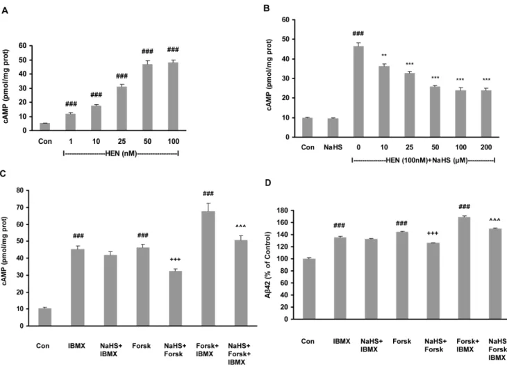
Figure 2. Effect of NaHS on Ab42 production involved cAMP signaling pathway. A: Dose-dependent effect of HENECA on cAMP production in SH-SY5Y cells expressing APPswe.B: Concentration-dependent effect of NaHS (10–200mM, 12 hours) on HENECA (100 nM, 24 hours)-stimulated cAMP upregulation.C–D: Effects of NaHS (100mM) on cAMP (C) and Ab42 production (D) in cells treated with forskolin (20mM) and/or IBMX (100mM). The intracellular cAMP and Ab42 levels in conditioned media were measured by sandwich ELISA kits. Control values were adjusted to 100% for Ab42 levels measurement. Data are given as means6S.E.M, n = 6.###
p,0.001 vs Con group,**p,0.01, ***p,0.001 vs HEN group,+++p,0.001 vs
forsk group,ˆˆˆp,0.001 vs forsk+IBMX group. Con, control; HEN, HENECA; Forsk, forskolin. doi:10.1371/journal.pone.0088508.g002
PCR conditions were set as 95uC (for 30 sec), 55uC (for 30 sec), and 72uC (for 30 sec) for 40 cycles. PCR products were separated on a 1% agarose gel and stained with ethidium bromide. The optical densities of the mRNA bands were analyzed with GelDoc-It Imaging System.
Western Blot Assay
Cells were washed twice with ice-cold PBS after treatment and solubilized in RIPA lysis buffer (150 mM sodium chloride, 1.0% Nonidet P-40, 0.5% sodium deoxycholate, 0.1% SDS, 50 mM Tris at pH 8.0, protease and phosphatase inhibitor cocktails). The cell lysates were shaken and kept on ice for 1 hour before being subjected to centrifugation at 12,000 g at 4uC for 10 min. Supernatants were collected and denatured by SDS sample buffer. Epitopes were exposed by boiling the protein samples at 95uC for 5 min. Protein concentrations were determined with a NanoDrop Spectrophotometer (ND-1000, NanoDrop Technology). Equal
amounts of the protein samples were separated by electrophoresis using a 10% sodium dodecyl sulphate-polyacrylamide (SDS/ PAGE) gel and transferred onto a nitrocellulose membrane (WhatmanH, Germany). After being blocked in 10% milk with TBST buffer (10 mM Tris-HCl, 120 mM NaCl, 0.1% Tween-20, pH 7.4) at room temperature for 1 hour, the membranes were incubated with primary antibodies of phospho-CREB (1:1000),b -actin (1:1000), A2A-R (1:1000), APP (clone 22C11) (1:1000) and APP C-terminus (1:1000) at 4uC overnight. Membranes were washed three times in TBST buffer, followed by incubation with 1:10000 dilutions of appropriate horseradish peroxidase-conjugat-ed (HRP) anti-mouse IgG or anti-rabbit IgG at 25uC for 1 hour, and washed three times in TBST. Visualization was carried out using ECLH(plus/advanced chemi-luminescence) kit (GE health-care, UK). The density of the bands on Western blots was quantified by Image J software.
Figure 3. Effect of NaHS on mRNA expression of AC isoforms and AC activity. A–B: Representative gels (A) and histogram (B) demonstrating the effect of pretreatment with NaHS (100mM, 12 hours) attenuated the effects of HENECA (100 nM, 24 hours) on mRNA expressions of AC isoforms.C: Effect of NaHS (100mM) on AC activity stimulated by forskolin (20mM).D: Effect of NaHS (100mM) on Ab42 production in SH-SY5Y cells preincubated with AC antagonist, SQ 22536 (300mM). Control values were adjusted to 100%. Data are given as means6S.E.M, n = 4–6.##
p,
0.01,###
p,0.001 vs Con group;+++p,0.001 vs Forsk group,*p,0.05,**p,0.01 vs HEN group. Con, control; HEN, HENECA; Forsk, forskolin.
Statistical Analysis
Values stated are mean 6 SEM of at least triplicate measurements. SPSS software for Windows was used to perform one-way analysis of variance (ANOVA) followed by a post hoc
(Bonferroni) test for multiple group comparison. The significance level was set atp,0.05.
Results
NaHS attenuates Adenosine A2A receptor agonist (HENECA) induced Ab42 production in SH-SY5Y cells
We first analyzed the effect of adenosine A2A receptor agonist, HENECA on Ab42 production by SY5Y cells. These
SH-Figure 4. Effect of NaHS on Ab42 production involved PKA and CREB. A: Effect of HENECA (100 nM) on Ab42 formation was abolished by a PKA inhibitor, H89 (5, 10 and 15mM).B–C: Representative gel (B) and histogram (C) depicting that pretreatment with NaHS (100mM, 12 hours) attenuated the effects of HENECA (100 nM, 24 hours) on phosphorylation of CREB. Control values were adjusted to 100%. Data are given as means6S.E.M, n = 4–6.###
p,0.001 vs Con group;*p,
0.05,**p,0.01, ***p,0.001 vs HEN group. Con, control; HEN, HENECA.
doi:10.1371/journal.pone.0088508.g004
Figure 5. Effect of NaHS on expression of A2A receptors. A–B: Representative gels (A) and histogram (B) demonstrating the effect of pretreatment with NaHS (100mM, 12 hours) did not attenuate the effects of HENECA (100 nM, 24 hours) on protein expression of A2A receptor.C: Effect of NaHS (100mM) on production of Ab42 in cells pre-treated with A2A receptor antagonist, ZM 241385 (50 nM). Control values were adjusted to 100%. Data are given as means6S.E.M, n = 4– 6.##
p,0.01;###p,0.001 vs Con group. Con, control; HEN, HENECA.
doi:10.1371/journal.pone.0088508.g005
SY5Y cells were transfected with APP harboring Swedish mutation which is known to elevate secretion of Ab42 [44]. Treatment of cells with HENECA at 10–200 nM for 24 hours increased the levels of Ab42 in conditioned medium in a concentration-dependent manner (Fig 1A). Pretreatment with
NaHS (10–200mM) for 12 hours attenuated the stimulatory effect of HENECA (100 nM) on Ab42 production in a dose-dependent manner (Fig 1B). However, pretreatment with NaHS (50–100mM)
alone did not reduce the basal levels of Ab42 significantly (Fig 1C). To confirm that the observed effects of NaHS and HENECA were not due to their effects on cell viability, MTT cell viability assay were performed. MTT results showed that both NaHS (Fig 1D) and HENECA (Fig 1E) at the concentration ranges used for the experiments had no effect on cell viability.
The effect of NaHS on HENECA-induced Ab42 production involves cAMP/PKA/CREB pathway
We further examined the underlying mechanism for the effect of NaHS. As shown in Fig 2A, HENECA (1–100 nM) in a dose-dependent manner increased intracellular cAMP levels. The increase in intracellular cAMP levels by HENECA was abrogated by NaHS (10–200mM) in a concentration-dependent manner (Fig 2B). However, NaHS (100mM) alone did not produce any
significant effect on cAMP levels (Fig 2B). In an attempt to investigate whether NaHS targets the synthesis or the decompo-sition of cAMP, the following series of experiments was conducted. Intracellular cAMP production was elevated by forskolin, an AC agonist and IBMX, a phosphodiesterase (PDE) antagonist. As shown in Fig 2C, NaHS significantly reduced the elevated cAMP production caused by either forskolin alone or forskolin in combination with IBMX, but had no effect on those caused by IBMX alone. Similarly, NaHS treatment significantly attenuated the elevated Ab42 production stimulated by either forskolin alone or forskolin in combination with IBMX, but had no effect on the effect caused by IBMX alone (Fig 2D).
Gene expression studies with RT-PCR (Fig 3A and 3B) showed that HENECA upregulated mRNA expressions of all three isoforms of AC (AC1, AC3 and AC8) and NaHS pretreatment was effective in reducing their upregulated expressions. Addition-ally, a direct measurement of enzymatic activity of AC showed that NaHS treatment significantly suppressed the AC activity elevated by forskolin (Fig 3C). To bolster the hypothesis further, we investigated the effect of NaHS on Ab42 generation by SH-SY5Y cells preincubated with AC antagonist SQ 22536. Neither HENECA nor NaHS was able to induce any kind of significant effect on Ab42 production (Fig 3D) in this situation.
Blockade of PKA with its selective inhibitor, H-89 (5–15mM) also dose-dependently attenuated the elevated Ab42 induced by HENECA (Fig 4A). To examine the involvement of CREB, we determined the phosphorylation of CREB using western blotting. As shown in Fig. 4B and 4C, NaHS treatment significantly attenuated HENECA-induced phosphorylation of CREB. These results suggest that the inhibitory effect of H2S on Ab42
production involves cAMP/PKA/CREB pathway.
Unlike its effect on AC, NaHS pretreatment was ineffective in inducing any significant effect on protein expression of A2A receptors (Fig 5A and 5B). Furthermore, we observed that Ab42 production was not affected by either HENECA or NaHS in cells preincubated with A2A receptor antagonist, ZM 241385 (Fig 5C). Therefore, these data suggest that NaHS may preferentially suppress AC activity when it is stimulated.
NaHS inhibits APP production and maturation
We next sought to monitor the potential effects of H2S on
post-translational modification (maturation), which is a major regula-tory step in Ab42 formation. During SDS-PAGE, both mature (mAPP) and immature (imAPP) forms of APP can be distinguished on the basis of their molecular weights. The immature form (N -glycosylated) is reported as,110 kD and mature form (N- and O-Figure 6. Effect of NaHS on production and maturation of APP.
Representative gel (A) and quantitative analysis (B–C) showing the effects of NaHS (100mM, 12 hours) on HENECA (100 nM, 24 hours) stimulated production (B) and maturation (C) of APP. The cell lysates were analysed by western blot technique with antibody against N-terminus of APP orb-actin. The extent of maturation of APP is shown as the ratio between mAPP to imAPP. mAPP and imAPP are represented by upper and lower bands in a blot respectively.b-actin was used as a loading control. Data are given as means6S.E.M, n = 4.###
p,0.001 vs
Con group; ***p,0.001 vs HEN group. Con, control; HEN, HENECA.
glycosylated) is ,130 kD peptide [45]. As shown in Fig 6,
HENECA significantly upregulated both holoprotein APP (imAPP +mAPP, Fig 6A & 6B) and the ratio of mAPP and imAPP (Fig 6A & 6C). Pretreatment with NaHS significantly abolished these effects. Thus, the effect of NaHS on HENECA-stimulated Ab42 production is mediated by inhibition of both production and maturation of APP.
Effect of NaHS on activities ofb– andc-secretases
b –secretase cleavage of APP gives rise to production ofb -C-terminal fragment, C99. Thus, the amount of C99 generated is considered as an index of b–secretase activity. As evident from Fig 7A and 7B, neither HENECA nor NaHS pretreatment (25– 100mM) was able to induce any significant change in C99 expression. This suggests that NaHS does not affect the activity of
b–secretase in SH-SY5Y cells.
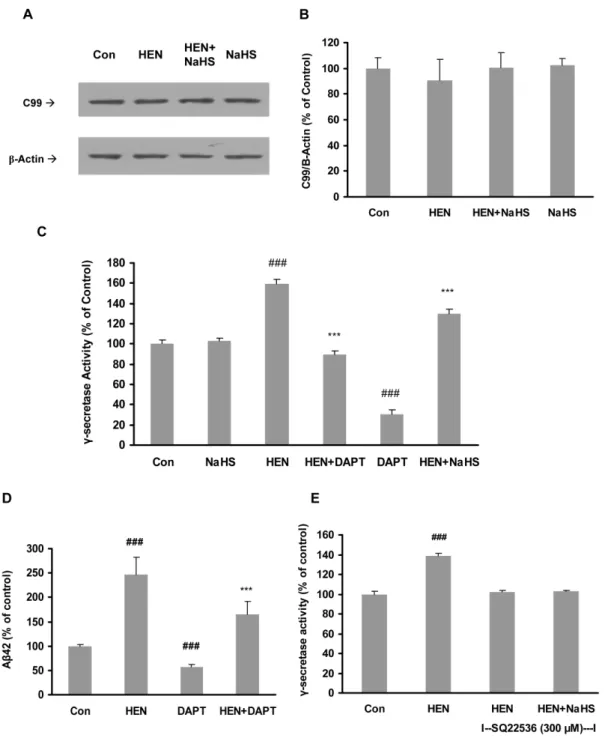
Figure 7. Effect of NaHS on activities ofb- andc-secretases. A–B: Representative gel (A) and quantitative analysis (B) showing NaHS (25– 100mM, 12 hours) and HENECA (100 nM, 24 hours) failed to affectb-CTF (C99) expression in SH-SY5Y cells. The membrane fractions were analysed by western blot with antibody against c-terminal fragment of APP (C99) orb-actin.C–D:Effect of NaHS (100mM) and ac-secretase inhibitor, DAPT (1mM, 1 hour) on HENECA (100 nM)-stimulatedc-secretase activity (C) and Ab42 formation (D) in SH-SY5Y cells expressing APPswe.E: Effect of NaHS (100mM) onc-secretase activity in SH-SY5Y cells preincubated with AC antagonist, SQ 22536 (300mM). Data are given as means6S.E.M, n = 4–6.
###
p,0.001 vs Con group; ***p,0.001 vs HEN group. Con, control; HEN, HENECA.
doi:10.1371/journal.pone.0088508.g007
c-secretase activity in the membrane fractions of SH-SY5Y cells was also measured. As shown in Fig 7C, HENECA significantly stimulatedc-secretase activity. Treatment with NaHS or DAPT, a
c-secretase inhibitor, significantly attenuated the effect of HE-NECA. It is also demonstrated that treatment of NaHS alone did not affectc-secretase activity significantly (Fig 7C). Furthermore,
DAPT reduced the production of Ab42 in HENECA-stimulated cells (Fig 7D). In accordance with our previous result in Fig 3D, the blocking of AC with its specific inhibitor did not have any effect onc-secretase activity (Fig 7E). We next investigated the effect of NaHS pretreatment on mRNA expressions of presenilins 1 (PS1) and 2 (PS2), which are the catalytic components ofc -secretase complex. We found that NaHS significantly suppressed HENECA-upregulated gene expression of PS2 while PS1 mRNA level was not significantly affected (Fig 8A, 8B and 8C). These results suggest that NaHS attenuates HENECA induced activation ofc-secretase. It ultimately results in lowered production of Ab42 in the conditioned medium of SH-SY5Y cells.
Discussion
Accumulating epidemiological, genetic and pharmaceutical studies have shown that there is a convincing role of adenosine signaling in controlling brain damage. While activation of A1A receptors is important in controlling the early events in case of brain damage, blocking of A2A receptors seems to be more important in context of the latter events. The efficiency of A1A receptors decreases as they are subjected to chronic noxious stimuli, whereas the efficiency of A2A receptors tend to remain unaffected or even increase in the similar situations [46]. It has been reported that the expression of A2A receptors is significantly increased in a transgenic mouse model of AD carrying the APP Swedish mutation [47]. The studies conducted in diverse animal models of AD showed that caffeine (a non-selective antagonist of adenosine receptors) improved memory performance in rodents with the protection against memory dysfunction [48]. It was also found that blockade of adenosine A2A receptors mimicked the neuroprotective effect of caffeine against Ab-induced neurotoxicity [49] and prevented the development of Ab-induced synaptotoxi-city leading to memory dysfunctionin vivo[50]. Antagonizing A2A receptor signaling, thus, seems to be one of the most promising therapeutic strategies for chronic brain pathologies such as AD [46,51].
The present study was designed to investigate the effect of H2S
on HENECA-induced synthesis of Ab42 in APPswe transfected SH-SY5Y cells, an established cell model of AD. As demonstrated in our study, A2A receptor stimulation with HENECA resulted in increased production of Ab42. Essentially, this is consistent with the findings in a previous study [47] where increased productions of both Ab40 and Ab42 were reported in AD transgenic mice and APPswe N2a neuronal cultures with higher adenosine receptor density. Albeit both Ab40 and Ab42 forms are pathological, predominantly present 42 amino-acid form of Ab(Ab42) peptide is readily collective and thus more pathogenic in nature [52]. We showed that NaHS inhibited HENECA-induced Ab42 release from SH-SY5Y cells in a dose-dependent manner.
Being positively linked to AC, adenosine A2A receptor stimulation results in the increase in intracellular cAMP level. Experimental studies in bothin vitroandin vivomodels of AD have reported that cAMP dependent pathway is critically important in APP processing. In a study conducted with astrocytes, Lee et al. observed that cAMP signaling can increase cellular levels of APP holoprotein by stimulating APP gene expression [53]. APP protein expression and processing are shown to be increased following the elevation of cAMP level in neuronal cells [54]. Su et al. demonstrated that direct administration of PKA antagonist into the brains of transgenic mice overexpressing human APP inhibited Ab production in the hippocampal region [55]. Our group has previously reported the inhibitory effect of H2S on cAMP
production in various tissues like JG cells of kidney, vascular
Figure 8. Effect of NaHS on mRNA expressions of presenilins 1 and 2.Representative gels (A) and histograms (B–C) demonstrating the effect of pretreatment with NaHS (100mM, 12 hours) on HENECA (100 nM, 24 hours) stimulated mRNA expression of presenilins 1 and 2 respectively. Control values were adjusted to 100% for mRNA expression. Data are given as means6S.E.M, n = 4.#
p,0.05 vs Con
group;*
p,0.05 vs HEN group. Con, control; HEN, HENECA.
smooth tissue and heart [39,56,57]. Based on these reports, it was reasonable to speculate the involvement of cAMP dependent pathway in the observed effects of H2S.
We observed the increase in intracellular cAMP level by stimulating A2A adenosine receptors. This increase was simulta-neous with stimulation in Ab42 production. This was also true when cAMP was elevated by forskolin-induced stimulation of AC (cAMP synthesis) and IBMX-induced inhibition of phosphodies-terase (cAMP decomposition). H2S inhibited both intracellular
cAMP production and Ab42 production in above mentioned conditions. Curiously, there are some incongruous reports demonstrating the increase in intracellular cAMP concentration after NaHS pretreatment [58]. It seems that there are number of factors which can determine the final effect of H2S on intracellular
cAMP levels in various cell types. The duration of treatment with and concentration of H2S donor could be key influences.
Moreover, the presence of different isoforms of AC and/or PDE and concurrent activation/deactivation of other intracellular signaling pathways in single cell type can also alter the final effect. Nine isoforms of AC (AC 1–9) have been identified in humans till date, of which AC1, AC3 and AC8 are exclusively
expressed in neurons [59]. We found that H2S decreased the
expressions of all 3 isoforms of AC in HENECA stimulated cells. This result is consistent with the findings of recent study where NaHS attenuated upregulated protein and mRNA expression of AC isoforms and cAMP production in the striatum of morphine-dependent mice and selective m-opioid receptor agonist treated SH-SY5Y cells [60]. Additionally, H2S suppressed augmented
activity of AC in HENECA stimulated cells.
CREB is a primary downstream target of cAMP for secondary intracellular signal transduction. We observed that pretreatment with NaHS inhibited HENECA induced CREB phosphorylation. Furthermore, the results from experiments using a specific blocker of PKA/CREB strongly favored our speculation as it also abolished the secretion of Ab42. Additionally, when AC was blocked with its specific inhibitor, no significant change in Ab42 production was detected. Unlike AC isoforms, H2S did not have
any direct inhibitory effect on A2A receptor protein expression. Furthermore, both HENECA and H2S failed to induce any
significant effect on Ab42 levels in the presence of A2A receptor antagonist. These results suggest that H2S inhibits activity and Figure 9. Schematic diagram showing the inhibitory effect of H2S on HENECA induced Abgeneration in SH-SY5Y cells.APP is an
integral membrane protein which undergoes post-translational modification such as glycosylation during its transfer through intracellular secretory pathway. The mature isoform of APP (i.e. APP holoprotein) is then acted upon byb- andc-secretases to generate Ab. The A2A receptor agonist, HENECA, induces production of Ab42 in SH-SY5Y cells via cAMP/PKA/CREB pathway. It enhances both synthesis and maturation processes of APP increasing total APP production. It also stimulatesc-secretase activity in mAPP cleavage resulting in Abgeneration. H2S not only interferes with the step of APP maturation, but also attenuates the production of APP holoprotein. By inhibiting AC (and subsequent cAMP production), H2S also inhibits c-secretase activity. It ultimately leads to decreased production in Ab.
doi:10.1371/journal.pone.0088508.g009
expression of AC only, thus downregulating its downstream pathway.
The production of Ab is strictly regulated by both post-translational modifications of APP and secretases activity. The immature form of APP (imAPP) is only N-glycosylated and confined to endoplasmic reticulum (ER). It then undergoes maturation in the Golgi complex by O-glycosylation to form mature APP (mAPP). After insertion into plasma membrane, some of mAPP is reinternalized into endosomes to generate Ab [61]. Any disruption in trafficking and maturation of APP might result into AD pathogenesis [61,62]. It was found that PKA inhibition results in imAPP accumulation and leads to reduced Ab42 production [55]. We found that H2S significantly abolished the
effect of HENECA on mAPP/imAPP ratio. Furthermore, H2S is
known to downregulate sarcoplasmic/endoplasmic reticulum calcium ATPase pump (SERCA) [63]. These findings suggest the possible alteration of APP metabolism by H2S at the level of
ER. It is also possible that H2S disrupts reinternalization of mAPP
into endosomes. Although inhibition of APP maturation by H2S
suggests its accountability for the decreased Ab42 production, the exact underlying mechanism is still elusive and needs further study. The first proteolytic cleavage of APP is carried out by an aspartyl protease named BACE1, the key rate limiting enzyme of Ab production. We could not find any evidence suggesting that H2S inhibits BACE1 as the expression of b -CTF, C99 was
unaffected. However, Zhang et al. have reported that H2S
downregulated BACE-1 expression and Ab42 production in unstimulated rat pheochromocytoma PC12 cells [64]. The apparent inconsistencies between these two reports may be explained on the basis of differences in experimental parameters such as cell type, APPswe transfection and duration of NaHS treatment. More importantly, directb- activity was not measured in our study.
On the other hand, H2S seems to affect the proteolytic
processing of APP by c-secretase. It is a high-molecular weight complex consisting of at least four components: presenilins (PS1
and PS2), nicastrin, anterior pharynx defective 1 (APH-1) and presenilin enhancer 2 (PEN-2) [65]. Each of these components has its own physiological function and are necessary together for full proteolytic activity of the complex. The relevance ofc-secretase complex to AD pathology became irrefutable when PS1 and PS2 were identified as major pathological genes in familial AD [66]. In the current study, we confirmed that up-regulated cAMP level and CREB phosphorylation by adenosine A2A receptor stimulation resulted in enhanced c-secretase activity. We found that H2S
suppressed HENECA-elevatedc-secretase activity as evident from direct measurement. Further, we also found that H2S significantly
attenuated the HENECA- stimulated gene expression of major catalytic subunit of c-secretase complex, PS2. This specific alteration of only PS2 mRNA level and not PS1’s may be explained by the probability of the existence of separated regulatory systems controlling the expression of PS1 and PS2 genes in human neural cells [42].
In a nutshell, current study suggests beneficial therapeutic role of H2S in AD as it interferes with HENECA stimulated Ab42
production by attenuating APP maturation and inhibiting c -secretase via a cAMP dependent pathway (Fig 9). By unraveling the underlying mechanisms of action, the inhibitory actions of H2S
on adenosine A2A receptor signal transduction can be deeply studied and thus can be tweaked to gain the maximum therapeutic efficiency.
Acknowledgments
The authors gratefully thank Shoon Mei Leng for her technical assistance and Dr. Sriram Gopu for his valuable suggestions on proofreading earlier version of this manuscript.
Author Contributions
Conceived and designed the experiments: BVN JSB. Performed the experiments: BVN. Analyzed the data: BVN JSB. Contributed reagents/ materials/analysis tools: JSB. Wrote the paper: BVN JSB.
References
1. Reitz C, Brayne C, Mayeux R (2011) Epidemiology of Alzheimer disease. Nat Rev Neurol 7: 137–152.
2. Castellani RJ, Rolston RK, Smith MA (2010) Alzheimer Disease. Disease-a-Month 56: 484–546.
3. Selkoe DJ (2002) Alzheimer’s disease is a synaptic failure. Science 298: 789–791. 4. Mattson MP (2004) Pathways towards and away from Alzheimer’s disease.
Nature 430: 631–639.
5. Selkoe DJ (1991) The molecular pathology of Alzheimer’s disease. Neuron 6: 487–498.
6. Cummings JL, Vinters HV, Cole GM, Khachaturian ZS (1998) Alzheimer’s disease: etiologies, pathophysiology, cognitive reserve, and treatment opportu-nities. Neurology 51: S2-17; discussion S65-17.
7. de Mendonc¸a A, Ribeiro JA (2001) Adenosine and synaptic plasticity. Drug Development Research 52: 283–290.
8. Ribeiro JA, Sebastia˜o AM, de Mendonc¸a A (2002) Adenosine receptors in the nervous system: pathophysiological implications. Progress in Neurobiology 68: 377–392.
9. Fredholm BB, Chen J-F, Cunha RA, Svenningsson P, Vaugeois J-M (2005) Adenosine and Brain Function. International Review of Neurobiology: Academic Press. pp. 191–270.
10. Cunha RA (2001) Adenosine as a neuromodulator and as a homeostatic regulator in the nervous system: different roles, different sources and different receptors. Neurochemistry International 38: 107–125.
11. Abbracchio MP, Cattabeni F (1999) Brain adenosine receptors as targets for therapeutic intervention in neurodegenerative diseases. Ann N Y Acad Sci 890: 79–92.
12. Albasanz JL, Perez S, Barrachina M, Ferrer I, Martin M (2008) Up-regulation of adenosine receptors in the frontal cortex in Alzheimer’s disease. Brain Pathol 18: 211–219.
13. Fuxe K, Marcellino D, Borroto-Escuela DO, Guescini M, Fernandez-Duenas V, et al. (2010) Adenosine-dopamine interactions in the pathophysiology and treatment of CNS disorders. CNS Neurosci Ther 16: e18–42.
14. Blum D, Hourez R, Galas MC, Popoli P, Schiffmann SN (2003) Adenosine receptors and Huntington’s disease: implications for pathogenesis and therapeutics. Lancet Neurol 2: 366–374.
15. Boison D (2006) Adenosine kinase, epilepsy and stroke: mechanisms and therapies. Trends in Pharmacological Sciences 27: 652–658.
16. Markesbery WR (1997) Oxidative stress hypothesis in Alzheimer’s disease. Free Radic Biol Med 23: 134–147.
17. Christen Y (2000) Oxidative stress and Alzheimer disease. Am J Clin Nutr 71: 621S–629S.
18. Park J, Gupta R (2013) Adenosine Metabolism, Adenosine Kinase, and Evolution. In: Masino S, Boison D, editors. Adenosine: Springer New York. pp. 23–54.
19. Headrick JP, Willis RJ (1990) Adenosine formation and energy metabolism: a 31P-NMR study in isolated rat heart. Am J Physiol 258: H617–624. 20. Ballard FJ (1970) Adenine nucleotides and the adenylate kinase equilibrium in
livers of foetal and newborn rats. Biochem J 117: 231–235.
21. Decking UK, Schlieper G, Kroll K, Schrader J (1997) Hypoxia-induced inhibition of adenosine kinase potentiates cardiac adenosine release. Circ Res 81: 154–164.
22. Bhatia M, Wong FL, Fu D, Lau HY, Moochhala SM, et al. (2005) Role of hydrogen sulfide in acute pancreatitis and associated lung injury. FASEB J 19: 623–625.
23. Jeong SO, Pae HO, Oh GS, Jeong GS, Lee BS, et al. (2006) Hydrogen sulfide potentiates interleukin-1beta-induced nitric oxide production via enhancement of extracellular signal-regulated kinase activation in rat vascular smooth muscle cells. Biochem Biophys Res Commun 345: 938–944.
24. Lee SW, Hu YS, Hu LF, Lu Q, Dawe GS, et al. (2006) Hydrogen sulphide regulates calcium homeostasis in microglial cells. Glia 54: 116–124. 25. Yong QC, Choo CH, Tan BH, Low CM, Bian JS (2010) Effect of hydrogen
sulfide on intracellular calcium homeostasis in neuronal cells. Neurochem Int 56: 508–515.
27. Yamada K, Inagaki N (2005) Neuroprotection by KATP channels. Journal of Molecular and Cellular Cardiology 38: 945–949.
28. Tan BH, Wong PTH, Bian J-S (2010) Hydrogen sulfide: A novel signaling molecule in the central nervous system. Neurochemistry International 56: 3–10. 29. Hu LF, Lu M, Hon Wong PT, Bian JS (2011) Hydrogen sulfide:
neurophysiology and neuropathology. Antioxid Redox Signal 15: 405–419. 30. Qu K, Lee SW, Bian JS, Low CM, Wong PT (2008) Hydrogen sulfide:
neurochemistry and neurobiology. Neurochem Int 52: 155–165.
31. Abe K, Kimura H (1996) The possible role of hydrogen sulfide as an endogenous neuromodulator. J Neurosci 16: 1066–1071.
32. Li L, Rose P, Moore PK (2011) Hydrogen sulfide and cell signaling. Annu Rev Pharmacol Toxicol 51: 169–187.
33. Hu LF, Lu M, Tiong CX, Dawe GS, Hu G, et al. (2010) Neuroprotective effects of hydrogen sulfide on Parkinson’s disease rat models. Aging Cell 9: 135–146. 34. Hu L-F, Wong PTH, Moore PK, Bian J-S (2007) Hydrogen sulfide attenuates
lipopolysaccharide-induced inflammation by inhibition of p38 mitogen-activated protein kinase in microglia. Journal of Neurochemistry 100: 1121–1128. 35. Lu M, Hu LF, Hu G, Bian JS (2008) Hydrogen sulfide protects astrocytes against
H(2)O(2)-induced neural injury via enhancing glutamate uptake. Free Radic Biol Med 45: 1705–1713.
36. Reiffenstein RJ, Hulbert WC, Roth SH (1992) TOXICOLOGY OF HYDROGEN-SULFIDE. Annual Review of Pharmacology and Toxicology 32: 109–134.
37. Mackay K, Mochly-Rosen D (2001) Arachidonic acid protects neonatal rat cardiac myocytes from ischaemic injury through epsilon protein kinase C. Cardiovasc Res 50: 65–74.
38. Pan TT, Neo KL, Hu LF, Yong QC, Bian JS (2008) H2S preconditioning-induced PKC activation regulates intracellular calcium handling in rat cardiomyocytes. Am J Physiol Cell Physiol 294: C169–177.
39. Yong QC, Pan T-T, Hu L-F, Bian J-S (2008) Negative regulation of b-adrenergic function by hydrogen sulphide in the rat hearts. Journal of Molecular and Cellular Cardiology 44: 701–710.
40. Farmery MR, Tjernberg LO, Pursglove SE, Bergman A, Winblad B, et al. (2003) Partial purification and characterization of gamma-secretase from post-mortem human brain. J Biol Chem 278: 24277–24284.
41. Kolachala V, Asamoah V, Wang L, Srinivasan S, Merlin D, et al. (2005) Interferon-gamma down-regulates adenosine 2b receptor-mediated signaling and short circuit current in the intestinal epithelia by inhibiting the expression of adenylate cyclase. J Biol Chem 280: 4048–4057.
42. Satoh J, Kuroda Y (1999) Constitutive and cytokine-regulated expression of presenilin-1 and presenilin-2 genes in human neural cell lines. Neuropathol Appl Neurobiol 25: 492–503.
43. Canals M, Angulo E, Casado V, Canela EI, Mallol J, et al. (2005) Molecular mechanisms involved in the adenosine A and A receptor-induced neuronal differentiation in neuroblastoma cells and striatal primary cultures. J Neurochem 92: 337–348.
44. Tomita S, Kirino Y, Suzuki T (1998) A Basic Amino Acid in the Cytoplasmic Domain of Alzheimer’s b-Amyloid Precursor Protein (APP) Is Essential for Cleavage of APP at thea-Site. Journal of Biological Chemistry 273: 19304– 19310.
45. Nordstedt C, Gandy SE, Alafuzoff I, Caporaso GL, Iverfeldt K, et al. (1991) Alzheimer beta/A4 amyloid precursor protein in human brain: aging-associated increases in holoprotein and in a proteolytic fragment. Proc Natl Acad Sci U S A 88: 8910–8914.
46. Cunha RA (2005) Neuroprotection by adenosine in the brain: From A(1) receptor activation to A (2A) receptor blockade. Purinergic Signal 1: 111–134.
47. Arendash GW, Schleif W, Rezai-Zadeh K, Jackson EK, Zacharia LC, et al. (2006) Caffeine protects Alzheimer’s mice against cognitive impairment and reduces brain beta-amyloid production. Neuroscience 142: 941–952. 48. Takahashi RN, Pamplona FA, Prediger RDS (2008) Adenosine receptor
antagonists for cognitive dysfunction: A review of animal studies. Frontiers in Bioscience 13: 2614–2632.
49. Dall’lgna OP, Porciu´ncula LO, Souza DO, Cunha RA, Lara DR (2003) Neuroprotection by caffeine and adenosine A2A receptor blockade ofb-amyloid neurotoxicity. British Journal of Pharmacology 138: 1207–1209.
50. Canas PM, Porciuncula LO, Cunha GM, Silva CG, Machado NJ, et al. (2009) Adenosine A2A receptor blockade prevents synaptotoxicity and memory dysfunction caused by beta-amyloid peptides via p38 mitogen-activated protein kinase pathway. J Neurosci 29: 14741–14751.
51. Stone TW (2002) Purines and neuroprotection. Adv Exp Med Biol 513: 249– 280.
52. Klein AM, Kowall NW, Ferrante RJ (1999) Neurotoxicity and oxidative damage of beta amyloid 1-42 versus beta amyloid 1–40 in the mouse cerebral cortex. Ann N Y Acad Sci 893: 314–320.
53. Lee RK, Araki W, Wurtman RJ (1997) Stimulation of amyloid precursor protein synthesis by adrenergic receptors coupled to cAMP formation. Proc Natl Acad Sci U S A 94: 5422–5426.
54. Kumar A, La Rosa FG, Hovland AR, Cole WC, Edwards-Prasad J, et al. (1999) Adenosine 39,59-cyclic monophosphate increases processing of amyloid precur-sor protein (APP) to beta-amyloid in neuroblastoma cells without changing APP levels or expression of APP mRNA. Neurochem Res 24: 1209–1215. 55. Su Y, Ryder J, Ni B (2003) Inhibition of Abeta production and APP maturation
by a specific PKA inhibitor. FEBS Lett 546: 407–410.
56. Lu M, Liu Y-H, Ho CY, Tiong CX, Bian J-S (2012) Hydrogen sulfide regulates cAMP homeostasis and renin degranulation in As4.1 and rat renin-rich kidney cells. American Journal of Physiology - Cell Physiology 302: C59–C66. 57. Lim JJ, Liu Y-H, Khin ESW, Bian J-S (2008) Vasoconstrictive effect of hydrogen
sulfide involves downregulation of cAMP in vascular smooth muscle cells. American Journal of Physiology - Cell Physiology 295: C1261–C1270. 58. Kimura H (2000) Hydrogen sulfide induces cyclic AMP and modulates the
NMDA receptor. Biochem Biophys Res Commun 267: 129–133.
59. Defer N, Best-Belpomme M, Hanoune J (2000) Tissue specificity and physiological relevance of various isoforms of adenylyl cyclase. American Journal of Physiology - Renal Physiology 279: F400–F416.
60. Yang HY, Wu ZY, Wood M, Whiteman M, Bian JS (2013) Hydrogen Sulfide Attenuates Opioid Dependence by Suppression of Adenylate Cyclase/cAMP Pathway. Antioxid Redox Signal.
61. Xia W, Zhang J, Kholodenko D, Citron M, Podlisny MB, et al. (1997) Enhanced Production and Oligomerization of the 42-residue Amyloid b-Protein by Chinese Hamster Ovary Cells Stably Expressing Mutant Presenilins. Journal of Biological Chemistry 272: 7977–7982.
62. Haass C, Lemere CA, Capell A, Citron M, Seubert P, et al. (1995) The Swedish mutation causes early-onset Alzheimer’s disease by beta-secretase cleavage within the secretory pathway. Nat Med 1: 1291–1296.
63. Chen Y, Zhao J, Du J, Xu G, Tang C, et al. (2012) Hydrogen sulfide regulates cardiac sarcoplasmic reticulum Ca2 +uptake via KATP channel and PI3K/Akt pathway. Life Sciences 91: 271–278.
64. Zhang H, Gao Y, Zhao F, Dai Z, Meng T, et al. (2011) Hydrogen sulfide reduces mRNA and protein levels of beta-site amyloid precursor protein cleaving enzyme 1 in PC12 cells. Neurochem Int 58: 169–175.
65. De Strooper B (2003) Aph-1, Pen-2, and Nicastrin with Presenilin generate an active gamma-Secretase complex. Neuron 38: 9–12.
66. St George-Hyslop PH (2000) Molecular genetics of Alzheimer’s disease. Biological psychiatry 47: 183–199.