AS-703026 Inhibits LPS-Induced TNF
Production through MEK/ERK Dependent and
Independent Mechanisms
Ping Li1, Yonghong Wu2, Manxiang Li3, Xiaojuan Qiu1, Xiaoyan Bai1, Xiaojing Zhao1*
1Department of Emergency, the Second Affiliated Hospital of Xi'an Jiao Tong University, Xi'an, China,
2Department of clinical immunology and pathogenic examination, Xi'an Medical University, Xi'an, China,
3The First Affiliated Hospital of Xi'an Jiao Tong University, Xi'an, China
Abstract
Chronic obstructive pulmonary disease (COPD) is characterized by intense lung infiltrations of immune cells (macrophages and monocytes). Lipopolysaccharide (LPS) activates mac-rophages/monocytes, leading to production of tumor necrosis factorα(TNFα) and other cytokines, which cause subsequent lung damages. In the current study, our results demon-strated that AS-703026, a novel MEK/ERK inhibitor, suppressed LPS-induced TNFαmRNA expression and protein secretion in RAW 264.7 murine macrophages, and in murine bone marrow-derived macrophages (BMDMs). Meanwhile, TNFαproduction in LPS-stimulated COPD patents’peripheral blood mononuclear cells (PBMCs) was also repressed by AS-703026. At the molecular level, we showed that AS-703026 blocked LPS-induced MEK/ ERK activation in above macrophages/monocytes. However, restoring ERK activation in AS-703026-treated RAW 264.7 cells by introducing a constitutive-actively (CA)-ERK1 only partially reinstated LPS-mediated TNFαproduction. Meanwhile, AS-703026 could still inhibit TNFαresponse in ERK1/2-depleted (by shRNA) RAW 264.7 cells. Significantly, we found that AS-703026 inhibited LPS-induced nuclear factorκB (NFκB) activation in above macrophages and COPD patients’PBMCs.In vivo, oral administration of AS-703026 inhib-ited LPS-induced TNFαproduction and endotoxin shock in BALB/c mice. Together, we show that AS-703026in vitroinhibits LPS-induced TNFαproduction in macrophages/mono-cytes, andin vivoprotects mice from LPS-induced endotoxin shock. Thus, it could be further studied as a useful anti-inflammatory therapy for COPD patients.
Introduction
Chronic obstructive pulmonary disease (COPD) is a major health problem in China and around the world. It is a progressive disorder characterized by massive airway inflammations
[1,2,3]. Physiologically, COPD is accompanied with expiratory airflow obstruction,i.e.
emphy-sema and chronic bronchitis [1,2,3]. Despite the fact that COPD’s incidence is increasing at an alarming rate, there has been no specific and/or effective therapies available to limit or prevent OPEN ACCESS
Citation:Li P, Wu Y, Li M, Qiu X, Bai X, Zhao X (2015) AS-703026 Inhibits LPS-Induced TNFα Production through MEK/ERK Dependent and Independent Mechanisms. PLoS ONE 10(9): e0137107. doi:10.1371/journal.pone.0137107
Editor:Cong Cao, Suzhou University, CHINA
Received:July 1, 2015
Accepted:August 12, 2015
Published:September 18, 2015
Copyright:© 2015 Li et al. This is an open access article distributed under the terms of theCreative Commons Attribution License, which permits unrestricted use, distribution, and reproduction in any medium, provided the original author and source are credited.
Data Availability Statement:All relevant data are within the paper.
Funding:This study is supported by the Natural Science Foundation of Xian. The funders had no role in study design, data collection and analysis, decision to publish, or preparation of the manuscript.
the progression or airway destruction of COPD [1,2,3]. Thus, there is an urgent need to explore novel agents targeting this disease.
Lung inflammation in COPD is often associated with an increased level of circulating patho-gen-associated molecular patterns (PAMPs), leading to local infiltrations of macrophages and monocytes, which produce several pro-inflammatory cytokines, including tumor necrosis
factor-α(TNFα), interleukin 1β(IL-1β) and many others [4,5]. One of the most prominent
PAMPs is lipopolysaccharide (LPS) or endotoxin, which is a gram-negative bacteria wall prod-uct [6,7]. In COPD, LPS is known as an important inducer and contributor of lung inflamma-tions and injuries [8,9]. Several anti-LPS strategies have displayed benefits against COPD inflammations [8,9].
Circulating LPS will be sensed by CD14 and LPS-binding protein (LBP), and binds to Toll-like receptor 4 (TLR-4) on the plasma surface of macrophages/monocytes [6,10]. After LPS-TLR-4 binding, a number of adaptor proteins [including myeloid differentiation factor 88 (MyD88), IL-1R-associated kinase (IRAK), and tumor necrosis factor receptor-associated fac-tor 6 (TRAF6)] will be recruited to the recepfac-tor complex, leading to activation of nuclear facfac-tor
κB (NFκB) and mitogen-activated protein kinases (MAPKs) signaling cascades [6,10].
Activa-tion of these signalings mediates expression and producActiva-tion of various pro-inflammatory
cyto-kines, including TNFα[6,10].
TNFαlevel is increased in bronchoalveolar lavage fluids, sputum, and plasma/lung tissues
of COPD patients, serving as an important contributor of COPD lung injuries [9,11,12]. The extracellular signal-regulated kinase (ERK) kinase (MEK)/ERK kinase pathway is important
for the activation of AP-1 and C/EBPβtranscription factors, required for the transcription of
TNFα[13,14]. Pharmacological or genetic inhibition of MEK-ERK signaling was shown to
inhibit TNFαproduction by LPS [13,14]. In this study, we investigated the potential effect
of AS-703026, a novel MEK/ERK inhibitor [15,16,17], against LPS-induced TNFα
produc-tionin vitroand endotoxin shockin vivo. The underlying signaling mechanisms were also investigated.
Materials and Methods
2.1. Chemicals, reagents and antibodies
LPS, D-galactosamine, PD98059 and U0126 were obtained from Sigma (St. Louis, MO).
Anti-MEK1/2, ERK1/2,β-tubulin and IKKα/βantibodies were purchased from Santa Cruz (Santa
Cruz, CA). All other antibodies were obtained from Cell Signaling Technology (Danvers, MA). Cell culture reagents were purchased from Gibco (Shanghai, China).
2.2. RAW 264.7 mouse macrophage culture
RAW 264.7 cells were from Dr. Shen’s group [18], and were cultured in DMEM supplemented with 10% heat-inactivated FBS, 3 mM glutamine and necessary antibiotics [18].
2.3. Bone marrow—derived macrophages (BMDMs) culture
2.4. Ex-vivo culture of human peripheral blood mononuclear cells
(PBMCs)
PBMCs of COPD patients (male, 52–65 years old) were collected by centrifugation over lym-phocyte separation medium, and were washed in PBS. PBMCs were cultured in DMEM plus 15% FBS, and necessary supplements (see [19]). PBMCs were serum starved for experimental use. Experiments and the protocols requiring clinical samples were approved by the ethics committee of the Second Affiliated Hospital of Xi'an Jiao Tong University (contact person: Dr. Wei Liu). The written informed consent was obtained from each participant. A total of five
COPD patients administrated in authors’hospital were enrolled. All investigations were
con-ducted according to the principles expressed in the Declaration of Helsinki.
2.5. MTT assay for cell viability
Cytotoxicity was analyzed by MTT (Sigma, Shanghai, China) assay. Briefly, macrophages/
monocytes were plated at a density of 1×10⁴cells/mL onto 96-well plates. After treatment,
MTT (5 mg/mL, 20μL/well) was added and further incubated for 4 h. Afterwards, cells
were resolved with 150μL DMSO per well, followed by optical density (OD) measurement at
490 nm with a ELX800-UV absorbance microplate reader (BioTek Instruments Inc., Winoo-ski, VT).
2.6. Cell death assay
After applied treatment, cell death was tested by trypan blue staining. Cells positive for trypan blue staining were considered dead [20].
2.7. TNF
α
enzyme-linked immunosorbent assay (ELISA)
After treatment, TNFαcontent in the conditional medium was measured with a TNFαELISA
kit (R&D Systems, Abingdon, UK) with the manufacturer’s instructions.
2.8. Total RNA isolation and real-time reverse transcriptase polymerase
chain reaction (RT-PCR)
Total RNA was extracted by the RNA-TRIZOL reagents (Gibco, Shanghai, China). RT-PCR was performed by using TOYOBO ReverTra Ace RT-PCR kit with the manufacturer’s
instruc-tions. A typical reaction (50μL) contained 1/50 of reverse transcription—generated cDNA and
200 nM of primer in 1× SYBR Green RealTime Master Mix (Toyobo, Shanghai, China) buffer. The PCR reactions were carried out on a Bio-Rad IQ5 multicolor detection system by using
2μg of synthesized cDNA. Following primers were utilized: TNFα-forward:50-ATGAGCACT
GAAAGCATGATC-30; reverse:50-CAGATGACCTAGTAACGGACT-30[18,19].
Glyceraldehyde-3-phosphate dehydrogenase (GAPDH)-forward:5'-CAATGACCCCTTCATTGACC-3';
reverse:5'-GACAAGCTTCCCGTTCTCAG-3'. All real-time PCRs were performed at least in
triplicate. The relative TNFαexpression was calculated by the comparative Ct method (2−ΔΔCt)
[21], using GAPDH as the reference gene. The TNFαmRNA expression level was expressed as
the fold change vs. control group.
2.9. Western blots
against indicated kinases or the loading control overnight at 4°C followed by peroxidase-conju-gated appropriate secondary antibodies and ECL detection. The bands were quantified by Bio-Rad Quantity One software. The intensities of the target bands were always normalized to cor-responding equal loadings.
2.10. ERK1/2 shRNA and stable cell selection
The ERK1/2 short hairpin RNA (shRNA)-containing lentiviral particles (29307-SH and sc-35335-SH) or the scramble non-sense control shRNA lentiviral particles (sc-108080) were
obtained from Santa Cruz Biotech (Shanghai, China). The lentiviral particles (15μL/mL) were
added to macrophages for 48 h, and stable colonies expressing targeted-shRNA were selected
by puromycin (0.25μg/mL, Sigma) for 8 days. The expression of ERK1/2 was tested by
West-ern blots in the resistant colonies.
2.11. Constitutively-active (CA) ERK1 plasmid and transfection
A constitutively-active (CA) ERK1 (T217D/Y221D)-puromycin-GFP plasmid was from Dr. Tong’s Lab [19]. Lipofectamine 2000 was applied to transfect CA-ERK1 plasmid or the empty
vector [19] into RAW 264.7 cells. Stable cells were again selected by puromycin (0.25μg/mL)
for 8 days. After selection, more than 90% of RAW 264.7 cells were GFP positive, indicating a decent transfection efficiency.
2.12. Measuring NF
κ
B (p65) DNA-binding activity
After treatment, nuclear proteins of macrophages/monocytes were extracted using commer-cially available reagents according to the manufacturer's instructions (Sigma, Shanghai, China).
NFκB (p65) DNA-binding activity was examined using the TransAM™ELISA kit (Active
Motif, Carlsbad, CA) according to the manufacturer's protocol. In brief, 1.0μg of nuclear
extract was subjected to the binding of NFκB to an immobilized consensus sequence in a
96-well plate, and the primary and secondary antibodies were added. After the colorimetric reaction, OD value of samples was measured in an ELISA reader at 450 nm.
2.13. LPS-induced endotoxin shock
BALB/c mice were maintainedad libitumunder the temperature of 24 ±1°C, humidity of 40–
80%, and a 12-h light/12-h dark cycle. All the animal procedures were in accordance with the guide for the Care and Use of Laboratory Animals published by the US National Institutes of
Health and authors’institutions. Mice were injected intraperitoneally (i.p.) with LPS (30 mg/kg
body weight) and D-galactosamine (300 mg/kg body weight) [19,22], the latter is a hepatotoxic
transcriptional inhibitor which sensitizes the animals to the cytotoxic effects of TNF-α, or plus
gastric lavage of AS-703026 (30 mg/kg mice body weight). Mice death was recorded 24 h and 48 h after LPS administration, tail vein serum samples were collected 4 h and 8 h after LPS
stimulation, TNFαcontent was determined by ELISA using the same protocol described in
2.14. Data analysis
Data were collected using a minimum of three experiments and used to calculate the mean ± S.
D. Statistical differences were analyzed by one-wayANOVAfollowed by multiple comparisons
performed with post hoc Bonferroni test (SPSS version 18). Values ofp<0.05 were considered
statistically significant.
Results
3.1. AS-703026 inhibits LPS-induced TNF
α
production in RAW 264.7
murine macrophages
In the current study, we aim to understand the potential effect of AS-703026, a novel MEK/
ERK inhibitor [16,17], on LPS-induced TNFαproduction in macrophages. Since AS-703026
displayed cytotoxic effect against multiple established human cancer cells [16,17], we first examined its activity on the survival of cultured macrophages. The murine macrophage RAW 264.7 cells were treated with indicated concentrations of AS-703026 for 24 h. MTT cell survival (Fig 1A) assay and trypan blue cell death assay (Fig 1B) results demonstrated that AS-703026
was safe to RAW 264.7 cells. Significantly, as demonstrated inFig 1C, AS-703026 at 10–250
nM dose-dependently inhibited LPS (100 ng/mL)-induced TNFαproduction in RAW 264.7
cells. Further, TNFαmRNA expression by the LPS treatment was also inhibited (Fig 1D).
AS-703026-mediated inhibition on TNFαexpression/production was also seen in response to
other LPS concentrations in RAW 264.7 cells (Fig 1E and 1F). Collectively, these results
dem-onstrate that AS-703026 inhibits LPS-induced TNFαproduction in RAW 264.7 macrophages.
3.2. AS-703026 inhibits LPS-induced TNF
α
production in murine bone
marrow-derived macrophages (BMDMs)
The potential effect of AS-703026 on murine BMDMs was also tested. AS-703026 at tested concentrations (10–250 nM) was again safe to primary murine BMDMs (Fig 2A and 2B). Cell viability (Fig 2A) and trypan blue staining (Fig 2B) was almost unchanged after applied
AS-703026 treatment. LPS treatment (100 ng/mL) in BMDMs induced significant TNFα
produc-tion, which was remarkably inhibited by AS-703026 (10–250 nM) co-treatment (Fig 2C). The activity of AS-703026 was dose-dependent, and was most significant at 250 nM (Fig 2C).
Real-time PCR results demonstrated that LPS-induced TNFαmRNA expression was also inhibited
by AS-703026 in BMDMs (Fig 2D). Together, these results demonstrate that AS-703026
inhib-its LPS-induced TNFαproduction in primary murine BMDMs.
3.3. AS-703026 inhibits LPS-mediated TNF
α
production in
ex-vivo
cultured PBMCs of COPD patients
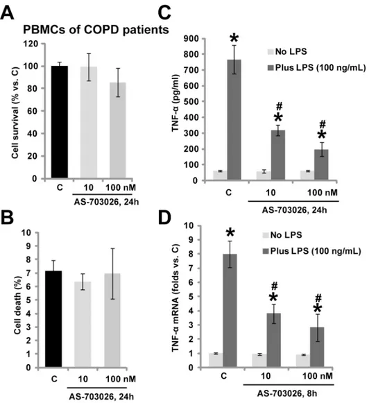
The potential role of AS-703026 in primary human monocytes was also analyzed. Weex-vivo
cultured primary PBMCs from COPD patients (Seemethods). Again, we failed to detect any
cytotoxic effects after applied AS-703026 treatment in above patients’PBMCs (Fig 3A and 3B).
Importantly, LPS-mediated TNFαproduction in the PBMCs was dramatically inhibited by
AS-703026 co-treatment (Fig 3C). Further, TNFαmRNA expression in response to LPS was also
inhibited (Fig 3D). Thus, in line with the macrophage data, in COPD patients’monocytes,
LPS-mediated TNFαproduction was again inhibited by AS-703026. We repeated those
Fig 1. AS-703026 inhibits LPS-mediated TNFαproduction in RAW 264.7 murine macrophages.RAW 264.7 cells were treated with applied
concentrations of AS-703026, and cultured for additional 24 h, cell survival was tested by MTT assay (A), while cell death was examined by trypan blue dye assay (B). RAW 264.7 cells were treated with LPS (10–250 ng/mL), or plus indicated AS-703026 co-treatment, TNFαcontent in conditional medium was tested by ELISA assay after 24 h (C and E); Relative TNFαmRNA expression was examined after 8 h (D and F).“C”stands for medium control group (Same for all figures). The results presented were representative of three independent experiments, for each assay, n = 5 (Same for all figures). Bars stand for means±SD (Same for all figures).*p<0.05 compared with“C”group (A-D).#p<0.05 vs. LPS only group (C-F).
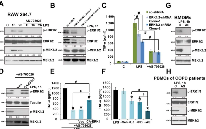
3.4. AS-703026 blocks LPS-induced MEK/ERK activation in murine
macrophages and human monocytes
Above results showed that AS-703026 inhibited LPS-induced TNFαproduction in mouse
mac-rophages and human monocytes. The underlying signaling mechanisms were also tested. As expected, LPS treatment in RAW 264.7 cells induced significant MEK/ERK activation/phos-phorylation, which was almost completely blocked by AS-703026 co-treatment (Fig 4A).
Inter-estingly, our results implied that AS-703026-mediteaed inhibition on TNFαwas not solely
dependent on MEK/ERK blockage. First, we showed that AS-703026 was still functional
(anti-TNFα) in ERK1/2 shRNA-silenced RAW264.7 cells, where LPS was less effective (Fig 4B and
4C). As expected, MEK phosphorylation was not affected by ERK1/2 shRNA (Fig 4B). Further, exogenous expression of constitutively-active (CA) ERK1, which restored ERK (but not MEK)
activation in AS-703026-treatd RAW264.7 cells (Fig 4D), only partially reinstated TNFα
Fig 2. AS-703026 inhibits LPS-induced TNFαproduction in murine BMDMs.Primary cultured murine BMDMs were treated with applied concentration of
AS-703026, and were cultured for additional 24 h, cell survival and cell death were tested by MTT assay (A) and trypan blue dye assay (B), respectively. Murine BMDMs were treated with LPS (100 ng /mL), or plus indicated concentrations of AS-703026, TNFαcontent in conditional medium (C, after 24 h) and relative TNFαmRNA expression in the BMDM cells (D, after 8 h) were tested.*p<0.05 compared with“C”group (C-D).#p<0.05 vs. LPS only group (C-D).
production by LPS (Fig 4E). Third, AS-70302 was more potent than traditional MEK/ERK
inhibitors, including PD98059 and U0126, in inhibiting LPS-mediated TNFαproduction (Fig
4F). Note that LPS-induced MEK/ERK activation was completely blocked the two traditional MEK/ERK inhibitors (Data not shown). In primary mouse BMDMs and human PMBCs, LPS-induced MEK/ERK activation was again completely blocked by AS-703026 (Fig 4G and 4H).
All these data indicate that AS-703026-induced activity against TNFαproduction appears not
solely dependent on MEK/ERK blockage.
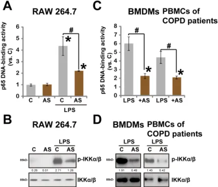
3.5. AS-703026 inhibits LPS-induced NF
κ
B activation
Above results indicate that AS-703026-exerted inhibition on TNFαproduction is not solely
dependent on MEK/ERK blockage. NFκB signaling is another important mediator of TNFα
Fig 3. AS-703026 inhibits LPS-mediated TNFαproduction inex-vivocultured PBMCs of COPD patients.Ex-vivocultured PBMCs form COPD patients were treated with applied concentration of AS-703026 (10/100 nM), cells were cultured for additional 24 h, cell survival and cell death were tested by MTT assay (A) and trypan blue dye assay (B), respectively. The PBMCs were treated with LPS (100 ng/mL), or plus AS-703026 (10/100 nM), TNFαproduction (C) and mRNA expression (D) were tested similarly.*
p<0.05 compared with“C”group (C-D).#p<0.05 vs. LPS only group (C-D).
production by LPS [6]. Thus, we studied the effect of AS-703026 on LPS-induced NFκB
activa-tion in macrophages/monocytes. To our surprise, we found that LPS-induced NFκB activation,
tested by p65 DNA-binding assay and Western blot assay of p-IKKα/β(Ser176/180), was
inhibited by AS-703026 in RAW264.7 cells (Fig 5A and 5B). Similar NFκB inhibition by
AS-703026 was also seen in BMDMs and COPD patients’monocytes (Fig 5C and 5D). Note that
LPS-induced NFκB activation was not affected by two traditional MEK/ERK inhibitors
PD98059 and U0126 (Data not shown). Meanwhile, CA-ERK1 failed to restore NFκB
activa-tion after AS-703026 treatment (Data not shown). Thus, AS-703026-mediated NFκB
inhibi-tion is unlikely associated with MEK/ERK blockage. Together, we demonstrate that AS-703026
inhibits LPS-induced NFκB activation, which could also be responsible, at least in part, for its
effect against LPS-induced TNFαproduction in macrophages/monocytes.
Fig 4. AS-703026 blocks LPS-induced MEK/ERK activation in murine macrophages and human monocytes.RAW 264.7 cells, BMDMs and PBMCs of COPD patients were treated with LPS (100 ng/mL), or plus AS-703026 (AS, 100 nM), after applied time, phosphorylated (p-) and regular MEK1/2 and ERK1/ 2 were tested by Western blots (A, G and H). Scramble shRNA (“sc-shRNA”)- or ERK1/2 shRNA (-1/-2)-expressing stable RAW 264.7 cells, pre-treated with AS-703026 (100 nM, 1 h), were stimulated with LPS (100 ng/mL), expressions of MEK1/2-ERK1/2 were tested after 1 h (B), TNFαproduction was tested by ELISA after 24 h (C). Scramble RAW 264.7 cells expressing empty vector or CA-ERK1 were stimulated with LPS (100 ng/mL), or plus AS-703026 (100 nM), expressions of indicated proteins were tested by Western blots after 1 h (D), TNFαproduction was tested after 24 h (E). TNFαproduction in RAW 264.7 cells treated with LPS (100 ng/mL, 24 h) or plus MEK/ERK inhibitors U0126 (U0, 5μM), PD98059 (PD, 5μM) or AS-703026 (AS, 100 nM) was tested (F). Kinase phosphorylation (vs. regular kinase) was quantified (A, B, D, G and H). ERK1/2 expression (vs. regular MEK1/2) was also quantified (B).*p<0.05 compared
with LPS only group (E and F).#p<0.05 (C, E and F).
3.6. AS-703026 inhibits LPS-induced endotoxin shock and TNF
α
production in BALB/c mice
Finally, the potential role of AS-703026 on LPS-induced inflammatory response was testedin
vivo. BALB/c mice were inoculatedi.p. with LPS (30 mg/kg body weight) plus D-galactosamine (300 mg/kg), the latter is a hepatotoxic transcriptional inhibitor which sensitizes the cytotoxic
effects of TNFα[22]. We demonstrated that LPS administration induced septic shock within
24 h to 48 h, causing dramatic mortality in BALB/c mice (Fig 6A). Importantly, co-administra-tion of AS-703026 (30 mg/kg) remarkably protected mice from LPS/D-galactosamine, and
endotoxin shock was largely inhibited (Fig 6A). The concentration of AS-703026in vivowas
determined based on previous studies [16,17], where no significant toxicities were observed to tested animals. Over 50% of AS-703026-co-administrated mice were still alive 48 h after LPS/ D-galactosamine challenge (Fig 6A). These surviving mice were observed for additional 5–6 days, and no late occurring toxic effects or mortality were observed (Data not shown). The
effect of AS-703026 on TNFαproduction was also analyzedin vivo. ELISA results analyzing
mice tail vein serum demonstrated that AS-703026 significantly inhibited
LPS/D-galactos-amine-induced TNFαproduction in BALB/c mice (Fig 6B). Together, we showed that
AS-703026 oral administration significantly inhibited LPS-induced TNFαproductionin vivo, and
protected mice from endotoxin shock.
Discussions
In the current study, we showed that AS-703026, a novel MEK/ERK inhibitor, dramatically
inhibited LPS-induced TNFαproduction in macrophages (primary cells and RAW 264.7 cells).
Fig 5. AS-703026 inhibits LPS-induced NFκB activation.RAW 264.7 cells, BMDMs and COPD patients’
PBMCs were treated with LPS (100 ng/mL, 2 h), or plus AS-703026 (AS, 100 nM), NFκB (p65) DNA-binding activity was analyzed, and the values were normalized to“C”group (A and C), p- and regular IKKα/βwere tested by Western blots (B and D). IKKα/βphosphorylation (vs. regular IKKα/β) was quantified (C and D).*
p<0.05 compared with“C”group (A and C).#p<0.05 vs. LPS only group (A and C).
Meanwhile, TNFαproduction in LPS-stimulated PBMCs of COPD patents was also inhibited
by AS-703026. At the molecular level, we showed that AS-703026-exerted anti-TNFαactivity
was likely mediated through MEK/ERK-dependent and -independent mechanisms.
AS-703026 is a potent MEK/ERK inhibitor [16,17]. Here we demonstrated that AS-703026 blocked MEK/ERK activation by LPS in murine macrophages and monocytes of COPD
patients. However, our results indicated that AS-703026-medaited anti-TNFαactivity
appeared not solely dependent on MEK/ERK blockage. First, we found that AS-703026 was more potent than traditional MEK/ERK inhibitors (PD98059 and U0126) in repressing
LPS-mediated TNFαproduction. Further, although exogenous CA-ERK1 restored ERK activation
in AS-703026-treated RAW 264.7 cells, it only partially reinstated TNFαproduction by LPS.
Third, AS-703026 could still inhibit LPS-induced TNFαproduction in ERK1/2-depleted RAW
264.7 cells. Thus, other mechanisms besides MEK/ERK inhibition could also be responsible for its activity in monocytes/macrophages.
As a matter of fact, one important finding of this study is that LPS-induced NFκB activation
was also inhibited by AS-703026 in macrophages and COPD patients’PMBCs. Thus,
AS-703026-mediated anti-TNFαactivity could also be due to its effect on NFκB signaling. At the
current stage, the detailed mechanisms of NFκB inhibition by AS-703026 were still under
investigation. However, it is unlikely that NFκB inhibition by AS-703026 is the consequence of
MEK/ERK blockage. Since restoring ERK activation by exogenously introducing CA-ERK1
had no effect on NFκB activation in RAW264.7 cells (Data not shown). Also, traditional MEK/
ERK inhibitors (PD98059 and U0126), or shRNA-mediated silencing of ERK1/2, were
ineffec-tive on NFκB activation in LPS-treated macrophages/monocytes (Data not shown). Thus,
NFκB inhibition is possibly an unique effect by this novel MEK/ERK inhibitor, and the detailed
signaling mechanisms warrant further investigations.
There are several advantages using this novel MEK/ERK inhibitor. AS-703026 displayed
superior efficiency in inhibiting LPS-induced TNFαproduction, more potently than traditional
Fig 6. AS-703026 inhibits LPS-induced TNFαproduction and endotoxin shock in BALB/c mice.BALB/c mice (4–6 weeks old, 6 mice per group) were
injectedi.p. with LPS (30 mg/kg body weight) and D-galactosamine (300 mg/kg body weight), or plus oral gavage of AS-703026 (30 mg/kg mice body weight), mice death was recorded 24 h and 48 h after LPS administration (A), tail vein serum samples were collected 4 h and 8 h after LPS stimulation, TNFα
content was determined by ELISA (B).In vivoexperiments were repeated three times.#p<0.05.
MEK/ERK inhibitors (PD9809 and U0126).In vitro, AS-703026 at nM concentrations could
significantly inhibit TNFαproduction in both primary and established
macrophages/mono-cytes. Further, besides exerting MEK/ERK blockage activity, AS-703026 could also inhibit
LPS-induced NFκB activation, which at least in part explains its superior activity. The another
advantage of using this novel MEK/ERK inhibitor is its oral availability. As a matter of fact, here we demonstrated that AS-703026 oral administration significantly inhibited LPS-induced endotoxin shock; Mice with AS-703026 administration were protected from LPS challenge.
Conclusions
Together, these results demonstrate that AS-703026in vitroinhibits LPS-induced TNFα
pro-duction in macrophages and COPD patients’monocytes, andin vivoprotects mice from
LPS-induced endotoxin shock. These results suggest that AS-703026 could be further studied as a useful anti-inflammatory therapy for COPD patients.
Author Contributions
Conceived and designed the experiments: PL XZ. Performed the experiments: PL YW ML XQ XB. Analyzed the data: PL YW ML XQ XB XZ. Contributed reagents/materials/analysis tools: PL XZ. Wrote the paper: PL XZ.
References
1. Barnes PJ (2013) New anti-inflammatory targets for chronic obstructive pulmonary disease. Nat Rev Drug Discov 12: 543–559. PMID:23977698
2. Brusasco V, Martinez F (2014) Chronic obstructive pulmonary disease. Compr Physiol 4: 1–31. doi:
10.1002/cphy.c110037PMID:24692133
3. Roversi S, Roversi P, Spadafora G, Rossi R, Fabbri LM (2014) Coronary artery disease concomitant with chronic obstructive pulmonary disease. Eur J Clin Invest 44: 93–102. doi:10.1111/eci.12181
PMID:24164255
4. Cosio MG, Saetta M, Agusti A (2009) Immunologic aspects of chronic obstructive pulmonary disease. N Engl J Med 360: 2445–2454. doi:10.1056/NEJMra0804752PMID:19494220
5. Lamela J, Vega F (2009) Immunologic aspects of chronic obstructive pulmonary disease. N Engl J Med 361: 1024.
6. Bryant CE, Spring DR, Gangloff M, Gay NJ (2010) The molecular basis of the host response to lipopoly-saccharide. Nat Rev Microbiol 8: 8–14. doi:10.1038/nrmicro2266PMID:19946286
7. Ruiz N, Kahne D, Silhavy TJ (2009) Transport of lipopolysaccharide across the cell envelope: the long road of discovery. Nat Rev Microbiol 7: 677–683. doi:10.1038/nrmicro2184PMID:19633680 8. Singh D, Smyth L, Borrill Z, Sweeney L, Tal-Singer R (2010) A randomized, placebo-controlled study of
the effects of the p38 MAPK inhibitor SB-681323 on blood biomarkers of inflammation in COPD patients. J Clin Pharmacol 50: 94–100. doi:10.1177/0091270009347873PMID:19880675
9. Ouagued M, Martin-Chouly CA, Brinchault G, Leportier-Comoy C, Depince A, Bertrand C, et al. (2005) The novel phosphodiesterase 4 inhibitor, CI-1044, inhibits LPS-induced TNF-alpha production in whole blood from COPD patients. Pulm Pharmacol Ther 18: 49–54. PMID:15607127
10. Miller SI, Ernst RK, Bader MW (2005) LPS, TLR4 and infectious disease diversity. Nat Rev Microbiol 3: 36–46. PMID:15608698
11. Rabinovich RA, Figueras M, Ardite E, Carbo N, Troosters T, Filella X, et al. (2003) Increased tumour necrosis factor-alpha plasma levels during moderate-intensity exercise in COPD patients. Eur Respir J 21: 789–794. PMID:12765422
12. Profita M, Chiappara G, Mirabella F, Di Giorgi R, Chimenti L, Costanzo G, et al. (2003) Effect of cilomi-last (Ariflo) on TNF-alpha, IL-8, and GM-CSF release by airway cells of patients with COPD. Thorax 58: 573–579. PMID:12832668
13. Tsai EY, Falvo JV, Tsytsykova AV, Barczak AK, Reimold AM, Glimcher LH, et al. (2000) A lipopolysac-charide-specific enhancer complex involving Ets, Elk-1, Sp1, and CREB binding protein and p300 is recruited to the tumor necrosis factor alpha promoter in vivo. Mol Cell Biol 20: 6084–6094. PMID:
14. Cho MK, Jang YP, Kim YC, Kim SG (2004) Arctigenin, a phenylpropanoid dibenzylbutyrolactone lig-nan, inhibits MAP kinases and AP-1 activation via potent MKK inhibition: the role in TNF-alpha inhibi-tion. Int Immunopharmacol 4: 1419–1429. PMID:15313439
15. Kim K, Kong SY, Fulciniti M, Li X, Song W, Nahar S, et al. (2010) Blockade of the MEK/ERK signalling cascade by AS703026, a novel selective MEK1/2 inhibitor, induces pleiotropic anti-myeloma activity in vitro and in vivo. Br J Haematol 149: 537–549. doi:10.1111/j.1365-2141.2010.08127.xPMID:
20331454
16. Park SJ, Hong SW, Moon JH, Jin DH, Kim JS, Lee CK, et al. (2013) The MEK1/2 inhibitor AS703026 circumvents resistance to the BRAF inhibitor PLX4032 in human malignant melanoma cells. Am J Med Sci 346: 494–498. PMID:24051957
17. Yoon J, Koo KH, Choi KY (2011) MEK1/2 inhibitors AS703026 and AZD6244 may be potential thera-pies for KRAS mutated colorectal cancer that is resistant to EGFR monoclonal antibody therapy. Can-cer Res 71: 445–453. doi:10.1158/0008-5472.CAN-10-3058PMID:21118963
18. Zhang JL, Xu Y, Shen J (2014) Cordycepin inhibits lipopolysaccharide (LPS)-induced tumor necrosis factor (TNF)-alpha production via activating amp-activated protein kinase (AMPK) signaling. Int J Mol Sci 15: 12119–12134. doi:10.3390/ijms150712119PMID:25007068
19. Du SL, Yuan X, Zhan S, Tang LJ, Tong CY (2015) Trametinib, a novel MEK kinase inhibitor, sup-presses lipopolysaccharide-induced tumor necrosis factor (TNF)-alpha production and endotoxin shock. Biochem Biophys Res Commun 458: 667–673. doi:10.1016/j.bbrc.2015.01.160PMID:
25684183
20. Sato AY, Tu X, McAndrews KA, Plotkin LI, Bellido T (2015) Prevention of glucocorticoid induced-apo-ptosis of osteoblasts and osteocytes by protecting against endoplasmic reticulum (ER) stress in vitro and in vivo in female mice. Bone 73C: 60–68.
21. Livak KJ, Schmittgen TD (2001) Analysis of relative gene expression data using real-time quantitative PCR and the 2(-Delta Delta C(T)) Method. Methods 25: 402–408. PMID:11846609