PONTIFÍCIA UNIVERSIDADE CATÓLICA DO RIO GRANDE DO SUL FACULDADE DE ODONTOLOGIA
PROGRAMA DE PÓS-GRADUAÇÃO EM ODONTOLOGIA MESTRADO EM ODONTOLOGIA
ÁREA DE CONCENTRAÇÃO EM ENDODONTIA
AVALIAÇÃO IN VITRO DO POTENCIAL DE DESCONTAMINAÇÃO DE CANAIS RADICULARES INFECTADOS COM ENTEROCOCCUS FAECALIS E
DA CAPACIDADE DE DISSOLUÇÃO TECIDUAL ATRAVÉS DO USO DA TERAPIA FOTODINÂMICA E DO HIPOCLORITO DE SÓDIO
MATHEUS ALBINO SOUZA
PONTIFÍCIA UNIVERSIDADE CATÓLICA DO RIO GRANDE DO SUL FACULDADE DE ODONTOLOGIA
PROGRAMA DE PÓS-GRADUAÇÃO EM ODONTOLOGIA MESTRADO EM ODONTOLOGIA
ÁREA DE CONCENTRAÇÃO EM ENDODONTIA
MATHEUS ALBINO SOUZA
AVALIAÇÃO IN VITRO DO POTENCIAL DE DESCONTAMINAÇÃO DE CANAIS RADICULARES INFECTADOS COM ENTEROCOCCUS FAECALIS E
DA CAPACIDADE DE DISSOLUÇÃO TECIDUAL ATRAVÉS DO USO DA TERAPIA FOTODINÂMICA E DO HIPOCLORITO DE SÓDIO
Dissertação apresentada como parte dos requisitos obrigatórios para a obtenção do título de Mestre em Odontologia, área de concentração em Endodontia.
ORIENTADOR: PROF. DR. JOSÉ ANTÔNIO POLI DE FIGUEIREDO
“Nas grandes batalhas
da vida, o primeiro passo para a
vitória é o desejo de vencer”.
AGRADECIMENTOS
A Deus, que me iluminou, me fez seguir em frente nas vitórias, me ergueu nas dificuldades e me encorajou a ir até o fim.
Aos meus pais, Antônio e Sandra, exemplos de vida que me deram educação, ensinaram valores, abdicaram de seus sonhos para que eu tivesse a oportunidade de realizar o meu, que não mediram esforços em mostrar o caminho correto e que me incentivaram fazendo seguir adiante fossem quais fossem os obstáculos – amor eterno.
Aos meus irmãos, Diogo e Tiago, que me ensinaram tantas coisas, em quem sempre me espelhei por tudo que conquistaram, pela amizade, companheirismo, incentivo, cumplicidade e torcida incondicional durante todo esse período.
Ao diretor da Faculdade de Odontologia da PUC/RS, Prof. Marcos Túlio Mazzini Carvalho, e ao coordenador do Programa de Pós-Graduação em Odontologia da PUC/RS, Prof. Dr. José Antônio Poli de Figueiredo, que viabilizaram esta oportunidade de aprendizado e crescimento.
Ao CNPq, pelo fornecimento da bolsa que me possibilitou suporte financeiro para a conclusão deste mestrado.
Ao meu orientador, Prof. Dr. José Antônio Poli de Figueiredo, cuja sabedoria, trabalho, conhecimento e dedicação à ciência são admiráveis. Agradeço pela acolhida e comprometimento com o desenvolvimento da dissertação. A contribuição para o meu crescimento pessoal e profissional foi imensurável neste período. Seus ensinamentos, idéias, teorias, conselhos, percepções e, principalmente, a sua amizade jamais serão esquecidos. Obrigado pelo convívio e pela honra de desenvolver esse trabalho em conjunto, estimado Mestre.
Aos professores, Dr. Caio Cézar Randi Ferraz e Dra. Brenda Paula Figueiredo de Almeida Gomes, pelos conhecimentos teóricos e práticos transmitidos durante o curso de especialização e que até hoje fazem parte do meu cotidiano acadêmico e clínico. É muito bom saber que, apesar da distância e do transcorrer do tempo, ainda posso contar com a amizade, o convívio, o trabalho, o incentivo e a torcida contínua de vocês.
Aos professores do Programa de Pós-Graduação em Odontologia, área de concentração em Endodontia, Dr. Eraldo Batista, Dra. Maria Martha Campos, Dr. João Webber e Dra. Patrícia Kopper, pelas aulas e ensinamentos transmitidos, pela disposição e atenção de sempre, sendo fundamentais para a conclusão de mais uma etapa. A presença de vocês foi gratificante para a obtenção do aprendizado.
Aos professores do departamento de Endodontia da Graduação da Faculdade de Odontologia, Ms. Alexandre Ghisi, Dra. Fabiana Vier, Dr. Márcio Menin, Dra. Maristela Borba e Dra. Simone Luisi, pelo convívio do dia-a-dia na clínica de Endodontia, pelas sugestões, questionamentos e propostas a cerca da realização deste trabalho. A amizade, experiência e o amparo de vocês foram indispensáveis nesta conquista.
À professora, Dra. Silvia Dias de Oliveira, e ao aluno de graduação e iniciação científica em Biologia, Valdir Barth Júnior, pelo auxílio na realização da parte microbiológica do experimento da dissertação. Agradeço pelo convívio, pela troca de conhecimentos e por não me deixarem desanimar quando as amostras contaminavam com microorganismos que não deveriam contaminar. Aprendi muito mais que biologia, microbiologia e bactérias, aprendi sobre amizade, paciência, cuidados, determinação e dedicação exaustiva beirando a perfeição. Sem vocês, essa dissertação jamais estaria sendo escrita.
Ao professor, Dr. Mario Wagner, pelo auxílio e competência na realização da análise estatística desse trabalho.
Ao funcionário do Centro de Microscopia e Microanálises, Maurício França, pelo auxílio no preparo, processamento e aquisição de imagens das amostras na microscopia eletrônica de varredura. Suas conversas e histórias ajudaram o tempo a passar um pouco mais rápido, quando a análise das imagens beirava o sono. Sua dedicação, agilidade e precisão foram peças-chave para complementação deste trabalho.
Fernanda Lopez, Grasiela Grundling, Janaina Kufner, Kathrein Tapia, Larissa Klassmann, Lenara Dondoni, Marilia Burgel, Pabla Sechi, Renata Morgental e Roberta Scarparo, pela amizade e companheirismo ao longo desses dois anos.
Aos amigos e colegas de Endodontia, Dr. Doglas Cecchin e Dr. Francisco Montagner, pelas conversas, troca de conhecimentos, pelos momentos de descontração, pelos trabalhos realizados juntos, pelo auxílio e atenção de sempre, e, acima de tudo, pela enorme amizade constituída e fortalecida ao longo desses anos. É um prazer e uma honra ter pessoas como vocês do meu lado. Não tenho dúvidas que sempre poderei contar com vocês pra tudo e tenham certeza que não medirei esforços pra retribuir. A consideração que tenho a vocês vai além de um simples muito obrigado. Devo muito a vocês, pessoas especiais que eternamente serei grato por tudo que me acrescentaram.
Aos alunos da graduação, ATO 2010 (turmas A e C), ATO 2011 (turmas A e C) e ATO 2012 (turma A), pela confiança e tranqüilidade transmitidas durante a realização do estágio docente, requisito obrigatório para a conclusão do Mestrado.
Aos funcionários da Secretaria do Programa de Pós-Graduação em Odontologia, Ana Prestes, Davenir Brusch, Marcos Corrêa e Paulo Silva, pela simpatia, competência e disponibilidade no trabalho realizado.
À funcionária do almoxarifado da clínica de Endodontia, Luciane Gonçalvez, sempre prestativa no dia-a-dia da clínica e que ajudava a amenizar o longo e exaustivo turno de trabalho.
Às funcionárias da esterilização, Daena Viegas e Denise Barbosa, pela organização, pontualidade e cuidado na esterilização das amostras utilizadas no experimento.
Ao amigo, Julio Vogt, com quem dividi residência e me transmitiu tudo aquilo que eu deveria saber sobre a capital no meu primeiro ano em Porto Alegre. Agradeço pela paciência, por dividir seu espaço, pelo convívio e pela amizade que dura até hoje.
E, concluindo, agradeço a todos que, de alguma forma, contribuíram para a realização deste trabalho.
ARTIGO 1 RESUMO
O objetivo deste estudo foi comparar, in vitro, a ação da terapia fotodinâmica com a ação do hipoclorito de sódio em canais radiculares infectados com Enterococcus
faecalis. Os canais radiculares de 45 dentes humanos unirradiculares foram ampliados
até a lima 60, autoclavados, inoculados com uma suspensão de Enterococcus faecalis e incubados por 60 dias, com renovação do meio de cultura de 48 em 48 horas. Após o período de contaminação, as amostras foram divididas aleatoriamente em cinco grupos, de acordo com o protocolo de descontaminação: G1 (n=5) controle positivo – nenhum procedimento; G2 (n=10) controle negativo – água destilada; G3 (n=10) – PAD; G4 (n=10) – NaOCl 2,5%; G5 (n=10) – NaOCl 2,5% + PAD. Posteriormente, as amostras foram fixadas em glutaraldeído 25%, desidratadas, secas, metalizadas e submetidas à microscopia eletrônica de varredura (MEV). A avaliação foi feita em imagens do MEV com o aumento de 5.000x para a luz do canal e de 10.000x para a interface dentinária, ambos em backscattering (BS), observando o efeito dos tratamentos propostos nos diferentes terços do canal radicular. A presença de bactérias foi classificada por intermédio de um rankeamento de posições de 1 a 45, onde quanto maior o valor, maior o grau de limpeza da amostra. Os dados de níveis de contaminação foram submetidos ao teste de ANOVA de uma via, seguido pelo post-hoc de Tukey (α=0,05). Os resultados mostraram que o grupo 5 apresentou os maiores valores na média de rankeamento de posições nos três terços do canal radicular, tanto na luz do canal quanto na interface dentinária. Com isso, foi o mais efetivo na eliminação de Enterococcus faecalis. Diante do exposto, é possível concluir que a associação NaOCl 2,5% + PAD foi eficiente na eliminação de Enterococcus faecalis presentes na parede dentinária e na profundidade de penetração dos túbulos dentinários.
ARTIGO 2 RESUMO
O objetivo deste estudo foi avaliar a capacidade de dissolução do tecido pulpar bovino através da ação da terapia fotodinâmica. Vinte fragmentos de tecido pulpar bovino foram pesados e divididos aleatoriamente dentro de quatro grupos (n=5), de acordo com o mecanismo de dissolução testado: G1 – água destilada (controle negativo); G2 – hipoclorito de sódio 1% (controle positivo); G3 – terapia fotodinâmica; G4 – hipoclorito de sódio + terapia fotodinâmica. A observação dos eventos de dissolução foi realizada por dois observadores cegados em relação ao teste, com lupa de 2x de aumento, sendo anotado, em minutos, o tempo até que ocorresse a completa dissolução do tecido. O período total de observação foi de 2 horas. O tempo necessário para dissolução foi registrado em minutos (min) e a velocidade de dissolução foi calculada dividindo o peso do fragmento pulpar (mg) pelo tempo de dissolução (mg/min). Os resultados mostraram que somente o grupo 2 (NaOCl) foi capaz de promover a dissolução completa do tecido pulpar. Nos demais grupos não houve a ocorrência da dissolução completa das amostras. A média do tempo de dissolução para as amostras do grupo 2 (NaOCl) foi de 1.26 mg/min. Em conclusão, somente o hipoclorito de sódio foi capaz de dissolver os fragmentos de tecido pulpar bovino e a terapia fotodinâmica não apresentou capacidade de dissolução tecidual.
LISTA DE TABELAS DO ARTIGO 1
Tabela 1 – média e desvio padrão dos ranks de posições dos grupos testados nos terços apical, médio e cervical da parede do canal e interface dentinária...21
LISTA DE FIGURAS DO ARTIGO 1
Figura 1 – gráficos mostrando as médias dos ranks de posições dos grupos testados nos terços apical, médio e cervical da parede do canal e interface dentinária...22
Figura 2 – microfotografias do MEV (5.000x – parede do canal) dos grupos testados no estudo...23
Figura 3 - microfotografias do MEV (10.000x – interface dentinária) dos grupos testados no estudo...23
LISTA DE TABELAS DO ARTIGO 2
SUMÁRIO
1 INTRODUÇÃO GERAL………...1
2 ARTIGO 1...5
2.1 Abstract...7
2.2 Introduction...8
2.3 Material and methods...9
2.4 Results...12
2.5 Discussion...13
2.6 References...16
2.7 Table 1...21
2.8 Figure 1...22
2.9 Figure 2...23
2.10 Figure 3...23
3 ARTIGO 2...24
3.1 Abstract...26
3.2 Resumo...27
3.3 Introduction...28
3.4 Material and methods...29
3.5 Results...30
3.6 Discussion...30
3.7 Conclusion...32
3.8 References...32
3.9 Table 1...35
4 DISCUSSÃO GERAL...36
5 REFERÊNCIAS...41
6 ANEXOS...47
1
1 INTRODUÇÃO GERAL
Diversos estudos têm sido conduzidos na endodontia em busca de um protocolo de limpeza que apresente quatro propriedades principais: atividade antimicrobiana, não-tóxico aos tecidos periapicais, solúvel em água e capacidade de dissolver matéria orgânica (KURUVILLA & KAMATH, 1998).
A grande maioria das alterações patológicas que acometem a polpa e os tecidos perirradiculares é de natureza inflamatória e de etiologia microbiana. Bactérias e seus produtos exercem um papel significativo na indução e, principalmente, na perpetuação de tais doenças (SUNDQVIST, 1976). Mais de cem espécies bacterianas diferentes, muitas potencialmente patogênicas, têm sido isoladas de canais radiculares infectados, com grande prevalência de bactérias anaeróbias estritas (HAAPASALO, 1989).
As espécies aeróbias ou anaeróbias facultativas também têm sido encontradas na microbiota endodôntica, associadas às infecções persistentes ou secundárias. Estas espécies podem comprometer o sucesso da terapia endodôntica, destacando-se entre elas o Enterococcus faecalis (MOLANDER et al., 1998; SUNDQVIST et al., 1998).
O Enterococcus faecalis é um microorganismo anaeróbio facultativo (RÔÇAS et al., 2004), altamente resistente e desempenha um papel importante na etiologia de lesões perirradiculares persistentes após o tratamento de canais radiculares, sendo freqüentemente encontrado nos casos de insucesso endodôntico (EVANS et al., 2002).
Tal microorganismo possui diversos fatores de virulência, incluindo enzimas líticas, citolisinas e ácido lipoprotéico, sendo capaz de catabolizar uma grande variedade de fontes de energia, permitindo, dessa forma, sua sobrevivência em ambientes de extremo pH alcalino (TENDOLKAR et al., 2003) e com ausência de nutrientes em um estado metabólico mínimo (FIGDOR et al., 2003). Além disso, é capaz de suprimir a ação de linfócitos, contribuindo potencialmente para o insucesso do tratamento endodôntico (LEE et al., 2004).
Não se limitando aos seus fatores de virulência, o Enterococcus faecalis é também capaz de distribuir esta virulência entre outras espécies bacterianas, formando um biofilme, que o ajuda a ser resistente à destruição por intermédio de células fagocitárias, perpetuando a patologia (JETT et al., 1994).
2
endodônticas persistentes. Nas infecções endodônticas primárias, o Enterococcus
faecalis está associado com lesões perirradiculares crônicas assintomáticas do que com
abscesso perirradicular agudo ou periodontite apical aguda, podendo ser encontrado em até 40% dos casos de infecção endodôntica primária (RÔÇAS et al., 2004). Nas infecções endodônticas persistentes, a percentagem de Enterococcus faecalis tem se mostrado muito mais elevada quando comparada às infecções endodônticas primárias, o que evidencia a sua capacidade de induzir patologias perirradiculares por sobreviver aos procedimentos intracanais de desinfecção e persistir como patógeno no interior dos canais radiculares (PINHEIRO et al., 2003), auxiliando, dessa forma, na reorganização de um novo biofilme (SEMENOFF et al., 2010).
Por outro lado, a permanência de tecido pulpar residual, dentina infectada ou bactérias no interior dos canais radiculares pode ser a responsável pelo insucesso do tratamento endodôntico (MOORER & WESSELINK, 1982). Os agentes irrigantes devem apresentar a capacidade de dissolver a polpa remanescente (ZEHNDER, 2006), uma vez que a remoção do tecido pulpar é inadequada com o preparo mecânico isoladamente, devido às complexidades anatômicas do sistema de canais radiculares (THÉ, 1979).
A maioria das bactérias encontradas na microflora dos canais radiculares pode ser removida, simplesmente, através da ação mecânica dos instrumentos endodônticos. No entanto, devido à complexidade anatômica do sistema de canais radiculares, bactérias e resíduos orgânicos localizados profundamente nos túbulos dentinários, bem como em regiões de istmos e reentrâncias, podem não ser alcançados (FERRAZ et al., 2001).
Diversas substâncias e recursos têm sido utilizados durante e imediatamente após o preparo dos canais radiculares no intuito de remover detritos e tecido pulpar necrótico, além de ajudar na neutralização dos microorganismos que não puderam ser eliminados pela instrumentação mecânica (SAFAVI et al., 1990).
O hipoclorito de sódio é a substância química auxiliar mais freqüentemente utilizada na endodontia atualmente (IZU et al., 2004). Este composto apresenta uma série de propriedades e vantagens, entre as quais se inclui a capacidade de dissolver matéria orgânica (ZEHNDER et al., 2003; OKINO et al., 2004) e o amplo espectro antibacteriano que possibilita a eliminação efetiva de microorganismos do canal radicular (JEANSONNE & WHITE, 1994) e do interior dos túbulos dentinários (BUCK
3
capacidade de dissolução tecidual do hipoclorito de sódio, onde quanto maior a concentração e a temperatura, maior a atividade antimicrobiana e mais rápida a dissolução tecidual (KOSKINEN et al., 1980; ABOU-RASS & JASTRAB, 1992).
Além disso, o hipoclorito de sódio apresenta outras propriedades e vantagens como baixa tensão superficial representando sua elevada capacidade de penetração, desodorizador, clareador de dentina, lubrificante e, ainda, detergente, pois promove a saponificação de lipídios (BLOOMFIELD & MILES, 1979).
Entre as suas desvantagens, é instável ao armazenamento e é inativado por matéria orgânica (LOPES & SIQUEIRA JR., 2004), é extremamente citotóxico quando extravasado no interior dos tecidos perirradiculares (CHANG et al., 2001), diminui a resistência à fratura dos dentes e a resistência de união dos materiais restauradores à dentina (CECCHIN et al., 2009).
Segundo alguns estudos, o hipoclorito de sódio é capaz de desinfetar os túbulos dentinários e a dentina radicular contaminada por Enterococcus faecalis (SIQUEIRA et
al., 1997; DUNAVANT et al., 2006; BERBER et al., 2006) e apresenta capacidade de
dissolução tecidual (ZEHNDER et al., 2003; OKINO et al., 2004).
Entre as alternativas existentes no intuito de realizar uma eficiente limpeza e eliminação dos agentes irritantes do interior do canal radicular está a terapia fotodinâmica, que consiste na aplicação de um feixe de laser de baixa intensidade de potência associado a um meio condutor (KIMURA & WILDER-SMITH, 2000).
Uma vantagem desta nova técnica é que as complexidades anatômicas não se constituem uma barreira no processo de eliminação dos agentes irritantes do interior do canal, uma vez que o feixe de laser aplicado a partir da superfície coronária pode ser orientado em múltiplas direções através dos túbulos dentinários, atuando efetivamente nessas regiões (ODOR et al., 1996).
4
estudada como um achado promissor no intuito de erradicar bactérias orais patogênicas que causam cáries dentárias e periodontites apicais (WILSON, 2004).
No entanto, assim como as demais alternativas de descontaminação do canal radicular, a terapia fotodinâmica apresenta algumas desvantagens. A quantidade de calor liberada, apesar de ser variável, apresenta potencial para carbonizar a dentina, anquilosar raízes, dissolver cemento e causar reabsorção radicular, bem como, necrose perirradicular, ao mesmo tempo em que microorganismos são fotosensitizados (BAHCALL et al., 1992; HARDEE et al., 1994).
Diversos estudos têm mostrado que a terapia fotodinâmica apresenta potencial para maximizar a desinfecção do canal radicular (BERGMANS et al., 2008; MEIRE et al., 2009; LIM et al., 2009; SOUZA et al., 2010). No entanto, a real capacidade de limpeza da terapia fotodinâmica é um aspecto ainda a ser elucidado. Além disso, não há pesquisas relatando o comportamento da terapia fotodinâmica na dissolução de tecido orgânico.
Diante do problema exposto, caracterizado pela presença de canais radiculares infectados por microorganismos mais resistentes, como o Enterococcus faecalis, bem como pela presença de tecido orgânico remanescente não removido pelo preparo químico-mecânico, torna-se necessária a busca de meios que promovam uma eficiente descontaminação do canal radicular e que apresentem uma contribuição com o processo de dissolução tecidual.
O presente estudo compreende dois trabalhos apresentados sob a forma de artigos científicos. O primeiro artigo teve como objetivo comparar, in vitro, a ação da terapia fotodinâmica com a ação do hipoclorito de sódio em canais radiculares infectados com
Enterococcus faecalis, analisando o potencial de descontaminação nas paredes
5
2 ARTIGO 1
6
In vitro effect of photodynamic therapy and sodium hypochlorite on root canal system infected with Enterococcus faecalis
Matheus Albino Souza1, Liviu Steier2, Giampiero Rossi-Fedele1,2, Silvia Dias de Oliveira3, Valdir Barth Júnior3, José Antônio Poli de Figueiredo1
1
School of Dentistry, Pontificial Catholic University of Rio Grande do Sul, Porto Alegre, RS, Brazil.
2
Warwick Dentistry, Warwick Medical School, Warwick, United Kingdom.
3
School of Biosciences, Pontificial Catholic University of Rio Grande do Soul, Porto Alegre, RS, Brazil.
Address to correspondence: José Antonio Poli de Figueiredo. Post-Graduate Program in Dentistry -
Pontifical Catholic University of Rio Grande do Sul – PUCRS. Av. Ipiranga 6681 Prédio 6 sala 507. CEP
7
ABSTRACT
Introduction: Photodynamic therapy has been proposed to improve to root canal desinfection, but its role on certain bacterial species needs to be further ellucidated. Objective: to compare, in vitro, the action of photodynamic therapy and sodium hypochlorite in root canals infected with Enterococcus faecalis.
Methods: The root canals of 45 single-rooted human extracted teeth were enlarged up to a file 60, autoclaved, inoculated with a suspension of Enterococcus faecalis and incubated for 60 days, with renewal of the culture medium every 48 hours. After that, samples were divided into five groups according to the protocol of decontamination: G1 (n = 5) positive control - no procedure; G2 (n = 10) negative control - distilled water; G3 (n = 10) - PAD, G4 (n = 10) – NaOCl 2.5%; G5 (n = 10) - NaOCl 2.5% + PAD. Subsequently, samples were fixed on 25% glutaraldehyde, dehydrated, dried, metalized and subjected to scanning electron microscopy (SEM). The assessment was made of images taken at every root third at 5.000x magnification for the canal wall and 10.000x for the exposed tubule area, both in backscattering (BS), noting the effect of proposed treatments to the different thirds of the root canal. The presence of bacteria in the images was scored by position ranks from 1 to 45, where the higher the value, the cleanliness of the sample. Data were subjected to One-way ANOVA followed by post-hoc Tukey test (α = 0.05).
Results: Group 5 had the highest mean position ranks for all thirds of the root canal, both in canal wall and in the exposed tubule area. Thus, it was the most effective in eliminating Enterococcus faecalis.
Conclusion: the association NaOCl 2,5% + PAD is an efficient protocol for the elimination of Enterococcus faecalis present in the dentinal surface and deep into the dentinal tubules.
8
INTRODUCTION
Most pathological changes affecting pulp and periradicular tissues are inflammatory in nature and of microbial etiology. Bacteria and their products play a significant role in the induction and especially the perpetuation of such conditions (1). Despite the prevalence of anaerobic bacteria within infected root canals (2), aerobic or facultative anaerobic species have also been found in endodontic microbiota, foremost amongst them Enterococcus faecalis (3,4).
Enterococcus faecalis is a facultative anaerobic microorganism highly resistant (5)
which plays an important role in the etiology of persistent periradicular lesions, frequently found in cases of endodontic failure (6), helping in the reorganization of a new biofilm (7). It has several virulence factors, including lytic enzymes, acid and lipoprotein cytolysin, being able to catabolize a wide variety of energy sources, enabling thus their survival in environments of extreme alkaline pH (8) and lack of nutrients in a minimum metabolic state (9). Furthermore, it is able to suppress the action of lymphocytes (10) and distribute their virulence among other bacterial species, forming a biofilm that becomes resistant to destruction by phagocytes (11).
Most bacteria found in root canal microbiota can be removed simply by mechanical action of endodontic instruments. However, due to anatomical complexities, bacteria and organic residues located deep in the dentinal tubules as well as in isthmus and recesses, may not be achieved (12), thus requiring the use of auxiliary chemical substances and alternative resources to promote decontamination.
Sodium hypochlorite is the most often used irrigating substance to assist current Endodontics (13). This compound has a number of properties and advantages, which include the broad spectrum antimicrobial activity (14,15,16) and the ability to dissolve organic matter (17,18). On the other hand, is highly cytotoxic when extravasated within the periradicular tissues (19), reduces fracture resistance of teeth (20) and affects negatively bond strength of restorative materials to dentin (21).
9
The laser beam applied from the coronal surface can be oriented in multiple directions through dentin tubules, acting effectively in these regions (26). However, the actual cleaning ability of photodynamic therapy needs further elucidation.
The aim of this study was to compare the in vitro action of photodynamic therapy and sodium hypochlorite in infected root canals with Enterococcus faecalis, analyzing the potential decontamination on canal walls and dentinal tubules.
MATERIAL AND METHODS
Sample obtaining and specimen preparation
The study was approved by the Ethics Committee of the Pontifical Catholic University of Rio Grande do Sul (protocol 1637/09). Forty five extracted human lower premolars with a single root canal were used for the present study, with straight roots, obtained from the tooth bank of the Faculty of Dentistry of the Pontifical Catholic University of Rio Grande do Sul. The teeth were stored in 4% formalin by the beginning of the experiment in order to preserve their properties. The crown portion was sectioned with diamond disk in the cement-enamel junction, so that the remaining roots presented a length of approximately 15 mm. The samples were sterilized in autoclave for 30 minutes at a temperature of 120o C.
Root canal preparation
10
Culture and inoculum preparation
The reference strain used, Enterococcus faecalis (ATCC 19433), was obtained and activated in the Laboratory of Immunology and Microbiology, Faculty of Biosciences, Pontifical Catholic University of Rio Grande do Sul. The bacteria were grown in BHI (Brain Heart Infusion), by 18 to 24 hours at 37 ° C in oven bacteriology.
The number of colony forming units (CFU) / mL of inoculum was determined by counting on blood agar plates. For this, the cultivation of Enterococcus faecalis was diluted serially at 10-8 in saline 0.85% and 0.1 mL aliquots of the dilutions 10-6, 10-7 and 10-8 were planted by scattering technique on blood agar, in duplicate. After incubation for 24 hours in a bacteriological incubator, the number of CFU / mL was determined.
The 45 samples were handled with sterile forceps in a laminar flow cabinet, immersed in closed test tubes containing 2 mL of BHI, which were inoculated 100 µ L of overnight culture of Enterococcus faecalis. The samples were kept immersed in the bacterial culture for 60 days at a temperature of 37oC, so there would be the formation of biofilms. One third of the volume of BHI was renewed every 48 hours, continuously providing the nutrients needed to sustain bacterial growth.
Classification of treatment groups
In a laminar flow cabinet, samples were fixed in a wax base previously decontaminated to receiving each of the specimens. Initially, all root canals were irrigated with 2 ml of distilled water to remove planktonic bacteria and dried by aspiration. The samples were then randomly divided into five groups according to the method used to promote clean root canal:
Group 1 (n = 5) positive control - no decontamination procedure was performed. Group 2 (n = 10) negative control - the root canal was filled with distilled water (Decloquimis, São Paulo, Brazil). Agitation of the solution with size 15 k-type hand file was performed for 60 seconds. The solution remained within the root canal for 3 minutes, being renewed after this period for another cycle. In total, there were four cycles of decontamination.
11
working length. Activation of low intensity laser was performed for a period of 120 seconds and an intensity of 120 mW, the tip remaining stationary, according to the manufacturer's instructions.
Group 4 (n = 10) - the same root canal irrigation protocol as performed in group 2 was adopted for this group, changing the irrigating substance, which was 2.5% sodium hypochlorite solution (Lírios, São Vicente, Brazil).
Group 5 (n = 10) - the root canal was filled with 2.5% sodium hypochlorite (Lírios, São Vicente, Brazil). Agitation of the solution with size 15 k-type hand file was performed for 60 seconds. The solution remained within the root canal for 3 minutes, being renewed after this period for another cycle. In total, there were four cycles of decontamination with this solution. After, neutralization of sodium hypochlorite was performed for photodynamic therapy protocol, which was done in the same manner as described for group 3.
For all groups, the protocol was completed by active irrigation with 2 ml of distilled water, aspiration and drying of root canals with absorbent paper cones.
SEM preparation
The samples were fixed in 25% glutaraldehyde, dehydrated in ascending series of ethanol and dried to critical point. Longitudinal grooves were performed on free surfaces of the roots with a diamond disc, being careful to not invade the root canal. A complete fracture occurred with a number 50 spatula (SS White, Rio de Janeiro, Brazil) providing two halves of each sample, which were placed on stubs for subsequent metallization with palladium gold.
Classification of results
12
The assessment was made on the BS images with 5.000x magnification for the canal wall and 10.000x for the exposed tubule area. Observations by thirds looked at the effect of treatments proposed to the full extent of the root canal.
A blind calibrated examiner assessed the presence of bacteria through a position rank score system in which a higher value of position would indicate a higher level of cleanliness in the canal wall and exposed tubule area, at each third of the root canal. Forty-five images were classified from 1 to 45, in each separate third.
Statistical analysis
Data on position rank score means of contamination were subjected One-way ANOVA followed by post-hoc Tukey test, with α set at 0.05. Data were analyzed using SPSS version 17.0.
RESULTS
The mean position ranks and standard deviation of samples subjected to different decontamination protocols are shown in Table 1. Figure 1 also demonstrates the difference in the mean position ranks between the groups in canal walls and exposed tubule areas respectively. Figures 2 and 3 show SEM photomicrographs of the groups tested at the canal wall (5.000x) and exposed tubule area (10.000x) respectively.
Group 5 showed the highest mean ranks in all three thirds of the root canal, both in canal walls and exposed tubule areas.
The post-hoc Tukey test denoted, in the canal wall at the apical third, higher mean values for group 5 than other groups, and group 4 had greater values than groups 1, 2 and 3 (p < 0.05). At the exposed tubule area in the same third, groups 3, 4 and 5 were higher that groups 1 and 2 (p < 0.05) with no statistical difference between them.
In the medium third, at the canal wall, groups 4 and 5 scored higher that groups 1, 2 and 3 (p < 0.05) but not statistically different between them. Group 3 had greater scores than groups 1 and 2 (p <0.05). At the level of the exposed tubule area, groups 3, 4 and 5 scored higher that groups 1 and 2 (p < 0.05) with no statistical difference between them.
13
< 0.05) and did not differ statistically from group 4. Groups 3 and 4 scored higher than groups 1 and 2 (p < 0.05) with no statistical difference between them.
DISCUSSION
One of the most important factors for achieving success in root canal therapy is an effective decontamination of root canal system. For this, chemical substances and alternative resources are available to perform their role concurrently with the mechanical action of endodontic instruments.
Enterococcus faecalis is a highly resistant facultative anaerobic microorganism (5)
which plays an important role in the etiology of persistent periradicular lesions, frequently found in cases of endodontic failure (6). In this study, the samples were contaminated with Enterococcus faecalis, coming widely used as a marker for in vitro microbiological studies. This is because it proves to be able to successfully colonize the root canal in the form of biofilm, and invade dentinal tubules resisting to some root canal decontamination procedures (27-29).
The time of contamination of samples is a highly variable factor in the literature. Periods of 24 hours (30,31), 48 hours (32,33), 7 days (15,34-36), 14 days (37) and 21 days (38) of contamination with Enterococcus faecalis were used previously. However, this study determined the contamination for a period of 60 days, similar to a previous study (39). It is believed that a prolonged period of contamination and spread of
Enterococcus faecalis is necessary to visualize the formation of a more consistent and
organized biofilm, allowing adequate colonization in all areas of the root canal system. Thus, decontamination protocols can be tested effectively.
In some models the contamination is carried out on bovine teeth (34,37). These teeth are commonly used because their anatomical characters of dentinal tubules are very similar to human teeth in quantity, morphology and density, plus the ease of obtaining (40,41). This study used extracted human teeth, similar to previous studies (30-33,36), since the diameter and length of the tubules are small compared to bovine teeth. Thus, the present study adequately simulates the real clinical and anatomical conditions found in root canal system.
14
decontamination (15,30,35,36,38), characterizing the chemo-mechanical preparation. The choice for previous preparation in the present study considered the assessment of the actual effect and intrinsic potential of decontamination protocols. Furthermore, it provided adequate space for carrying out the inoculation phase, since premolars can contain constricted canals. Together with that, adequate penetration and adaptation of the tip of the laser equipment was easily achieved.
SEM images in backscattering (BS) at magnifications of 5.000x for the canal walls and 10.000x for exposed tubule areas were used to evaluate the effect of the treatments proposed in the study. It differed from other many studies, where samples were collected from the root canal, plated, followed by culturing and counting of colony forming units (CFUs) (30-39). The use of SEM for evaluation can be justified by the fact that in many cases the failure of root canal treatment is due to the persistence of bacteria in areas that are inaccessible by chemo-mechanical preparation. Therefore, this method of assessment allows a better visualization of microorganisms remaining in these areas. This does not occur when the counting of CFUs, where the collection is limited to bacteria located in the canal wall and space.
The use of BS feature provides a clearer vision and accuracy of microbial components tested in the study, allowing more easily differentiate the bacteria found in dirt samples. Moreover, the evaluation on canal wall and in exposed tubule area, allows a real demonstration of the effect of the treatments proposed, since an efficient decontamination should cover not only the microbial content existing in walls of the main canal, but also microorganisms that penetrate deep into the dentinal tubules.
The results of this study showed that the group of 2.5% sodium hypochlorite + photodynamic therapy had the highest position rank means of the groups tested in the apical, middle and coronal thirds, on the level of canal walls and exposed tubule areas. Even when there was no statistically significant difference when comparing groups with similar results, the mean ranks of group 5 were the greatest among them. This shows that was the group which had been more effective in eliminating Enterococcus faecalis from inside the root canal system. Similar results were found in a previous study (36), which assessed the antimicrobial effect of photodynamic therapy as an adjunct to root canal preparation using 2.5% sodium hypochlorite as auxiliary chemical substance. The groups which the association was performed were more effective in eliminating
15
sodium hypochlorite and by directing the light source through dentinal tubules. Thus, the complexities do not constitute an anatomical barrier in the process of removal of irritants from inside the root canal.
Similar results of this study were also found in a previous study (33), where evaluating the single use of 2.5% sodium hypochlorite and photodynamic therapy was found that these decontamination protocols showed higher results than control groups, no statistically significant difference between them. As in the present study, although not denoting much difference, the groups where 2.5% sodium hypochlorite was used as a decontamination protocol showed the highest decontamination in samples inoculated with Enterococcus faecalis.
The best results for the association of sodium hypochlorite and PAD occured at the apical third. Possibly, the fact that the light source was placed at 2 mm short of working length could have influenced these results. It is feasible to speculate that similar light exposures should contemplate the medium and coronal thirds. Although this could increase time of the procedure, it could be a therapeutic choice considering the results found in the apical third.
Photodynamic therapy alone was inferior to 2.5% sodium hypochlorite and association of 2.5% sodium hypochlorite + photodynamic therapy. These findings are contrary to those found by previous study (31), which found superior results with sodium hypochlorite. This can be explained by the low concentration of sodium hypochlorite used in that study, which was 0.5%. Still, there was no statistically significant difference between groups.
In the groups that used PAD alone or following the use of sodium hypochlorite, figures of Enterococcus faecalis could be found with ruptured membranes and empty spaces (Figure 3e). Those voids seem to suggest that, in some cases the PAD acts partially, possibly where the photosensitizer and/or the light source did not reach the area completely.
16
commonly found in cases of endodontic infection resistant to traditional decontamination would facilitate procedures on root canal system.
In conclusion, the present study demonstrated that the use of 2.5% sodium hypochlorite as auxiliary chemical substance associated with the action of photodynamic therapy as a supplementary resource, translates into an efficient protocol for the elimination of Enterococcus faecalis present in the dentin surface and the depth of penetration of dentinal tubules.
REFERENCES
1. Sundqvist G. Bacteriological studies of necrotic dental pulps [dissertation]. Umea (Sweden): University of Umea; 1976.
2. Haapasalo M. Bacteroides spp in dental root canal infections. Endod Dent Traumatol 1989;5:1-10.
3. Molander A, Reit C, Dahlén G, Kvist T. Microbiological status of root-filled teeth with periapical periodontitis. Int Endod J 1998;31:1-7.
4. Sundqvist G, Figdor D, Persson S, Sjögren U. Microbiological analysis of teeth with failed endodontic treatment and the outcome of conservative retreatment. Oral Surg Oral Med Oral Pathol Oral Radiol Endod 1998;85:86-93.
5. Rôças IN, Siqueira JF, Santos KRN. Association of Enterococcus faecalis with different forms of periradicular diseases. J Endod 2004;30:315–320.
6. Evans M, Davies JK, Sundqvist G, Figdor D. Mechanisms involved in the resistance of Enterococcus faecalis to calcium hydroxide. Int Endod J 2002;35:221– 228.
7. Semenoff TADV, Ferreira WRS, Semenoff-Segundo A, Biasoli ER. In vitro effectiveness of Aloe vera in natura, 0.12% chlorhexidine gel, and 2% chlorhexidine gel against Enterococccus faecalis. J Dent Sci 2008;23:283-286.
17
9. Figdor D, Davies JK, Sundqvist G. Starvation survival, growth and recovery o f Enterococcus faecalis in human serum. Oral Microbiol Immunol 2003;18(4):234-239.
10. Lee W, Lim S, Son H, Bae K. Sonicated extract of Enterococcus faecalis induces irreversible cell cycle arrest in phytohemagglutinin-activated human lymphocytes. J Endod 2004;30:209–212.
11. Jett BD, Huycke MM, Gilmore MS. Virulence of Enterococci. Clin Microbiol Rev 1994;7:462–478.
12. Ferraz CCR, Gomes BPFA, Zaia AA, Teixeira FB, Souza-Filho FJ. In vitro assessment of the antimicrobial action and the mechanical ability of chlorhexidine gel as an endodontic irrigant. J Endod 2001;27:452-455.
13. Izu KH, Thomas SJ, Zhang P, Izu AE, Michalek S. Effectiveness of sodium hypochlorite in preventing inoculation of periapical tissues with contaminated patency files. J Endod 2004;30:92-4.
14. Dunavant TR, Regan JD, Glickman GN, Solomon ES, Honeyman AL. Comparative evaluation of endodontic irrigants against Enterococcus faecalis biofilms. J Endod 2006;32:527-531.
15. Oliveira DP; Barbizam JV; Trope M; Teixeira FB. In vitro antibacterial efficacy of endodontic irrigants against Enterococcus faecalis. Oral Surg Oral Med Oral Pathol Oral Radiol Endod 2007;103:702-706.
16. Martinho FC, Gomes BPFA. Quantification of endotoxins and cultivable bacteria in root canal infection before and after chemomechanical preparation with 2.5% sodium hypochlorite. J Endod 2008;34:268-272.
17. Zehnder M, Grawehr M, Hasselgren G, Waltimo T. Tissue-dissolution capacity and dentin-disinfecting potential of calcium hydroxide mixed with irrigating solutions. Oral Surg Oral Med Oral Pathol Oral Radiol Endod 2003;96:608–613.
18
19. Chang YC, Huang FM, Tai KW, Chou MY. The effect of sodium hypochlorite and chlorhexidine on cultured human periodontal ligament cells. Oral Surg Oral Med Oral Pathol Oral Radiol Endod 2001;92:446–50.
20. Derek-White J, Lacefield WR, Chavers LS, Eleazer PD. The effect of three commonly used endodontic materials on the strength and hardness of root dentin. J Endod 2002;28:828-830.
21. Cecchin D, Farina AP, Barbizam JVB, Paranhos MPG, Carlini Júnior B. Effects of endodontic irrigants solutions on the adhesive bond strength to dentin. Braz Dent J In press 2009.
22. Kimura Y, Wilder-Smith P. Lasers in endodontic: a review. Int Endod J 2000;33:173-185.
23. Hamblin MR, Hasan T. Photodynamic therapy: a new antimicrobial approach to infectious disease? Photochem Photobiol Sci 2004;3:436–450.
24. Wainwright M. Photodynamic antimicrobial chemotherapy (PACT). J Antimicrob Chemother 1998;42:13–28.
25. Demidova TN, Hamblin MR. Photodynamic therapy targeted to pathogens. Int J Immunopathol Pharmacol 2004;17:245–254.
26. Odor TM, Watson TF, Pitt Ford TR, McDonald F. Pattern of transmission of laser light in teeth. Int Endod J 1996;29:228-234.
27. Love RM. Enterococcus faecalis – a mechanism for its role in endodontic failure. Int Endod J 2001;34:399–405.
28. Sedgley CM, Lennan SL, Appelbe OK. Survival of Enterococcus faecalis in root canals ex vivo. Int Endod J 2005;38:735–742.
19
30. Siqueira-Jr JF, Rôças IN, Favieri A, Lima KC. Chemomechanical reduction of the bacterial population in the root canal after instrumentation and irrigation with 1%, 2.5%, and 5.25% sodium hypochlorite. J Endod 2000;16:331-334.
31. Garcez AS, Núñez SC, Lage-Marques JL, Jorge AOC, Ribeiro MS. Efficiency of NaOCl and laser-assisted photosensitization on the reduction of Enterococcus faecalis in vitro. Oral Surg Oral Med Oral Pathol Oral Radiol Endod 2006;102:93-98.
32. Bergmans L, Moisiadis P, Huybrechts B, Van Meerbeek B, Quirynen M, Lambrecths P. Effect of photo-activated disinfection on endodontic pathogens ex vivo. Int Endod J 2008;41:227-239.
33. Meire MA, De Prijcks K, Coenye T, Nelis HJ, De Moor JG. Effectiveness of different laser systems to kill Enterococcus faecalis in aqueous suspension and in an infected tooth model. Int Endod J 2009;42:351-359.
34. Gomes BPFA. Souza SFC, Ferraz CCR, Teixeira FB, Zaia AA, Valdrighi L, Souza-Filho FJ. Effectiveness of 2% chlorhexidine gel and calcium hydroxide against Enterococcus faecalis in bovine root dentine in vitro. Int Endod J 2003;36:267-275.
35. Dametto FR, Ferraz CCR, Gomes BPFA, Zaia AA, Teixeira FB, Souza-Filho FJ. In vitro assessment of the immediate and prolonged antimicrobial action of chlorhexidine gel as an endodontic irrigant against Enterococcus faecalis. Oral Surg Oral Med Oral Pathol Oral Radiol Endod 2005;99:768-772.
36. Souza LC, Brito PRR, Oliveira JCM, Alves FRF, Moreira JL, Sampaio-Filho HR, et al. Photodynamic therapy with two different photosensitizers as a supplement to instrumentation/irrigation procedures in promoting intracanal reduction of Enterococcus faecalis. J Endod 2010;36:292-296.
37. Krause TA, Liewehr FR, Hahn CL. The antimicrobial effect of MTAD, sodium hypochlorite, doxycycline, and citric acid on Enterococcus faecalis. J Endod 2007;33:28-30.
20
39. Gurgel-Filho ED, Vivacqua-Gomes N, Gomes BPFA, Ferraz CCR, Zaia AA, Souza-Filho FJ. In vitro evaluation of the effectiveness of the chemomechanical preparation against Enterococcus faecalis after single- or multiple-visit root canal treatment. Braz Oral Res 2007;21:308-313.
40. Haapasalo M, Orstavik D. In vitro infection and disinfection of dentinal tubules. J Dent Res 1987;66:1375-1379.
21 Table 1 – mean and standard deviation of position ranks of the groups tested in the apical, middle and coronal thirds of the canal walls and exposed tubule area.
Group
Apical third Medium third Coronal third
Canal wall Exposed
tubule Canal wall
Exposed
tubule Canal wall
Exposed tubule
1 6.2±4.9 5.8±5.5 7.0±9.6 6.0±3.1 3.0±1.5 7.8±8.2
2 17.6±11.1 10.1±5.5 10.9±6.0 11.7±7.1 18.1±10.0 11.6±8.9
3 16.3±9.2 30.7±11.8 20.6±7.5 24.4±11.7 21.5±10.3 25.1±10.4
4 27.8±7.8 26.6±7.4 32.5±8.9 30.3±6.5 26.8±11.0 26.5±8.8
22
Figure 1 - graphs showing position rank means of the groups tested at the apical, middle and coronal thirds of the canal wall and
exposed tubule area / Co = positive control, W = distilled water, P = PAD; Hy = 2,5% sodium hypochlorite; Hy + P = 2,5% sodium
23
Figure 2 – SEM photomicrographs (5000x – canal wall) of the groups under study – a: 1 (positive control); b: 2 (distilled water); c: 3 (PAD); d: 4 (2.5% sodium hypochlorite); e: 5 (2.5% sodium hypochlorite + PAD).
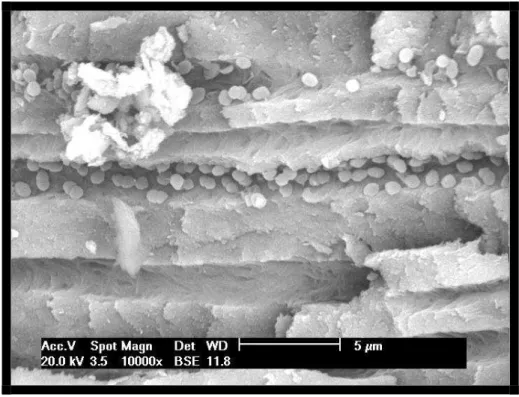
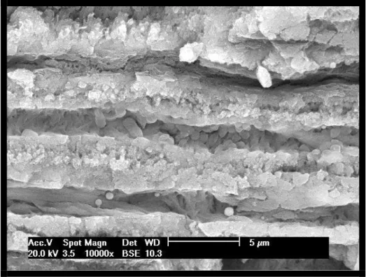
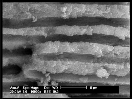
Figure 3 – SEM photomicrographs (10.000x – exposed tubule area) of the groups under study – a: 1 (positive control); b: 2 (distilled water); c: 3 (PAD); d: 4 (2.5% sodium hypochlorite); e: 5 (2.5% sodium hypochlorite + PAD). Please note the figures of
24
3 ARTIGO 2
25
Analysis of bovine pulp tissue dissolution ability by photodynamic therapy – in vitro study
Análise da capacidade de dissolução de tecido pulpar bovino através da terapia fotodinâmica: estudo in vitro
Liviu Steier1, Giampiero Rossi-Fedele1,2, Monique Acauan1, Priscila Bianchini1, Matheus Albino Souza1, José Antônio Poli de Figueiredo2
1
Warwick Dentistry, Warwick Medical School, Warwick, United Kingdom
2
School of Dentistry, Pontificial Catholic University of Rio Grande do Sul, PUCRS, Porto Alegre, RS, Brazil
Address to correspondence: José Antonio Poli de Figueiredo. Post-Graduate Program in Dentistry -
26
ABSTRACT
Purpose: Evaluate the bovine pulp tissue dissolution ability of photodynamic therapy. Methods: Twenty pieces of bovine pulp tissue were weighed and divided randomly into four groups (n = 5), according to the cleaning protocol: G1 - distilled water (negative control), G2 - sodium hypochlorite 1% (positive control), G3 - photodynamic therapy, G4 - sodium hypochlorite 1% + photodynamic therapy. The observation of the events of dissolution was performed by two observers blinded in relation to the test using 2x loupe magnification, recording time in minutes until complete tissue dissolution. The total observation time was 2 hours. The dissolution rate was calculated dividing the weight of the fragment pulp (mg) by the time of dissolution (mg / min).
Results: only group 2 (NaOCl) was able to promote complete dissolution of pulp tissue. In the other groups there was no occurrence of complete dissolution of the samples. The mean dissolution time for samples from group 2 (NaOCl) was 1.26 mg / min.
Conclusion: photodynamic therapy was not able to dissolve the fragments of bovine pulp tissue and was not able to accelerate the rate of dissolution.
27
RESUMO
Objetivo: Avaliar a capacidade de dissolução de tecido pulpar bovino com o uso da terapia fotodinâmica.
Metodologia: Vinte fragmentos de polpas bovinas foram pesados e distribuídos randomicamente em quatro grupos (n=5) de acordo com os seguintes protocolos de limpeza: G1 – água destilada (controle negativo), G2 – hipoclorito de sódio 1% (controle positivo), G3 – terapia fotodinâmica, G4 - hipoclorito de sódio 1% + terapia fotodinâmica. A observação dos eventos de dissolução foi realizada por dois observadores, cegados quanto aos grupos experimentais, com lupa de 2x de aumento, que registravam o tempo em minutos até a completa dissolução de tecido. O tempo total de observação foi de 2 horas. O cálculo da taxa de dissolução foi feito dividindo o peso do fragmento pelo tempo de dissolução (mg/min).
Resultados: somente o grupo 2 (NaOCl 1%) foi capaz de promover dissolução completa do tecido pulpar, tendo como média de dissolução 1.26 mg/min..
Conclusão: a terapia fotodinâmica não tem capacidade de dissolver tecido pulpar, nem de acelerar a velocidade de dissolução de tecido pulpar.
28
INTRODUCTION
The persistence of residual pulp tissue, infected dentin or bacteria inside the root canals may be responsible for the failure of endodontic treatment (1). Irrigating agents must present capacity of dissolving pulp remnants (2), since the removal of pulp tissue is inadequate with mechanical preparation alone, due to the complexities of the anatomical root canal system (3). Moreover, it is suggested that postoperative pain is more prevalent in cases of vital pulp than in cases of non-vital pulp (4) and the remaining pulp can cause postoperative pain (5).
Several studies have been conducted in search of an irrigant to provide four major properties: antimicrobial activity, non-toxicity to periapical tissues, solubility in water and ability to dissolve organic matter (6).
Sodium hypochlorite (NaOCl) is considered the main irrigating substance in endodontics because of its broad antimicrobial spectrum, its ability to prevent the formation and dissolving the organic part of the smear layer, and its ability to dissolve organic tissue remnants (2). However, it has been shown a cytotoxic effect on vital tissues, causing severe inflammatory reactions to the periapex, with the concentration of 5.25% producing more toxic and caustic solutions than 0.5 and 1% (7). Moreover, low concentrations of sodium hypochlorite have reduced the ability to dissolve tissue (8), although this can be improved by increasing the temperature (4,5).
Photodynamic therapy (PDT) or photoactivated disinfection uses light of a specific wavelength to activate a non-toxic photoactive dye, known as the photosensitizer in the presence of oxygen (9). The energy transferred from the activated photosensitizer to available oxygen results in the formation of highly reactive oxygen species, which may eliminate microorganisms by damaging their essential cellular molecules, including proteins, nucleic acids and lipid membranes (10). In vitro (11,12) and in vivo studies (13,14) using photodynamic therapy have shown that this resource has the potential to maximize the disinfection of root canals.
29
MATERIAL AND METHODS
The study was approved by the Ethics Committee of the Pontifical Catholic University of Rio Grande do Sul (protocol 0024/10).
Tissue preparation
Ten bovine incisors were extracted, immersed in distilled water and stored at a temperature of – 20oC until required. The teeth were thawed at room temperature and two longitudinal grooves were prepared in buccal and lingual surfaces using a diamond disc (KG Sorensen, Barueri, Brazil), running from the crown portion to the apex. The teeth were split in half. The pulp tissue was removed and washed with distilled water. Each pulp sample was divided into two pieces of similar volume, resulting in twenty pieces.
Preparation of solutions
Solution of 1% sodium hypochlorite was prepared at the Endodontic Laboratory, School of Dentistry, Pontifical Catholic University of Rio Grande do Sul, from the solution of 2% sodium hypochlorite (Plus Virex - Johnson Diversey, Sturtevant, USA) diluted in distilled water in the proportion 1:1. A viscous solution of tolonium chloride was provided by the manufacturer of the photodynamic therapy device (PAD Plus, Denfotex Light Systems Ltd., Inverkeithing, Scotland).
Dissolution process
The pulp tissue fragments were weighed on a high precision balance (Sartorius BP61S, Göttingen, Germany), placed in transparent plastic pots and were divided randomly into four groups (n=5) according to initial weight:
Group 1 – immersion in 1.5 ml of distilled water (negative control).
Group 2 - immersion in 1.5 ml of 1% sodium hypochlorite (positive control). Group 3 - immersion in 1.5 ml of tolonium chloride, introduction of the tip of the PAD Plus (Denfotex Light Systems Ltd., Inverkeithing, Scotland) and activation of low power laser during a period of 120 seconds on the power of 120 mW.
30
(Denfotex Light Systems Ltd., Inverkeithing, Scotland) and activation of low power laser during a period of 120 seconds on the power of 120 mW.
The observation of the events of dissolution was performed by two observers blinded in relation to the test using 2x loupes magnification, recording in minutes the time until complete tissue dissolution occurred. The total observation time was 2 hours. The time required for dissolution was recorded in minutes (min) and the dissolution rate was calculated by dividing the weight of the fragment pulp (mg) by the time of dissolution (mg / min).
RESULTS
Results were presented in a table with average weights and dissolution times. Since only one experimental group showed total dissolution, no further statistical analysis was needed.
The weight and time of dissolution of each pulp fragment are shown in Table 1. Only group 2 (NaOCl) was able to promote complete dissolution of pulp tissue. In the other groups there was no occurrence of complete dissolution of the samples.
The dissolution rate was calculated on the weight of the fragment of pulp tissue (mg) divided by the dissolution time (min), obtaining a value in mg / min. The mean dissolution time for samples from group 2 (NaOCl) was 1.26 mg / min.
DISCUSSION
Previous studies have demonstrated the importance of the ability of an endodontic solvent and emphasized that the elimination of pulp tissue of the root canal was primordial to the success of endodontic treatment (15,16). The bovine pulp tissue was used in our study because it is compared to human pulp tissue despite some minor differences (17). Moreover, previous studies have used the bovine pulp tissue to assess the capability of dissolving various endodontic irrigants (18,19).
31
The tissue dissolution is dependent on three factors: frequency of agitation, amount of organic matter in relation to the amount of irrigating and surface area of contact (1). The present study was standardized by using the same volume of irrigant and dye for the samples of the respective groups, besides the fact that all samples of bovine pulp tissue fragments showed similar mean weights (+ - 90 mg).
The results were calculated in dissolution rates and not only time of dissolution, to compensate any variations in the weights of the fragments. The results of this study appear to be in line with previous investigations where only sodium hypochlorite showed ability to dissolve tissue (18,20), although in a previous study the average speed of tissue dissolution to sodium hypochlorite was lower (18). This can be explained by differences in the volume of irrigant in contact with the pulp fragment, specifically the size and weight of the fragments, the concentration of sodium hypochlorite solutions, and perhaps a difference in temperature of the solutions.
Photodynamic therapy is a new antimicrobial strategy that involves the use of low-intensity laser, which operates through photosensitizing agents (21). The low-low-intensity laser, beyond been considered harmless to human tissue, has anti-inflammatory and analgesic effects. Its basic mechanism of the operation is based on biostimulation that occurs at the molecular level. The laser penetrates through the tissues and faces a photosensitizer in mitochondria of cells (22).
Disinfection by photo-activation method proves effective against endodontic bacteria, and has proved less toxic and faster than sodium hypochlorite (23). Furthermore, the infiltration of dentin tubules by sensitizers was evaluated microscopically, indicating the effectiveness of this therapy within the tubules (11). There are still records that indicate oral bacteria as susceptible to photodynamic therapy (12).
32
The idea was that photodynamic therapy could exert an ability to dissolve in a previously unstructured tissue by initial action provided by previous immersion in sodium hypochlorite. However, this could not be observed, since the dissolution process was interrupted from the laser action, without such a development.
CONCLUSION
In conclusion, only the sodium hypochlorite showed ability to dissolve the fragments of bovine pulp tissue and photodynamic therapy does not aid to provide further dissolution.
REFERENCES
1. Moorer WR, Wesselink PR. Factors promoving the tissue dissolving capability of sodium hypochlorite. Int Endod J 1982;15:187-196.
2. Zehnder. Root canal irrigants. J Endod 2006;32:389-398.
3. Thé SD. The solvent action of sodium hypochlorite on fixed and unfixed necrotic tissue. J Endod 1979;47:558-561.
4. Abou-Rass M, Oglesby SW. The effects of temperature, concentration and tissue type on the solvent ability of sodium hypochlorite. J Endod 1981;7:376-377.
5. Cunningham WT, Balekjian BA. Effect of temperature on collagen-dissolving ability of sodium hypochlorite irrigant. Oral Surg Oral Med Oral Pathol 1980;49:175-177.
6. Kuruvilla JR, Kamath MP. Antimicrobial activity of 2.5% sodium hypochlorite and 0.2% chlorhexidine gluconate separately and combined as endodontic irrigants. J Endod 1998;24:472-476.
7. Pashley EL, Birdsong NL, Bowman K, Pashley DH. Cytotoxic effect of NaOCl on vital tissue. J Endod 1985;11:525-528.
33
9. Souza LC, Brito PRR, Oliveira JCM, Alves FRF, Moreira EJL, Sampaio-Filho HR et al. Photodynamic therapy with two different photosensitizers as a supplement to instrumentation/irrigation procedures in promoting intracanal reduction of Enterococcus faecalis. J Endod 2010;2:292-296.
10. Konopka K, Goslinski T. Photodynamic therapy in dentistry. J Dent Res 2007;86:694-707.
11. Soukos NS, Chen PS, Morris JT, Ruggiero K, Albernethy AD, Som S, Foschi D et al. Photodynamic therapy for endodontic disinfection. J Endod 2006;32:979-984.
12. Fimple JL, Fontana CR, Foshi F, Ruggiero K, Song X, Pagonis TC et al. Photodynamic treatment of endodontic polymicrobial infection in vitro. J Endod 2008;34:728-734.
13. Garcez AS, Nunez SC, Hamblin MR, Ribeiro MS. Antimicrobial effects of photodynamic therapy on patients with necrotic pulps and periapical lesion. J Endod 2008;34:138-142.
14. Bonsor SJ, Nichol R, Reid TM. Microbiological evaluation of photo-activated disinfection in endodontics (an in vivo study). Br Dent J 2006;200:337-341.
15. Callahan JR. Sulfuric acid for opening root canals. Dent Cos 1894;36:957-959.
16. Grossman LI, Meiman B. Solution of pulp tissue by chemical agents. J Am Dent Assoc 1941;28:223-225.
17. Koskinen KP, Stenvall H, Uitto VJ. Dissolution of bovine pulp tissue by endodontic irrigants. Scan J Dent Res 1980;88:406-411.
18. Okino LA, Siqueira EL, Santos M, Bombana AC, Figueiredo JAP. Dissolution of pulp tissue by aqueous solution of chlorhexidine digluconate and chlorhexidine digluconate gel. Int Endod J 2004;37:38-41.
34
20. Naenni N, Thoma K, Zehnder M. Soft tissue dissolution capacity of currently used and potential irrigants. J Endod 2004;30:785-787.
21. Garcez AS, Núñes SC, Lage-Marques JL, Jorge AOC, Ribeiro MS. Efficiency of NaOCl and laser-assisted photosensitization on the reduction of Enterococcus faecalis in vitro. Oral Surg Oral Med Oral Pathol Oral Radiol Endod 2006;102:93-98.
22. Eduardo CP, Gouw-Soares S. The use of lasers for endodontic applications in dentistry. Med Laser Appl 2001;16:231–243.
35
Table 1 – Weight and time dissolution of dissolution of each pulp tissue sample
Sample 1 Sample 2 Sample 3 Sample 4 Sample 5
Group
Weight (mg)
Time (min)
Weight (mg)
Time (min)
Weight (mg)
Time (min)
Weight (mg)
Time (min)
Weight (mg)
Time (min)
1 99,7 nildiss 138,7 nildiss 71,7 nildiss 69,7 nildiss 83,0 nildiss
2 67,8 55 140,2 90 83,2 75 102,1 85 79,3 70
3 66,8 nildiss 109,4 nildiss 108,2 nildiss 72,8 nildiss 102,2 nildiss
36
4 DISCUSSÃO GERAL
O sucesso do tratamento endodôntico está relacionado, entre outros fatores, a uma eficiente descontaminação e à dissolução de matéria orgânica do interior do sistema de canais radiculares. Para tal, dispomos de substâncias químicas auxiliares e recursos alternativos que desempenham suas funções concomitantemente à ação mecânica dos instrumentos endodônticos.
Diversos estudos têm sido conduzidos na endodontia em busca de um protocolo de limpeza que atinja tais propriedades. O presente estudo, através da realização de dois artigos científicos, procurou avaliar essas duas variáveis tão relevantes na realização do tratamento endodôntico.
O primeiro artigo teve como objetivo comparar, in vitro, a ação da terapia fotodinâmica com a ação do hipoclorito de sódio em canais radiculares infectados com
Enterococcus faecalis, analisando o potencial de descontaminação nas paredes
dentinárias e no sistema de túbulos dentinários.
As amostras foram contaminadas com Enterococcus faecalis, por se tratar de um microorganismo anaeróbio facultativo altamente resistente (ROÇAS et al., 2004), que vem sendo amplamente utilizado como marcador microbiológico para estudos in vitro. Isso se deve ao fato dele demonstrar ser capaz de colonizar com sucesso o canal radicular em forma de biofilme, bem como invadir túbulos dentinários e resistir a alguns procedimentos de descontaminação do sistema de canais radiculares (LOVE, 2001; SEDGLEY et al., 2005; GEORGE et al., 2005).
O tempo de contaminação das amostras é um fator bastante variável na literatura. Esse período varia em estudos prévios de 24 horas (SIQUEIRA JR. et al., 2000; GARCEZ et al., 2006) a 21 dias (BERBER et al., 2006). No entanto, o presente estudo realizou a contaminação pelo período de 60 dias, de forma semelhante a estudo anterior (GURGEL-FILHO et al., 2007). Acredita-se que um período mais prolongado de contaminação e proliferação de Enterococcus faecalis seja necessário para que haja a formação de um biofilme mais consistente e organizado, permitindo uma adequada colonização. Dessa forma, os protocolos de descontaminação estariam sendo efetivamente testados.