Correspondence to:
Nikola STOJANOVIĆ Department of Restorative Dentistry and Endodontics Faculty of Medicine University of East Sarajevo Studentska 5, 73300 Foča Bosnia and Herzegovina
[email protected] SUMMARY
Introduction Because apical periodontitis is recognizably an infectious disease, elimination or reduction of intracanal bacteria is of utmost importance for optimum treatment outcome.
Objective The prevalence of Enterococcus faecalis and Porphyromonas gingivalis in infected root canals was studied Also, the effect of endodontic therapy by using intracanal medicaments, calcium hydroxide paste (CH) or gutta-percha points containing calcium hydroxide (CH-GP) or chlorhexidine (CHX-GP) on these microorganisms was assessed by polymerase chain reaction (PCR) assay.
Methods Fifty-one patients with chronic apical periodontitis were randomly allocated in one of the fol-lowing groups according to the intracanal medicament used: CH, CH-GP and CHX-GP group. Bacterial samples were taken upon access (S1), after chemomechanical instrumentation (S2) and after 15-day medication (S3). PCR assay was used to detect the presence of selected bacteria.
ResultsE. faecalis was detected in 49% (25/51) and P. gingivalis in 17.6% (9/51) of the samples. Samples which showed no bacterial presence at S1 were excluded from further analysis. Overall analysis of all 29 samples revealed significant differences between S1 and S2 (p<0.001), S2 and S3 (p<0.05), and S1 and S3 (p<0.001). When distinction was made between the intracanal medications, there was a significant difference in the number of PCR positive samples between S1 and S2, S1 and S3, but not between S2 and S3 samples.
ConclusionE. faecalis is more prevalent than P. gingivalis in primary endodontic infection. Intracanal medication in conduction with instrumentation and irrigation efficiently eliminates E. faecalis and P. gingivalis from infected root canals.
Keywords: antibacterial treatment; calcium hydroxide; chlorhexidine; medicated gutta-percha points; polymerase chain reaction
Prevalence of
Enterococcus faecalis
and
Porphyromonas gingivalis
in Infected Root Canals
and Their Susceptibility to Endodontic Treatment
Procedures: A Molecular Study
Nikola Stojanović1, Jelena Krunić1, Branka Popović2, Sonja Stojičić3, Slavoljub Živković4
1Department of Restorative Dentistry and Endodontics, Faculty of Medicine, University of East Sarajevo,
Foča, Bosnia and Herzegovina;
2Institute of Human Genetics, School of Dental Medicine, University of Belgrade, Belgrade, Serbia; 3Division of Endodontics, Department of Oral Biological and Medical Sciences, Faculty of Dentistry,
University of British Columbia, Vancouver, BC, Canada;
4Department of Restorative Dentistry and Endodontics, School of Dental Medicine,
University of Belgrade, Belgrade, Serbia
INTRODUCTION
Chronic apical periodontitis is a lesion formed by the periradicular host defense system as re-sponse to microorganisms present in the root canal system [1]. Although no specific micro-organism has been identified as the principal etiologic agent of pulpal and periapical patho-sis, some species have been more frequently reported in the root canal space. Culture studies have shown that species of the genera
Eubacterium, Fusobacterium, Peptostreptococ cus, Porphyromonas and Prevotella are com-monly encountered in endodontic infection [1, 2]. Sensitive and accurate molecular biol-ogy techniques have provided significant ad-ditional knowledge regarding the composition of microbiota associated in root canal infection [2, 3, 4]. Consequently, some uncultivable and difficult-to-cultivate species have been detected in higher prevalence values in samples from the canal with pulp necrosis [2]. The prevalence of
some species in endodontic samples can sig-nificantly vary between patients from differ-ent locations [5, 6]. Nevertheless, there is only scarce information as to whether these varia-tions are restricted to certain species or involve the whole profile of bacterial communities [7].
536
Stojanović N. et al. Prevalence of E. faecalis and P. gingivalis in Infected Root Canals and Their Susceptibility to Endodontic Treatment Procedures
canal bacteria, some microorganisms have proved to be resistant to calcium hydroxide [12]. So, alternative agents capable to predictably eliminate root canal bacteria have been investigated. Chlorhexidine is considered as an ef-fective medicament against endodontic pathogens. It is a broad-spectrum antimicrobial agent with prolonged sub-stantial effect. When used as interappointment dressing, chlorhexidine has been shown as more effective than cal-cium hydroxide in the elimination of Enterococcus faecalis
[13], the species that has been implicated in the treatment failures.
Calcium hydroxide has been commonly used as a root canal dressing in the form of paste. However, complete removal of paste may be a challenge, raising concern about the potential influence of calcium hydroxide residue on the setting of root canal sealers [14]. To sidestep that potential difficulty, gutta-percha points impregnated with calcium hydroxide have been introduced as another means for cal-cium hydroxide delivery. Chlorhexidine containing gutta-percha points that allow easy introduction and retrieval from the root canal have been also marketed. Studies in-vestigating antimicrobial activity of calcium hydroxide and chlorhexidine containing gutta-percha points have gener-ated conflicting results and have been almost exclusively conducted in vitro [13, 15-18].
OBJECTIVE
The purpose of this study was to investigate the prevalence of E. faecalis and Porphyromonas gingivalis in teeth with pulp necrosis and periradicular lesion and to evaluate the effects of endodontic therapy associated with calcium hy-droxide paste or gutta-percha points containing calcium hydroxide or chlorhexidine as intracanal medicaments on these microorganisms by polymerase chain reaction (PCR).
METHODS
Patients and teeth
Fifty-one healthy patients (mean age 36.53 years, SD 12.97 years), range 18-76 years, 21 males, 30 females, who have been referred to the Endodontic Clinic of the Faculty of Medicine Foča, University of East Sarajevo, Bosnia and Herzegovina, for the treatment of chronic apical periodon-titis were selected for a study according to the following criteria: asymptomatic single-rooted and single-canalled teeth with necrotic pulps (as confirmed by negative re-sponse to the electric pulp test), and present radiographic evidence of chronic apical periodontitis. In all teeth the presence of periapical radiolucency was assessed using the periapical index (PAI), and teeth with PAI score equal to or greater than 3 were included. Teeth that could not be prop-erly isolated with a rubber dam, had present periodontal pockets (>4 mm), crown or root fracture as well as retreat-ment cases and patients who received antibiotic therapy
during previous 6 months were excluded from the study. Only one tooth was included from each patient. Before treatment the teeth were randomly assigned into one of the following three groups (17 teeth per each group) accord-ing to the intracanal medicament used: calcium hydroxide paste (Calxyl; OCO Products, Dirnstein, Germany; CH group), calcium hydroxide containing gutta-percha points (Calcium hydroxide Plus, Roeko Langenau, Germany; CH-GP group) or chlorhexidine containing gutta-percha points (Active Point, Roeko, Langenau, Germany; CHX-GP group). The study was conducted in accordance with the World Medical Association Declaration of Helsinki of 1975 (revised in 1983) and had been approved by the Ethical Committee of the Faculty of Medicine Foča. The procedures and the purpose of the study were explained to all patients. Informed consents were obtained before entering the study.
Endodontic treatment and root canal samples
Each tooth was polished with pumice and isolated with a rubber dam. The operative field including the tooth crown, rubber dam and clamps was disinfected with 30% hydro-gen peroxide for 60 seconds followed by 2.5% sodium hy-pochlorite for additional 60 seconds [19]. Caries and/or coronal restorations removal and access cavity preparation were accomplished using sterile high-speed and low-speed burs under sterile saline irrigation. Before entering the pulp chamber, decontamination procedure was repeated in the same way as described above. Disinfecting agents were neutralized with 5% sodium thiosulphate solution and sterility of operative field was checked using sterile paper points. All of these samples were tested negative. The subsequent procedures were performed aseptically.
Upon the initial access into the root canal, the first sample was taken (S1). Three sterile paper points were sequentially introduced to the level approximately 1 mm shorter to the radiographic apex of the tooth and each was maintained in place for 1 min. If the root canal was dry, a small amount of sterile saline was introduced into the canal and a file was used to disperse the canal content. Paper points were transferred in sterile tubes containing 1 mL of RTF and immediately frozen at -20°C until they were further processed.
taken in the same manner as the first sample. To remove smear layer, the root canal was flushed with 17% EDTA followed by 5 mL of 1% sodium hypochlorite. After inac-tivation of sodium hypochlorite and drying the canal with sterile paper points, intracanal medicament was placed in the root canal. Calcium hydroxide paste was introduced into the canal using the Lentulo spiral and packed with a cotton pellet to the level of canal entrance. A medicated gutta-percha point was selected to the full working length and inserted into the canal with a drop of sterile water, ac-cording to the manufacturer’s instructions. Following the placement of intracanal medicament, the access cavity was sealed with temporary filling (Cavit, 3M ESPE AG, Seefeld, Germany) and glass ionomer cement (Fuji IX, GC, Tokyo, Japan). A radiograph was taken to assure the proper place-ment of the medicaplace-ment.
At the second appointment, 15 days later, a rubber dam was placed and the root canal was accessed respecting the strict aseptic protocol as previously described. Calcium hydroxide paste was removed using a master apical file and sterile saline irrigation while calcium hydroxide and chlorhexidine containing gutta-percha points were re-moved with tweezers. Neutralization of calcium hydrox-ide dressing (paste and gutta-percha point) was achieved with 2 mL of 0.5% citric acid. The root canal containing chlorhexidine gutta-percha point was rinsed with 2 mL of 3% Tween 80 and 0.3% L-α-lecithin to inactivate chlo-rhexidine. The canal was additionally flushed with sterile saline solution and the third, postmedication sample (S3) was obtained. Subsequently, the root canal was obturated with gutta-percha and AH Plus sealer (Dentsply, DeTrey, GmbH, Konstanz, Germany) using the cold lateral com-paction technique. All teeth were treated by the same per-son, an endodontic specialist.
Microbiological assessment
The detection of bacterial DNA was performed using the PCR assay (polymerase chain reaction). The extraction of potentially present bacterial DNA was performed by boiling the collected material at 100˚C for 10 min followed by 5 min centrifugation in a microfuge (mini Spin, Eppen-dorf, Hamburg, Germany) to pellet the cell debris. Tested bacteria were detected by means of multiplex PCR using the following primers: Universal 16S rDNA forward
prim-er Escherichia coli5’ AGA GTT TGA TCC TGG CTC AG
3’ and species specific reverse primer 5’CAA TAC TCG TAT CGC CCG TTA TTC 3’ for Porphyromonas gingiva lis. Primers used for detection Enterococcus faecalis were: forward, 5’ TACTGACAAACCATTCATGATG 3’ and re-verse, 5’ AACTTCGTCACCAACGCGAAC 3’. The size of amplified products for P. gingivalis and E. faecalis were: 400 bp, and 110 bp, respectively. PCR was performed in vol-umes of 25 μL containing PCR buffer (Fermentas, Vilnius, Lithuania), 0.2 mM of each dNTP, 0.2 μM of each primer, 1U Taq DNA polymerase (Fermentas, Vilnius, Lithuania) and 3-5 μL of template DNA containing supernatant.
The amplification was carried out in the thermal cy-cler (PCR Express, Thermo Hybaid, California, USA), and PCR reactions for analysis of P. gingivalis under the follow-ing conditions: initial denaturation at 94°C for 3 min, 35 cycles at 94°C for 1 min, 55°C for 1 min, 72°C for 1.5 min and a final extension at 72°C for 5 min. For the detection of E. faecalis PCR cycle conditions were as follows: initial denaturation at 94°C for 3 min, 35 cycles at 94°C for 30 sec, 55°C for 30 sec, 72°C for 1 min, and final extension at 72°C for 5 min. In the negative control, DNA sample was replaced by distilled water.
The amplicons were visualized on 8% native polyacry-lamide gel stained with ethidium bromide in an UV tran-silluminator.
Statistical analysis
Statistical analysis was performed using SPSS 19.0 for Windows (IBM Corp., Armonk, NY). The Fischer Exact test was used to evaluate reduction in the number of PCR positive samples from S1 to S3. The difference between groups at various sampling points was compared using the chi-square test. The level of significance in all analyses was set at 5% (p<0.05).
RESULTS
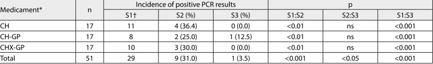
Upon the initial entering into the root canal (S1 samples), 56.9% (29/51) of all cases were PCR positive. Teeth with no initial presence of bacteria being evaluated were excluded from further analysis (Table 1). Of the teeth that yielded tested bacteria at S1, 69% (20/29) were bacteria-free at S2. A significant reduction in the number of PCR positive canals from S1 to S2 was observed (p<0.001). All samples
Table 1. Incidence of positive PCR results for the tested bacterial species at various sampling points (S1, S2, S3)
Medicament* n Incidence of positive PCR results p
S1† S2 (%) S3 (%) S1:S2 S2:S3 S1:S3
CH 17 11 4 (36.4) 0 (0.0) <0.01 ns <0.001
CH-GP 17 8 2 (25.0) 1 (12.5) <0.01 ns <0.001
CHX-GP 17 10 3 (30.0) 0 (0.0) <0.01 ns <0.001
Total 51 29 9 (31.0) 1 (3.5) <0.001 <0.05 <0.001
* No significant difference in bacterial prevalence between the groups at S1, S2 and S3. † 22 samples did not show initial bacterial presence, and they were excluded from the study.
538
that were PCR positive at S2 were PCR negative at S3, ex-cept one sample in CH-GP group. With no distinction of the medication used, significant differences were found between S2 and S3 (p<0.05), and S1 and S3 (p<0.001).
Within the groups, reduction in the number of PCR positive S2 samples for 63.6% in CH, 75% in CH-GP and 70% in CHX-GP group as compared to S1 was observed. After 15-day medication, the number of positive PCR sam-ples further decreased in all groups (Table 1). Intragroup analysis showed that the number of canals yielded signifi-cantly decreased tested bacteria from S1 to S2 (p<0.01 for each group) and from S1 to S3 (p<0.001 for each group), but not from S2 to S3 (p>0.05 for each group). Consider-ing the intergroup comparisons, no significant differences were observed at the start of the treatment (S1), after ch-emomechanical preparation (S2) and intracanal medica-tion (S3).
Regarding bacterial species, E. faecalis was found in 49% (25/51) and P. gingivalis in 17.6% (9/51) of S1 sam-ples (Table 2). Postinstrumentation samsam-ples (S2) and post-medication samples (S3) contained E. faecalis only in 31% and 3.4% of cases, respectively.
DISCUSSION
The present study evaluated the prevalence of E. faeca lis and P. gingivalis in untreated teeth with asymptomatic chronic periradicular lesions. The antimicrobial effects of chemomechanical procedures and various intracanal medicaments were investigated as well.
Endodontic infections are mixed infections of pol-ymicrobial etiology, with a predominance of obligate and facultative anaerobic species. In this study the occurrence of E. faecalis and P. gingivalis in a tooth with necrotic pulp and periradicular lesions was determined for several rea-sons. Data concerning molecular detection of these patho-gens in primary endodontic infection are diverse [2, 3, 4, 20, 21, 22]. Also, some studies found that the prevalence of some species in infections of endodontic origin may sig-nificantly differ from one geographic location to another [5, 6], and no clinical studies have been performed in the Bosnian population detecting the presence of these two pathogens in primary infection. In addition to the gram positive bacteria, at least P. gingivalis has been reported for its ability to invade dentinal tubules [23]. Furthermore, facultative bacteria, such as E. faecalis have been consid-ered one of the most resistant species in the oral cavity and a possible cause of root canal treatment failure. The effects of endodontic therapy associated with calcium hy-droxide paste or medicated gutta-percha points as intra-canal medicaments on these pathogens were assessed as well. To the best of our knowledge, no clinical study has investigated antibacterial effects of medicated gutta-percha points against E. faecalis and P. gingivalis.
Different methods are used for the detection and iden-tification of endodontic microbiota, including cultivation of microorganisms and molecular techniques. Molecular methods have proved to be more rapid, more sensitive, and
more accurate than culture and can provide more reliable results with regard to the composition of root canal micro-biota and effects of endodontic treatment procedures. PCR assay has been increasingly used to identify endodontic bacteria in clinical samples and is able to detect culture dif-ficult and even as-yet-uncultivated bacteria [2]; therefore it was used in the present study. One should also consider the fact that PCR is unable to differentiate living bacteria and DNA from nonviable or lysed cells, which is particu-larly relevant when effects of intracanal procedures are being evaluated. However, a previous study suggested that antimicrobial agents such as sodium hypochlorite and cal-cium hydroxide used during endodontic treatment might destroy DNA from dead cells [24].
Studies using culture methods have reported that E. fae calis, closely found in association with root-filled teeth, is present in very low numbers of the untreated canals [21, 25]. Culture-independent molecular studies also failed to isolate this species in higher prevalence from the primary infected root canals. Rôças et al. [20] and Fouad et al. [26] using standard PCR, and Sequira et al. [4] by DNA-DNA hybridization detected this species in 18%, 14% and 14.3% of cases, respectively. In this study E. faecalis was found in about one-half (25/51) of the infected canals. Findings for this species is in line with results from several other recent molecular studies that demonstrate a high presence of E. faecalis in untreated root canals and its association with primary endodontic infection. Sassone et al. [3] were able to detect E. faecalis in 89.3% of cases using DNA-DNA hybridization, whereas Sedgley et al. [21] and Gomes et al. [25] revealed this species in 67.5% and 82% of cases by real-time PCR and PCR, respectively. It is apparent that prevalence data for E. faecalis are quite variable, and is probably influenced by the molecular method of the de-tection used in each investigation, as well as by existence of geography-dependent variation in the oral microbiota.
Molecular methods usually detected higher prevalence of black-pigmented bacteria in endodontic infection than the traditional culture methods [27]. Sassone et al. [3] and de Souza et al. [2] using DNA-DNA hybridization detected
P. gingivalis in 67% and 75% of samples, respectively. In this study P. gingivalis was observed in 17.6% of patients by PCR, an estimate closer to those obtained by Siqueira et al. [4], who used the DNA-DNA hybridization method (17.9%), and Seol et al.[22], who used PCR (22.5%) to detect this species. Foschi et al. [28] found P. gingivalis in
Table 2. Number of PCR positive samples for the selected species at various sampling points (S1, S2, S3)
Medicament Bacterial species PCR positive samples
S1 S2 S3
CH E. faecalis 8 4
P. gingivalis 6
CH-GP E. faecalis 7 2 1
P. gingivalis 1
CHX-GP E. faecalis 10 3
P. gingivalis 2
S1 – initial sample; S2 – postinstrumentation sample; S3 – postmedication sample; CH – calcium hydroxide; CH-GP – calcium hydroxide containing gutta-percha; CHX-GP – chlorhexidine containing gutta-percha
13% of the samples from Italian patients using PCR, while Baumagartner et al. [6] found P. gingivalis in 72% and 30% of the endodontic abscesses from patients in the United States and Brazil, respectively. These results suggest that variation in the prevalence of P. gingivalis may be caused by the differences in clinical diagnosis, sampling method and sample analysis, as well as differences in the endodon-tic microbiota and several host and environmental factors, such as genetic background, socioeconomic status, psy-chological stress, smoking, and the nature of the species colonizing other individuals in the same country [5, 6].
Sodium hypochlorite is most widely used root canal solution because of its pronounced antimicrobial activ-ity and tissue dissolving capacactiv-ity. However, no general agreement exists regarding its optimal concentration, which ranges from 0.5% to 5.25%. In this study the 1% of sodium hypochlorite solution was selected because it provides efficient antimicrobial action with acceptable cy-totoxic level [29]. A substantial reduction in the prevalence rate of selected species was observed after irrigation with 1% sodium hypochlorite (S2). This finding is in agreement with previous studies [10, 30], confirming the essential role of chemomechanical procedures in eliminating root canal microbiota. P. gingvalis was completely eliminated, corroborating the results from other studies that found gram-negative bacteria usually eliminated after the ch-emomechanical debridement [24, 30]. However, 17.6% of cases still yielded E. faecalis. It has been shown that irrespective of concentration used, sodium hypochlorite is highly effective in eliminating E. faecalis including its existence as a biofilm in vitro[31]. For instance, 1% so-dium hypochlorite eliminated E. faecalis to levels below the detection after contact time ranging from 10 to 30 min [32, 33]; the time that does not exceed the average time usually spent for chemomechanical preparation. Nevertheless, its clinical efficacy may be influenced by the complexity of root canal anatomy and inability of sodium hypochlorite to penetrate into confined areas of the root canal. In ad-dition, the interaction of sodium hypochlorite with tissue fluids, blood, dentine and other organic debris inactivates irrigant and reduces its antibacterial capacity. This may explain the persistence of E. faecalis after chemomehanical preparation in the present study.
An interappointment medication has been recom-mended to supplement antibacterial effect of chemome-chanical procedures and eliminate residual bacteria [9, 11]. In our study, the placement of either CH paste, CH-GP or CHX-GP as intracanal dressing successfully eliminated E. faecalis (except in one sample of CHX-GP group) but these reductions did not reach statistical significance when com-pared to the samples obtained after the chemomechanical procedure (S2). Low prevalence of tested species in root canals before medication as well as a small sample size in each group may have influenced the results. Because the bacterial reduction was similar in all treatment pro-tocols, ranging from 87.5% to 100%, a study with a large sample size than the present study would be required. To compensate for this shortcoming, we performed power calculation to determine the number of samples required
in order to reach significance for the observed differences. A sample of 125 teeth per group would have resulted in a significant difference between the groups at the 5% level and 80% power. Since this number is difficult to achieve in a prospective, controlled studies, with stricter inclusion/ exclusion criteria for selection of study population, our finding may still be clinically relevant.
Although the observed difference between S2 and S3 within the groups was not significant, decrease in bacterial prevalence from S2 to S3 in overall sample highlights the importance of intracanal medication after chemomechani-cal procedures to predictably control the root canal infec-tion. Calcium hydroxide is effective in killing the majority of bacteria in the root canal system; however, some contro-versies exist about its effectiveness against E. faecalis [12]. Several theories have been proposed to explain the survival of E. faecalis after treatment with calcium hydroxide [12, 34]. It has been shown that E. faecalis has ability to main-tain pH homeostasis passively and actively by proton pump [34]. Moreover, it can survive harsh environment, includ-ing a high pH value of 11.5, and is capable of penetratinclud-ing into the dentine tubules and escaping from the effective concentration of medicament. Furthermore, the buffering effects of dentine may not allow a sufficiently high pH to be achieved in the dentine tubules [35]. Our findings are in accordance with studies done by Sjögren et al. [9] and de Souza et al. [2] who confirmed the effectiveness of calcium hydroxide paste against E. faecalis. Considering antibac-terial activity of intracanal medicament vehicle may have a significant impact affecting both physical and chemical properties of carrying compounds. Some in vitro studies have shown that CH-GP may not be the effective deliv-ery system of calcium hydroxide [16, 17]. This difference may be explained by a lower hydroxyl-releasing potential of medicated points in comparison to calcium hydroxide paste. In addition, CH-GP may not act as a good physical and chemical barrier as calcium hydroxide paste. However, findings from the present study demonstrate that, in addi-tion to the chemomechanical instrumentaaddi-tion, intracanal medication with calcium hydroxide, despite the vehicle, efficiently eliminates E. faecalis from infected root canals.
In response to the ambivalent efficacy of calcium hy-droxide, chlorhexidine has been proposed as alternative intracanal medicament that can be delivered in a variety of vehicles. In the present study CHX-GP was effective in eliminating E. faecalis, corroborating the results of previ-ous in vitro studies. Namely, a complete elimination of
540
CONCLUSION
Within the limitations of the study, the obtained results showed that E. faecalis is more prevalent than P. gingi valis in untreated teeth with asymptomatic chronic per-iradicular lesions. In addition, intracanal medication in
conduction with instrumentation and irrigation effi-ciently eliminate E. faecalis and P. gingivalis from infected root canals. Further investigation should be conducted to elucidate antimicrobial potential of medicated points against a wider range of root canal bacteria in clinical condition.
1. Kakehashi S, Stanley HR, Fitzgerald RJ. The effects of surgical exposures of dental pulps in germ-free and conventional laboratory rats. Oral Surg Oral Med Oral Pathol Oral Radiol Endod. 1965; 20:340-9.
2. de Souza CA, Teles RP, Souto R, Chaves MA, Colombo AP. Endodontic therapy associated with calcium hydroxide as an intracanal dressing: microbiologic evaluation by the checkerboard DNA-DNA hybridization technique. J Endod. 2005; 31:79-83.
3. Sassone L, Fidel R, Figueiredo L, Fidel S, Faveri M, Feres M. Evaluation of the microbiota of primary endodontic infections using checkerboard DNA-DNA hybridization. Oral Microbiol Immunol. 2007; 22:390-7.
4. Siqueira JF Jr, Rôças IN, Souto R, de Uzeda M, Colombo AP. Checkerboard DNA-DNA hybridization analysis of endodontic infections. Oral Surg Oral Med Oral Pathol Oral Radiol Endod. 2000; 89:744-8.
5. Rôças IN, Baumgartner JC, Xia T, Siqueira JF Jr. Prevalence of selected bacterial named species and uncultivated phylotypes in endodontic abscesses from two geographic locations. J Endod. 2006; 32:1135-8.
6. Baumgartner JC, Siqueira JF Jr, Xia T, Rôças IN. Geographical differences in bacteria detected in endodontic infections using polymerase chain reaction. J Endod. 2004; 30:141-4.
7. Siqueira JF Jr, Rôças IN, Debelian GJ, Carmo FL, Paiva SS, Alves FR, et al. Profiling of root canal bacterial communities associated with chronic apical periodontitis from Brazilian and Norwegian subjects. J Endod. 2008; 34:1457-61.
8. Sjögren U, Figdor D, Persson S, Sundqvist G. Influence of infection at the time of root filling on the outcome of endodontic treatment of teeth with apical periodontitis. Int Endod J. 1997;
30:297-306.
9. Sjögren U, Figdor D, Spångberg L, Sundqvist G. The antimicrobial effect of calcium hydroxide as a short-term intracanal dressing. Int Endod J. 1991; 24:119-25.
10. Shuping GB, Ørstavik D, Sigurdsson A, Trope M. Reduction of intracanal bacteria using nickel-titanium rotary instrumentation and various medications. J Endod. 2000; 26:751-5.
11. McGurkin-Smith R, Trope M, Caplan D, Sigurdsson A. Reduction of intracanal bacteria using GT rotary instrumentation, 5.25% NaOCl, EDTA, and Ca(OH)2. J Endod. 2005; 31:359-63.
12. Evans M, Davies JK, Sundqvist G, Figdor D. Mechanisms involved in the resistance of Enterococcus faecalis to calcium hydroxide. Int Endod J. 2002; 35:221-8.
13. de Lucena JM, Decker EM, Walter C, Boeira LS, Löst C, Weiger R. Antimicrobial effectiveness of intracanal medicaments on Enterococcus faecalis: chlorhexidine versus octenidine. Int Endod J. 2013; 46:53-61.
14. Hosoya N, Kurayama H, Iino F, Arai T. Effects of calcium hydroxide on physical and sealing properties of canal sealers. Int Endod J. 2004; 37:178-84.
15. Lin S, Zuckerman O, Weiss EI, Mazor Y, Fuss Z. Antibacterial efficacy of a new chlorhexidine slow release device to disinfect dentinal tubules. J Endod. 2003; 29:416-8.
16. Ebert J, Roggendorf MJ, Frank K, Petschelt A. Antimicrobial activity of various „active“ gutta-percha points against Enterococcus faecalis in simulated root canals. Int Endod J. 2008; 41:249-57. 17. Podbielski A, Spahr A, Haller B. Additive antimicrobial activity of
calcium hydroxide and chlorhexidine on common endodontic bacterial pathogens. J Endod. 2003; 29:340-5.
18. Oztan MD, Kiyan M, Gerçeker D. Antimicrobial effect, in vitro, of gutta-percha points containing root canal medications against yeasts and Enterococcus faecalis. Oral Surg Oral Med Oral Pathol Oral Radiol Endod. 2006; 102:410-6.
19. Ng YL, Spratt D, Skriskantharajah S, Gulabivala K. Evaluation of protocols for field decontamination before bacterial sampling of root canals for contemporary microbiology techniques. J Endod. 2003; 29:317-20.
20. Rôças IN, Siqueira Jr JF, Santos KRN. Association of Enterococcus faecalis with different forms of periradicular diseases. J Endod. 2004; 30:315-20.
21. Sedgley C, Nagel A, Dahlén G, Reit C, Molander A. Real-time quantitative polymerase chain reaction and culture analyses of Enterococcus faecalis in root canals. J Endod. 2006; 32:173-7. 22. Seol JH, Cho BH, Chung CP, Bae KS. Multiplex polymerase chain
reaction detection of black-pigmented bacteria in infections of endodontic origin. J Endod. 2006; 32:110-4.
23. Peters LB, Wesselink PR, Buijs JF, van Winkelhoff AJ. Viable bacteria in root dentinal tubules of teeth with apical periodontitis. J Endod. 2001; 27:76-81.
24. Sakamoto M, Siqueira JF Jr, Rôças IN, Benno Y. Bacterial reduction and persistence after endodontic treatment procedures. Oral Microbiol Immunol. 2007; 22:19-23.
25. Gomes BP, Pinheiro ET, Sousa EL, Jacinto RC, Zaia AA, Ferraz CC, et al. Enterococcus faecalis in dental root canals detected by culture and by polymerase chain reaction analysis. Oral Surg Oral Med Oral Pathol Oral Radiol Endod. 2006; 102:247-53.
26. Fouad AF, Barry J, Caimano M, Clawson M, Zhu Q, Carver R, et al. PCR-based identification of bacteria associated with endodontic infections. J Clin Microbiol. 2002; 40:3223-31.
27. Tomazinho LF, Avila-Campos MJ. Detection of Porphyromonas gingivalis, Porphyromonas endodontalis, Prevotella intermedia, and Prevotella nigrescens in chronic endodontic infection. Oral Surg Oral Med Oral Pathol Oral Radiol Endod. 2007; 103:285-8. 28. Foschi F, Cavrini F, Montebugnoli L, Stashenko P, Sambri V, Prati C.
Detection of bacteria in endodontic samples by polymerase chain reaction assays and association with defined clinical signs in Italian patients. Oral Microbiol Immunol. 2005; 20:289-95.
29. Zehnder M. Root canal irrigants. J Endod. 2006; 32:389-98. 30. Byström A, Sundqvist G. The antibacterial action of sodium
hypochlorite and EDTA in 60 cases of endodontic therapy. Int Endod J. 1985; 18:35-40.
31. Dunavant TR, Regan JD, Glickman GN, Solomon ES, Honeyman AL. Comparative evaluation of endodontic irrigants against Enterococcus faecalis biofilms. J Endod. 2006; 32:527-31.
32. Vianna ME, Gomes BP, Berber VB, Zaia AA, Ferraz CC, de Souza-Filho FJ. In vitro evaluation of the antimicrobial activity of chlorhexidine and sodium hypochlorite. Oral Surg Oral Med Oral Pathol Oral Radiol Endod. 2004; 97:79-84.
33. Radcliffe CE, Potouridou L, Qureshi R, Habahbeh N, Qualtrough A, Worthington H, et al. Antimicrobial activity of varying concentrations of sodium hypochlorite on the endodontic microorganisms Actinomyces israelii, A. naeslundii, Candida albicans and Enterococcus faecalis. Int Endod J. 2004; 37:438-46. 34. Stuart CH, Schwartz SA, Beeson TJ, Owatz CB. Enterococcus faecalis:
Its role in root canal treatment failure and current concepts in retreatment. J Endod. 2006; 32:93-8.
35. Haapasalo HK, Siren EK, Waltimo TM, Ørastavik D, Haapasalo MP. Inactivation of local root canal medicaments by dentine: an in vitro study. Int Endod J. 2000; 33:126-31.
REFERENCES
КРАТАК САДРЖАЈ
Увод Бу ду ћи да је апек сни па ро дон ти тис обо ље ње ко је на-ста је услед по сто ја ња ин фек ци је, за по сти за ње оп ти мал ног ис хо да ле че ња нај зна чај ни је је укла ња ње бак те ри ја из ка на-ла ко ре на зу ба или ба рем сма ње ње њи хо вог бро ја.
Циљ ра да Циљ овог ра да је био да се ис пи та пре ва лен ци ја бак те ри ја En te ro coc cus fa e ca lis и Porphyro mo nas gin gi va lis у ин фи ци ра ним ка на ли ма ко ре на зу ба и уста но ви ефе кат ен-до ен-донт ског ле че ња при ме ном ин тра ка нал них ме ди ка ме на-та – кал ци јум-хи дрок сид не су спен зи је (CH), гу та пер ка-по е на на ба зи кал ци јум-хи дрок си да (CH-GP) или гу та пер ка-по е на на ба зи хлор хек си ди на (CHX-GP) – на ове ми кро ор га ни зме ме то дом лан ча не ре ак ци је по ли ме ри за ци је (PCR).
Ме то де ра да Ис тра жи ва њем је об у хва ће на 51 осо ба ко-ја је има ла зуб с хро нич ним апек сним па ро дон ти ти сом. Ис пи та ни ци су ме то дом слу чај ног узор ка свр ста ни у три гру пе у за ви сно сти од вр сте при ме ње ног ин тра ка нал ног ме ди ка мен та (CH, CH-GP и CHX-GP). Бак те риј ски узор ци из ка на ла ко ре на са ку пља ни су при ини ци јал ном ула ску у
ка-нал ко ре на (S1), по сле хе мо ме ха нич ке об ра де (S2) и по сле пет на е сто днев ног ле че ња (S3). PCR ана ли за је ко ри шће на за од ре ђи ва ње при су ства ис пи ти ва них бак те ри ја.
Ре зул та тиE. fa e ca lis је изо ло ван из 25 (49%) узо ра ка, а P. gin gi va lis из де вет (17,6%). Узор ци у ко ји ма ни су на ђе не бак-те ри је у S1 фа зи ис кљу че ни су из да ље ана ли зе. Ана ли за свих 29 узо ра ка је ука за ла на ста ти стич ки зна чај ну раз ли ку из ме ђу S1 и S2 узо ра ка (p<0,001), S2 и S3 (p<0,05) и S1 и S3
(p<0,001). Ка да се узме у об зир вр ста ин тра ка нал ног ме ди-ка мен та, ста ти стич ки зна чај на раз ли ди-ка у бро ју PCR-по зи тив-них узо ра ка за бе ле же на је из ме ђу S1 и S2, S1 и S3, али не и из ме ђу S2 и S3.
За кљу чак У при мар ној ен до донт ској ин фек ци ји E. fa e ca lis
се че шће ја вља од P. gin gi va lis. Ин тра ка нал на ме ди ка ци ја за-јед но с ин стру мен та ци јом и ири га ци јом ефи ка сно укла ња E. fa e ca lis и P. gin gi va lis из ин фи ци ра них ка на ла ко ре на.
Кључ не ре чи: ан ти бак те риј ски трет ман; кал ци јум-хи дрок-сид; хлор хек си дин; ме ди ко ва ни гу та пер ка-по е ни; лан ча на ре ак ци ја по ли ме ри за ци је (PCR)
Преваленција бактерија
Enterococcus faecalis
и
Porphyromonas gingivalis
у
инфицираним каналима корена зуба и њихова осетљивост на ендодонтско
лечење: молекуларна студија
Никола Стојановић1, Јелена Крунић1, Бранка Поповић2, Соња Стојичић3, Славољуб Живковић4
1Катедра за болести зуба и ендодонцију, Медицински факултет Фоча, Универзитет у Источном Сарајеву, Фоча, Босна и Херцеговина; 2Институт за хуману генетику, Стоматолошки факултет, Универзитет у Београду, Београд, Србија;
3Одељење за ендодонцију, Катедра за оралне биолошке и медицинске науке, Стоматолошки факултет,
Универзитет Британске Колумбије, Ванкувер, Британска Колумбија, Канада;
4Клиника за болести зуба, Стоматолошки факултет, Универзитет у Београду, Београд, Србија