ISSN 2231-4261
Speciation and Biofilm Production of Coagulase Negative Staphylococcal Isolates
from Clinically Significant Specimens and their Antibiogram
1* 1 1
S. S. Vijayasri Badampudi , Surya Kirani KRL , RajyalakshmiGunti
1
Department of Microbiology, Rangaraya Medical College, Kakinada-533001
(Andhra Pradesh) India
Abstract:
Background: Coagulase Negative Staphylococci (CONS) are increasingly recognized as significant nosocomial pathogens. Their ability of biofilm formation and multiple drug resistance are causing serious human infections. Aim and Objectives: To isolate, identify, speciate clinically significant CONS from various specimens, to study antibiotic resistance pattern and biofilm production. Material and Methods: Specimens were collected aseptically, processed and identified upto the species level by a simple scheme of tests urease, novobiocin resistance, mannose and mannitol fermentation, ornithine decarboxylase. Antibiotic sensitivity was done with special reference to methicillin resistance. Biofilm formation was detected by Congo Red Agar (CRA) method and Tube Method (TM). Results: Study group-Of 100 isolates majority were pus (40), followed by urine (28), blood (16), CSF (5), body fluids (4) and catheter tips and implants (7). The most common species isolated was S. epidermidis (40%) followed by S. haemolyticus (26%), S. saprophyticus (15%), S. schleiferi (13%), S. simulans (2%), S. cohnii (2%) and S. warneri and S. capitis each 1%. Resistance to penicillin was 91% followed by ampicillin (79%), cotrimoxazole (67%). Methicillin resistance was 72%. Biofilm producers were 69% by CRA method and 33% by TM with majority species S. epidermidis (82.5%-CRA and 55%-TM). Biofilm production was significantly associated with MRCONS (p value 0.0036). Control group-Of 30 isolates were S. epidermidis 66.6% followed by S. haemolyticus (16.66%). Biofilm producers were 53.33% by CRA method and 26.65% by TM with majority species S.
epidermidis (65%-CRA and 30%-TM).Methicillin resistance was 26.6%. Conclusion: Clinical significance of CONS is increasing day by day, so there is a need for accurate identification to species level and their antibiogram to avoid multidrug resistance. Biofilm producing CONS species pose a risk and CRA method for screening biofilm can be used in all conventional microbiology laboratories.
Keywords: Coagulase Negative Staphylococcus, Speciation, Methicillin resistance, Biofilm, Congo Red.
Introduction:
Coagulase Negative Staphylococci (CONS) are common colonizers of the skin, anterior nares and ear canals of human beings [1].They are opportunistic pathogens that cause infection in debilitated or compromised patients. CONS have emerged as predominant pathogens in hospital acquired infections and vary in pathogenic potential [2].More than 30 species of CONS are recognised but only a few are commonly incriminated in human infections [3]. S. epidermidis is most frequently isolated from infections of wound, urogenital tract, respiratory tract, meninges, conjunctiva and skin [4].
S.saprophyticus was shown to be an important cause of urinary tract infections in young females [5]. Identification of CONS by reference method Kloos and Schleifer is cumbersome and requires expensive reagents not available in most clinical laboratories [6]. Commercial kit identification
ORI GI N AL ART I CLE
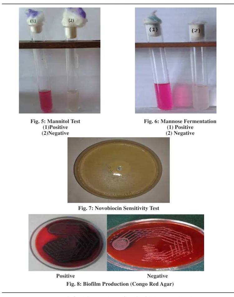
blood agar and nutrient broth. The isolates were considered clinically significant when isolated in pure culture from infected sites. The isolates collected were initially identified by colony morphology, Gram staining, catalase, slide and tube coagulase test (Fig. 1, 2) and anaerobic acid formation from mannitol (Fig.5) [2, 12]. Speciation of CONS was done by urease test, mannose fermentation test, Novobiocin sensitivity test, ornithine decarboxylase test [3] (Fig. 3, 4, 6, 7). These simple, inexpensive and easy to perform tests were selected from the scheme of Kloos and Schleifer to identify CONS species [3] (Table 1). Biofilm production was performed by culture of the CONS isolates on Congo Red Agar (CRA) plates as proposed by Freeman et al which is an alternative method of screening biofilm formation which requires the use of a specially prepared solid medium-Brain Heart Infusion (BHI) broth supplemented with 5% sucrose and Congo red (Fig.8) [13].
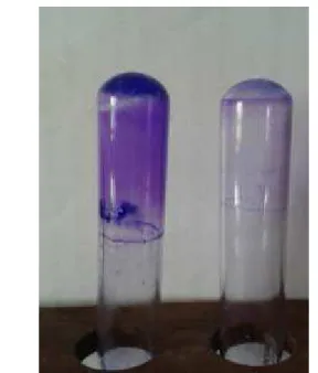
A qualitative assessment of biofilm formation was determined as previously described by Christensen et al [15]. A positive result was defined as the presence of a layer of stained material adhered to the inner wall of the tubes. The exclusive observation of a stained ring at the liquid air interface was not considered to be positive (Fig. 9). The antimicrobial susceptibility CONS are capable of forming biofilm on
polymeric surfaces. Biofilm is a structured community of microorganism encapsulated with a self-developed polymeric matrix and adherent to a living or inert surface. Biofilm consists of multi-layered cell clusters embedded in a matrix of extracellular polysaccharide which facilitates the adherence of these microorganisms to biomedical surfaces and protect them from host immune system and antimicrobial therapy [10].A working knowledge of the biology and antimicrobial susceptibility of CONS may be necessary to distinguish infections from contaminating isolates and to device appropriate therapy [11]. In the present study an attempt was made to isolate and speciate CONS with simple, inexpensive and easy to perform tests selected from the scheme of Kloos and Schleifer [6]. Antibiotic resistance patterns and biofilm production was done for all the isolates.
Material and Methods:
JKIMSU, Vol. 5, No. 2, April-June 2016
Fig. 1: Slide Coagulase Test Fig. 2: Tube Coagulase Test
S. S. Vijayasri Badampudi et. al.
Fig. 3: Urease Test
(1) Test Positive (2) Negative Control
(3) Positive Control
Fig. 4: Ornithine Decarboxylase Test (1) Positive
Fig. 5: Mannitol Test
(1)Positive (2)Negative
Fig. 6: Mannose Fermentation (1) Positive
(2) Negative
JKIMSU, Vol. 5, No. 2, April-June 2016
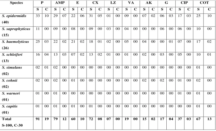
72(72%), cotrimoxazole 67(67%). Methicillin resistant CONS were 72 (72%) (Table 3). Of 100 isolates 69 (69%) were Biofilm producers by CRA method and 33% by TM with majority species S. epidermidis (82.5%-CRA and 55%-TM) (Table 4). Biofilm production was associated with MRCONS (p – Value 0.0036) (Table 5). In control group, of 30 isolates most common CONS species isolated was S. epidermidis 20(66.6%) followed by S. haemolyticus 05(16.66%) (Table 6). Majority showed resistance to Penicillin19 (63.3%) followed by cotrimoxazole 13(43.3%), Ampicillin 12(40%) and Erythromycin 10(33.3%). Methicillin resistance CONS were 08 (26.6%) (Table 3). Biofilm producers were 16 (53.33%) and 26.65% by TM with majority species S. epidermidis (65%-CRA and 30%-TM) (Table 4).
Results:
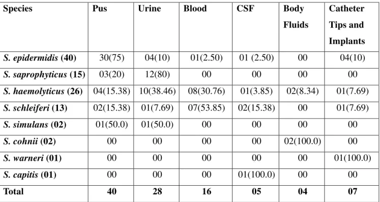
Among 100 isolates of CONS, 40 were isolated from pus, 28 from urine, 16 Blood, 05 CSF, 04 Body fluids and07 Catheter Tips and implants (Table 1). Majority of isolates were from males 63 (63%) and remaining were from females 37 (37%). Most common age group in both males and females was 15-45 years. Simple scheme for the Identification of CONS (Table-2) showed S. epidermidis as the most common isolate (40%),followed by S. haemolyticus 26 (26%), S. saprophyticus 15 (15%), S. schleiferi 13 (13%), S. simulans and S. cohnii each 02 (2%) and S. warneri and S. capitis each 01 (1%). Maximum isolates of S. epidermidis were from pus 30 (75%),
S. saprophyticus from urine 12 (80%), S. haemolyticus from blood 08 (53.85%) (Table 2). Majority showed resistance to penicillin 91 (91%) followed by ampicillin 79 (79%), cefoxitin
S. S. Vijayasri Badampudi et. al.
Fig. 9: Biofilm Production (Tube method)
Table 2: Simple Scheme for the Identification of CONS
Species Clumping
factor
Tube
Coagulase
Ornithine
Decarboxylase
Urease Mannose Mannitol Novobiocin
(5 µg )
S. epidermidis - - + + + - S
S. saprophyticus - - - + - + R
S. haemolyticus - - - V S
S. schleiferi - - - - + - S
S. simulans - - - + ± V S
S. cohnii - - - + + + R
S. warneri - - - + - V S
S. capitis - - - - + V* S
S. schleiferi(13) 02(15.38) 01(7.69) 07(53.85) 02(15.38) 00 01(7.69)
S. simulans (02) 01(50.0) 01(50.0) 00 00 00 00
S. cohnii (02) 00 00 00 00 02(100.0) 00
S. warneri (01) 00 00 00 00 00 01(100.0)
S. capitis (01) 00 00 00 01(100.0) 00 00
JKIMSU, Vol. 5, No. 2, April-June 2016
Table 3: Resistance Patterns of CONS to Different Antibiotics in Study(S) and Control(C) groups
P AMP E CX LZ VA AK G CIP COT
Species
S C S C S C S C S C S C S C S C S C S C
S. epidermidis
(40)
33 10 29 07 22 06 31 05 01 00 09 00 07 02 06 03 17 03 25 10
S. saprophyticus
(15)
11 00 09 00 08 00 09 00 03 00 04 00 00 00 06 00 06 00 10 00
S. haemolyticus
(26)
25 03 22 02 21 02 18 01 02 00 05 00 04 00 00 01 07 00 17 02
S. schleiferi
(13)
16 04 13 03 07 02 13 02 01 00 01 00 02 00 03 00 05 00 10 01
S. simulans
(02)
02 01 02 00 00 00 00 00 00 00 00 00 00 00 00 00 00 00 01 00
S. cohnii
(02)
02 00 02 00 01 00 00 00 00 00 00 00 02 00 02 00 01 00 02 00
S. warneri
(01)
01 00 01 00 00 00 00 00 00 00 00 00 00 00 00 00 01 00 01 00
S. capitis
(01)
01 00 01 00 01 00 01 00 00 00 00 00 00 00 00 00 00 00 01 00
Total S-100, C-30
91 19 79 12 60 10 72 08 07 00 19 00 15 02 17 04 37 03 67 13
Table 4: Detection of Biofilm in CONS by Two Phenotypic Methods in Study(S) and Control (C) Groups
CONS Species
S. epidermidis S. saprophyticus S. haemolyticus S. schleiferi S. simulans
Total
Biofilm
Method
S C S S C S C C S C
Congo RedAgar Method
Positive 33(82.5) 13(65) 12(80) 18(69.2) 02(40) 06(46.1) 01(25) 00 69 16
Negative 07(17.5) 07(35) 03(20) 08(30.7) 03(60) 07(53.8) 03(75) 1(100) 25 14
Tube Method
Positive 22(55) 06(30) 05(33.3) 04(15.3) 02(40) 02(15.3) 00 00 33 08
Negative 18(45) 14(70) 10(66.6) 22(84.6) 03(60) 11(84.6) 04(100) 01(100) 61 22
Total 40 20 15 26 05 13 04 01 94 30
earlier studies by Singh et al [15] and Asangi et al
[12]. S. epidermidis has been the most common isolate in 40(40%) and correlates to earlier studies by Singh et al (41%) [15] and Shubhra et al (40%) [16]. The next most common species in our study has been 26% of S. haemolyticus nearly correlates to Usha et al (17.64%) [17].
Majority of S. epidermidis isolates are from pus (75%) followed by urine (10%), catheter tips and implants (10%) and blood and CSF (2.5%) each which is similar to other studies of Cunha et al
[18], Sheikh and Mehdinejad et al [19].
Discussion:
Infections with CONS have been reported with increasing frequency [9], they must now be individually evaluated as potentially true pathogens and identified to the species level by simple, reliable and preferably inexpensive methods [14]. Many of the CONS species are commonly resistant to antibiotics that are being indicated for Staphylococcal infections. Biofilm may function as a penetration barrier to antibiotics and hence the high level of resistance [13].
In our study majority, i.e., 40(40%) CONS isolates
Species Nares Ear Skin
S. epidermidis (20) 07 07 06
S. saprophyticus(00) 00 00 00
S. haemolyticus(05) 00 02 03
S. schleiferi(04) 02 01 01
S. simulans(01) 01 00 00
S. cohnii(00) 00 00 00
S. warneri(00) 00 00 00
S. capitis(00) 00 00 00
JKIMSU, Vol. 5, No. 2, April-June 2016
biofilm production can be considered as associated with MRCONS. Control group of 30 CONS isolates from nares, ear, and skin from healthy individuals in our study have shown the most common species of S. epidermidis 35% which correlates with the study by Shubha et al
[20]. Antibiotic susceptibility testing has shown multidrug resistance with control group also and methicillin resistant CONS have been 8 (26.6%). Biofilm producers have been 16 (53.33%) by CRA method.
Conclusion:
The clinical significance of CONS is increasing day by day and there is a need for accurate identification to species level by simple, inexpensive methodology and their antibiotic sensitivity to avoid decreased susceptibility to glycopeptides. Pathogenecity of CONS strains may be decided by screening for biofilm production. CRA method is simple and is very easy to perform and interpret, can be used as screening test for biofilm detection in all conventional microbiology laboratories. Biofilm producing CONS species from healthy controls determine the risk of acquiring more infections in hospitals and pose a major concern. Multidrug resistant nature of MRCONS necessitates every effort to be made to eradicate infections by them through a strict hospital policy.
[20] and majority of S. haemolyticus isolates have been from urine (38.46%) followed by blood (30.76%) which is similar to the study by Asangi
et al (31.57%) in the urine and (15.78%) in the blood [12].
In our study biofilm production by CRA method is seen in 69 (69%) specimens, which is nearly correlating with the study by Cunha et al 73 (73%) [18] and majority of biofilm producers are the species S. epidermidis 82.5 (82.5%) which correlates with the study by Shubha et al [20], followed by S. saprophyticus 80 (80%) and S. haemolyticus 69.2 (69.2%). Biofilm production by TM has been 33 (33%) is correlating with the study of Sharvari et al 32(32%) [21].
Antibiotic susceptibility testing has shown variability and multidrug resistance with maximum resistance to penicillin 91 (91%) isolates followed by ampicillin 79 (79%), cefoxitin 72 (72%), cotrimoxazole 67 (67%), erythromycin 60 (60%), ciprofloxacin 37 (37%). Singh et al, Sharma et al, Bouchami et al, Asangi
et al, Shubha et al and Usha et al have shown maximum resistance to penicillin and ampicillin which correlates with our study [12,15,17,20, 21,22].
Among 100 isolates 72 (72%) have shown methicillin resistance which nearly correlates with Asangi et al (67.7%) of which 56 (77.7%) MRCONS have been biofilm producers with a p-value of 0.00369. It is statistically significant, so
S. S. Vijayasri Badampudi et. al.
Mitchell CJ. Epidemiological typing of Coagulase negative Staphylococci from nosocomial infections. J
Med Microbiol 1997; 46:195-203.Joshi JR, Pawar S,
Joshi PJ, Samuel A. Biological characters and antimicrobial sensitivity of Staphylococcus epidermidis. Indian J Pathol Microbiol 1987; 30:89-96. 4. Marrie TJ, Kwan C, Noble MA, West A, Duffield L.
Staphylococcus saprophyticus as a cause of urinary
tract infections. J Clin Microbiol 1982; 16(3):427-431. 1. Silvia Natoli, Carla Fontana et al. Characterization of
coagulase-negative Staphylococcal isolates from blood with reduced susceptibility to glycopeptides and therapeutic options. J Antimicrob Chemother 1992, 29:459-466.
2. Badwi JA, Memon AH, Soomro AA. Coagulase Negative Staphylococci (CONS) is the contaminant in the clinical specimen. Med Channel 2012; 19:23-7. 3. Geary C, Jordens JZ, Richardson JF, Howcraft DM,
18. Adilson Oliveira, Maria de Lourdes RS Cunha, Comparison of methods for the detection of biofilm production in coagulase-negative Staphylococci. Oliveira andCunha BMC Research notes 2010, 3:260 19. Sheikh AF, Mehdinejad M. Identification and
determination of coagulase negative Staphylococci species and antimicrobial susceptibility pattern of isolates from clinical specimens. Afr J Microbiol Res
2012; 6:1669-74.
20. Shubha DS, Banoo Sageera Shashidar, Fatima Farheen, Venkatesha D. Speciation and Antibiogram of Coagulase Negative Staphylococci (CONS) from various clinical specimens. IJPHRD 2012; 3(1):91-95. 21. Samant Sharvari A and Pai Chitra G, Evaluation of
Different Detection Methods of biofilm Formation in Clinical Isolates of Staphylococci. Int J Pharm Bio Sci 2012 Oct; 3(4): (B) 724 – 733
22. Sharma P, Lahiri KK, Kapila K. Conventional and Molecular characterization of coagulase negative Staphylococcus in hospital isolates. Indian J Pathol
Microbiol 2011; 54(1):85-9.
23. Bouchami O, Achour W, Hassen AB. Species distribution and antibiotic sensitivity pattern of coagulase-negative Staphylococci other than
Staphylococcus epidermidis isolated from various
clinical specimens. Afri J Microb Res 2011; 5(11):1298-1305.
8. Caierao J, Musskopf M, Superti S, Rosech E, Dias CG, d'Azevedo PA. Evaluation of phenotypic methods for methicillin resistance characterization in coagulase-negative Staphylococci. J Med Microbiol 2004; 53:1195-99.
9. Stewart PS, Costerton JW. Antibiotic resistance of bacteria in biofilms. Lancet 2001; 358: 135-8.
10. Archer GL, Climo MW Mandell, Douglas and Bennett's Principle and Practice of infectious diseases. 6th ed. Pennsylvania: Elsevier Churchill Livingstone; 2005.
11. Seetha KS, Santosh PK, Shivananda PG. Study of coagulase negative Staphylococci isolated from blood and CSF cultures. Indian J Pathol Microbiol 2000; 43(1):41-45.
12. Surekha Y Asangi, Mariraj J, Sathyanarayan MS, Nagabhushan, Rashmi. Speciation of clinically significant Coagulase Negative Staphylococci and their antibiotic resistant patterns in a tertiary care hospital.
Int J Biol Med Res 2011; 2(3): 735-739.
13. Freeman DJ, Falkiner FR, Keane CT. New method for detecting slime production by coagulase negative staphylococci. J Clin Pathol 1989; 42: 872–4.
14. Christensen GD, Bisno AL, Simpsom WA, Beachey EH: Adherence of slime producing strains of Staphylococcus epidermidis to smooth surfaces. Infect
Immun 1982; 37:318-326.
15. Goyal R, Singh NP, Kumar A, Kaur I, Singh M, Sunita N, Mathur M. Simple and economical method for
*
Author for Correspondence: Dr. S. S. Vijayasri Badampudi, Department of Microbiology, Rangaraya Medical