UNIVERSIDADE ESTADUAL PAULISTA
FACULDADE DE ODONTOLOGIA DE ARARAQUARA
Rodrigo Cavassim
A v a l i a ç ã o
d a
d e s c o n t a m i n a ç ã o
c o m
ácido cítrico e tetraciclina e da rugosidade,
na adesão de coágulo a superfícies radiculares
submetidas a diferentes tipos de instrumentação
ϭ
52'5,*2&$9$66,0
52'5,*2&$9$66,0
52'5,*2&$9$66,0
52'5,*2&$9$66,0
s>/K ^KEdD/EK KD /K 1dZ/K
ddZ/>/E Zh'K^/͕ E ^K K'h>K
^hWZ&1/^ Z/h>Z^ ^hDd/^ /&ZEd^ d/WK^
/E^dZhDEdK
$5$5$48$5$
81,9(56,'$'((67$'8$/3$8/,67$
81,9(56,'$'((67$'8$/3$8/,67$
81,9(56,'$'((67$'8$/3$8/,67$
81,9(56,'$'((67$'8$/3$8/,67$
)$&8/'$'('(2'2172/2*,$'($ )$&8/'$'('(2'2172/2*,$'($ )$&8/'$'('(2'2172/2*,$'($
)$&8/'$'('(2'2172/2*,$'($5$5$48$5$5$5$48$5$5$5$48$5$5$5$48$5$
7HVH DSUHVHQWDGD DR 3URJUDPD GH 3yV*UDGXDomR HP 2GRQWRORJLD ² ÉUHD GH 3HULRGRQWLD GD )DFXOGDGH GH 2GRQWRORJLDGH$UDUDTXDUDGD8QLYHUVLGDGH(VWDGXDO 3DXOLVWD ´-~OLR GH 0HVTXLWD )LOKRµ FRPR SDUWH GRV UHTXLVLWRV SDUD REWHQomR GR 7tWXOR GH 'RXWRU HP 3HULRGRQWLD
2ULHQWDGRU3URI'U-RVp(GXDUGR&H]DU6DPSDLR
Cavassim, Rodrigo
Avaliação da descontaminação com ácido cítrico e tetraciclina e da rugosidade, na adesão de coágulo a superfícies radiculares submetidas a diferentes tipos de instrumentação / Rodrigo Cavassim.-- Araraquara: [s.n.], 2012.
146 f. ; 30 cm.
Tese (Doutorado) – Universidade Estadual Paulista,
Faculdade de Odontologia
Orientador: Prof. Dr. José Eduardo Cezar Sampaio
1. Raiz dentária 2. Cálculos dentários 3. Raspagem dentária 4. Coagulação sanguínea 5. Microscopia eletrônica de varredura I. Título
RODRIGO CAVASSIM
AVALIAÇÃO DA DESCONTAMINAÇÃO COM ÁCIDO CÍTRICO E TETRACICLINA E DA RUGOSIDADE NA ADESÃO DE COÁGULO A SUPERFÍCIES RADICULARES
SUBMETIDAS A DEFERENTES TIPOS DE INSTRUMENTAÇÃO
COMISSÃO JULGADORA
TESE PARA OBTENÇÃO DO GRAU DE DOUTOR
Presidente e Orientador ...Prof. Dr. José Eduardo Cezar Sampaio 2º Examinador...Prof. Dra. Juliana Rico Pires
3º Examinador ...Prof. Dr. Fábio André dos Santos
4º Examinador ...Prof. Dra. Rosemary Adriana Chierici Marcantonio 5º Examinador ...Prof. Dr. Carlos Rossa Junior
'
'
'
'
'
'
'
'
$
$
$
$
$
$
$
$
'
'
'
'
'
'
'
'
2
2
2
2
2
2
2
2
6666
6666
&
&
&
&
&
&
&
&
8
8
8
8
8
8
8
8
5
5
5
5
5
5
5
5
5
5
5
5
5
5
5
5
,,,,
,,,,
&
&
&
&
&
&
&
&
8
8
8
8
8
8
8
8
/
//
/
/
//
/
$
$
$
$
$
$
$
$
5
5
5
5
5
5
5
5
(
(
(
(
(
(
(
(
6666
6666
5
5
5
5
5
5
5
5
22222222''''''''55555555,,,,,,,,********22222222&
&
&
&
&
&
&
&
$$$$$$$$99999999$$$$$$$$6666666666666666,,,,,,,,00000000Nascimento 22/05/1980 Prudentópolis – PR
Filiação Agostinho Cavassim
Janete Maria Cavassim
1999/2003 Graduação em Odontologia
Universidade Estadual de Ponta Grossa – UEPG
2005/2007 Curso de Especialização em Periodontia
Associação Brasileira de Odontologia – ABO Regional de Ponta Grossa – PR
2006/2008 Curso de Pós-Graduação em Odontologia
Área de Periodontia, Nível Mestrado, Faculdade de Odontologia de Araraquara – UNESP
2008/2012 Curso de Pós-Graduação em Odontologia
Área de Periodontia, Nível Doutorado, Faculdade de Odontologia de Araraquara - UNESP
2010/2012 Curso de Especialização em Implantodontia
'
'
'
'
'
'
'
'
(
(
(
(
(
(
(
(
'
'
'
'
'
'
'
'
,,,,
,,,,
&
&
&
&
&
&
&
&
$
$
$
$
$
$
$
$
77
7
77
7
7
7
Ð
Ð
Ð
Ð
Ð
Ð
Ð
Ð
5
5
5
5
5
5
5
5
,,,,
,,,,
$
$
$
$
$
$
$
$
$
$
$
$
$
$
$
$
RRRR
RRRR
VVVV
VVVV
P
P
P
P
P
P
P
P
HHHH
HHHH
XXXX
XXXX
VVVV
VVVV
SSSS
SSSS
DDDD
DDDD
LLLL
LLLL
VVVV
VVVV
----
----
DDDD
DDDD
QQQQ
QQQQ
HHHH
HHHH
WWWW
WWWW
HHHH
HHHH
HHHH
HHHH
$
$
$
$
$
$
$
$
JJJJ
JJJJ
RRRR
RRRR
VVVV
VVVV
WWWW
WWWW
LLLL
LLLL
QQQQ
QQQQ
KKKK
KKKK
RRRR
RRRR
TTTT
TTTT
XXXX
XXXX
HHHH
HHHH
LLLL
LLLL
QQQQ
QQQQ
FFFF
FFFF
RRRR
RRRR
QQQQ
QQQQ
GGGG
GGGG
LLLL
LLLL
FFFF
FFFF
LLLL
LLLL
RRRR
RRRR
QQQQ
QQQQ
DDDD
DDDD
OOOO
OOOO
P
P
P
P
P
P
P
P
HHHH
HHHH
QQQQ
QQQQ
WWWW
WWWW
HHHH
HHHH
DDDD
DDDD
SSSS
SSSS
RRRR
RRRR
LLLL
LLLL
DDDD
DDDD
UUUU
UUUU
DDDD
DDDD
P
P
P
P
P
P
P
P
WWWW
WWWW
RRRR
RRRR
GGGG
GGGG
DDDD
DDDD
VVVV
VVVV
DDDD
DDDD
VVVV
VVVV
P
P
P
P
P
P
P
P
LLLL
LLLL
QQQQ
QQQQ
KKKK
KKKK
DDDD
DDDD
VVVV
VVVV
GGGG
GGGG
HHHH
HHHH
FFFF
FFFF
LLLL
LLLL
VVVV
VVVV
}}}}
}}}}
HHHH
HHHH
VVVV
VVVV
HHHH
HHHH
TTTT
TTTT
XXXX
XXXX
HHHH
HHHH
P
P
P
P
P
P
P
P
HHHH
HHHH
VVVV
VVVV
P
P
P
P
P
P
P
P
RRRR
RRRR
GGGG
GGGG
LLLL
LLLL
VVVV
VVVV
WWWW
WWWW
DDDD
DDDD
QQQQ
QQQQ
WWWW
WWWW
HHHH
HHHH
VVVV
VVVV
VVVV
VVVV
HHHH
HHHH
P
P
P
P
P
P
P
P
SSSS
SSSS
UUUU
UUUU
HHHH
HHHH
HHHH
HHHH
VVVV
VVVV
WWWW
WWWW
LLLL
LLLL
YYYY
YYYY
HHHH
HHHH
UUUU
UUUU
DDDD
DDDD
P
P
P
P
P
P
P
P
DDDD
DDDD
RRRR
RRRR
P
P
P
P
P
P
P
P
HHHH
HHHH
XXXX
XXXX
OOOO
OOOO
DDDD
DDDD
GGGG
GGGG
RRRR
RRRR
$
$
$
$
$
$
$
$
P
P
P
P
P
P
P
P
LLLL
LLLL
QQQQ
QQQQ
KKKK
KKKK
DDDD
DDDD
LLLL
LLLL
UUUU
UUUU
P
P
P
P
P
P
P
P
mmmm
mmmm
'
'
'
'
'
'
'
'
DDDD
DDDD
LLLL
LLLL
DDDD
DDDD
QQQQ
QQQQ
HHHH
HHHH
SSSS
SSSS
HHHH
HHHH
OOOO
OOOO
DDDD
DDDD
IIII
IIII
RRRR
RRRR
UUUU
UUUU
oooo
oooo
DDDD
DDDD
HHHH
HHHH
LLLL
LLLL
QQQQ
QQQQ
FFFF
FFFF
HHHH
HHHH
QQQQ
QQQQ
WWWW
WWWW
LLLL
LLLL
YYYY
YYYY
RRRR
RRRR
VVVV
VVVV
HHHH
HHHH
P
P
P
P
P
P
P
P
SSSS
SSSS
UUUU
UUUU
HHHH
HHHH
TTTT
TTTT
XXXX
XXXX
HHHH
HHHH
SSSS
SSSS
UUUU
UUUU
HHHH
HHHH
FFFF
FFFF
LLLL
LLLL
VVVV
VVVV
HHHH
HHHH
LLLL
LLLL
$
$
$
$
$
$
$
$
P
P
P
P
P
P
P
P
LLLL
LLLL
QQQQ
QQQQ
KKKK
KKKK
DDDD
DDDD
QQQQ
QQQQ
RRRR
RRRR
LLLL
LLLL
YYYY
YYYY
DDDD
DDDD
'
'
'
'
'
'
'
'
LLLL
LLLL
XXXX
XXXX
OOOO
OOOO
LLLL
LLLL
HHHH
HHHH
*
*
*
*
*
*
*
*
UUUU
UUUU
DDDD
DDDD
]]]]
]]]]
LLLL
LLLL
HHHH
HHHH
OOOO
OOOO
DDDD
DDDD
3
33
3
33
3
3
HHHH
HHHH
OOOO
OOOO
RRRR
RRRR
DDDD
DDDD
P
P
P
P
P
P
P
P
RRRR
RRRR
UUUU
UUUU
LLLL
LLLL
QQQQ
QQQQ
FFFF
FFFF
RRRR
RRRR
QQQQ
QQQQ
GGGG
GGGG
LLLL
LLLL
FFFF
FFFF
LLLL
LLLL
RRRR
RRRR
QQQQ
QQQQ
DDDD
DDDD
OOOO
OOOO
GGGG
GGGG
HHHH
HHHH
P
P
P
P
P
P
P
P
RRRR
RRRR
QQQQ
QQQQ
VVVV
VVVV
WWWW
WWWW
UUUU
UUUU
DDDD
DDDD
GGGG
GGGG
RRRR
RRRR
VVVV
VVVV
HHHH
HHHH
P
P
P
P
P
P
P
P
SSSS
SSSS
UUUU
UUUU
HHHH
HHHH
TTTT
TTTT
XXXX
XXXX
HHHH
HHHH
QQQQ
QQQQ
HHHH
HHHH
FFFF
FFFF
HHHH
HHHH
VVVV
VVVV
VVVV
VVVV
LLLL
LLLL
WWWW
WWWW
HHHH
HHHH
LLLL
LLLL
3
33
3
3
33
3
RRRR
RRRR
UUUU
UUUU
HHHH
HHHH
VVVV
VVVV
WWWW
WWWW
DDDD
DDDD
UUUU
UUUU
VVVV
VVVV
HHHH
HHHH
P
P
P
P
P
P
P
P
SSSS
SSSS
UUUU
UUUU
HHHH
HHHH
DDDD
DDDD
RRRR
RRRR
P
P
P
P
P
P
P
P
HHHH
HHHH
XXXX
XXXX
OOOO
OOOO
DDDD
DDDD
GGGG
GGGG
RRRR
RRRR
GGGG
GGGG
DDDD
DDDD
QQQQ
QQQQ
GGGG
GGGG
RRRR
RRRR
IIII
IIII
RRRR
RRRR
UUUU
UUUU
oooo
oooo
DDDD
DDDD
VVVV
VVVV
SSSS
SSSS
DDDD
DDDD
UUUU
UUUU
DDDD
DDDD
HHHH
HHHH
OOOO
OOOO
DDDD
DDDD
EEEE
EEEE
RRRR
RRRR
UUUU
UUUU
DDDD
DDDD
UUUU
UUUU
HHHH
HHHH
VVVV
VVVV
WWWW
WWWW
HHHH
HHHH
WWWW
WWWW
UUUU
UUUU
DDDD
DDDD
EEEE
EEEE
DDDD
DDDD
OOOO
OOOO
KKKK
KKKK
RRRR
RRRR
6
6HHPPYYRRFFrrVVQQDDGDGDGLGLVVVVRRVVHHUULLDDSSRRVVVVttYYHHOO
0
$
$
$
$
$
$
$
$
*
*
*
*
*
*
*
*
5
5
5
5
5
5
5
5
$
$
$
$
$
$
$
$
'
'
'
'
'
'
'
'
(
(
(
(
(
(
(
(
&
&
&
&
&
&
&
&
,,,,
,,,,
0
0
0
0
0
0
0
0
(
(
(
(
(
(
(
(
1
1
1
1
1
1
1
1
7
77
7
77
7
7
2
2
2
2
2
2
2
2
6666
6666
$
$
$
$
$
$
$
$
'
'
'
'
'
'
'
'
HHHH
HHHH
XXXX
XXXX
VVVV
VVVV
Pela vida...
Pela sabedoria...
Pelo Seu infinito amor...
Por ter me permitido chegar até aqui...
Por ter me permitido voar e alcançar todos os meus sonhos
Ao orientador e amigo José Eduardo Cezar Sampaio. Pela confiança depositada em todas as atividades que a mim confiou no curso de pós-graduação. Agradeço pela participação e incentivo durante todo o curso.
Pela postura exemplar como professor e orientador.
Aos professores da Disciplina de Periodontia da Faculdade de Odontologia de Araraquara: AAddrriiaannaa,, CCaarrlliinnhhooss,, EEggbbeerrtt,, ÉÉllcciioo,, JJoonnii,,
J
Joosséé EEdduuaarrddoo,, SSiillvvaannaa ee RRiiccaarrddoo, sempre incentivando a pesquisa e desenvolvimento da área.
Ao coordenador do curso de Pós-Graduação em Odontologia, PPrrooff.. DDrr..
M
MaarriiooTTaannoommaarruuFFiillhhoo, pela competência e dedicação como conduz este curso de Pós-Graduação.
Aos pprrooffeessssoorreess ddoo CCuurrssoo ddee PPóóss--GGrraadduuaaççããoo da Faculdade de Odontologia de Araraquara - UNESP, pela atenção dedicada.
A todos os funcionários do Departamento de Diagnóstico e Cirurgia, em especial, à CCllááuuddiiaa,,MMaarriiaaddooRRoossáárriioo,,RReeggiinnaaLLúúcciiaa,,TTeerreezziinnhhaa,,ZZeezzéé,, E
Essttéérr ee LLeeaannddrroo,, pelos momentos agradáveis que me proporcionam durante nosso convívio.
Aos funcionários da Seção de Pós-Graduação, AAlleexxaannddrree,,MMaarraa,, SSyyllvviiaa e
eRRoossâânnggeellaa, por serem sempre prestativos e atenciosos.
Aos funcionários da Biblioteca, em especial a MMaarrlleeyy que prontamente tirou todas as dúvidas em relação às normas de redação.
Aos meus colegas de turma:: AAnnaa LLúúcciiaa,, MMaarriinnaa,, RRoobbeerrttaa,, RRuubbeennss,,
S
Saabbrriinnaa,,WWaaggnneerr,,ÁÁnnddrreess,,YYeeoonn,,HHuummbbeerrttooeeSShheelloonn..
À
Ànnoossssaaeeqquuiippeeddeettrraabbaallhhoo::
S
ShheelloonnCCrriissttiinnaaddeeSSoouuzzaaPPiinnttoo, sempre pronta a ajudar em todas as etapas da realização deste trabalho.
L
Luuccaass AAmmaarraall FFoonnttaannaarrii,, que auxiliou em importantes etapas deste trabalho.
A todos os aammiiggooss ddoo ccuurrssoo ddee PPóóss--GGrraadduuaaççããoo ee ddaa ddiisscciipplliinnaa ddee P
Peerriiooddoonnttiiaa, pelos agradáveis momentos que passamos ao longo destes anos.
Ao LLuuííss,, pela amizade e ajuda na revelação das fotomicrografias.
Aos pacientes que cederam seus dentes ao Banco de Dentes da FOAr/UNESP, dando condições para realização desta pesquisa.
A toda a equipe envolvida na criação e manutenção do Banco de Dentes da FOAr/UNESP.
A CCAAPPEESS, pelo apoio financeiro.
A todos aqueles que direta ou indiretamente contribuíram para a realização deste trabalho.
0
0
0
0
0
0
0
VYH]HVDYLGDWHDFHUWDQDFDEHoDFRPXPWLMROR
1mRSHUFDDIp
(VWRXFRQYHQFLGRGHTXHD~QLFDFRLVDTXHPHSHUPLWLXVHJXLUDGLDQWHIRLR PHXDPRUTXHHXIL]
9RFrWHPTXHHQFRQWUDURTXHYRFrDPD
(LVVRpWmRYHUGDGHLURSDUDRVHXWUDEDOKRFRPRpSDUDVHXVDPDQWHV
6HXWUDEDOKRYDLSUHHQFKHUXPDSDUWHJUDQGHGDVXDYLGDHD~QLFDPDQHLUD GHILFDUUHDOPHQWHVDWLVIHLWRpID]HURTXHYRFrDFUHGLWDVHUXPyWLPR WUDEDOKR
(D~QLFDPDQHLUDGHID]HUXPH[FHOHQWHWUDEDOKRpDPDURTXHYRFrID]
6HYRFrDLQGDQmRHQFRQWURXDLQGDFRQWLQXHSURFXUDQGR
1mRVHDFRPRGH
&RPRHPWRGRVRVDVVXQWRVGRFRUDomRYRFrVDEHUiTXDQGRHQFRQWUiOR
(FRPRTXDOTXHUJUDQGHUHODFLRQDPHQWRVyILFDPHOKRUHPHOKRUjPHGLGD TXHRVDQRVSDVVDP
(QWmRFRQWLQXHSURFXUDQGRDWpHQFRQWUiOR
1mRVHDFRPRGH
°
LISTA DE FIGURAS...10
RESUMO...12
ABSTRACT...16
INTRODUÇÃO...19
PROPOSIÇÃO...28
CAPÍTULO 1...29
CAPÍTULO 2...41
CAPÍTULO 3...59
MATERIAL E MÉTODOS...80
CONSIDERAÇÕES FINAIS...116
CONCLUSÃO...127
REFERÊNCIAS...128
ANEXOS...142
LISTA DE FIGURAS
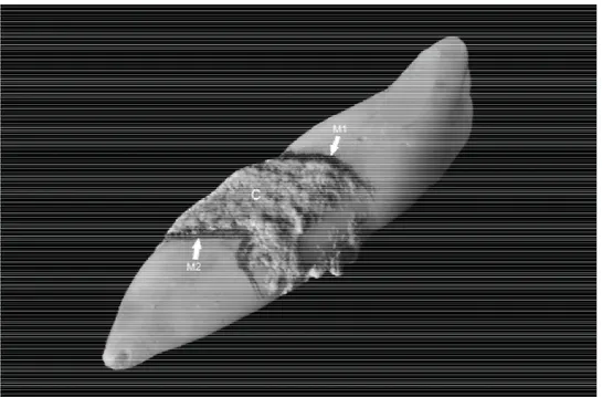
FIGURA 1 - A) Seleção dos dentes. B) Demarcação dos sulcos. C) Área de trabalho. D) Raspagem e alisamento radicular...82 FIGURA 2 - A) Corte transversal ao longo eixo do dente, na altura do primeiro sulco. B) Coroa seccionada. C) Corte longitudinal ao longo eixo do dente, separando as faces preparadas. D) Corte longitudinal ao longo eixo do dente, na altura do segundo sulco...82 FIGURA 3 - Espécimes preparados...83 FIGURA 4 - A) Aplicação passiva. B) Fricção vigorosa. C) Fricção sua...85 FIGURA 5 - A) Desidratação em concentrações crescentes de álcool. B) e C) Placas de acrílico codificadas para aplicação do HMDS...86 FIGURA 6 - . A) Secagem em papel de filtro. B) Espécime tratado, colado em “stub” metálico. C) Dessecador a vácuo. D) Espécime metalizado....88 FIGURA 7 - Fotomicrografia com aumento de 1500x representando o grau 1...89 FIGURA 8 - Fotomicrografia com aumento de 3500x representando o grau 1...89 FIGURA 9 - Fotomicrografia com aumento de 1500x representando o grau 2...89 FIGURA 10 - Fotomicrografia com aumento de 3500x representando o grau 2...89 FIGURA 11 - Fotomicrografia com aumento de 1500x representando o grau 3...90 FIGURA 12 - Fotomicrografia com aumento de 3500x representando o grau 3...90 FIGURA 13 - Fotomicrografia com aumento 1500x representando o grau 4...90 FIGURA 14 - Fotomicrografia com aumento de 3500x representando o grau 4...90 FIGURA 15 - Fotomicrografia com aumento de 1500x representando o grau 5...91 FIGURA 16 - Fotomicrografia com aumento de 3500x representando o grau 5...91 FIGURA 17 - Fotomicrografia com aumento de 1500x representando o grau 6...91 FIGURA 18 - Fotomicrografia com aumento de 3500x representando o grau 6...91 FIGURA 19 - Fotomicrografia com aumento de 1500x representando o grau 7...92 FIGURA 20 - - Fotomicrografia com aumento de 3500x representando o grau 7...92 FIGURA 21 - Fotomicrografia com aumento de 1500x representando o grau 8...92 FIGURA 22 - - Fotomicrografia com aumento de 3500x representando o grau 8...92 FIGURA 23 - Dente com cálculo radicular selecionado para o estudo. M1 – delimitação da porção mais coronário do cálculo.
FIGURA 24 - Fluxograma, mostrando os grupos e seus subgrupos utilizados nos estudos II e III...96 FIGURA 25 - A) Corte com disco diamantado para remoção da coroa. B) Coroa removida...98 FIGURA 26 - A) Corte separando a face de trabalho do restante do dente. B) Vista lateral após separação das faces. C) Visualização aproximada após o corte...99 FIGURA 27 - A) e B) Corte transversal, separando o primeiro espécime. C) Primeiro espécime separado...99 FIGURA 28 - A) Corte separando o segundo espécime. B) Os dois espécimes originados do mesmo dente, de área adjacente...100 FIGURA 29 - Aplicação do ácido cítrico a 25%, com pincel suave pelo tempo de 3 minutos...102 FIGURA 30 -Aplicação do cloridrato de tetraciclina na concentração de 50 mg/mL por fricção de bolinha de algodão pelo tempo de 3 minutos...102 FIGURA 31 - Coleta do sangue. B) Aplicação de uma gota de sangue sobre o espécime. C) Espécime metalizado...103 FIGURA 32 -Índice de adesão de elementos sanguíneos. A) Escore 1 – ausência de fibrina e de células sanguíneas. B) Escore 2 – escassa rede de fibrina e/ou de células sanguíneas. Escore 3 – moderada quantidade de células sanguíneas e rede de fibrina mais fina com pequeno entrelaçamento. Escore 4 – densa rede de fibrina com grande entrelaçamento e células sanguíneas aprisionadas...105 FIGURA 33 - Aplicação do ácido cítrico a 25%, com pincel suave pelo tempo de 3 minutos...108 FIGURA 34 - Aplicação do cloridrato de tetraciclina na concentração de 50 mg/mL por fricção de bolinha de algodão pelo tempo de 3 minutos..108 FIGURA 35 - Direção das três mensurações realizadas utilizado-se o rugosímetro...110
FIGURA 36 - Ra – rugosidade média. É a distância média de um perfil
desde sua linha média, sobre um comprimento medido (lm). Média aritmética entre picos e vales...112 FIGURA 37 - Rt – rugosidade total. Distância vertical entre o pico mais
alto e o vale mais baixo no percurso de medição lm, medido por duas linhas paralelas à linha média independente dos valores de Zi.
Rt = Rp + Rv...112 FIGURA 38 - Ry – rugosidade máxima. maior rugosidade entre as
rugosidades parciais (Zi) que se apresenta no percurso de medição (lm) Ry = Z2 ...112 FIGURA 39 - Rz (DIN) – é a média aritmética dos valores de rugosidade
parcial (Zi) medidos por linhas paralelas à linha média.
Cavassim R. Avaliação da descontaminação com ácido cítrico e
tetraciclina e da rugosidade, na adesão de coágulo a superfícies
radiculares submetidas a diferentes tipos de instrumentação [Tese de
Doutorado]. Araraquara: Faculdade de Odontologia da UNESP; 2012.
O tratamento periodontal engloba o processo de
raspagem e alisamento radicular, podendo ser realizado por diferentes
meios que além de remover o cálculo dental, produzem diferentes
características na superfície radicular como ranhuras e concavidades e
também a formação de smear layer. O uso de agentes químicos é
proposto na literatura para remover essa smear layer, e descontaminar a
superfície radicular, aumentando assim as chances de formação de nova
inserção conjuntiva. O objetivo do Estudo 1 foi avaliar, por meio de
microscopia eletrônica de varredura, a influência de diferentes
concentrações, modos e tempos de aplicação de ácido cítrico na
biomodificação de superfícies radiculares submetidas à raspagem e
alisamento radicular. Neste estudo, 270 amostras foram divididas em 6
grupos (45 amostras/grupo): soro fisiológico (controle), ácido cítrico (0.5%,
1%, 2%, 15% e 25%), com tempos de 1, 2 ou 3 minutos para cada grupo,
nos modos de aplicação: a) aplicação passiva (bolinha de algodão); b)
fricção suave (pincel); c) fricção vigorosa (bolinha de algodão), com
submetidas à desidratação em concentrações crescentes de álcool etílico
e HMDS, sendo em seguidas metalizadas e levadas para observação em
microscopia eletrônica de varredura. Um examinador treinado, calibrado
(kappa=0,93) e cego avaliou as fotomicrografias obtidas. A análise
estatística foi realizada utilizando-se os testes de Kruskal- Wallis e Dunn.
No estudo 2, investigou-se a influência da biomodificação radicular
associada a diferentes meios de instrumentação na adesão de coágulo e
elementos sanguíneos. Cento e cinquenta dentes afetados
periodontalmente foram divididos em 5 grupos: Grupo I, instrumentação
com curetas; Grupo II, instrumentação com curetas removendo cálculo
superficial; Grupo III, remoção do cálculo superficial com ultrassom;
Grupo IV, remoção do cálculo superficial com instrumento ultrassônico
seguido pela instrumentação com curetas Grupo V, superfície com
cálculo. Estes cinco grupos foram divididos em três subgrupos (10
amostras cada) de acordo com o condicionamento químico: a) sem
condicionamento químico; b) condicionamento químico com ácido cítrico;
c) condicionamento químico com cloridrato de tetraciclina. Em seguida,
uma gota de sangue humano da circulação periférica foi aplicada à
superfície radicular das amostras. Foi aguardado o período para
coagulação e as amostras foram então preparadas para microscopia
eletrônica de varredura. As fotomicrografias obtidas foram avaliadas por
um examinador treinado, calibrado (kappa=0,87) e cego e os escores
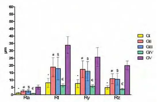
estudo 3 , investigou-se a influência dos diferentes meios de
instrumentação e da biomodificação na rugosidade e morfologia radicular.
Neste estudo foram utilizados cento e cinquenta dentes comprometidos
periodontalmente. Os dentes foram divididos em cinco grupos: Grupo I,
instrumentação com curetas; Grupo II, instrumentação com curetas
removendo cálculo superficial; Grupo III, remoção do cálculo superficial
com ultrassom; Grupo IV, remoção do cálculo superficial com instrumento
ultrassônico seguido pela instrumentação com curetas Grupo V,
superfície com cálculo. Estes cinco grupos foram divididos em três
subgrupos (10 amostras cada) de acordo com o condicionamento
químico: a) sem condicionamento químico; b) condicionamento químico
com ácido cítrico; c) condicionamento químico com cloridrato de
tetraciclina. As amostras obtidas foram avaliadas por meio de
rugosímetro, obtendo-se valores de rugosidade para os parâmetros (Ra,
Rt, Ry e Rz) e por meio de microscopia eletrônica de varredura para
avaliação da morfologia. Os resultados mostraram que: o ácido cítrico a
25% aplicado por pincel, por 1 ou 3 minutos foi mais eficiente na obtenção
da biomodificação radicular; o condicionamento químico favoreceu a
formação de rede de fibrina e adesão de componentes sanguíneos após
raspagem e alisamento radicular, apesar de não haver diferenças entre os
meios de instrumentação avaliados; os grupos instrumentados por
raspagem vigorosa apresentaram menores valores de rugosidades que os
morfologia mostrou um aspecto mais rugoso da superfície radicular foi
observado no grupo controle e nos grupos que tiveram o cálculo apenas
destacado. Presença de smear layer e hiperdesmineralização, com
aspecto de maior lisura de superfície foi observadas nos grupos após
raspagem, com e sem condicionamento químico da superfície.
Palavras-chave: Raiz dentária, Cálculos dentários, Raspagem dentária,
Coagulação sanguínea, Microscopia eletrônica de varredura
Cavassim R. Evaluation of decontamination with citric acid and
tetracycline and roughness on clot adhesion to root surfaces after different
types of instrumentation [Tese de Doutorado]. Araraquara: Faculdade de
Odontologia da UNESP; 2012.
Periodontal treatment encompasses the scaling and root
planning process and can therefore be carried out by various means. In
addition to removing dental calculus, the root instrumentation produces
different characteristics in the radicular surface as the formation of
concavities and smear layer. The use of chemical agents is proposed in
the literature to remove the smear layer, and detoxify the radicular surface,
thereby increasing the chances of forming new connective attachment.
The aim of the Study 1 was to assess, through scanning electron
microscopy, the influence of different concentrations, application methods
and application times of citric acid in the root conditioning after scaling and
root planning. Two hundred seventy (270) samples were equally divided
into six groups (n=45) for treatment with saline solution (control) and five
different concentrations of citric acid (0.5, 1, 2, 15, and 25 percent). Three
acid application methods were used (passive, brushing, and burnishing)
as well as three application periods (1, 2, and 3 minutes). A previously
trained, calibrated (kappa score = 0.93), and blind examiner subsequently
analyses were performed by using Kruskal-Wallis and Dunn’s post-hoc
tests. In Study 2, was investigated the influence of root conditioning
associated with different modes of instrumentation in the adhesion of clot
and blood elements. One hundred and fifty periodontally affected teeth
were divided into five groups: Group I, instrumentation with curettes;
Group II, instrumentation with curettes removing the superficial calculus;
Group III, ultrasonic scaler with removal of the superficial calculus; Group
IV, ultrasonic scaler for removal of the superficial calculus and after that,
instrumentation with curettes; Group V, calculus surface. These five
groups were further divided into three subgroups (10 samples each)
according to the root conditioning method used: a) no root conditioning; b)
root conditioning with citric acid; c) root conditioning with tetracycline
hydrochloride. After treatments, a drop of fresh human whole peripheral
blood was applied to the external root surface. The blood was allowed to
clot onto the root samples and then, the samples were prepared for SEM
analyses. In Study 3, was investigated the influence of the different means
of instrumentation and of root conditioning in root morphology and
roughness. One hundred and fifty human teeth lost due to periodontal
disease were use in this study The teeth were divided into five groups:
Group I, instrumentation with curettes; Group II, instrumentation with
curettes removing the superficial calculus; Group III, ultrasonic scaler with
removal of the superficial calculus; Group IV, ultrasonic scaler for removal
Group V, calculus surface. These five groups were further divided into
three subgroups (10 samples each) according to the root conditioning
method used: a) no root conditioning; b) root conditioning with citric acid;
c) root conditioning with tetracycline hydrochloride. After that, teeth were
evaluated according to roughness parameters (Ra, Rt, Ry, Rz) and
surface morphology. The results showed that exposure of collagen fibers
were obtained with application of citric acid at 25 percent by brushing for
one or three minutes; chemical root conditioning favored the fibrin network
formation and adhesion of blood components after scaling and root
planning, although there are no differences between the Instrumentation
means; the groups instrumented by vigorous scaling and root planning
have smaller roughness values than the groups that had only the
superficial dental calculus removed. Morphology evaluation showed a
rough aspect of radicular surface, observed in the control group and the
groups that had only the superficial dental calculus removed. Presence of
smear layer and chemical demineralization, with respect to greater surface
smoothness were observed in the groups after scaling, with and without
chemical conditioning of the surface.
Keywords: dental: root, calculus, scaling and root planning, clot
"³
As doenças periodontais inflamatórias crônicas acometem
os tecidos de proteção e sustentação dos dentes e, segundo a Academia
Americana de Periodontologia, compreendem respectivamente a
gengivite e periodontite8.
A periodontite é uma doença infecciosa que resulta na
inflamação dos tecidos de suporte dos dentes com perda progressiva de
inserção, formação de bolsa periodontal e presença de processo
inflamatório ativo41.
A etiologia da periodontite é multifatorial, estando
relacionada aos periodontopatógenos presente na placa bacteriana e no
cálculo dental7,65, e um hospedeiro susceptível. A adsorção de minerais e
proteínas salivares, bem como proteínas séricas leva à mineralização da
placa bacteriana e formação do cálculo dental. Este se adere às
superfícies dentárias por imbricamento mecânico e atua como
reservatório das bactérias responsáveis pela inflamação dos tecidos
periodontais7,11,34.
Após a instalação da periodontite, ocorre uma sucessão
de eventos nos tecidos periodontais, incluindo a perda de inserção de
tecido conjuntivo ao elemento dental, perda do osso alveolar de suporte e
a migração apical do epitélio juncional ao longo da superfície
ϮϬ
O tratamento periodontal básico baseia-se na remoção
mecânica do cálculo dental por meio de procedimentos clínicos de
raspagem e alisamento radicular (RAR), que pode ser realizada por
instrumentos manuais, rotatórios, sônicos ou ultrassônicos62-63.
Os principais objetivos do tratamento periodontal são a
completa remoção do cálculo dental e o restabelecimento das estruturas
de suporte (ligamento periodontal, cemento e osso alveolar) perdidas em
decorrência do processo inflamatório2-3,6-7. Entretanto, estudos têm
demonstrado dificuldades em alcançar esses objetivos5,52,71,78,81-82.
Mesmo após raspagem vigorosa e alisamento radicular ainda são
encontrados remanescentes de cálculo aderidos à superfície radicular33,55.
Além disso, enzimas e metabólitos bacterianos presentes na superfície
radicular contaminada levam a alterações na composição e densidade
mineral, reabsorção localizada, perda da matriz colágena e proteínas não
colágenas, retenção de bactérias e adsorção de endotoxinas2,6,93. Além
disso, endotoxinas e bactérias também podem ser encontradas no interior
de túbulos dentinários2-3,6,77,79-80.
Uma vez que o tratamento periodontal não restaura a
anatomia original do periodonto, resultando apenas em reparo periodontal
no qual os tecidos não reproduzem a arquitetura original dos tecidos
perdidos5,26,65, sugeriu-se a utilização de outros procedimentos com intuito
de complementar o tratamento periodontal básico. Tais procedimentos
Ϯϭ
condicionar a superfície radicular hipermineralizada e também expor as
fibras colágenas da matriz dentinária. A esse procedimento denomina-se
biomodificação radicular16,19,47.
A biomodificação radicular assim como a regeneração
tecidual guiada, os enxertos ósseos, os fatores de crescimento e a
combinação dos mesmos são modalidades terapêuticas que buscam
estabelecer condições favoráveis à regeneração periodontal107.
Além da remoção da smear layer formada durante a
raspagem, as substâncias químicas utilizadas para se obter a
biomodificação radicular teriam uma função secundária e de grande
importância à terapia periodontal, que seria a descontaminação da
superfície radicular. Uma vez que não se obtém a remoção completa do
cálculo dentário, a biomodificação radicular poderia atuar sobre o cálculo
residual, promovendo sua descontaminação ou mesmo sua remoção por
desmineralização e favorecendo ao reparo periodontal55.
Após a instrumentação periodontal, inicia-se o processo
de reparo dos tecidos adjacentes. O primeiro evento a ocorrer é a
formação de um coágulo sobre a superfície radicular, como resposta
imediata a qualquer tipo de trauma, constituindo assim a fase inicial do
processo de cicatrização dos tecidos periodontais80,106.
Dentro do processo cicatricial, o coágulo exerce duas
funções: protege temporariamente os tecidos e atua como matriz para
ϮϮ
promove a formação de uma barreira capaz de diminuir a migração apical
do tecido epitelial26,105.
No entanto, o processo de reparo é bastante complexo
nesta situação em que há o tecido conjuntivo em contato a superfície
radicular, após a instrumentação periodontal. A raiz dental apresenta-se
desprovida das células do ligamento periodontal e encontra-se
hipermineralizada e recoberta por smear layer42,76,78.
A formação do coágulo na interface raiz tecido conjuntivo
inicia-se quando elementos do sangue aderem-se à superfície radicular,
representando os eventos iniciais do processo de adesão do coágulo e de
reparo. Neste estágio temos a absorção e adesão de proteínas
plasmáticas à superfície radicular, seguida da formação de um coágulo
fibrinoso ligado à raiz. Dentro de algumas horas pode ser observada uma
fase inflamatória inicial, onde predominam neutrófilos e monócitos. Dentro
de aproximadamente três dias pode-se observar uma fase inflamatória
com migração de macrófagos e formação de um tecido de granulação.
Após sete dias pode ser observado um início de nova inserção conjuntiva
à raiz, porém, remanescente do coágulo e fibrina ainda estão presentes76.
Polson, Proye (1983)80 mostraram, em um modelo
experimental com macacos, a importância da adesão e maturação do
coágulo no reparo em retalhos periodontais. Os resultados deste estudo
mostraram que raízes instrumentadas e biomodificadas com ácido cítrico
Ϯϯ
conjuntiva, enquanto raízes apenas instrumentadas (controle)
apresentaram formação de epitélio juncional longo. Aparentemente, a
biomodificação radicular proporciona uma ancoragem estável do coágulo
sanguíneo e rede de fibrina à superfície radicular instrumentada,
proporcionando assim a maturação de nova inserção conjuntiva.
Reafirmando os autores anteriores, Baker et al.10 (2005)
sugerem que a smear layer interposta entre a raiz e o tecido conjuntivo
pode impedir a aderência do coágulo sanguíneo à superfície radicular,
podendo atuar como uma barreira física à nova inserção periodontal45.
Rocha et al. (dados não publicados) demonstrou que a
smear layer formada após instrumentação manual passa por duas fases
de eliminação. Em um primeiro momento ocorre uma rápida redução da
quantidade de smear layer formada sobre a superfície radicular e esta
continua a ser eliminada de forma mais lenta ao longo do tempo,
subsequente à instrumentação periodontal. Estes autores mostraram que
após raspagem e alisamento radicular, a smear layer é reabsorvida ao
longo do tempo, sendo que ao final da avaliação de 28 dias houve a
reabsorção de cerca de 60% da smear layer formada sobre a superfície
radicular. Por outro lado, no período inicial de 7 dias após raspagem
radicular, foi observado a presença de smear layer em cerca de 70% dos
dentes avaliados. Este resultado sugere que a presença de smear layer
sobre a superfície radicular pode ser um fator desfavorável ao processo
Ϯϰ
fase inflamatória e somente após 7 dias pode-se observar o início de
formação de nova inserção76.
Outros autores45,65,78 também sugerem que a
permanência da smear layer demonstrou ser desfavorável à estabilização
da rede de fibrina e formação de coágulo sanguíneo, atrasando essa fase
inicial da regeneração periodontal.
Ao que parece, a formação de um coágulo estável aderido
à superfície radicular por meio de uma rede de fibrina, é de vital
importância para o desenvolvimento de nova inserção conjuntiva. A
biomodificação radicular, além de remover a smear layer e expor fibras
colágenas da matriz dentinária, atuaria na inativação das endotoxinas
bacterianas, favorecendo a agregação plaquetária e formação de uma
rede de fibrina insolúvel, a qual atua como arcabouço para adesão e
proliferação de células do tecido conjuntivo10,48,95.
As características da superfície radicular parecem
influenciar diretamente ao processo cicatricial e de regeneração
periodontal, no entanto ainda não há na literatura estudos que avaliaram
os diferentes meio de instrumentação quanto às características e
rugosidade de superfícies produzidas, associadas à biomodificação
radicular.
A instrumentação periodontal pode ser realizada
utilizando-se instrumentos manuais, dispositivos sônicos e ultrassônicos
Ϯϱ
diferentes meios de instrumentação mostraram que dispositivos
ultrassônicos são tão efetivos quanto a instrumentação manual na
remoção de cálculo e placa bacteriana21,25,37,58,72,88. Outros estudos que
utilizaram instrumentos sônicos e ultrassônicos relatam o efeito
cavitacional ocasionado pela oscilação de bolhas de ar em meio líquido,
entretanto, esse efeito cavitacional parece não ter influência em estudos
in vivo, tendo contribuído para remoção de placa bacteriana em
avaliações in vitro apenas38,49,57,72,88,101,103,110.
Durante a instrumentação periodontal é considerado
adequado que a superfície radicular se apresente tão lisa quanto
possível61, uma vez que estudos demonstraram que a rugosidade da
superfície radicular influencia significativamente no estabelecimento do
biofilme dental53,61.
Estudos que avaliaram a instrumentação radicular
realizada por meio de instrumentos manuais, sônicos e ultrassônicos,
mostraram diferenças nas características de rugosidade obtidas. Estes
estudos apresentam controvérsias quanto ao tipo de instrumentação
capaz de produzir uma superfície mais regular. Na grande maioria dos
estudos, os instrumentos ultrassônicos produziram superfícies com maior
rugosidade que os instrumentos manuais ou sônicos50,56,69,102. Outros
estudos mostraram resultados em que não houve diferença na
instrumentação manual ou ultrassônica43,89, ou ainda que a
Ϯϲ
instrumentação manual37,51,74, sendo que este meio de instrumentação
produziu sulcos e ranhuras na superfície radicular54,87.
Clinicamente é muito difícil detectar pequenas variações
na lisura de superfície, visto que os instrumentos utilizados (sonda
exploradora) não possuem precisão para isso50, porém uma superfície
com maior rugosidade parece favorecer ao acúmulo de placa61,83,
enquanto uma superfície mais lisa parece favorecer a cicatrização dos
tecidos periodontais pois, possivelmente, apresenta uma menor
quantidade de depósitos aderidos, o que resulta em uma condição mais
aceitável biologicamente35. Esta condição é particularmente importante
quando observada em uma região radicular próxima à margem gengival e
de grande importância quando sua localização é em uma porção
supragengival, visto que nestas localizações uma superfície de maior
rugosidade facilitaria também o acúmulo do biofilme dental e dificultaria o
controle mecânico de placa realizado pelo paciente. Por outro lado, uma
região de maior rugosidade localizada no fundo de uma bolsa periodontal,
pode não afetar significativamente a cicatrização periodontal.
Assim, considerando-se o conjunto de fatores
relacionados à regeneração periodontal não se encontram completamente
elucidados, parece oportuno avaliar alguns dos fatores envolvidos neste
processo, tais como a adesão do coágulo sobre a superfície radicular,
após a descontaminação química, de modo a analisar o comportamento
Ϯϳ
ou cálculo residual e consequente contaminação. Também, julga-se
oportuno avaliar o efeito da descontaminação radicular química e
"³
Capítulo 1: o objetivo deste estudo in vitro foi avaliar, por
meio de microscopia eletrônica de varredura, a influência de diferentes
concentrações, modos e tempos de aplicação de ácido cítrico na
biomodificação de superfícies radiculares submetidas à raspagem e
alisamento radicular.
Capítulo 2: o objetivo deste estudo foi avaliar a influência
da descontaminação química radicular e de diferentes meios de
instrumentação, na adesão de coágulo e elementos sanguíneos.
Capítulo 3: o objetivo deste estudo foi avaliar a influência
dos diferentes meios de descontaminação, mecânica e química, na
rugosidade e morfologia radicular.
ϯϭ
ϯϮ
ϯϯ
ϯϰ
ϯϱ
ϯϲ
ϯϳ
ϯϴ
ϯϵ
ϰϬ
ϰϮ
Influence of different root instrumentation procedures associated with chemical root decontamination with citric acid and tetracycline hydrochloride on the clot adhesion on periodontally affected root surfaces - A SEM Study
* Rodrigo CAVASSIM * Shelon C. S. PINTO * Lucas A. FONTANARI * José E. C. SAMPAIO
Corresponding author:
Prof. Dr. José Eduardo Cezar Sampaio Department of Oral Diagnostic and Surgery
Araraquara Dental School, UNESP – Univ. Estadual Paulista, 1680 Humaitá St – ZIP Code 14801-903 - Araraquara-SP, BRAZIL Fone: 55-16-3301-6374
Fax: 55-16-3301-6369
E-mail: jsampaio@foar.unesp.br
Running title: Blood cells adhesion on affected teeth
Keywords: root instrumentation; root conditioning, periodontal disease, blood component adhesion, periodontal regeneration, SEM evaluation
ϰϯ
SUMMARY
The aim of this study was to evaluate the influence of root chemical decontamination and different instrumentation means, in clot and blood elements adhesion. One hundred and fifty periodontally affected teeth were divided into five groups: Group I, instrumentation with curettes; Group II, instrumentation with curettes removing the superficial calculus; Group III, ultrasonic scaler with removal of the superficial calculus; Group IV, ultrasonic scaler for removal of the superficial calculus and after that, instrumentation with curettes; Group V, calculus surface. These five groups were further divided into three subgroups (10 samples each) according to the root conditioning method used: a) no root conditioning; b) root conditioning with citric acid; c) root conditioning with tetracycline hydrochloride. After treatments, a drop of fresh human whole peripheral blood was applied to the external root surface. The blood was allowed to clot onto the root samples and then, the samples were prepared for SEM analyses. Chemical root conditioning favored considerable fibrin network formation and blood components adhesion in groups I and IV. No differences were observed when comparing instrumentation means. These results indicate that scaling and root planning associated with root conditioning were able to encourage the formation of fibrin network and adhesion of blood components.
ϰϰ
circulação periférica foi aplicada à superfície radicular das amostras. Foi aguardado o período para coagulação e as amostras foram então preparadas para microscopia eletrônica de varredura. O condicionamento químico favoreceu considerável formação de rede de fibrina e adesão de células sanguíneas nos grupos I e IV. Nenhuma diferença foi observada na comparação dos métodos de instrumentação. Os resultados indicaram que a raspagem e alisamento radicular associada ao condicionamento químico favoreceu a formação de rede de fibrina e adesão de células sanguíneas.
INTRODUCTION
Periodontal disease is characterized by chronic inflammatory processes caused by the presence of specific pathogen microorganisms, which trigger host response, progressive destruction of alveolar bone and apical migration of connective and epithelial attachments over time (1, 2). Scaling and root planning (SRP) is the main treatment for periodontal disease. One of the objectives of the periodontal therapy is the reduction of bacterial deposits and calculus on tooth surfaces (3). These procedures may be accomplished by using sonic, ultrasonic and manual hand instruments and has proved to be efficient in the control of gingivitis and periodontitis, producing a root that is biologically acceptable for healing process (4, 5).
The main objective is the removal of the subgingival biofilm, calculus and endotoxins from the root surface (5, 6) and also to accomplish a shift in microbial flora (7). However, it is well known that the root instrumentation results in a 2-15
ȝm in thickness layer formed of organic and mineralized debris. This layer is known as smear layer, and it covers the surface of teeth that underwent scaling and root planning. These layer as well as residual calculus may impair healing and soft tissue regeneration following periodontal treatment. So, there is an attempt to remove all the mineralized deposits from the root surface, thus improving periodontal regeneration.
endotoxin-ϰϱ
contaminated root cementum (6, 8). This condition is more favorable to optimize healing and enhance periodontal regeneration.
Ultrasonic instruments are as effective in removing subgingival plaque and subgingival calculus as hand instruments. However, complete removal of subgingival calculus with hand or machine instruments is impossible (9).
Once it is not possible to complete remove calculus and the instrumentation of the root surface produces a contaminated smear layer that may impair periodontal regeneration, we decide to conduct a study in order to evaluate the blood cell attachment to differently instrumentation methods of the root surface. In addition, two substances for decontamination were planned to be evaluated in this study. Citric acid and tetracycline hydrochloride are able to remove the smear layer produced after root instrumentation or even to demineralize it (10, 11). In an attempt to evaluate the effect of root conditioning in the residual calculus left after incomplete root instrumentation and decontamination of the root surface, these two substances were included in this study.
In sum, the objective of this study was to evaluate the blood cell adhesion to periodontally affected root surfaces submitted to mechanical instrumentation performed using conventional curettes or piezoelectric scalers when used singlehandedly or with a combined technique and the association of chemical root decontamination to the mechanical instrumentation. The blood cell adhesion was evaluated by means of scanning electron microscopy.
MATERIAL AND METHODS
ϰϲ
Experimental Design
After extraction, teeth were stored in water at room temperature for no longer than 3 months. All teeth were randomly divided into five groups (n=30 for each group) and a region of interest (ROI) which contained subgingival calculus was defined on one of the root surface aspects of each tooth. The ROI was instrumented by a single operator according to the groups:
Group I (curette only): each experimental surface was instrumented by applying sufficient strokes until complete calculus removal and achievement of a hard and smooth surface, clinically tested with an explorer. It was done using a new and sharpened Gracey’s curette 7-8 (Hu-Friedy Mfg. Co., Inc.).
Group II (curette removing superficial calculus): each experimental surface was instrumented with a new and sharpened Gracey’s curette 7-8 (Hu-Friedy Mfg. Co., Inc.), only removing the superficial calculus. The root planning was not performed in this group.
Group III (ultrasonic scaler removing superficial calculus): each experimental surface was instrumented with a periodontal tip mounted on an ultrasonic handpiece (Profi II Ceramic – Dabi Atlante, RibeirãoPreto, SP, Brazil) working at 24 to 28 kHz in a vertical direction under copious water irrigation. In this group, only the superficial calculus was removed using the ultrasonic tip.
Group IV (ultrasonic scaler/curette): each experimental surface was first instrumented with a periodontal tip mounted on an ultrasonic handpiece (Profi II Ceramic - DabiAtlante, Ribeirão Preto, SP, Brazil) working at 24 to 28 kHz in a vertical direction under copious water irrigation. Only the superficial calculus was removed using the ultrasonic tip. After that, each experimental surface was instrumented by applying sufficient strokes until complete calculus removal and achievement of a hard and smooth surface clinically tested with an explorer. It was done using a new and sharpened Gracey’s curette 7-8 (Hu-Friedy Mfg. Co., Inc.).
ϰϳ
Sample preparation
The ROI was marked at the most coronal and apical portion using a pencil (Figure 1).
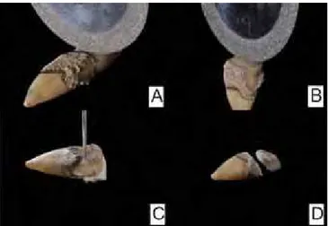
After undergoing one of the proposed treatments (Figure 2), the teeth were sectioned using a flexible double faced diamond disc (# 7020 – Ø 0.22 mm – thickness: 0.15 mm) (KG Sorensen, Barueri, SP, Brazil) at low speed. The roots were crosscut in the first mark, separating them from the crown (Figure 3-a). All roots were immediately cut lengthwise in the bucco-lingual orientation and then in the mesio-distal orientation (Figure 3-b).Finally, the sample was obtained by sectioning at the apical mark (Figure 3-c and 3-d).
Root Conditioning
These five groups were further divided into three subgroups (10 samples each) according to the root conditioning method used. These subgroups are listed below:
Subgroup a: no substance was applied to the root surface.
Subgroup b: the samples were root conditioned with citric acid 25% by brushing application with a soft brush (Disposable Brush Tips Ø2, 3M ESPE, Seefeld, Germany) for 3 minutes (10).
Subgroup c: the samples were root conditioned with tetracycline hydrochloride 50 mg/mL by burnishing with a small cotton pellet for 3 minutes (11).
Thereafter each sample was rinsed with 10 ml of saline solution and identified with a code for blind scoring of the SEMs.
Root preparation with blood tissue
ϰϴ
in PBS and rinsed again. The samples were post fixed in 2.5% glutaraldehyde in PBS for 30 minutes and rinsed again.
All samples were then dehydrated in an increasingly graded ethanol series (25%, 50%, 75%, 95%) for 10 minutes each. Following, the samples underwent dehydrationin 100% percent ethanol for 3 times of 10 minutes each.
Finally, samples were dried overnight in a dehydration jar (Corning, Corning Life Sciences, São Paulo, SP, Brazil), mounted on metallic stubs (Senai, Sao Paulo, SP, Brazil) and sputter-coated with a thin 25 nm layer of 99.99% pure gold (Balt-Tec SCD-050, Balt-(Balt-Tec, Gnathole Farm, Kettleshulme, High Peak, Cheshire - UK).
Scanning electron microscopy evaluation
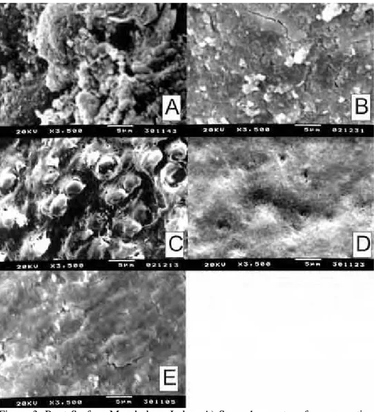
Two photomicrographs were obtained from the center area of each sample with 500X and 2,000X magnifications, using a scanning electron microscope operated at an accelerating voltage of 20 kV (Jeol T330 A, Jeol Ltd., Peabody, MA, USA). After the photomicrographs were obtained, they were identified and scored as follows to verify the blood component adhesion (BCA) and to analyze the morphologic characteristics obtained in the treatment, as follows: Score 1 -Root surface showing absence of blood components and complete inhibition of fibrin network formation. Score 2 -Poorly organized fibrin network with scarce cells covering the conditioned root surface. Score 3 - Root surface covered by a moderate thin fibrin network with a few quantity of blood cells. Score 4 - Organized fibrin network with large amount of entrapped blood cells.
ϰϵ
Statistical analysis
Data were analyzed using GraphPad Prism 5 statistical software (GraphPad, La Jolla, CA, USA). Level of significance was set at Į = 0.05 (two-sided).Once the data did not showed normal distribution, non-parametric analysis were used for comparisons. Differences among groups and among subgroups were tested using Kruskal-Wallis test and Dunn´s post hoc test.
RESULTS
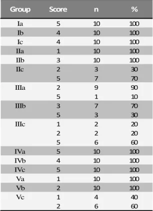
Basically four different morphological patterns of clot adhesion were observed in the evaluated groups. These patterns range from the absence of fibrin network to the presence of a dense network of fibrin with entrapment of blood components. The results for clot adhesion according to the root chemical conditioning are shown in Table 1. This table shows the comparison between citric acid, tetracycline hydrochloride and subgroups that received no chemical conditioning. The results for the comparison of the effect of the root instrumentation are shows in Figure 4.
Descriptive analyses for blood components adhesion
In group I, subgroup Ib presented the best results. In this subgroup, three samples (30%) had fibrin network formation and adhesion of blood elements and other four samples (40%) showed development of an organized fibrin network of. In the subgroup Ic only one sample (10%) showed formation of fibrin network organized with the adhesion of blood elements. The vast majority of samples (8 samples - 80%) showed only a poor network of fibrin on the root surface.
In group II, although not present significant differences, subgroup IIb showed better results for formation of a fibrin network. There are two samples (20%) with moderate fibrin network formation and seven samples (70%) with poorly organized fibrin network. Subgroup IIc had nine samples (90%) with poorly organized fibrin network.
ϱϬ
In group IV there was a subgroup superiority of IVc, which showed eight samples (80%) with formation of dense fibrin network. The subgroups IVa and IVb were predominant in samples with poor formation of fibrin network.
In group V, was the predominance of samples without the formation of fibrin network in all subgroups.
DISCUSSION
Periodontally-affected root surfaces treated with conventional non-surgical periodontal treatment might be not completely free from contaminants due to the presence of a residual smear layer created during instrumentations and residual calculus of an incomplete instrumentation (12). Since this residual calculus present on the tooth root is prone to contamination, discusses the importance of smear layer removal and decontamination of residual calculus.
The presence of the smear layer and residual calculus on the instrumented root surfaces has been shown to act as a contaminated physical barrier between the periodontal tissues and the root surface (13, 14) unsuitable for reintegration in periodontal connective tissue (15). Thus, this study investigated the effect of different means used for scaling and root planning and chemical root conditioning in the decontamination of the rood surface as a mean to remove contamination from the root surface and let possible the creation of a suitable root surface for the development of a reintegration in periodontal connective tissue.
ϱϭ
In this study, intragroup comparison showed that the formation of a fibrin network and adhesion of blood components was favored by vigorous scaling and root planning, when in association to chemical root conditioning. In group I the chemical conditioning with citric acid favored the formation of fibrin network and blood components adhesion. In group IV the chemical root conditioning with tetracycline hydrochloride favors the formation of fibrin network and blood components adhesion.
In intergroup comparison (Figure 4), differences among the means of root instrumentation in relation to the formation of fibrin network and adhesion of blood elements were not significant, except between subgroups IIc and IVc. One factor that may have influenced the samples which undergone chemical conditioning in group I to increase formation of fibrin network and adhesion of blood components is that were used teeth lost due to chronic periodontitis. These teeth were exposed to injuries and have their root surface with a higher degree of mineralization (18). The vigorous scaling and root planning is able to remove this mineralized layer, thereby providing the conditioning agents may act by removing the smear layer produced by scaling and root planning and expose the collagen matrix of dentin. On the other hand, groups in which only the superficial dental calculus was removed, a mineralized layer with endotoxins was kept on the root surface, hindering the action of chemical agents (19, 20).
ϱϮ
CONCLUSION
Within the limitations of the methodology, it can be concluded that scaling and root planning with curettes associate with chemical root decontamination were able to encourage the formation of fibrin network and adhesion of blood components, independent of having been held in isolation or after the use of ultrasound scalers.
REFERENCES
ϭ͘ ƌŵŝƚĂŐĞ'͘ŝĂŐŶŽƐŝƐŽĨƉĞƌŝŽĚŽŶƚĂůĚŝƐĞĂƐĞƐ͘:WĞƌŝŽĚŽŶƚŽů͘ϮϬϬϯ͖ϳϰ͗ϭϮϯϳͲϰϳ͘ Ϯ͘ ĂLJĂŶ^͕^ƚĂƐŚĞŶŬŽW͕EŝĞĚĞƌŵĂŶZ͕<ƵƉƉĞƌd^͘KƌĂůĞƉŝƚŚĞůŝĂůŽǀĞƌĞdžƉƌĞƐƐŝŽŶŽĨ />ͲϭĂůƉŚĂĐĂƵƐĞƐƉĞƌŝŽĚŽŶƚĂůĚŝƐĞĂƐĞ͘:ĞŶƚZĞƐ͘ϮϬϬϰ͖ϴϯ͗ϳϴϲͲϵϬ͘
ϯ͘ džĞůƐƐŽŶ W͕ >ŝŶĚŚĞ :͕ ELJƐƚƌŽŵ ͘ KŶ ƚŚĞ ƉƌĞǀĞŶƚŝŽŶ ŽĨ ĐĂƌŝĞƐ ĂŶĚ ƉĞƌŝŽĚŽŶƚĂů ĚŝƐĞĂƐĞ͘ ZĞƐƵůƚƐ ŽĨ Ă ϭϱͲLJĞĂƌ ůŽŶŐŝƚƵĚŝŶĂů ƐƚƵĚLJ ŝŶ ĂĚƵůƚƐ͘ : ůŝŶ WĞƌŝŽĚŽŶƚŽů͘ ϭϵϵϭ͖ϭϴ͗ ϭϴϮͲϵ͘
ϰ͘ ŚĞĞƚŚĂŵ t͕ tŝůƐŽŶ D͕ <ŝĞƐĞƌ :͘ ZŽŽƚ ƐƵƌĨĂĐĞ ĚĞďƌŝĚĞŵĞŶƚͲͲĂŶ ŝŶ ǀŝƚƌŽ ĂƐƐĞƐƐŵĞŶƚ͘:ůŝŶWĞƌŝŽĚŽŶƚŽů͘ϭϵϴϴ͖ϭϱ͗ϮϴϴͲϵϮ͘
ϱ͘ ^ŚĞƌŵĂŶ WZ͕ ,ƵƚĐŚĞŶƐ >,͕ :ƌ͕͘ :ĞǁƐŽŶ >'͘ dŚĞ ĞĨĨĞĐƚŝǀĞŶĞƐƐ ŽĨ ƐƵďŐŝŶŐŝǀĂů ƐĐĂůŝŶŐĂŶĚƌŽŽƚƉůĂŶŝŶŐ͘//͘ůŝŶŝĐĂůƌĞƐƉŽŶƐĞƐƌĞůĂƚĞĚƚŽƌĞƐŝĚƵĂůĐĂůĐƵůƵƐ͘:WĞƌŝŽĚŽŶƚŽů͘ ϭϵϵϬ͖ϲϭ͗ϵͲϭϱ͘
ϲ͘ ƌŝƐŬŽ,͘EŽŶƐƵƌŐŝĐĂůƉĞƌŝŽĚŽŶƚĂůƚŚĞƌĂƉLJ͘WĞƌŝŽĚŽŶƚŽůϮϬϬϬ͘ϮϬϬϭ͖Ϯϱ͗ϳϳͲϴϴ͘ ϳ͘ 'ƌĞĞŶƐƚĞŝŶ '͕ WŽůƐŽŶ ͘ DŝĐƌŽƐĐŽƉŝĐ ŵŽŶŝƚŽƌŝŶŐ ŽĨ ƉĂƚŚŽŐĞŶƐ ĂƐƐŽĐŝĂƚĞĚ ǁŝƚŚ ƉĞƌŝŽĚŽŶƚĂůĚŝƐĞĂƐĞƐ͘ƌĞǀŝĞǁ͘:WĞƌŝŽĚŽŶƚŽů͘ϭϵϴϱ͖ϱϲ͗ϳϰϬͲϳ͘
ϴ͘ ^ĂƚŽ<͕zŽŶĞLJĂŵĂd͕KŬĂŵŽƚŽ,͕ĂŚůĞŶ'͕>ŝŶĚŚĞ:͘dŚĞĞĨĨĞĐƚŽĨƐƵďŐŝŶŐŝǀĂů ĚĞďƌŝĚĞŵĞŶƚŽŶƉĞƌŝŽĚŽŶƚĂůĚŝƐĞĂƐĞƉĂƌĂŵĞƚĞƌƐĂŶĚƚŚĞƐƵďŐŝŶŐŝǀĂůŵŝĐƌŽďŝŽƚĂ͘:ůŝŶ WĞƌŝŽĚŽŶƚŽů͘ϭϵϵϯ͖ϮϬ͗ϯϱϵͲϲϱ͘
ϵ͘ ƵƐƐůŝŶŐĞƌ͕>ĂŵƉĞ<͕ĞƵĐŚĂƚD͕>ĞŚŵĂŶŶ͘ĐŽŵƉĂƌĂƚŝǀĞŝŶǀŝƚƌŽƐƚƵĚLJŽĨĂ ŵĂŐŶĞƚŽƐƚƌŝĐƚŝǀĞ ĂŶĚ Ă ƉŝĞnjŽĞůĞĐƚƌŝĐ ƵůƚƌĂƐŽŶŝĐ ƐĐĂůŝŶŐ ŝŶƐƚƌƵŵĞŶƚ͘ : ůŝŶ WĞƌŝŽĚŽŶƚŽů͘ ϮϬϬϭ͖Ϯϴ͗ϲϰϮͲϵ͘
ϭϬ͘ ĂǀĂƐƐŝŵZ͕>ĞŝƚĞ&Z͕ĂŶĚŝŵ>͕ĂŶƚĂƐ͕^ĂŵƉĂŝŽ:͘^ŵĞĂƌůĂLJĞƌƌĞŵŽǀĂů ĨŽƌ ĐŽůůĂŐĞŶ ĨŝďĞƌ ĞdžƉŽƐƵƌĞ ĂĨƚĞƌ ĐŝƚƌŝĐ ĂĐŝĚ ĐŽŶĚŝƚŝŽŶŝŶŐƐ͘ : ŽŶƚĞŵƉ ĞŶƚ WƌĂĐƚ͘ ϮϬϭϬ͖ϭϭ͗ϬϬϭͲϴ͘
ϭϭ͘ /ƐŚŝW͕ĂŶƚĂƐ͕ĂƚŝƐƚĂ>,͕KŶŽĨƌĞD͕^ĂŵƉĂŝŽ:͘^ŵĞĂƌůĂLJĞƌƌĞŵŽǀĂůĂŶĚ ĐŽůůĂŐĞŶĨŝďĞƌĞdžƉŽƐƵƌĞƵƐŝŶŐƚĞƚƌĂĐLJĐůŝŶĞŚLJĚƌŽĐŚůŽƌŝĚĞĐŽŶĚŝƚŝŽŶŝŶŐ͘:ŽŶƚĞŵƉĞŶƚ WƌĂĐƚ͘ϮϬϬϴ͖ϵ͗ϮϱͲϯϯ͘
ϭϮ͘ 'ĂŵĂů z͕ DĂŝůŚŽƚ :D͘ ĨĨĞĐƚƐ ŽĨ d ŐĞů ƉƌĞĐŽŶĚŝƚŝŽŶŝŶŐ ŽĨ ƉĞƌŝŽĚŽŶƚĂůůLJ ĂĨĨĞĐƚĞĚ ŚƵŵĂŶ ƌŽŽƚ ƐƵƌĨĂĐĞƐ ŽŶ ĐŚůŽƌŚĞdžŝĚŝŶĞ ƐƵďƐƚĂŶƚŝǀŝƚLJ Ͳ ĂŶ ^D ƐƚƵĚLJ͘ : WĞƌŝŽĚŽŶƚŽů͘ϮϬϬϳ͖ϳϴ͗ϭϳϱϵͲϲϲ͘