Patients with Sickle Cell Anemia
Ted Wun1,2,3,4*, Lori Styles5, Laura DeCastro6, Marilyn J. Telen6, Frans Kuypers5, Anthony Cheung3, William Kramer7, Henry Flanner8, Seungshin Rhee9, John L. Magnani8, Helen Thackray8
1Division of Hematology Oncology, University of California Davis School of Medicine, Sacramento, California, United States of America,2Clinical and Translational Sciences Center, UC Davis School of Medicine, Sacramento, California, United States of America,3Department of Pathology and Laboratory Medicine, UC Davis School of Medicine, Sacramento, California, United States of America,4VA Northern California Health Care System, Sacramento, California, United States of America,5Children’s Hospital and Research Institute Oakland, Oakland, California, United States of America,6Division of Hematology and Oncology, University of Pittsburgh, Pittsburgh, Pennsylvania, United States of America,7Kramer Consulting LLC, North Potomac, Maryland, United States of America,8GlycoMimetics, Inc, Gaithersburg, Maryland, United States of America,9Rho, Inc., Chapel Hill, North Carolina, United States of America
Abstract
Background: Sickle cell anemia is an inherited disorder of hemoglobin that leads to a variety of acute and chronic complications. Abnormal cellular adhesion, mediated in part by selectins, has been implicated in the pathophysiology of the vaso-occlusion seen in sickle cell anemia, and selectin inhibition was able to restore blood flow in a mouse model of sickle cell disease.
Methods:We performed a Phase 1 study of the selectin inhibitor GMI 1070 in patients with sickle cell anemia. Fifteen patients who were clinically stable received GMI 1070 in two infusions.
Results:The drug was well tolerated without significant adverse events. There was a modest increase in total peripheral white blood cell count without clinical symptoms. Plasma concentrations were well-described by a two-compartment model with an elimination T1/2of 7.7 hours and CLr of 19.6 mL/hour/kg. Computer-assisted intravital microscopy showed transient increases in red blood cell velocity in 3 of the 4 patients studied.
Conclusions:GMI 1070 was safe in stable patients with sickle cell anemia, and there was suggestion of increased blood flow in a subset of patients. At some time points between 4 and 48 hours after treatment with GMI 1070, there were significant decreases in biomarkers of endothelial activation (sE-selectin, sP-selectin, sICAM), leukocyte activation (MAC-1, LFA-1, PM aggregates) and the coagulation cascade (tissue factor, thrombin-antithrombin complexes). Development of GMI 1070 for the treatment of acute vaso-occlusive crisis is ongoing.
Trial Registration:ClinicalTrials.gov NCT00911495
Citation:Wun T, Styles L, DeCastro L, Telen MJ, Kuypers F, et al. (2014) Phase 1 Study of the E-Selectin Inhibitor GMI 1070 in Patients with Sickle Cell Anemia. PLoS ONE 9(7): e101301. doi:10.1371/journal.pone.0101301
Editor:Robert K. Hills, Cardiff University, United Kingdom
ReceivedDecember 10, 2013;AcceptedJune 3, 2014;PublishedJuly 2, 2014
Copyright:ß2014 Wun et al. This is an open-access article distributed under the terms of the Creative Commons Attribution License, which permits unrestricted use, distribution, and reproduction in any medium, provided the original author and source are credited.
Funding:Glycomimetics, Inc. funded this clinical trial. Part of this research was carried out at the UC Davis CTSC Clinical Research Center, supported by the grant UL1 TR 000002, NCATS, NIHGlycomimetics, Inc. (H.T.) participated in the study design, data collection and analysis, decision to publish, and preparation of the manuscript in collaboration with the academic authors.
Competing Interests:H.F., J.M., and H.T. are employees and have equity interest in Glycomimetics, Inc., which owns the patents to GMI 1070. W.K. has equity interest in Kramer Consulting, LLC, but has no commercial interest in GMI 1070. S.R. is an employee of Rho, Inc., but has no commercial interest in GMI 1070. This does not alter the authors’ adherence to PLOS ONE policies on sharing data and materials.
* Email: ted.wun@ucdmc.ucdavis.edu
Introduction
Sickle cell disease (SCD) results from a mutation in theb-globin gene that leads to a substitution of valine for glutamic acid at position 6 of the b globin chain. The most common genotypes associated with disease are homozygous S (HbSS), and compound heterozygous hemoglobin SC and S/b-thalassemia.[1,2] The prevalence in the United States is between 70,000–100,000, but is much greater in Africa and other parts of the world. Complications are protean and affect every major organ system including acute and chronic pain; ischemic and hemorrhagic stroke; infections; acute chest syndrome and pulmonary hyper-tension; congestive heart failure; azotemia, proteinuria, renal
concentrating defects, papillary necrosis, and priapism; osteomy-elitis and avascular necrosis of the bone; and leg ulcers. Vascular occlusion (or vaso-occlusive crisis – VOC) with ischemia-reperfu-sion injury is thought to underlie most, if not all, of these complications.
sub-endothelial matrix underlie this enhanced adhesiveness.[6–12] Furthermore, sRBC populations differ in their adhesiveness (young RBCs or reticulocytes being more adherent than older, more dense cells) and the degree of adhesiveness directly correlates with clinical severity.[4] The model of VOC that emerged involved initial adherence of sRBC reticulocytes to activated endothelium with secondary adherence and capture of more dense, rigid sRBC. Decreased adhesion molecule expression may partially underlie the beneficial effect of hydroxyurea in patients with SCD.[13] More recently, it has been suggested that platelets and leukocytes (specifically neutrophils and monocytes) play a role in acute and chronic morbidity as elucidated by in vitro and in vivo studies.[14–18] Heterotypic adhesive events between red cells, platelets, leukocytes, and endothelial cells, are emerging as a model for sickle cell vaso-occlusion.
Several lines of clinical evidence suggest a role for leukocytes in SCD pathogenesis.[19] Elevated leukocyte counts have been associated with increased mortality[20] and acute chest syn-drome.[21] The clinical benefit of hydroxyurea may be in part due to a reduction of leukocytes in addition to other mechanisms (increased fetal hemoglobin, decreased red cell adhesion molecule expression, decreased leukocyte adhesion.[22–30] In transgenic sickle cell mice, there is increased adherence of leukocytes to endothelium compared with non-sickle control mice following hypoxia or inflammatory stimuli.[14,31] This adherence, with secondary capture of sickle red blood cells, is the initiating event of VOC in this mouse model.
Turhan and colleagues demonstrated in a sickle mouse that adherent leukocytes bound to post-capillary cremasteric ve-nules.[14] Rolling and capture are mediated by selectins and integrins, respectively. Leukocyte capture onto endothelium stimulates signal transduction mediated conformational changes in leukocyte integrins that further increase competence to bind sickle red blood cells.
The selectins comprise a family of three members mediating adhesion events between blood cells and the endothelium. L-selectin is constitutively expressed on leukocytes and mediates lymphocyte recruitment in lymph nodes and secondary tethers between leukocytes and inactivated venules. Endothelial cells express two selectins, P-selectin which is stored in Weibel-Palade bodies and can be rapidly translocated to the cell surface upon stimulation, and E-selectin whose expression is induced by inflammatory cytokines such as TNF-aor IL-1b. Selectins mediate leukocyte rolling along on the endothelium, allowing circulating leukocytes to rapidly decelerate and come into close contact with chemokines and induce firm adhesion.[32] Transgenic sickle mice deficient in both P-and E- selectins exhibit severe defects in leukocyte adhesion[33,34] and are protected from VOC.[14] Studies of the individual function of single selectins in a mouse model of SCD have revealed a key role for E-selectin, but not P-selectin, in sending activating signals leading to the upregulation of theb2 integrin, Mac-1, specifically at the leading edge of crawling neutrophils in inflamed venules.[35]
Selectin ligands are composed of a trisaccharide domain common to both sialyl Lea and sialyl Lex (sLea/x)[36]. GMI 1070 is a novel small molecule glycomimetic that was rationally designed to contain both a more potent sLea/xmimetic and an extended sulfated domain to accommodate the binding require-ments of P-and L-selectins and confer drug-like properties to the molecule.[37]
In mouse models containing human sickle hemoglobin, GMI 1070 prevented and reversed VOC when administered well after initiation of the crisis in a clinically relevant treatment
proto-col.[38]. These data strongly suggest that targeting leukocyte adhesion has therapeutic potential in sickle cell disease.
Based on these animal studies, we performed an open-label, Phase 1 dose-ranging study of IV GMI 1070 in adults with stable SCD. The primary objective was to evaluate the safety of two intravenous (IV) doses of GMI 1070 in adults with sickle cell disease (SCD). The secondary objectives were to evaluate the pharmacokinetics (PK) of GMI 1070; measure the microvascular blood flow before and after infusion with IV GMI 1070; and, determine serial biomarkers of endothelial activation, inflamma-tion, coagulainflamma-tion, and downstream selectin effect in the blood before and after treatment with IV GMI 1070.
Methods
Ethics Statement
The Institutional Review Boards of the University of California, Davis; Duke University; and Oakland Children’s Hospital approved the study, and all patients gave signed, informed consent. The study was conducted according to the principles of Good Clinical Practice. The protocol for this trial and supporting CONSORT checklist are available as supporting information: see Checklist S1 and Protocol S1.
Eligible subjects were adults aged 18–50 years with an established diagnosis of sickle cell disease/homozygous hemoglo-bin S (SCD-SS) or sickle cell disease hemoglohemoglo-binb0
-thalassemia (SCD-Sb0
-thal). Disease activity had to be at the level of the subject’s medical baseline, with no evidence of worsening over the last 3 months (e.g. any acute complication of SCD that required unscheduled medical attention or intervention) as determined by the investigator.
This was an open-label, dose-ranging study of IV GMI 1070 in adults with stable SCD. The initial dose level (A) was selected based on preclinical data, and subsequent dose levels could be adjusted in response to in-study data. Dose Level A, consisted of a 20 mg/kg loading dose of IV GMI 1070 (0 hours) followed by a single 10 mg/kg dose of IV GMI 1070 1061 hours later. The plan was to double the dose if target drug concentrations were not achieved. Because target concentrations were achieved at Level A, no dose escalation was done.
Sampling for PK, biomarkers, and IVM (to assess microvascular blood flow) was performed immediately prior to the loading dose and at specified intervals after the first and second doses of IV GMI 1070.
Outcome Measures
Safety. The primary objective of the study was to evaluate the safety of GMI 1070 in adults with SCD. Safety was assessed by adverse event (AE) reporting, clinical laboratory test results, physical examination, electrocardiogram (ECG), and concomitant medication use. Subjects were to be followed for a total of 28 days after dosing of study drug: a clinic visit at day 7 (62 days) and a telephone follow-up at day 28 (63 days).
Pharmacodynamics. Computer-assisted intravital microsco-py (CAIM) was performed at the UC Davis site as per previously published methods[39–42] before dosing (on the day of dosing) and at the following intervals following the first dose of IV GMI 1070: 30 minutes, 2, 4, 8, and 24 hours. Plasma sampling for biomarkers of adhesion, inflammation, and downstream selectin effect in the blood was performed before dosing and 4, 8, 24, and 48 hours after administration of the first dose of IV GMI 1070. Analytes measured included: human sE-selectin, sICAM -1, ICAM-3, sPECAM-1, sP-selectin and sVCAM-1 by multiplex bead assay (eBioscience, San Diego, CA; D-dimer by ELISA, tissue factor in plasma and thrombin-antithrombin complexes (TAT), all by ELISA (Seikisui, Stamford, CT); and, surface expression of monocyte b2 integrins MAC-1 & LFA-1; and
platelet-monocyte aggregates (PMA) by flow cytometry all performed using modifications of previously described tech-niques[17,43].
Briefly, samples were pre-treated with or without ADP, labeled with or without anti-CD14-PE, and either anti-cd41a-APC or anti-CD11b-APC, and fixed prior to shipment on ice to the lab. Upon receipt, samples were centrifuged at 1000xg for 3–5 minutes at room temperature to remove the fixative. Samples were then washed once with 0.5ml Hepes Buffered Saline (HBS) and transferred to FACS tubes. One aliquot of the cd14 labeled cells was labeled on site with anti-CD11a-APC for 30 minutes at room temperature. All samples were analyzed on the BD LSR Fortessa Special Order Research Product flow cytometer (Becton Dick-inson, San Jose, CA) with DIVA acquisition software and FlowJo
analysis software version 8.8.7 (Tree Star Inc. Ashland, OR). Platelet-monocyte aggregates are defined as double positive events (CD14+
/CD41a+
) and results are presented as percent of total monocytes (CD14+
). MAC-1 (CD11b) and LFA-1 (CD11a) results are presented as mean fluorescence intensity (MFI) as compared to the unlabeled cells.
Analysis Plan
The analysis populations included the following 3 populations:
1. Safety population – all subjects who received at least 1 dose of investigational product (i.e., the loading dose) and had at least 1 post-baseline safety measurement (e.g., vital signs). The safety population was used for summarizing baseline characteristics and safety data.
2. Efficacy population – all subjects who received at least 1 dose of investigational product (i.e., the loading dose) and had at least subsequent 1 CAIM or biomarker measurement. The efficacy population was used for the primary and secondary efficacy analyses (CAIM and biomarkers of adhesion, inflammation, coagulation, and downstream selectin effect).
3. Pharmacokinetic population – all subjects who completed the study and had sufficient data for the PK analyses. The PK population was used to summarize the plasma concentrations of GMI 1070 over time, amount of GMI 1070 excreted in the urine, and PK parameters.
Safety. Safety results were summarized descriptively and presented in listings.
Pharmacokinetics. GMI 1070 plasma concentration, uri-nary excretion, and PK parameters were summarized by treatment group using descriptive statistics. Pharmacokinetic parameters were estimated by fitting a 2-compartment IV infusion model to each subject’s data. The primary PK parameters estimated in fitting the model included total plasma clearance (CL), intercompartmental clearance (CLD2), volume of the central compartment (V1), and volume of the peripheral compartment (V2). These primary parameters were used to calculate the following secondary PK parameters: maximum observed drug concentration (Cmax), time of maximum drug concentration (Tmax), area under the plasma concentration-time curve to infinity (AUC[inf]), apparent first-order distribution rate constant (a), apparent first-order terminal elimination rate constant (b), elimination half-life (tK), and volume of distribution at steady state (Vss). Urine data were used in conjunction with the PK model to estimate renal clearance for the collection period (CLr). The total amount of drug excreted in the urine during the collection period (Ue) was calculated from the urine concentration of drug (Cu) and the volume of urine collected (Vu), and expressed as milligrams of drug and percentage of administered dose recovered in the urine (Fe). The AUC for the urine collection period was estimated from the PK model, and CLr was calculated as CLr = Ue/AUC. All modeling was done using WinNonlin Professional Version 5.2.
Pharmacodynamics. Serial red blood flow velocity from the UC Davis site was calculated, tabulated, and presented in graphical form. Other efficacy (pharmacodynamic) outcome measures included biomarkers of endothelial activation, inflam-mation, and downstream selectin effect in the blood measured at 4, 8, 24, and 48 hours after administration of the first dose of IV GMI 1070.
Expression levels were compared against pre-treatment, and stratified by HU use. Variables evaluated were the mean changes in microvascular blood flow from baseline to each post-baseline
Table 1.Baseline Demographic Characteristics (Safety Population).
GMI-1070
20 mg/kg+10 mg/kg
Characteristic N = 15
Statistic or Category n (%)
Age (years)
n 15
Mean 32.1
SD 10.62
Median 28.0
Min, max 19, 50
Gender – n (%)
Male 9 (60.0)
Female 6 (40.0)
Baseline weight (kg)
n 15
Mean 64.79
SD 9.90
Median 65.5
Min, max 48.3, 90.7
Genotype – n (%)
HbSS 13 (86.7)
HbSb0-thal 2 (13.3)
Hb = hemoglobin; max = maximum; min = minimum; SD = standard deviation; thal = thalassemia.
assessment time point, and the changes in biomarkers from baseline to each post-baseline time point.
The hypothesis of no difference between mean baseline value and mean post-baseline value at each time point was assessed using a mixed-effects model. The change from baseline in microvascular blood flow and other biomarkers was modeled longitudinally using a mixed-effects model, with subject as a random effect and time, reader, interaction of time and reader, and baseline value as fixed effects. For continuous variables with repeated measures such as the biomarkers used in this trial, the linear mixed model approach allows use of all the data available.
Results
A total of 15 patients were enrolled (Table 1 and Figure 1) between May 28, 2009 (first subject enrolled) to July 6, 2010 (last patient last visit) when adequate data was collected to allow for planning of a Phase 2 study. All were African-American; nine were males, and ages ranged from 19 to 50 years. Thirteen of the 15 had homozygous hemoglobin S disease. Five patients were on a stable dose of hydroxyurea.
Safety
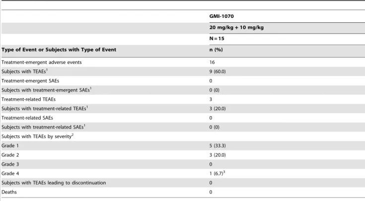
A total of 9 (60.0%) subjects reported 16 treatment-emergent adverse events (TEAEs) during the study (Tables 2 and 3). The most frequently reported were headache (4/15, 26.7%) and vaso-occlusive crisis (VOC) (2/15, 13.3%). Three (20.0%) subjects had TEAEs considered at least possibly related to study drug (headache in 2 subjects and leukocytosis in 1 subject). Except for grade 4 anemia (pre-existing as Grade 4, and worsening slightly while on study) in one subject, all TEAEs were rated grade 1 or 2 in severity. One subject reported a TEAE involving skin. One subject reported grade 1 pruritus of abdomen and back on Day 8, which resolved with no treatment after 3 days.
No deaths, serious adverse events, or TEAEs leading to discontinuation were reported during the study.
Eight (53.3%) subjects reported 10 TEAEs involving pain. These included the headache and VOC (13 and 23 days after infusion) already mentioned, and 1 (6.7%) subject each with infusion site pain, arthralgia, and oropharyngeal pain. All of the events were grade 1 in severity except for grade 2 headache in one subject and grade 2 VOC in another. The headache began on study Day 1 and was considered probably related to study drug. A headache in another subject (grade 1, onset Day 2) was considered possibly related. The remaining pain events were considered unlikely related (7 subjects) or unrelated (oropharyngeal pain), with onset days ranging from Day 1 to Day 24.
No notable trends in vital signs were observed during the study. For each body system, the majority ($73.3%) of subjects had no changes in physical examination findings, and those with changes had findings that were not considered clinically significant.
Mean values for aspartate aminotransferase (AST) and lactate dehydrogenase (LDH) were elevated at baseline and showed
decreases at subsequent time points, but they remained close to or above the upper limit of reference range throughout the study. At the Day 7 Visit, the mean (SD) changes in AST and LDH from baseline were -13.9 (24.4) U/L and -98.9 (134.7) U/L, respec-tively, and the mean (SD) observed values were 41.9 (14.6) U/L and 629.9 (378.2) U/L. Mean values for total bilirubin were also elevated at baseline (3.76 [3.08] mg/dL), and subsequent values were generally similar to those at the Baseline Visit. The elevations in AST, LDH, and total bilirubin were consistent with expected abnormalities in individuals with SCD.
Hematocrit and hemoglobin values for the safety population showed small mean increases from baseline at the Day 2 and 3 Visits. At the Day 3 Visit, the mean (SD) changes in hematocrit and hemoglobin from baseline were 1.56 (1.80)% and 0.35 (0.62) g/dL, respectively.
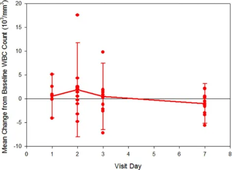
Moderate increases in neutrophil and total white blood cell (WBC) counts were observed at the Day 2 Visit (24 hours), when the mean (SD) values were 7.5 (5.5)6103/mm3 and 11.6 (6.6)6103/mm3, respectively (Figures 2–3). The corresponding changes from baseline were significant: 3.2 (4.5)6103/mm3 (p-value,0.001) for neutrophil count and 2.3 (4.8)6103/mm3 (p-value = 0.013) for WBC count. Mean (p-values for neutrophil and WBC counts returned to baseline values by Day 7. This pattern of increase in neutrophil and WBC counts at the Day 2 Visit was observed in the subjects who were not taking hydroxyurea during the study. There was no significant increase in neutrophil counts at 24 hours in the patients taking hydroxyurea group (p = 0.2). However, these data should be interpreted with caution given the small sample size.
The high sensitivity C-reactive protein (hsCRP) level increased from 4.3 (4.1) mg/L at the Baseline Visit to 9.0 (15.7) mg/L at the
Table 2.Overall Summary of Adverse Events (Safety Population).
GMI-1070
20 mg/kg+10 mg/kg N = 15
Type of Event or Subjects with Type of Event n (%)
Treatment-emergent adverse events 16
Subjects with TEAEs1 9 (60.0)
Treatment-emergent SAEs 0
Subjects with treatment-emergent SAEs1 0 (0)
Treatment-related TEAEs 3
Subjects with treatment-related TEAEs1 3 (20.0)
Treatment-related SAEs 0
Subjects with treatment-related SAEs1 0 (0)
Subjects with TEAEs by severity2
Grade 1 5 (33.3)
Grade 2 3 (20.0)
Grade 3 0
Grade 4 1 (6.7)3
Subjects with TEAEs leading to discontinuation 0
Deaths 0
SAE = serious adverse event; TEAE = treatment-emergent adverse event. 1 Subjects who experienced 1 or more adverse event were counted once.
2 If a subject experienced more than 1 adverse event, the subject was counted only once for the worst (or maximum) severity. 3 Event was grade 4 anemia (hemoglobin 5.9 g/dL), which was not considered serious by the investigator.
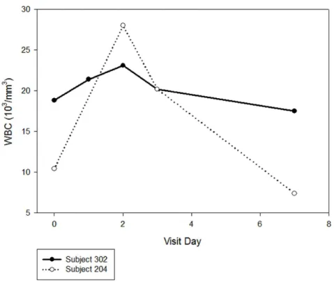
Day 3 Visit, and then decreased to a level near baseline (4.2 [5.4] mg/L) at the Day 7 Visit. Two subjects had notable increases in hsCRP during the study, with peak values of 50.4 and 40.7 mg/ L, respectively, at the Day 3 Visit. In both subjects, the increases were associated with significant increases in WBC count, and in one, with a pre-existing asymptomatic urinary tract infection (Figure 4). Neither subject had symptoms of infection or increased pain after treatment with GMI 1070.
Clinically significant laboratory test results (reported as adverse events and requiring clinical follow-up) were reported in 3 subjects. One subject had a hemoglobin value of 5.9 g/dL (Day 7 Visit), decreased from a baseline of 6.0 g/dL. Another subject had a WBC count of 28.06103/mm3and 20.26103/mm3(Day 2 and 3 Visits, respectively), and remained asymptomatic during this time. Another subject had an elevated LDH (476 U/L at baseline); decreased serum potassium (2.6 mEq/L at Day 3 Visit); increased WBC count (18.8, 21.4, 23.1, 20.2, and 17.56103/mm3 at the Baseline and Day 1 [post dose], 2, 3, and 7 Visits, respectively); increased neutrophil count (12.8, 14.5, and 11.66103/mm3 at Day 1 [post dose], 2, and 3, respectively); and several microscopic urinalysis variables consistent with a pre-existing asymptomatic urinary tract infection.
Pharmacokinetic Results
Fourteen (14) of the 15 subjects had sufficient data for PK analysis. The PK of GMI 1070 in subjects with SCD was consistent with a 2-compartment IV infusion model, and the estimated clearances, volumes of distribution, T1/2(7.7 hours) and
CLr (19.6 mL/hour/kg) were consistent with those observed previously in healthy volunteers.[44–46] The model-predicted plasma GMI 1070 concentrations for all 14 subjects in the PK population showed excellent agreement with the observed concentrations; this is illustrated with the mean data in Figure5. These results also demonstrated that the use of a loading dose achieves immediate steady-state plasma concentrations of GMI 1070 in individuals with SCD and that the use of this dose level (20 mg/kg loading dose) followed by a 10 mg/kg maintenance dose achieves plasma concentrations expected to have activity in this population.
Pharmacodynamic Results
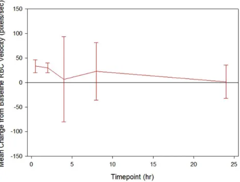
At the UC Davis site, the mean observed RBC velocity showed an increase over the baseline value at 30 minutes, 2 hours, 4 hours, 8 hours and 24 hours after dosing. The largest mean (SD) increase in RBC velocity from baseline occurred 30 minutes post dosing, with a value of 33.30 (13.52) pixels/sec (Figure6). All four subjects had detectable plasma levels of GMI 1070, consistent with
Table 3.Treatment-emergent Adverse Events by System Organ Class and Preferred Term (Safety Population).
GMI-1070
20 mg/kg+10 mg/kg
System Organ Class N = 15
Preferred Term n (%)
Any treatment-emergent adverse event 9 (60.0)
Nervous system disorders 4 (26.7)
Headache 4 (26.7)
Blood and lymphatic system disorders 2 (13.3)
Anemia 1 (6.7)
Leukocytosis 1 (6.7)
Congenital, familial, and genetic disorders 2 (13.3)
Sickle cell anemia with crisis1 2 (13.3)
Gastrointestinal disorders 1 (6.7)
Vomiting 1 (6.7)
General disorders and administration site conditions 1 (6.7)
Infusion site pain 1 (6.7)
Metabolism and nutrition disorders 1 (6.7)
Hypokalemia 1 (6.7)
Musculoskeletal and connective tissue disorders 1 (6.7)
Arthralgia 1 (6.7)
Respiratory, thoracic, and mediastinal disorders 1 (6.7)
Cough 1 (6.7)
Oropharyngeal pain 1 (6.7)
Skin and subcutaneous tissue disorders 1 (6.7)
Pruritus 1 (6.7)
Events are listed in descending order of frequency of preferred terms, with grouping by system organ class.
If a subject experienced more than 1 preferred term of adverse event, the subject was counted only once for that preferred term. Urinary tract infection reported for one subject was not included because the event occurred on study Day -3.
plasma levels predicted by the PK model, during velocity measurements. This trend toward an increase in RBC velocity from baseline may indicate improved blood flow in small blood vessels, but it did not reach statistical significance (all p-values.
0.05).
Statistically significant reduction of multiple biomarkers of adhesion, activation, and coagulation was observed after treatment with GMI 1070. Soluble adhesion markers were reduced after 8 hrs (soluble E-selectin), and after 4 and 8 hrs (soluble P-selectin; ICAM-1). Tissue factor (TF) was reduced at 4 and 8 hrs (Table 4). Thrombin-antithrombin complex (TAT) levels were reduced at all
time points (4 hr, 8 hr, 24 hr, 48 hr). The percentage of PMA was reduced at 8 hrs. The expression of MAC-1 and LFA-1 was reduced at all time points. When HU use was considered (HU–No; HU–Yes), the levels of sEsel, sPsel, ICAM-1, TF, TAT, MAC-1, and neutrophil counts were lower and more variable at baseline in the HU-No group, significantly so for ICAM-1 (p = 0.048) and MAC-1 (p = 0.001). After GMI 1070, significant reduction from baseline was seen in both groups: in the HU-No group for ICAM-1, TF, PMA, LFA-ICAM-1, and in the HU-Yes group for sEsel, MAC-ICAM-1, TF, TAT. However, again due to the small sample size caution should be exercised in interpreting these results.
Figure 2. Change in absolute neutrophil count from baseline. doi:10.1371/journal.pone.0101301.g002
Discussion
The primary objective of this study was to evaluate the safety of two intravenous doses of GMI 1070 in clinically stable adults with SCD. No serious adverse events were reported, and no subject discontinued the study because of an adverse event. All adverse events except one were grade 2 or less in severity. The single grade 4 event was anemia, which was considered to be non-serious by the investigator and consistent with underlying sickle cell anemia.
Three subjects reported TEAEs considered possibly or probably related to study drug, which included headache (2 subjects) and leukocytosis (1 subject). Laboratory values showed no notable trends of worsening over time. Overall no serious concerns were identified in this study with regard to the safety of GMI 1070.
Moderate and significant mean increases in neutrophil (p-value
,0.001) and WBC (p-value = 0.013) counts were observed at the Day 2 Visit (24 hours), followed by a return to values similar to Figure 4. Change in total white blood cell count (WBC) in the two subjects with marked leukocytosis.
doi:10.1371/journal.pone.0101301.g004
baseline by Day 7. Thus, administration of GMI 1070 was associated with neutrophilia and leukocytosis that were moderate in intensity, temporally associated with plasma levels of the drug, and reversible after elimination of the drug from the plasma. These findings are consistent with the expected anti-adhesive effect of selectin inhibition, and likely represent release of adherent leukocytes from the vascular endothelium into the peripheral circulation. Similar increases in neutrophil and WBC count were not observed in the subgroup of subjects who took hydroxyurea during the study, although small fluctuations in these counts were seen in individuals.
The WBC and neutrophil results from this study suggests that leukocyte adhesion can be inhibited at pharmacological levels in patients with sickle cell disease, although whether this will be enough to alleviate established vascular occlusion in patients presenting in VOC remains to be seen. Leukocytosis could theoretically have detrimental effect in patients with sickle cell disease, such as increased incidence of acute chest syndrome or infection. These potential adverse effects will have to be carefully followed in further clinical studies.
A secondary study objective was to evaluate the PK of two IV doses of GMI 1070 in adults with SCD. The plasma concentra-tions were concordant with a 2-compartment IV infusion model, and the estimates for clearances, volumes of distribution, tK, and CLr were consistent with those observed previously in healthy volunteers. The model-predicted plasma GMI 1070 concentra-tions for all 14 subjects in the PK population showed excellent agreement with the observed concentrations. The results support the use of a 20 mg/kg loading dose followed by a 10 mg/kg dose to rapidly reach and maintain plasma concentrations of GMI 1070 expected to be effective in individuals with SCD.
Another secondary objective was to evaluate microvascular blood flow before and after administration of IV GMI 1070. Previous work by our group has demonstrated that acute changes
in blood flow in response to various therapies are detectable by this method.[39,47] Although subjects showed mean increases in RBC velocity compared to baseline at 30 minutes and 2, 4, 8 and 24 hours post dosing, which may indicate improved blood flow, the trend toward an increase in RBC velocity did not reach statistical significance. However, the sample size (n = 4) may have precluded finding significant differences. The timing was consis-tent with observed WBC/ANC changes, suggesting pharmaco-logic response of increased WBC and ANC may correspond with improved microvascular flow.
The effects of GMI 1070 were evaluated on biomarkers known to be elevated in sickle cell disease and mechanistically affected by the target molecule, E-selectin. Markers of leukocyte activation (MAC-1; LFA-1); platelet activation (soluble P-selectin; platelet-monocyte aggregates or PMA); vascular inflammation (soluble E-selectin; soluble ICAM-1); monocyte activation (PMA); and coagulation system activation (tissue factor and thrombin-anti-thrombin complexes) were serially measured. At some time point between 4 and 48 hours after intravenous infusion of GMI 1070, there were significant decreases in all these various markers consistent with downstream inhibition of cellular activation and an overall anti-inflammatory effect. In some individuals these changes persisted at a time when drug plasma levels were below 10mg/mL. As the pathophysiology of VOC involves both leukocyte adhesion and downstream inflammation,[15,16,48] these findings provide further rationale for use of GMI 1070 in VOC.
When administered to adults with SCD at steady state, the pan-selectin inhibitor GMI 1070 had safety and PK profiles similar to those seen in healthy volunteers. This study provides mechanism-based evidence of effect of GMI 1070 in this population and supports further evaluation of the drug in the treatment of vaso-occlusive crisis. A prospective randomized double-blinded Phase 2 trial of GMI 1070 in patients with SCD and VOC has been conducted based on results of this Phase 1 study.
4 Hours 8 Hours 24 Hours 48 Hours Biomarker Baseline Level (SD) LS Mean (CI) Change from Baseline, LS
Mean (CI) p-value LS Mean (CI)
Change from Baseline, LS
Mean (CI) p-value
LS Mean (CI)
Change from Baseline, LS
Mean (CI) p-value LS Mean (CI)
Change from Baseline, LS
Mean (CI) p-value
sE-sel (ng/mL)
117.2 (48.6) 104.0 (93.1, 115.0)
210.53 (221.4, 0.4)
0.058 99.2 (89.7, 108.7)
215.4 (224.9,25.9)
0.004 105.5 (95.5, 115.4)
29.11 (219.1, 0.8)
0.070 109.4 (99.6, 119.1)
25.21 (214.9, 4.5)
0.272
sP-sel (ng/mL)
180.4 (154.0) 122.6 (94.1, 151.1)
231.2 (259.6,22.7)
0.034 122.9 (96.1, 149.7)
230.9 (257.7,24.1)
0.028 134.0 (106.7, 161.3)
219.8 (247.1, 7.5)
0.141 138.5 (111.4, 165.5)
215.3 (242.3, 11.7)
0.241
ICAM-1 (ng/mL)
239.6 (177.8) 195.2 (179.1, 211.3)
219.4 (235.5,23.3)
0.021 190.6 (175.7, 205.4)
224.1 (238.9,29.3)
0.004 203.3 (188.1, 218.5)
211.3 (226.5, 3.9)
0.133 202.5 (187.5, 217.6)
212.1 (227.1, 2.9)
0.105
TF (pg/mL)
466.7 (313.4) 323.8 (239.1, 408.4)
2120.5 (2205.1,235.8)
0.009 364.1 (283.9, 444.2)
280.2 (2160.3,20.1)
0.050 374.9 (293.5, 456.4)
269.3 (2150.8, 12.1)
0.087 374.6 (293.8, 455.4)
269.6 (2150.4, 11.2)
0.083
TAT (ng/mL)
145.4 (94.5) 40.3 (0.7, 79.8)
2104.0 (2143.5,264.5)
,0.001 83.6 (54.9, 112.2)
260.7 (289.3,232.0)
,0.001 63.1 (31.6, 94.6)
281.2 (2112.7,249.7)
,0.001 93.2 (63.2, 123.3)
251. 0 (281.1,221.0)
0.002 MAC-1 (MFI) 3278.5 (1302.4) 2271.5 (1559.3, 2983.8) 21235.7 (21948.0,2523.4)
0.002 2475.7 (1999.3, 2952.1)
21031.5 (21507.9,2555.1)
0.001 2742.8 (2227.5, 3258.0)
2764.5 (21279.7,2249.1)
0.008 2641.6 (2146.6, 3136.7)
2865.6 (21360.6,2370.6)
0.004
LFA-1 (MFI)
815.3 (271.8) 571.6 (386.3, 757.0)
2294.3 (2479.7,
2109.0)
0.004 695.8 (593.2, 798.5)
2170.1 (2272.8,267.4)
0.004 705.0 (584.8, 825.1)
2161.0 (2281.1,240.8)
0.012 704.0 (593.6, 814.5)
2161.9 (2272.3,251.5)
0.008
PMA (%) 82.1 (20.2) 72.8 (48.9, 96.7)
29.2 (233.1, 14.7)
0.425 61.0 (42.3, 79.7)
220.9 (239.6,22.2)
0.033 75.3 (55.8, 94.9)
26.6 (226.2, 13.0)
0.464 76.9 (57.8, 96.0)
25.0 (224.1, 14.1)
Supporting Information
Checklist S1 CONSORT Checklist. (DOC)
Protocol S1 Trial Protocol. (PDF)
Acknowledgments
The authors would like to acknowledge the efforts of the following: Clinical Research Coordinators that made this research possible: Carmen Wilberg,
R.N., Rebecca Seufert, R.N., Martina Garcia, B.S., Kathy Stewart, B.S., Charlene Flahiff, M.S.; Our patients who volunteered for this study; and Shanti Rodriguez for editorial assistance.
Author Contributions
Conceived and designed the experiments: TW MT AC FK HF JM HT. Performed the experiments: TW LS LD MT AC HF FK. Analyzed the data: TW AC WK FK SR HT. Contributed reagents/materials/analysis tools: AC HF FK. Wrote the paper: TW FK JM HT.
References
1. Platt OS (1994) Easing the suffering caused by sickle cell disease. N Engl J Med 330: 783–784.
2. Rees DC, Williams TN, Gladwin MT (2010) Sickle-cell disease. Lancet 376: 2018–2031.
3. Hebbel RP (1991) Beyond hemoglobin polymerization: the red blood cell membrane and sickle disease pathophysiology. Blood 77: 214–237.
4. Hebbel RP, Boogaerts MA, Eaton JW, Steinberg MH (1980) Erythrocyte adherence to endothelium in sickle-cell anemia. A possible determinant of disease severity. N Engl J Med 302: 992–995.
5. Hebbel RP, Boogaerts MA, Koresawa S, Jacob HS, Eaton JW, et al. (1980) Erytrocyte adherence to endothelium as a determinant of vasocclusive severity in sickle cell disease. Trans Assoc Am Physicians 93: 94–99.
6. Zennadi R, De Castro L, Eyler C, Xu K, Ko M, et al. (2008) Role and regulation of sickle red cell interactions with other cells: ICAM-4 and other adhesion receptors. Transfus Clin Biol 15: 23–28.
7. Zennadi R, Moeller BJ, Whalen EJ, Batchvarova M, Xu K, et al. (2007) Epinephrine-induced activation of LW-mediated sickle cell adhesion and vaso-occlusion in vivo. Blood 110: 2708–2717.
8. Brittain HA, Eckman JR, Swerlick RA, Howard RJ, Wick TM (1993) Thrombospondin from activated platelets promotes sickle erythrocyte adherence to human microvascular endothelium under physiologic flow: a potential role for platelet activation in sickle cell vaso-occlusion. Blood 81: 2137–2143. 9. Swerlick RA, Eckman JR, Kumar A, Jeitler M, Wick TM (1993) Alpha 4 beta
1-integrin expression on sickle reticulocytes: vascular cell adhesion molecule-1-dependent binding to endothelium. Blood 82: 1891–1899.
10. Walmet PS, Eckman JR, Wick TM (2003) Inflammatory mediators promote strong sickle cell adherence to endothelium under venular flow conditions. Am J Hematol 73: 215–224.
11. Parise LV, Telen MJ (2003) Erythrocyte adhesion in sickle cell disease. Curr Hematol Rep 2: 102–108.
12. Embury SH, Matsui NM, Ramanujam S, Mayadas TN, Noguchi CT, et al. (2004) The contribution of endothelial cell P-selectin to the microvascular flow of mouse sickle erythrocytes in vivo. Blood 104: 3378–3385.
13. Johnson C, Telen MJ (2008) Adhesion molecules and hydroxyurea in the pathophysiology of sickle cell disease. Haematologica 93: 481–485.
14. Turhan A, Weiss LA, Mohandas N, Coller BS, Frenette PS (2002) Primary role for adherent leukocytes in sickle cell vascular occlusion: a new paradigm. Proc Natl Acad Sci U S A 99: 3047–3051.
15. Frenette PS (2004) Sickle cell vasoocclusion: heterotypic, multicellular aggregations driven by leukocyte adhesion. Microcirculation 11: 167–177. 16. Frenette PS (2002) Sickle cell vaso-occlusion: multistep and multicellular
paradigm. Curr Opin Hematol 9: 101–106.
17. Lum AFH, Wun T, Staunton D, Simon SI (2004) Inflammatory potential of neutrophils detected in sickle cell disease. American Journal of Hematology 76: 126–133.
18. Wun T, Cordoba M, Rangaswami A, Cheung AW, Paglieroni T (2002) Activated monocytes and platelet-monocyte aggregates in patients with sickle cell disease. Clin Lab Haematol 24: 81–88.
19. Okpala I (2004) The intriguing contribution of white blood cells to sickle cell disease - a red cell disorder. Blood Rev 18: 65–73.
20. Platt OS, Brambilla DJ, Rosse WF, Milner PF, Castro O, et al. (1994) Mortality in sickle cell disease. Life expectancy and risk factors for early death. N Engl J Med 330: 1639–1644.
21. Vichinsky EP, Styles LA, Colangelo LH, Wright EC, Castro O, et al. (1997) Acute chest syndrome in sickle cell disease: clinical presentation and course. Cooperative Study of Sickle Cell Disease. Blood 89: 1787–1792.
22. Canalli AA, Franco-Penteado CF, Saad ST, Conran N, Costa FF (2008) Increased adhesive properties of neutrophils in sickle cell disease may be reversed by pharmacological nitric oxide donation. Haematologica 93: 605–609. 23. Benkerrou M, Delarche C, Brahimi L, Fay M, Vilmer E, et al. (2002) Hydroxyurea corrects the dysregulated L-selectin expression and increased H(2)O(2) production of polymorphonuclear neutrophils from patients with sickle cell anemia. Blood 99: 2297–2303.
24. Charache S, Dover GJ, Moore RD, Eckert S, Ballas SK, et al. (1992) Hydroxyurea: effects on hemoglobin F production in patients with sickle cell anemia. Blood 79: 2555–2565.
25. Charache S (1997) Mechanism of action of hydroxyurea in the management of sickle cell anemia in adults. Semin Hematol 34: 15–21.
26. Charache S, Terrin ML, Moore RD, Dover GJ, Barton FB, et al. (1995) Effect of hydroxyurea on the frequency of painful crises in sickle cell anemia. Investigators of the Multicenter Study of Hydroxyurea in Sickle Cell Anemia. N Engl J Med 332: 1317–1322.
27. Steinberg MH, Lu ZH, Barton FB, Terrin ML, Charache S, et al. (1997) Fetal hemoglobin in sickle cell anemia: determinants of response to hydroxyurea. Multicenter Study of Hydroxyurea. Blood 89: 1078–1088.
28. Saleh AW, Hillen HF, Duits AJ (1999) Levels of endothelial, neutrophil and platelet-specific factors in sickle cell anemia patients during hydroxyurea therapy. Acta Haematol 102: 31–37.
29. Haynes J Jr, Obiako B, Hester RB, Baliga BS, Stevens T (2008) Hydroxyurea attenuates activated neutrophil-mediated sickle erythrocyte membrane phos-phatidylserine exposure and adhesion to pulmonary vascular endothelium. Am J Physiol Heart Circ Physiol 294: H379–385.
30. Canalli AA, Franco-Penteado CF, Traina F, Saad ST, Costa FF, et al. (2007) Role for cAMP-protein kinase A signalling in augmented neutrophil adhesion and chemotaxis in sickle cell disease. Eur J Haematol 79: 330–337. 31. Kaul DK, Finnegan E, Barabino GA (2009) Sickle red cell-endothelium
interactions. Microcirculation 16: 97–111.
32. Simon SI, Green CE (2005) Molecular mechanics and dynamics of leukocyte recruitment during inflammation. Annu Rev Biomed Eng 7: 151–185. 33. Bullard DC, Kunkel EJ, Kubo H, Hicks MJ, Lorenzo I, et al. (1996) Infectious
susceptibility and severe deficiency of leukocyte rolling and recruitment in E-selectin and P-E-selectin double mutant mice. J Exp Med 183: 2329–2336. 34. Frenette PS, Wagner DD (1997) Insights into selectin function from knockout
mice. Thromb Haemost 78: 60–64.
35. Hidalgo A, Chang J, Jang JE, Peired AJ, Chiang EY, et al. (2009) Heterotypic interactions enabled by polarized neutrophil microdomains mediate thromboin-flammatory injury. Nat Med 15: 384–391.
36. Berg EL, Robinson MK, Mansson O, Butcher EC, Magnani JL (1991) A carbohydrate domain common to both sialyl Le(a) and sialyl Le(X) is recognized by the endothelial cell leukocyte adhesion molecule ELAM-1. J Biol Chem 266: 14869–14872.
37. Leppanen A, White SP, Helin J, McEver RP, Cummings RD (2000) Binding of glycosulfopeptides to P-selectin requires stereospecific contributions of individual tyrosine sulfate and sugar residues. J Biol Chem 275: 39569–39578. 38. Chang J, Patton JT, Sarkar A, Ernst B, Magnani JL, et al. (2010) GMI-1070, a
novel pan-selectin antagonist, reverses acute vascular occlusions in sickle cell mice. Blood 116: 1779–1786.
39. Cheung AT, Chan MS, Ramanujam S, Rangaswami A, Curl K, et al. (2004) Effects of poloxamer 188 treatment on sickle cell vaso-occlusive crisis: computer-assisted intravital microscopy study. J Investig Med 52: 402–406.
40. Cheung AT, Chen PC, Larkin EC, Duong PL, Ramanujam S, et al. (2002) Microvascular abnormalities in sickle cell disease: a computer-assisted intravital microscopy study. Blood 99: 3999–4005.
41. Cheung AT, Harmatz P, Wun T, Chen PC, Larkin EC, et al. (2001) Correlation of abnormal intracranial vessel velocity, measured by transcranial Doppler ultrasonography, with abnormal conjunctival vessel velocity, measured by computer-assisted intravital microscopy, in sickle cell disease. Blood 97: 3401– 3404.
42. Cheung AT, Miller JW, Craig SM, To PL, Lin X, et al. (2010) Comparison of real-time microvascular abnormalities in pediatric and adult sickle cell anemia patients. Am J Hematol 85: 899–901.
43. Wun T, Paglieroni T, Rangaswami A, Gosselin R, Cheung ATW (1999) Platelet-monocyte aggregates (PMA) in patients with sickle cell disease. Blood 94: 198A–198A.
44. Styles L, Wun T, De Castro LM, Telen MJ, Kramer W, et al. (2010) GMI-1070, a Pan-Selectin Inhibitor: Safety and PK In a Phase 1/2 Study In Adults with Sickle Cell Disease. Blood 116: 685–686.
45. Flanner H, Kramer W, Magnani JL, Thackray H (2009) Single and Multiple Ascending Dose Pharmacokinetics of the Novel Pan Selectin Antagonist GMI-1070 after Intravenous Infusions of 2, 5, 10, 20 and 40 Mg/Kg to Healthy Volunteers. Journal of Clinical Pharmacology 49: 1105–1105.
Intravenous Infusions to Adults with Sickle Cell Disease. Journal of Clinical Pharmacology 50: 1081–1081.
47. Cheung AT, Miller JW, Miguelino MG, To WJ, Li J, et al. (2012) Exchange transfusion therapy and its effects on real-time microcirculation in pediatric
sickle cell anemia patients: an intravital microscopy study. J Pediatr Hematol Oncol 34: 169–174.


