hematol transfus cell ther.2 0 1 8;4 0(2):166–181
w w w . r b h h . o r g
Hematology, Transfusion and Cell Therapy
Review article
Genetic, laboratory and clinical risk factors in the
development of overt ischemic stroke in children
with sickle cell disease
André Rolim Belisário
a,b,∗, Célia Maria Silva
c, Cibele Velloso-Rodrigues
d,
Marcos Borato Viana
baCentro de Tecidos Biológicos de Minas Gerais, Fundac¸ão Hemominas, Lagoa Santa, MG, Brazil bUniversidade Federal de Minas Gerais (UFMG), Belo Horizonte, MG, Brazil
cFundac¸ão Hemominas, Belo Horizonte, MG, Brazil
dUniversidade Federal de Juiz de Fora (UFJF), Juiz de Fora, MG, Brazil
a r t i c l e
i n f o
Article history:
Received 25 October 2016 Accepted 30 August 2017
Available online 26 November 2017
Keywords:
Sickle cell disease Cerebrovascular disease Stroke
Risk factors
Transcranial Doppler ultrasonography
a b s t r a c t
Cerebrovascular disease, particularly stroke, is one of the most severe clinical complica-tions associated with sickle cell disease and is a significant cause of morbidity in both children and adults. Over the past two decades, considerable advances have been made in the understanding of its natural history and enabled early identification and treatment of children at the highest risk. Transcranial Doppler screening and regular blood transfu-sions have markedly reduced the risk of stroke in children. However, transcranial Doppler has a limited positive predictive value and the pathophysiology of cerebrovascular disease is not completely understood. In this review, we will focus on the current state of knowl-edge about risk factors associated with ischemic stroke in patients with sickle cell disease. A search of PubMed was performed to identify studies. Full texts of the included articles were reviewed and data were summarized in a table. The coinheritance of alpha-thalassemia plays a protective role against ischemic stroke. The influence of other genetic risk factors is controversial, still preliminary, and requires confirmatory studies. Recent advances have established the reticulocyte count as the most important laboratory risk factor. Clinical features associated with acute hypoxemia as well as silent infarcts seem to influence the development of strokes in children. However, transcranial Doppler remains the only avail-able clinical prognostic tool to have been validated. If our understanding of the many risk factors associated with stroke advances further, it may be possible to develop useful tools to detect patients at the highest risk early, improving the selection of children requiring intensification therapy.
© 2018 The Authors. Published by Elsevier España, S.L.U. on behalf of Associação Brasileira de Hematologia, Hemoterapia e Terapia Celular. This is an open access article under the CC BY-NC-ND license (http://creativecommons.org/licenses/by-nc-nd/4.0/).
∗ Corresponding author at: Centro de Tecidos Biológicos de Minas Gerais, Fundac¸ão Hemominas, Rua das Goiabeiras, 779, 33400-000 Lagoa
Santa, MG, Brazil.
E-mail address:[email protected](A.R. Belisário).
https://doi.org/10.1016/j.bjhh.2017.08.008
hematol transfus cell ther.2 0 1 8;4 0(2):166–181
167
Introduction
Sickle cell disease (SCD) is a group of autosomal recessive genetic disorders characterized by the presence of at least oneSallele (HBB:c.20A→T) of theHBBgene that encodes the beta chain of hemoglobin (Hb).1,2The translation of aSallele
generates Hb S which results from the substitution of a nor-mal hydrophilic amino acid (glutamic acid) by a hydrophobic amino acid (valine) at position six in the variant beta globin chain. As the valine residue interacts with adjacent comple-mentary sites of globin chains, the resulting protein is prone to polymerization.3,4
In certain situations such as hypoxia, acidosis, and dehy-dration, Hb S molecules form elongated polymers that modify the cytoskeleton of red blood cells (RBCs), originating the characteristic ‘sickle’ shape (sickling). Polymerization of Hb S causes several physical and chemical changes in RBCs, and is the primary event essential for the pathogenesis of SCD.4,5
When a critical concentration of the Hb S polymer is reached in RBCs, cell damage occurs and, consequently, the pheno-typic manifestations of SCD, characterized by chronic severe hemolytic anemia and vaso-occlusion, arise.6
Cerebrovascular disease (CVD) is one of the most severe complications of SCD, affecting about 50% of individuals by 14 years of age.7 Without early therapeutic intervention, overt
ischemic stroke (hereafter, stroke), the most severe type of CVD, occurs in about 11% of individuals before 20 years of age.8
The natural history of stroke in SCD is well described9,10;
how-ever, its pathophysiology is not fully understood.11 Few risk
factors are established8except for the increased cerebral blood
flow in the arteries of the Willis circle detected by transcranial Doppler ultrasonography (TCD).12
Although TCD is recognized as a sensitive predictor of stroke risk, the specificity of the technique is relatively low, and the positive predictive value is low. About 60% of indi-viduals at high-risk of stroke detected by TCD will not have a stroke13and it is unnecessary to subject them to prophylactic
blood transfusions12or hydroxyurea therapy.14
There is no available method to predict which children with high-risk TCD will not have a stroke and thus would not benefit from prophylactic blood transfusions or hydroxyurea therapy. Recent data from Nigeria showed that none out of 17 children who had high-risk TCD and whose parents or guardians had refused a prophylactic blood transfusion program developed a stroke in a mean follow-up of 27.3±11.1 months.15 Only
about 10% of individuals who had high-risk TCD will suffer from stroke within one year after the confirmatory test.16
Fur-thermore, it is estimated that to prevent the occurrence of an episode of stroke, it would be necessary to put seven children into the prophylactic blood transfusion program.17
Stroke still occurs in children with normal TCD.13,16There
is a relatively large variability in blood flow velocities in the same children examined at regular intervals.18Furthermore,
access to TCD screening and to prophylactic blood transfu-sion programs is often absent or limited,19,20 especially in
developing countries.20,21 Additionally, TCD screening
pro-grams have poor adherence all over the world,19,21–25 and,
in some services, a TCD screening program is not available at all.
Prophylactic blood transfusion programs have several side effects, such as transfusion-transmitted infections, alloim-munization, and iron overload, among others. We emphasize the high prevalence of alloimmunization. Recently, data from Philadelphia showed that 57.7% of individuals with SCD in pro-phylactic blood transfusion programs become alloimmunized despite transfusion from Rh-matched minority donors.26The
risk of iron overload and the high cost of chelation therapy also deserve a mention when evaluating the disadvantages of a prophylactic blood transfusion program.27There are no data
about the effect of prophylactic blood transfusion and iron overload on mortality in individuals with SCD.28Some families
and hematologists refuse long-term transfusion therapy. The reasons for refusing a prophylactic blood transfusion program are diverse, and include the high cost of treatment, unavail-ability of blood, and the unlimited duration of the program.15
Due to the phenotypic heterogeneity of SCD, there is inter-est in predicting which individuals would be most severely affected. However, physicians are still unable to certainly pre-dict which children will have clinically more severe disease during childhood.29As mentioned before, early identification
of children at the highest risk of developing a stroke would allow early interventions such as a prophylactic blood trans-fusion program,12 hydroxyurea therapy,14 or bone marrow
transplantation,30 before the development of motor and/or
neurocognitive sequelae. Conversely, more accurate risk prediction would avoid the indication of risky and potentially toxic therapies in individuals with low risk. Moreover, it would be possible to avoid the considerable increase in the costs of treatment and management of individuals with stroke. The cost of prophylactic blood transfusion programs has been estimated at US$40,000 per year with deferoxamin,31 and D45,000 per year with deferasirox.7 Additionally, a stroke
event requires additional rehabilitation costs of US$40,000 per year.32Also, it would be possible to reduce the incidence,
morbidity, and mortality derived from stroke and, conse-quently, to improve the life expectancy and quality of life in children with SCD.
Several studies have been conducted to identify risk factors associated with CVD in individuals with SCD. In this review, we identify and compile data about genetic, laboratory and clinical risk factors associated with the development of stroke in individuals with SCD.
Methods
168
hema
tol
transfus
cell
ther.
2018
;
4
0(2)
:166–181
Table 1 – Published factors reported to contribute to the risk of stroke in individuals with sickle cell disease.
References Study design Studied outcomes Study population characteristics Factors studieda
n Genotype Mean age (years) Risk factors Protective factors
Powars et al., 1991101 Cohort Stroke 785 SS Cases: 13.1 (0.6–47.1) SCAR haplotype Alpha-thalassemia
Balkaran et al., 1992135 Cohort Stroke 310 SS 9–17 ↑Hb A2,↑WBC
Adams et al., 1992136 Cohort Cerebral infarction 190 SS 8.9±4.2 (3–18) High velocity of cerebral
blood flow Positive
ultrasonography (≥170 cm/s)
Cerebral infarction
Rodgers et al., 1993137 Cohort Stroke 89 SS 7–44 ↑systolic blood
pressure,↑diastolic blood pressure de Montalembert et al.,
1993105
Cohort Cerebrovascular
accident
444 SS or SC 1–43 Past history of bacterial
meningitis
Adams et al., 1994138 Cohort Stroke 300 SS Stroke: 7.5±3.5
No stroke: 7.7±4.7
Absence of alpha-thalassemia, ↓RBC,↓Hct,↑MCH, ↑MCHC,↑reticulocytes, ↓Hb F
Gill el al., 199575 Cohort Cerebrovascular
accident
310 SS Entry age: 3.0±1.4
(Follow-up 4.2±2.6)
Alpha-thalassemia
Tam 199757 Cross-sectional Stroke 13 SS 12.6±2.9 ↓Protein C activity,↓
protein S activity
Houston et al., 1997139 Cross-sectional Ischemic stroke 99 SS or S0-thal 19 (1–58) ↑Homocysteine
Adams et al., 199713 Cohort Stroke 315 SS or S0-thal 8.8±4.2 (3–18.8) TAMMV (MCA/ICA)≥
200 cm/s
TAMMV≥200 cm/s ↓age,↓Hct,↑WBC,
↑reticulocytes
Pegelow et al., 1997140 Cohort Occlusive strokes 3317 SS or SC 2–44 ↑systolic blood pressure
Kahn et al., 1997141 Cohort History of stroke 82 SS, SC or S+-thal 10.5 (48 days to 31 years)↑age, SS genotype SC genotype Ohene-Frempong et al.,
19988
Cohort Infarctive stroke 3.943 SS, SC, S+, or S0 14.2±12.7 Prior TIA,↓steady-state
Hb, ACS,↑systolic blood pressure
Alpha-thalassemia
Neonato et al., 200081 Cohort Stroke (occlusion or hemorrhage)
299 SS 10.1±5.8 Alpha-thalassemia
Styles et al., 2000142 Case–control Cerebral infarction 53 SS 12.9±5.6 HLA B*5301, DRB1*0301,
DRB1*0302, DQB1*0201 alleles
HLA B*4501, DRB1*1501, DRB1*1503, DQB1*0602 alleles
Hoppe et al., 2001100 Case–control Cerebral infarction 69 SS Cases: 7.1±3.5
Controls: 16.5±8.7
CBS 278thr
Kirkham et al., 2001126 Cohort Central nervous system events (stroke, TIA or seizure)
149 SS, SC or S-thal Median 7.7 (1–23.1) ↑right or left ICA or MCA velocity at time of sleep study, SS genotype,↑Hb, ↓nocturnal SaO2
↑Mean oxygen
Tang et al., 2001143 Case–control Cerebrovascular accident
63 Not informed 2–21 sz22 and/or sz24
hema
tol
transfus
cell
ther.
2018
;
4
0(2)
:166–181
169
Table 1 – (Continued)
References Study design Studied outcomes Study population characteristics Factors studieda
n Genotype Mean age (years) Risk factors Protective factors
Sarnaik et al., 2001102 Case series Cerebrovascular accident
41 SS 5.6±3.2 Female gender;S
Ben/CAR, atypical, CAR/CAR haplotypes
Alpha-thalassemia
Miller et al., 2001127 Cohort Stroke 248 SS 8.3±1.9 Silent infarcts, prior TIA
(marginal association), bacterial meningitis (marginal association), ↑Hct,↑aspartate aminotransferase
Taylor et al., 2002144 Case–control Clinical stroke 102 SS 17.1±7.4 (cases) VCAM-1 G1238C
Taylor et al., 200244 Case–control Clinical stroke 102 SS 17.1±7.4 (cases) ↑WBC
Hoppe et al., 200373 Cohort Small vessel stroke 231 SS 13.1±2.9 HLA HLA-A*0102 and
HLA-A*2612 alleles
HLA HLA-A*3301 alleles
Large vessel stroke HLA DPB1*0401 alleles HLA DPB1*1701 alleles
Hsu et al., 200376 STOP study data Abnormal TCD (≥200 cm/s)
225 SS or S0-thal 2–16 ↓Age,↓Hb Alpha-thalassemia-2,
↓MCV
Driscoll et al., 200371 Cohort Clinical stroke 2353 SS ≤21 Siblings with stroke
Kwiatkowski et al., 200372
Cohort ‘Positive’ TCD
(≥170 cm/s)
249 SS or S0-thal 10.1±4.9 (1.9–20.9) Sibling with a positive TCD
↑Hb
Hoppe el al., 200496 Cohort Large vessel stroke 230 SS 8.4±1.7 IL4R 503P, HLA-A ADRB2 27E, TNF-␣-308A
Small vessel stroke VCAM1 (-1594)C, HLA
locus homozygosity
LDLR (exon18)
Ncol-Romana et al., 2004145 Cohort Cerebrovascular
accident
156 SS 2–18 Allele sz28 of the
angiotensinogen gene
Sebastiani et al., 200589 Cohort Stroke 1398 SS A Bayesian network
describing the joint association of 69 SNPs in 20 genes with stroke was established. Of these, 25 SNPs in 11 genes were directly associated with stroke. Kwiatkowski et al.,
2006146
STOP study data Stroke 1975 SS or S0-thal 8.1 ACA≥170 cm/s
Hoppe et al., 200797 STOP study data plus local institution subjects
Large vessel subtype of stroke
96 SS 9.5±4.2 (1.8–17.7) IL4R 503P allele TNF(-308)A and
LTC4S(-444)C alleles
Bernaudin et al., 200877 Cohort Abnormal TCD
(≥200 cm/s)
373 SS Median at TCD
examination: 3.1 (1.5–8.3)
Absence of alpha-thalassemia, G6PD deficiency,↑LDH
↑Hb
Rees et al., 200819 Cohort TAMMV 96 SS 2–16 Positive correlation: AST
Negative correlation: Hb, age
Quinn et al., 2008129 Nested case–control Clinically overt stroke 412 SS or S0-thal Cases: 8.5 Controls: 9.5
170
hema
tol
transfus
cell
ther.
2018
;
4
0(2)
:166–181
Table 1 – (Continued)
References Study design Studied outcomes Study population characteristics Factors studieda
n Genotype Mean age (years) Risk factors Protective factors
Chang Milbauer et al., 200874
Experimental Abnormal TCD (≥200 cm/s), abnormal MRA or clinical stroke
20 SS or S0-thal 4–19 Inflammation biological
system
Hellani et al., 2009147 Case–control Abnormal TCD 48 SS 37.6 G6PD deficiency
Rees et al., 2009108 Cohort Cerebrovascular disease (abnormal TCD, conditional TCD or stroke and stenosed vessels on MRA).
218 SS Children ↑MCH,↑LDH
Quinn et al., 2009131 Cross-sectional TAMMV 181 SS or S0-thal 8.0 (3.2–13.5) Positive correlation:
proxy measure for degree of stenosis Negative correlation: SpO2, age, Hct
Abnormal TCD ↓SpO2
Makani et al., 2009130 Cross-sectional “High cerebral blood flow velocity” (≥150 cm/s)
105 SS 7.4±4.0 SpO2≤95% and history
of fever (3 or more episodes of fever in past year)
Belisario et al., 201051 Cohort Cerebrovascular disease (ischemic stroke or TCD≥170 cm/s)
208 SS 6.5±2.3 (2.5–10.4) Absence of
alpha-thalassemia
Pavlakis et al., 2010148 BABY HUG data TAMMV 192 SS or S0-thal 12.6 months (7–17) Positive correlation: age,
reticulocytes
Negative correlation: Hb Deane et al., 2010149 Cohort Extracranial internal carotid
artery velocities
236 SS 2–16 Positive correlation: LDH
Negative correlation: age
Clinical stroke Extracranial stenosis
Silva et al., 201183 Cross-sectional Cerebrovascular disease (abnormal TCD or ischemic stroke)
262 SS or S0-thal Median 6.2 (2–11.2) ↑reticulocytes
Flanagan et al., 201178 Case–control Ischemic stroke 233 SS Cases: 5.8±2.8
Controls: 10.2±3.5
ANXA2 (rs11853426), TEK (rs489347), TGFBR3 (rs284875)
Alpha-thalassemia, ADCY9 (rs2238432)
Filho et al., 2011103 Case–control Cerebrovascular disease (abnormal TCD, TIA or ischemic stroke)
94 SS 6.6 (3.2–15) Car/AtpShaplotype
Bernaudin et al., 20117 Cohort Abnormal TCD 217 SS, S0, or SD-Punjab Mean follow-up:
7.7±5.0
G6PD deficiency, absence of alpha-thalassemia, ↑reticulocytes
Abnormal MRA 132 G6PD deficiency,↑LDH
Cerebral vasculopathy (stroke or abnormal TCD or abnormal MRA or silent stroke)
hema
tol
transfus
cell
ther.
2018
;
4
0(2)
:166–181
171
Table 1 – (Continued)
References Study design Studied outcomes Study population characteristics Factors studieda
n Genotype Mean age (years) Risk factors Protective factors
Vicari et al., 201199 Cross-sectional 49 SS Median 23 (13–55)
Abnormal MRA ↓Hb,↑LDH
Hyacinth et al., 2012109 Cross-sectional, nested prospective study
Abnormal DTC 40 Cases: SS or S0
Controls: SS and health controls
Controls: 9.6±1.7 SS with normal TCD: 8.8±2.3
SS with abnormal TCD: 8.1±3.1
↑BDNF,↑PDGF-AA,↑ reticulocytes,↓Hb
Stroke ↑PDGF-AA,↑WBC
Ataga et al., 201258 Cohort History of stroke 52 SS, S0or SD 37.5 (26.75–46.25) ↑D-dimer
Thangarajh et al., 2012122
SIT trial data Magnetic resonance angiography-defined intracranial vasculopathy
516 (genetic analysis: 191 male participants)
SS or S0-thal 9.1 (5–15) Silent infarct, G6PD deficiency
Leite et al., 2012150 Cross-sectional Conditional or abnormal TCD
773 SS, SC or S0-thal 6.5 (1.8–15.8) SS genotype,
“complications of SCD”, “laboratory
abnormalities”, “TCD as a screening test”
Coexisting thalassemia
Flanagan et al., 201390 GWAS and WES Clinical stroke 677 SS Stroke group: 12.1±4.1
Non-stroke group: 12.9±3.7
22 non-synonymous variants were identified; GOLGB1 Y1212C and ENPP1 K173Q were validated.
Domingos et al., 201479 Cohort 261 SS Age at stroke: 12.4 (1–44)
Stroke susceptibility (stroke or TCD velocities ≥170 cm/s)
Female gender,↓RBC, ↓Hb,↑reticulocytes,↓ indirect bilirubin,↑LDH, ↓Hb F
Stroke Shaplotype CAR/CAR Hb F, Alpha-thalassemia
Cox et al., 201480 Cohort Cerebral blood flow
velocity
601 SS 9.76±3.86 (0.6–22.6) Negative correlation:
age, Hb
Alpha-thalassemia
Meier et al., 2014111 Cohort Stroke 354 SS 145±33 days at entry ↑reticulocytes
Lagunju et al., 201421 Cohort Elevated TAMMV 237 SS 101.8±47.9 months ↓age,↓Hb,↓Hct,↓SpO
2
Belisário et al., 201598 Cohort Ischemic stroke 386 SS 9.63±2.99 TNF-␣−308G>A Alpha-thalassemia
Cerebrovascular disease Alpha-thalassemia
Joly et al., 2015151 Cohort Cerebral vasculopathy (stroke, silent infarct or abnormal TCD)
121 SS Group without cerebral
vasculopathy: 8.6±4.3 Group without cerebral vasculopathy: 9.1±4.5
Absence of alpha-thalassemia
172
hema
tol
transfus
cell
ther.
2018
;
4
0(2)
:166–181
Table 1 – (Continued)
References Study design Studied outcomes Study population characteristics Factors studieda
n Genotype Mean age (years) Risk factors Protective factors
Meier et al., 2015110 Cohort Conditional or
abnormal TCD
121 SS 5.8±3.0 ↑reticulocytes,↓Hb
Belisário et al., 201591 Cohort Stroke 395 SS 6–16 ENPP1 K173Q
Sommet et al., 2016112 Cohort Cerebral
macrovasculopathy (abnormal TCD, two high conditional TCDs with abnormal MRA or overt stroke)
375 SS or S0-thal Median follow-up: 6.8 Upper-airway
obstruction, Bronchial obstruction and ↑reticulocytes
↑Hb F
Belisário et al., 201682 Cohort Abnormal TCD 395 SS Mean follow-up period:
9.04±0.17
↑reticulocytes, TEK rs489347 and TGFBR3 rs284875
Acute cerebral ischemia (Ischemic stroke or TIA)
↑reticulocytes,↑WBC, ↑ACS rate, TEK rs489347 and TNF-␣rs1800629
a The effects showed in the table were those described by the authors of the papers; possible methodological biases were not taken into account. The heterogeneity between populations of individuals
with sickle cell disease, the age of the patients, and the number of individuals in each study may lead to controversial interpretation of results. When both univariate and multivariate analysis were presented in the studies, only results of multivariate analysis were considered.
hematol transfus cell ther.2 0 1 8;4 0(2):166–181
173
to provide readers with more details and references about the topics covered by the review.
Pathophysiology of cerebrovascular disease in SCD
The pathophysiology of stroke in individuals with SCD involves multiple mechanisms.33 The chronology and
hier-archy, however, are not well established.34 The two main
mechanisms responsible for stroke in individuals affected by the disease are: (1) occlusive vasculopathy characterized by the proliferation of smooth muscle cells and increased fibro-blasts in the intima layer of artery walls and, consequently, progressive segmental narrowing of the distal internal carotid artery and proximal branches of the main intracranial arteries (circle of Willis) and (2) sickled RBC aggregation, and con-sequent occlusion of small vessel lumen.35 Some previously
proposed models11,33,35,36provide an overview of the
patho-physiology of cerebral vasculopathy in SCD.
Sickled RBCs firmly adhere to the vascular endothelium of intramural small vessels of the arteries of Willis circle through several RBC-endothelial bridges.37,38 It is believed that the
triggering factor in the pathogenesis of stroke in individuals with SCD is the adhesion of sickled RBCs and/or reticulo-cytes to the vascular endothelium, generating endothelial activation and damage.36 High expressions of endothelial
adhesion molecules and RBC adhesion molecules have been observed in individuals with SCD, including integrins,39,40
endothelial selectins,41,42soluble adhesion molecules,43and
immunoglobulin superfamily members.44 The adhesion of
RBCs in the microvasculature causes the entrapment of denser and less deformable cells, decreasing the blood flow, and increasing the capillary transit time.45 This favors further
polymerization of Hb S and causes vaso-occlusion.
Adhesion molecules, cytokines, and chemoattractants attract white blood cells (WBCs) to the site of dam-aged endothelium, causing microvascular obstruction and ischemia.46,47 The activated state of WBCs caused by the
chronic inflammatory state characteristic of SCD causes a sig-nificant number of WBCs exhibiting high levels of molecules that can bind to the endothelium. Abnormal adhesion of RBCs and WBCs occurs mainly in the post-capillary venules where shear stress is sufficiently low to allow blood cell adhesion to the vascular wall. Cell adhesion to endothelium may also occur in larger arteries. However, it is improbable that the abnormal adhesion of RBCs and WBCs to the endothelium occurs in large cerebral arteries of individuals with SCD, result-ing in stroke.11Alternatively, abnormal adhesion happens in
the venules of large arteries and the pathophysiological pro-cess occurs from the wall into the lumen of large arteries.
The role of hemolysis is evident in the pathophysi-ology of CVD.11 Chronic and acute hemolysis results in
free hemoglobin that interferes with the nitric oxide (NO) metabolism. The release of arginase derived from hemoly-sis consumes l-arginine, a substrate for the production of
NO. Free Hb, heme, and heme iron catalyze the production of oxygen radicals, potential NO scavengers and endothe-lium activators.48 The reduction in NO bioavailability and
increase of oxygen free radical formation lead to endothe-lial dysfunction, increasing inflammation and contributing to a hypercoagulable state associated to SCD. Furthermore,
reduced NO bioavailability limits smooth muscle relaxation and increases vascular resistance.11,49
Reduced NO bioavailability reduces vasodilation and impairs the inhibition of platelet activation and aggrega-tion mediated by NO, and also inhibits the repression of cell adhesion molecule transcription.49The role of vascular tone
regulation on the pathophysiology of stroke is not clear, but there is evidence supporting its involvement, such as the reduction of plasma free Hb levels and hemolysis secondary to chronic transfusion therapy.50The co-inheritance of
alpha-thalassemia also decreases hemolysis51 and preserves the
benefit of higher NO bioavailability.34
Adherent platelets aggregate at the site of endothelial injury, forming a web of cells together with WBCs and RBCs.33,36,52–54 Coagulation system abnormalities have been
reported in individuals with SCD, generating a hypercoagu-lable state. Coagulation and fibrinolysis markers are elevated at steady state and are more elevated during vaso-occlusive crises.55,56Low concentrations of protein C and S were
associ-ated with history of stroke,57as well as high concentrations of
the D-dimer.58However, the relationship of coagulation and
fibrinolysis markers with stroke has not been fully elucidated. In response to chronic severe anemia, cerebral blood flow and cerebral blood velocity are increased in individuals with SCD.59,60Any decrease in cerebral blood flow by physiological
or pathological reasons leads to a risk of imbalance between demand and supply of oxygen in the brain, increasing the risk of stroke.11 Increased hypoxia and the inability of the
brain vasculature to dilate lead to ischemia.61Furthermore,
hypoxia and ischemia may increase the expression of sev-eral adhesion molecule receptors in the vascular endothelium of the human brain. Increased transcription of several genes involved in angiogenesis, inflammation, vascular tone regula-tion, cell proliferaregula-tion, apoptosis, and coagulation in hypoxic situations could contribute to cerebral vasculopathy.11
After temporary hypoxia followed by re-oxygenation in order to induce reversible sickling, transgenic mice exhibit an excessive inflammatory response characterized by increased WBC adhesion and extravasation in the microvasculature, and evidence of oxidant production in the vascular endothelium.62
This pro-inflammatory state leads to intimal hyperplasia and proliferation of smooth muscle cells and fibroblasts, progres-sive stenosis of the affected intracranial artery and, finally, occlusion.63,64
Diagnosis of stroke risk
Transcranial Doppler ultrasound measures the blood flow velocity in the brain arteries and is an important tool to detect the risk of stroke in children with SCD. Results of Transcra-nial Doppler assist physicians to include high-risk individuals on chronic transfusion or hydroxyurea therapy to prevent the occurrence of the first stroke (primary prevention).12,14,30
174
hematol transfus cell ther.2 0 1 8;4 0(2):166–181(observational). Children with TAMMV≥200 cm/s have a 10% risk per year of developing stroke, which may be reduced to less than 1% with chronic transfusion therapy.12 The STOP
study was halted earlier than planned after this clear-cut evidence was found. Based on these results, the National Insti-tutes of Health (NIH) recommended TCD screening of children with SCA to assess the risk of stroke development, and chronic transfusion therapy to reduce risk of stroke in children at high risk. Consequently, there was a 45% reduction in the incidence of hospitalizations due to stroke in the USA, as well as a 45% reduction of hospital stays and 24% decrease in hospital fees attributable to stroke, when comparing the pre- and post-STOP published data.65
After the publication of STOP, several studies have been released reporting reduction of stroke incidence in children with SCD.7,66–69 Data from theCentre Hospitalier
Intercommu-nalin France showed that TCD and intensification therapy (chronic transfusion therapy, bone marrow transplantation, or hydroxyurea therapy) reduced the cumulative risk of stroke before 18 years of age from 11% to 1.9%.7
The STOP-2 trial evaluated the possibility of interrupt-ing chronic transfusion therapy after TAMMV normalization in children with SCA and high risk of stroke development detected by TCD. STOP-2 was also halted early after some evi-dence that discontinuing chronic transfusion therapy results in reversion to high-risk TCD or stroke in many children.70
Recently, the NIH also halted early the Transcranial Doppler with Transfusions Changing to Hydroxyurea (TWiTCH) trial. The aim of the study was to evaluate whether hydroxyurea therapy would lower the TCD TAMMV in children with SCD to a similar degree as chronic transfusion therapy. Children on chronic transfusion therapy to prevent the first stroke (at least one year; mean time of 4.6 years) were randomized to keep receiving transfusions or discontinuing chronic transfu-sion therapy and initiating hydroxyurea therapy. Hydroxyurea
therapy was not inferior to chronic transfusion therapy in reducing TCD TAMMV in children with SCD at high-risk for stroke development and no magnetic resonance angiography (MRA)-defined severe vasculopathy at the entry of the study.14
Genetic, laboratory, and clinical risk factors for the development of cerebrovascular disease and stroke
Several studies have been conducted to establish risk factors for the development of stroke in individuals with SCD. How-ever, the association of the underlying vasculopathy of stroke with these risk factors is not well set.Table 1summarizes char-acteristics and results of studies evaluating the influence of genetic, laboratory, and clinical factors on the occurrence of stroke in individuals with SCD. Data from these studies sug-gest that certain characteristics are mainly responsible for the occurrence of stroke in SCD (Table 2). Studies that evaluated some risk factors with no significant association with stroke were not included inTable 1due to the limited space. Studies aimed to evaluate risk factors for silent infarcts and hemor-rhagic stroke alone were not included because they were out of the scope of this review.
The pathogenesis of stroke in SCD probably involves a com-bination of theSmutation of theHBBgene, genetic modifiers, and environmental factors. Genetic predisposition to stroke in SCD has been suggested by the fact that the occurrence of stroke among siblings and twins is increased.71,72
Stroke risk in individuals with SCD probably involves many genes, within and outside of theHBBlocus. Genes involved in the process of inflammation, immune response, coagulation, cell adhesion, lipid metabolism, blood pressure regulation, hypoxia, among others, are candidates involved in the devel-opment of stroke. These genes have been associated with increased susceptibility or protection against the occurrence of the event.73Evidence has been shown that inflammation is
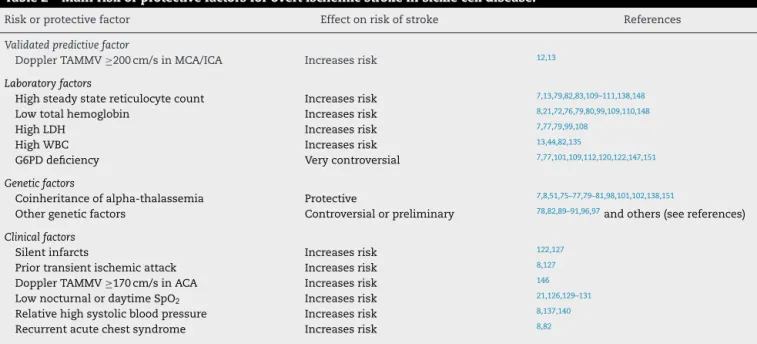
Table 2 – Main risk or protective factors for overt ischemic stroke in sickle cell disease.
Risk or protective factor Effect on risk of stroke References
Validated predictive factor
Doppler TAMMV≥200 cm/s in MCA/ICA Increases risk 12,13
Laboratory factors
High steady state reticulocyte count Increases risk 7,13,79,82,83,109–111,138,148
Low total hemoglobin Increases risk 8,21,72,76,79,80,99,109,110,148
High LDH Increases risk 7,77,79,99,108
High WBC Increases risk 13,44,82,135
G6PD deficiency Very controversial 7,77,101,109,112,120,122,147,151
Genetic factors
Coinheritance of alpha-thalassemia Protective 7,8,51,75–77,79–81,98,101,102,138,151
Other genetic factors Controversial or preliminary 78,82,89–91,96,97and others (see references)
Clinical factors
Silent infarcts Increases risk 122,127
Prior transient ischemic attack Increases risk 8,127
Doppler TAMMV≥170 cm/s in ACA Increases risk 146
Low nocturnal or daytime SpO2 Increases risk 21,126,129–131
Relative high systolic blood pressure Increases risk 8,137,140
Recurrent acute chest syndrome Increases risk 8,82
hematol transfus cell ther.2 0 1 8;4 0(2):166–181
175
the most important process in the arteries of the Willis circle related to the development of stroke.74
The protective effect of alpha-thalassemia against the development of stroke is well established in the literature.7,8,51,75–81 This association has been attributed
to both hematologic and rheologic factors. Due to the lower production of alpha globin chains, the co-inheritance of alpha-thalassemia reduces the intracellular concentration of Hb and, consequently, reduces polymerization, modulating other hematological characteristics. Most studies show an increase in the number of RBCs, total Hb and hematocrit levels, and decreased levels of mean corpuscular volume, mean corpuscular Hb, mean corpuscular Hb concentration, reticulocytes, leukocytes and hemolysis. Currently, a high reticulocyte count is considered the most important risk factor for stroke, as analyzed later in this review. A high retic-ulocyte count was a risk factor for acute cerebral ischemia and high-risk TCD in a multivariate analysis.82,83 Thus, the
reduction of reticulocyte count caused by the co-inheritance of alpha-thalassemia51 may be crucial to protect children
with SCA from stroke.
The deformability of sickled cells in individuals who also have alpha-thalassemia is greater than that of patients with-out it.84,85 In addition, individuals with alpha-thalassemia
have reduced numbers of dense cells and irreversibly sick-led cells.86–88Furthermore, the presence of alpha-thalassemia
could reduce the adhesion of sickled cells to the endothelium
in vivo.39,40Thus, the improvement in rheological features of
RBCs conferred by the co-inheritance of alpha-thalassemia may also contribute to reducing the risk of stroke in children with SCA.
In the last few years, some studies have suggested a role of other genes in the pathogenesis of stroke in children with SCA. In a candidate gene association study, Sebastiani et al. used Bayesian network modeling that tested 108 single nucleotide polymorphisms (SNPs) in 39 genes and found that 31 SNPs in 12 genes interact with Hb F to modulate the risk of stroke. The predictive value of the model was assessed in an independent validation set of patients with SCA and it predicted the occur-rence of stroke with a 100% true positive rate and a 98.14% true negative rate giving an overall predictive accuracy of 98.2%. Polymorphisms in theADCY9,ANXA2,BMP6,CCL2,CSF2,ECE1,
ERG,MET,SELP,TEKandTGFBR3genes were associated with stroke and had a major statistically independent effect on the risk of event. Furthermore, polymorphisms in genes previ-ously associated with stroke in populations without SCA, such as transforming growth factor (TGF)-beta pathway genes, were also included in the network.89
In a genome-wide association and exome study using a dis-covery cohort of 677 children and an independent validation cohort of 288 children, two mutations in theGOLGB1(Y1212C) andENPP1(K173Q) genes were significantly associated with a decreased risk for stroke. TheGOLGB1Y1212C mutation was also associated with protection from silent infarcts and abnor-mal TCD.90 In a recent longitudinal study by our group, the
ENPP1K173Q was associated with an increased risk of stroke and trends toward increased risk for high-risk TCD.91 The
ENPP1is a class II glycoprotein known to influence insulin sensitivity, binding, and, consequently, inhibiting the insulin receptor. The variantENPP1173Q is a more potent inhibitor
of the insulin receptor than the wild variant, and it has been associated with insulin resistance and diabetes type II.66,92
Insulin stimulates the activation of protein kinase B and this protein signals the release of NO from endothelial cells. This pathway is impaired in individuals with theENPP1173Q vari-ant. This variant has also been associated with increased blood pressure, cardiovascular events, and reduced activity of enzyme nitric oxide synthase.93,94We have proposed that NO
pathway impairment is the possible mechanism for theENPP1
K173Q modulation of stroke in children with SCA.91Further
studies are needed to shed additional light on the role ofENPP1
K173Q in the pathogenesis of stroke in pediatric patients with SCA.
Although several studies have reported that other genetic markers are associated with risk of stroke in individuals with SCD (Table 1), there is controversy over the different stud-ies. Most of these genetic associations are still preliminary and require confirmatory studies.2 Few studies have been
validated, mainly because the interpretation of these stud-ies is hampered by the relatively small sample size and/or absence of cohorts to validate the results.95In addition, there
is a wide variation in the definition of the outcomes studied. Furthermore, stroke in children with SCD seems to be a com-plex multifactorial and polygenic disorder that is influenced by many characteristics, each with only modest effects. The small effect of each marker also contributes to the controver-sial results. The influence of the tumor necrosis factor-alpha (TNF-␣) G-308A (rs1800629) polymorphism is a good exam-ple of these controversies. Some reports have indicated that homozygous for the−308G allele is associated with increased risk of stroke.96,97 On the other hand, the−308A allele was
reported to be associated with increased risk of stroke in two recent reports from our group82,98 whereas two other
stud-ies reported no association.99,100Similarly, some studies have
identified an association of the Central African Republic (CAR) haplotype with stroke in subjects with SCD.79,101–103However,
reliable replication of this potential association has not been reached in other independent validation cohorts.78,104–107
Regarding laboratory parameters, the relationship of hemolysis markers, such as low Hb concentration, high lactate dehydrogenase (LDH), and high reticulocyte count, with the occurrence of stroke stands out in the literature.7,8,13,19,76,77,79,80,83,99,108,109Recently, the reticulocyte
count has gained prominence as possibly the most important laboratory risk factor for increased risk of stroke.7,82,83,110,111
In recently published reports, including two by our group, reticulocyte count was the most important predictor of stroke or high-risk TCD.82,83,110–112 The re-analyzed data from the
Cooperative Study of Sickle Cell Disease report, designed to assess the impact of very early detection of reticulocytosis, anemia, or leukocytosis on prediction of future major adverse events, showed that a high reticulocyte count was significantly associated with increased risk of stroke and death during childhood.111 More recently, data from a French cohort of
children demonstrated a substantial independent association of high reticulocyte count [Hazard ratio: 1.82 per 50×109/L
increase; 95% confidence interval (95% CI): 1.10–3.01] with development of cerebral macrovasculopathy.112In a Brazilian
176
hematol transfus cell ther.2 0 1 8;4 0(2):166–181risk of acute cerebral ischemia (stroke or transient ischemic attack – TIA) or high-risk TCD increased by approximately 1.3% (95% CI: 1.13–1.47%) and 1.5% (95% CI: 1.27–1.69%), respectively.82 Reticulocytes probably have an essential role
in the pathogenesis of cerebral vasculopathy in children with SCA. Hyperhemolysis, as indicated by high steady state reticulocyte count, releases Hb, free heme, arginase, and other molecules from RBCs, which generate reactive oxygen species, scavenge NO, and inhibit NO production, promoting endothelial damage, platelet activation and induction of inflammation in the vascular endothelium.113–116Endothelial
dysfunction and inflammation stimulate selective release of mediators, which further promote expression of adhesion molecules on the endothelial and blood cells,117contributing
to a series of pathophysiological events that culminate in vasculopathy involving large cerebral arteries. Additionally, data from the French cohort showed that the serum LDH level and reticulocyte count were significant independent factors associated with stroke in multivariate analyses,7 suggesting
that hemolysis is not the single event associated with the pathophysiology of stroke but high steady state reticulo-cyte count per se is also probably involved. The tendency of reticulocytes to adhere to endothelial cells, resulting in endothelial activation and damage, might be the first stage of a series of pathophysiological events resulting in cerebral vasculopathy.36 The benefits of hydroxyurea therapy in the
prevention of stroke14,118 could be partially attributed to its
effect on decreasing reticulocyte adhesion to endothelial cells.119
Despite the relatively large number of studies, the role of concomitant glucose-6-phosphate dehydrogenase (G6PD) deficiency on risk of stroke remains controversial. In our experience, the prevalence of stroke or high-risk TCD was not significantly different in the groups with and with-out G6PD deficiency in a retrospective cohort study.120 This
absence of association has been reported in other studies that used molecular analysis as the diagnostic method for G6PD deficiency.78,121However, Thangarajh et al.,122showed that the
presence of the 376G (rs1050829) or 202A (rs1050828) allele was a significant and independent risk factor for intracra-nial MRA-arteriopathy in males with SCA. The 376G allele leads to a very mild reduction in G6PD activity, as demon-strated in one study.120Children with the 376G allele (isoform
A in males or AA in females) showed 85.2% of the enzy-matic activity of G6PD when compared to individuals with the wild 376A allele (isoform B in males or BB in females). On the other hand, studies that reported an association between G6PD deficiency and abnormal TCD or intracranial stenosis used the measurement of G6PD activity as the method to define G6PD deficiency.7,77One plausible explanation for these
divergent results is that transcriptional and epigenetic fac-tors that influence G6PD expression123 may be involved in
the modulation of stroke in children with SCA. Thus, children without pathogenic missense mutations, but with downreg-ulation ofG6PDexpression might have a higher risk for CVD. Similarly, children with pathogenic missense mutations and upregulation of G6PD expression might have a lower risk. However, some studies108,120,121,124 did not detect any
differ-ence in the mean G6PD activity in groups with and without
ischemic stroke or high-risk TCD. The association of G6PD defi-ciency and stroke seems to be unlikely. The most important laboratory predictor of stroke, high steady state reticulocyte count,83,111is associated with raised G6PD activity, as
demon-strated in one study.120 Another reason for the improbable
association between G6PD deficiency and stroke is that G6PD deficiency is an X-linked inherited disease and the effects of deficiency should be more common in males. However, available data show no influence of gender.108 It is obvious
that genetic background heterogeneity among populations may also lead to contradictory results. G6PD deficiency may be a risk factor for some children, but not for others from different ethnic backgrounds. Further large-scale prospective longitudinal studies controlled for ancestry are warranted to elucidate the relationship of G6PD deficiency with stroke sus-ceptibility in children with SCA. Furthermore, G6PD seems to have a critical antioxidant role in endothelial cells.7Further
studies measuring the G6PD activity in circulating endothelial cells may provide a better understanding of the relationship between G6PD deficiency and cerebrovascular vasculopathy in SCA.
About clinical features associated to the development of stroke, factors that cause imbalance between demand and supply of oxygen in the brain are critical. For example, aplas-tic crisis secondary to erythrovirus B19 infection,125nocturnal
hypoxemia,126and the occurrence of acute chest syndrome8
have been reported to be associated with stroke. Addition-ally, in a large study of 516 children with SCA, silent cerebral infarct was the single factor associated with intracranial MRA-vasculopathy in the final multivariate logistic regression model.122Another study of 248 children with SCA reported a
strong association between silent infarcts identified at age of six years or older and subsequent development of stroke.127
Although both events share some pathophysiological back-ground, the reason for this association is not clear. More convincingly, patients with a history of TIA were much more likely to have an overt stroke.8,127 According to data from
the Cooperative Study of Sickle Cell Disease (CSSD), chil-dren who have had a TIA have a 56-times higher chance to subsequently develop stroke when compared to those with-out prior TIA (95% CI: 12.0–285).8 In our experience,82 four
children had TIA and subsequently developed strokes and another three children had TIA and did not evolve with strokes; of the latter group the TIA was followed by a high-risk TCD (two cases) or an inconclusive TCD (one case) due to difficulty of insonation in the presence of a ‘good’ transtemporal window, probably due to severe stenosis.128
Then, recognizing and treating TIA certainly reduces the risk of an overt stroke. Children with a TIA event should be intensively monitored by TCD and/or MRA to evaluate the indication of intensification therapy such as prophy-lactic blood transfusion program or hydroxyurea therapy. Another clinical factor that has been associated with the occurrence of stroke is nocturnal126 or daytime129–131 Hb
hematol transfus cell ther.2 0 1 8;4 0(2):166–181
177
Conclusions
The scientific literature is controversial in relation to the risk factors associated with the development of stroke in individ-uals with SCD. The absence of uniformity and standardization in the definition of distinct CVD events (ischemic stroke, TIA, hemorrhagic stroke, silent infarcts, vascular stenosis detected by MRA, and moya-moya disease) makes the interpretation and comparison of study results difficult and is probably a major factor that explains the controversies.
To date, the reticulocyte count in the peripheral blood is probably the most important laboratory marker to predict the occurrence of stroke in individuals with SCD. Clinical factors associated with hypoxia and reduced availability of oxygen in the brain play an important role in the pathophys-iology of stroke and may act as a triggering factor for sudden acute cerebrovascular events. Although promising, genetic factors have a small effect on the occurrence of stroke when assessed individually. These factors may be used as a prog-nostic clinical tool in personalized medicine and may assist in the early detection of stroke risk in individuals with SCD, improving the selection of children for intensification ther-apy. For this purpose, they must be used all together, for example, as in the Bayesian network proposed by Sebastiani et al.89
Currently, the only validated prognostic clinical tool avail-able for assessing the risk of stroke is TCD. Prospective validation studies must be conducted before including other biomarkers in the guidelines for clinical management of chil-dren with SCD.
r e f e r e n c e s
1. Serjeant GR. The natural history of sickle cell disease. Cold Spring Harb Perspect Med. 2013;3(10):a011783.
2. Rees DC, Williams TN, Gladwin MT. Sickle-cell disease. Lancet. 2010;376(9757):2018–31.
3. Quinn CT. Sickle cell disease in childhood: from newborn screening through transition to adult medical care. Pediatr Clin North Am. 2013;60(6):1363–81.
4. Stuart MJ, Nagel RL. Sickle-cell disease. Lancet. 2004;364(9442):1343–60.
5. Steinberg MH. Management of sickle cell disease. N Engl J Med. 1999;340(13):1021–30.
6. Steinberg MH. Sickle cell anemia, the first molecular disease: overview of molecular etiology, pathophysiology, and therapeutic approaches. Sci World J. 2008;8:1295–324. 7. Bernaudin F, Verlhac S, Arnaud C, Kamdem A, Chevret S,
Hau I, et al. Impact of early transcranial Doppler screening and intensive therapy on cerebral vasculopathy outcome in a newborn sickle cell anemia cohort. Blood.
2011;117(4):1130–40, quiz 1436.
8. Ohene-Frempong K, Weiner SJ, Sleeper LA, Miller ST, Embury S, Moohr JW, et al. Cerebrovascular accidents in sickle cell disease: rates and risk factors. Blood. 1998;91(1):288–94. 9. Ohene-Frempong K. Stroke in sickle cell disease:
demographic, clinical, and therapeutic considerations. Semin Hematol. 1991;28(3):213–9.
10. Powars D, Wilson B, Imbus C, Pegelow C, Allen J. The natural history of stroke in sickle cell disease. Am J Med.
1978;65(3):461–71.
11. Connes P, Verlhac S, Bernaudin F. Advances in understanding the pathogenesis of cerebrovascular vasculopathy in sickle cell anaemia. Br J Haematol. 2013;161(4):484–98.
12. Adams RJ, McKie VC, Hsu L, Files B, Vichinsky E, Pegelow C, et al. Prevention of a first stroke by transfusions in children with sickle cell anemia and abnormal results on transcranial Doppler ultrasonography. N Engl J Med. 1998;339(1):5–11. 13. Adams RJ, McKie VC, Carl EM, Nichols FT, Perry R, Brock K,
et al. Long-term stroke risk in children with sickle cell disease screened with transcranial Doppler. Ann Neurol. 1997;42(5):699–704.
14. Ware RE, Davis BR, Schultz WH, Brown RC, Aygun B, Sarnaik S, et al. Hydroxycarbamide versus chronic transfusion for maintenance of transcranial doppler flow velocities in children with sickle cell anaemia-TCD With Transfusions Changing to Hydroxyurea (TWiTCH): a multicentre, open-label, phase 3, non-inferiority trial. Lancet. 2016;387(10019):661–70.
15. Lagunju IA, Brown BJ, Sodeinde OO. Chronic blood
transfusion for primary and secondary stroke prevention in Nigerian children with sickle cell disease: a 5-year appraisal. Pediatr Blood Cancer. 2013;60(12):1940–5.
16. Adams RJ, Brambilla DJ, Granger S, Gallagher D, Vichinsky E, Abboud MR, et al. Stroke and conversion to high risk in children screened with transcranial Doppler ultrasound during the STOP study. Blood. 2004;103(10):3689–94. 17. Jordan LC, Casella JF, Debaun MR. Prospects for primary
stroke prevention in children with sickle cell anaemia. Br J Haematol. 2012;157(1):14–25.
18. Brambilla DJ, Miller ST, Adams RJ. Intra-individual variation in blood flow velocities in cerebral arteries of children with sickle cell disease. Pediatr Blood Cancer. 2007;49(3): 318–22.
19. Rees DC, Dick MC, Height SE, O’Driscoll S, Pohl KR, Goss DE, et al. A simple index using age, hemoglobin, and aspartate transaminase predicts increased intracerebral blood velocity as measured by transcranial Doppler scanning in children with sickle cell anemia. Pediatrics. 2008;121(6):e1628–32. 20. Ali SB, Moosang M, King L, Knight-Madden J, Reid M. Stroke
recurrence in children with sickle cell disease treated with hydroxyurea following first clinical stroke. Am J Hematol. 2011;86(10):846–50.
21. Lagunju I, Sodeinde O, Brown B, Akinbami F, Adedokun B. Transcranial Doppler ultrasonography in children with sickle cell anemia: clinical and laboratory correlates for elevated blood flow velocities. J Clin Ultrasound. 2014;42(2): 89–95.
22. Fullerton HJ, Gardner M, Adams RJ, Lo LC, Johnston SC. Obstacles to primary stroke prevention in children with sickle cell disease. Neurology. 2006;67(6):1098–9. 23. Eckrich MJ, Wang WC, Yang E, Arbogast PG, Morrow A,
Dudley JA, et al. Adherence to transcranial Doppler screening guidelines among children with sickle cell disease. Pediatr Blood Cancer. 2013;60(2):270–4.
24. Raphael JL, Shetty PB, Liu H, Mahoney DH, Mueller BU. A critical assessment of transcranial doppler screening rates in a large pediatric sickle cell center: opportunities to improve healthcare quality. Pediatr Blood Cancer. 2008;51(5):647–51.
25. Adams RJ, Lackland DT, Brown L, Brown D, Voeks J, Fullerton HJ, et al. Transcranial doppler re-screening of subjects who participated in STOP and STOP II. Am J Hematol.
2016;91(12):1191–4.
26. Chou ST, Jackson T, Vege S, Smith-Whitley K, Friedman DF, Westhoff CM. High prevalence of red blood cell
alloimmunization in sickle cell disease despite transfusion from Rh-matched minority donors. Blood.
178
hematol transfus cell ther.2 0 1 8;4 0(2):166–18127. Payne KA, Desrosiers MP, Caro JJ, Baladi JF, Lordan N, Proskorovsky I, et al. Clinical and economic burden of infused iron chelation therapy in the United States. Transfusion. 2007;47(10):1820–9.
28. Cherry MG, Greenhalgh J, Osipenko L, Venkatachalam M, Boland A, Dundar Y, et al. The clinical effectiveness and cost-effectiveness of primary stroke prevention in children with sickle cell disease: a systematic review and economic evaluation. Health Technol Assess. 2012;16(43):1–129. 29. Meier ER, Miller JL. Sickle cell disease in children. Drugs.
2012;72(7):895–906.
30. Bernaudin F, Verlhac S, Arnaud C, Kamdem A, Hau I, Leveillé E, et al. Long-term treatment follow-up of children with sickle cell disease monitored with abnormal transcranial Doppler velocities. Blood. 2016;127(14):1814–22.
31. Wayne AS, Schoenike SE, Pegelow CH. Financial analysis of chronic transfusion for stroke prevention in sickle cell disease. Blood. 2000;96(7):2369–72.
32. Lo W, Zamel K, Ponnappa K, Allen A, Chisolm D, Tang M, et al. The cost of pediatric stroke care and rehabilitation. Stroke. 2008;39(1):161–5.
33. Switzer JA, Hess DC, Nichols FT, Adams RJ. Pathophysiology and treatment of stroke in sickle-cell disease: present and future. Lancet Neurol. 2006;5(6):501–12.
34. De Montalembert M, Wang W. Cerebrovascular
complications in children with sickle cell disease. Handb Clin Neurol. 2013;113:1937–43.
35. Hillery CA, Panepinto JA. Pathophysiology of stroke in sickle cell disease. Microcirculation. 2004;11(2):195–208.
36. Platt OS. Preventing stroke in sickle cell anemia. N Engl J Med. 2005;353(26):2743–5.
37. Hebbel RP, Yamada O, Moldow CF, Jacob HS, White JG, Eaton JW. Abnormal adherence of sickle erythrocytes to cultured vascular endothelium: possible mechanism for
microvascular occlusion in sickle cell disease. J Clin Invest. 1980;65(1):154–60.
38. Hoover R, Rubin R, Wise G, Warren R. Adhesion of normal and sickle erythrocytes to endothelial monolayer cultures. Blood. 1979;54(4):872–6.
39. Swerlick RA, Eckman JR, Kumar A, Jeitler M, Wick TM. Alpha 4 beta 1-integrin expression on sickle reticulocytes: vascular cell adhesion molecule-1-dependent binding to
endothelium. Blood. 1993;82(6):1891–9.
40. Joneckis CC, Ackley RL, Orringer EP, Wayner EA, Parise LV. Integrin alpha 4 beta 1 and glycoprotein IV (CD36) are expressed on circulating reticulocytes in sickle cell anemia. Blood. 1993;82(12):3548–55.
41. Natarajan M, Udden MM, McIntire LV. Adhesion of sickle red blood cells and damage to interleukin-1 beta stimulated endothelial cells under flow in vitro. Blood.
1996;87(11):4845–52.
42. Embury SH, Matsui NM, Ramanujam S, Mayadas TN, Noguchi CT, Diwan BA, et al. The contribution of endothelial cell P-selectin to the microvascular flow of mouse sickle erythrocytes in vivo. Blood. 2004;104(10):3378–85. 43. Hillery CA, Scott JP, Du MC. The carboxy-terminal
cell-binding domain of thrombospondin is essential for sickle red blood cell adhesion. Blood. 1999;94(1): 302–9.
44. Taylor JG, Tang D, Foster CB, Serjeant GR, Rodgers GP, Chanock SJ. Patterns of low-affinity immunoglobulin receptor polymorphisms in stroke and homozygous sickle cell disease. Am J Hematol. 2002;69(2):109–14.
45. French JA 2nd, Kenny D, Scott JP, Hoffmann RG, Wood JD, Hudetz AG, et al. Mechanisms of stroke in sickle cell disease: sickle erythrocytes decrease cerebral blood flow in rats after nitric oxide synthase inhibition. Blood. 1997;89(12): 4591–9.
46. Frenette PS. Sickle cell vasoocclusion: heterotypic, multicellular aggregations driven by leukocyte adhesion. Microcirculation. 2004;11(2):167–77.
47. Okpala I. Leukocyte adhesion and the pathophysiology of sickle cell disease. Curr Opin Hematol. 2006;13(1):40–4. 48. Morris CR, Kato GJ, Poljakovic M, Wang X, Blackwelder WC,
Sachdev V, et al. Dysregulated arginine metabolism, hemolysis-associated pulmonary hypertension, and mortality in sickle cell disease. JAMA. 2005;294(1):81–90. 49. Kato GJ, Gladwin MT, Steinberg MH. Deconstructing sickle
cell disease: reappraisal of the role of hemolysis in the development of clinical subphenotypes. Blood Rev. 2007;21(1):37–47.
50. Lezcano NE, Odo N, Kutlar A, Brambilla D, Adams RJ. Regular transfusion lowers plasma free hemoglobin in children with sickle-cell disease at risk for stroke. Stroke.
2006;37(6):1424–6.
51. Belisario AR, Rodrigues CV, Martins ML, Silva CM, Viana MB. Coinheritance of alpha-thalassemia decreases the risk of cerebrovascular disease in a cohort of children with sickle cell anemia. Hemoglobin. 2010;34(6):516–29.
52. Kaul DK, Fabry ME, Costantini F, Rubin EM, Nagel RL. In vivo demonstration of red cell-endothelial interaction, sickling and altered microvascular response to oxygen in the sickle transgenic mouse. J Clin Invest. 1995;96(6):2845–53.
53. Tomer A, Harker LA, Kasey S, Eckman JR. Thrombogenesis in sickle cell disease. J Lab Clin Med. 2001;137(6):398–407. 54. Brittain HA, Eckman JR, Swerlick RA, Howard RJ, Wick TM.
Thrombospondin from activated platelets promotes sickle erythrocyte adherence to human microvascular
endothelium under physiologic flow: a potential role for platelet activation in sickle cell vaso-occlusion. Blood. 1993;81(8):2137–43.
55. Ataga KI, Moore CG, Hillery CA, Jones S, Whinna HC, Strayhorn D, et al. Coagulation activation and inflammation in sickle cell disease-associated pulmonary hypertension. Haematologica. 2008;93(1):20–6.
56. Chantrathammachart P, Mackman N, Sparkenbaugh E, Wang JG, Parise LV, Kirchhofer D, et al. Tissue factor promotes activation of coagulation and inflammation in a mouse model of sickle cell disease. Blood. 2012;120(3):636–46. 57. Tam DA. Protein C and protein S activity in sickle cell
disease and stroke. J Child Neurol. 1997;12(1):19–21. 58. Ataga KI, Brittain JE, Desai P, May R, Jones S, Delaney J, et al.
Association of coagulation activation with clinical complications in sickle cell disease. PLoS ONE. 2012;7(1):e29786.
59. Prohovnik I, Pavlakis SG, Piomelli S, Bello J, Mohr JP, Hilal S, et al. Cerebral hyperemia, stroke, and transfusion in sickle cell disease. Neurology. 1989;39(3):344–8.
60. Brass LM, Prohovnik I, Pavlakis SG, DeVivo DC, Piomelli S, Mohr JP. Middle cerebral artery blood velocity and cerebral blood flow in sickle cell disease. Stroke. 1991;22(1):27–30. 61. Wang WC. The pathophysiology, prevention, and treatment
of stroke in sickle cell disease. Curr Opin Hematol. 2007;14(3):191–7.
62. Kaul DK, Hebbel RP. Hypoxia/reoxygenation causes inflammatory response in transgenic sickle mice but not in normal mice. J Clin Invest. 2000;106(3):411–20.
63. Rothman SM, Fulling KH, Nelson JS. Sickle cell anemia and central nervous system infarction: a neuropathological study. Ann Neurol. 1986;20(6):684–90.
64. Merkel KH, Ginsberg PL, Parker JC Jr, Post MJ.
Cerebrovascular disease in sickle cell anemia: a clinical, pathological and radiological correlation. Stroke. 1978;9(1): 45–52.
hematol transfus cell ther.2 0 1 8;4 0(2):166–181
179
children with sickle cell disease. Pediatr Blood Cancer. 2013;60(5):823–7.
66. McCarville MB, Goodin GS, Fortner G, Li CS, Smeltzer MP, Adams R, et al. Evaluation of a comprehensive transcranial doppler screening program for children with sickle cell anemia. Pediatr Blood Cancer. 2008;50(4):818–21. 67. Fullerton HJ, Adams RJ, Zhao S, Johnston SC. Declining
stroke rates in Californian children with sickle cell disease. Blood. 2004;104(2):336–9.
68. Armstrong-Wells J, Grimes B, Sidney S, Kronish D, Shiboski SC, Adams RJ, et al. Utilization of TCD screening for primary stroke prevention in children with sickle cell disease. Neurology. 2009;72(15):1316–21.
69. Enninful-Eghan H, Moore RH, Ichord R, Smith-Whitley K, Kwiatkowski JL. Transcranial Doppler ultrasonography and prophylactic transfusion program is effective in preventing overt stroke in children with sickle cell disease. J Pediatr. 2010;157(3):479–84.
70. Adams RJ, Brambilla D. Discontinuing prophylactic transfusions used to prevent stroke in sickle cell disease. N Engl J Med. 2005;353(26):2769–78.
71. Driscoll MC, Hurlet A, Styles L, McKie V, Files B, Olivieri N, et al. Stroke risk in siblings with sickle cell anemia. Blood. 2003;101(6):2401–4.
72. Kwiatkowski JL, Hunter JV, Smith-Whitley K, Katz ML, Shults J, Ohene-Frempong K. Transcranial Doppler ultrasonography in siblings with sickle cell disease. Br J Haematol.
2003;121(6):932–7.
73. Hoppe C, Klitz W, Noble J, Vigil L, Vichinsky E, Styles L. Distinct HLA associations by stroke subtype in children with sickle cell anemia. Blood. 2003;101(7):2865–9.
74. Chang Milbauer L, Wei P, Enenstein J, Jiang A, Hillery CA, Scott JP, et al. Genetic endothelial systems biology of sickle stroke risk. Blood. 2008;111(7):3872–9.
75. Gill FM, Sleeper LA, Weiner SJ, Brown AK, Bellevue R, Grover R, et al. Clinical events in the first decade in a cohort of infants with sickle cell disease. Cooperative study of sickle cell disease. Blood. 1995;86(2):776–83.
76. Hsu LL, Miller ST, Wright E, Kutlar A, McKie V, Wang W, et al. Alpha Thalassemia is associated with decreased risk of abnormal transcranial Doppler ultrasonography in children with sickle cell anemia. J Pediatr Hematol Oncol.
2003;25(8):622–8.
77. Bernaudin F, Verlhac S, Chevret S, Torres M, Coic L, Arnaud C, et al. G6PD deficiency, absence of alpha-thalassemia, and hemolytic rate at baseline are significant independent risk factors for abnormally high cerebral velocities in patients with sickle cell anemia. Blood. 2008;112(10):4314–7. 78. Flanagan JM, Frohlich DM, Howard TA, Schultz WH, Driscoll
C, Nagasubramanian R, et al. Genetic predictors for stroke in children with sickle cell anemia. Blood. 2011;117(24): 6681–4.
79. Domingos IF, Falcao DA, Hatzlhofer BL, Cunha AF, Santos MN, Albuquerque DM, et al. Influence of the beta haplotype and alpha-thalassemia on stroke development in a Brazilian population with sickle cell anaemia. Ann Hematol.
2014;93(7):1123–9.
80. Cox SE, Makani J, Soka D, L’Esperence VS, Kija E,
Dominguez-Salas P, et al. Haptoglobin, alpha-thalassaemia and glucose-6-phosphate dehydrogenase polymorphisms and risk of abnormal transcranial Doppler among patients with sickle cell anaemia in Tanzania. Br J Haematol. 2014;165(5):699–706.
81. Neonato MG, Guilloud-Bataille M, Beauvais P, Begue P, Belloy M, Benkerrou M, et al. Acute clinical events in 299
homozygous sickle cell patients living in France. French Study Group on Sickle Cell Disease. Eur J Haematol. 2000;65(3):155–64.
82. Belisário AR, Sales RR, Toledo NE, Muniz MBdSR, Velloso-Rodrigues C, Silva CM, et al. Reticulocyte count is the most important predictor of acute cerebral ischemia and high-risk transcranial Doppler in a newborn cohort of 395 children with sickle cell anemia. Ann Hematol.
2016;95(11):1869–80.
83. Silva CM, Giovani P, Viana MB. High reticulocyte count is an independent risk factor for cerebrovascular disease in children with sickle cell anemia. Pediatr Blood Cancer. 2011;56(1):116–21.
84. Serjeant BE, Mason KP, Kenny MW, Stuart J, Higgs DR, Weatherall DJ, et al. Effect of alpha thalassaemia on the rheology of homozygous sickle cell disease. Br J Haematol. 1983;55(3):479–86.
85. Ballas SK, Larner J, Smith ED, Surrey S, Schwartz E, Rappaport EF. Rheologic predictors of the severity of the painful sickle cell crisis. Blood. 1988;72(4):1216–23. 86. Embury SH, Clark MR, Monroy G, Mohandas N. Concurrent
sickle cell anemia and alpha-thalassemia. Effect on pathological properties of sickle erythrocytes. J Clin Invest. 1984;73(1):116–23.
87. Fabry ME, Mears JG, Patel P, Schaefer-Rego K, Carmichael LD, Martinez G, et al. Dense cells in sickle cell anemia: the effects of gene interaction. Blood. 1984;64(5):1042–6. 88. Noguchi CT, Dover GJ, Rodgers GP, Serjeant GR, Antonarakis
SE, Anagnou NP, et al. Alpha thalassemia changes
erythrocyte heterogeneity in sickle cell disease. J Clin Invest. 1985;75(5):1632–7.
89. Sebastiani P, Ramoni MF, Nolan V, Baldwin CT, Steinberg MH. Genetic dissection and prognostic modeling of overt stroke in sickle cell anemia. Nat Genet. 2005;37(4):435–40. 90. Flanagan JM, Sheehan V, Linder H, Howard TA, Wang YD,
Hoppe CC, et al. Genetic mapping and exome sequencing identify 2 mutations associated with stroke protection in pediatric patients with sickle cell anemia. Blood. 2013;121(16):3237–45.
91. Belisario AR, Sales RR, Toledo NE, Velloso-Rodrigues C, Silva CM, Viana MB. Association between ENPP1 K173Q and stroke in a newborn cohort of 395 Brazilian children with sickle cell anemia. Blood. 2015;126(10):1259–60.
92. Goldfine ID, Maddux BA, Youngren JF, Reaven G, Accili D, Trischitta V, et al. The role of membrane glycoprotein plasma cell antigen 1/ectonucleotide pyrophosphatase phosphodiesterase 1 in the pathogenesis of insulin resistance and related abnormalities. Endocr Rev. 2008;29(1):62–75.
93. Bacci S, Di Paola R, Menzaghi C, Di Fulvio P, Di Silvestre S, Pellegrini F, et al. ENPP1 Q121 variant, increased pulse pressure and reduced insulin signaling, and nitric oxide synthase activity in endothelial cells. Arterioscler Thromb Vasc Biol. 2009;29(10):1678–83.
94. Bacci S, Rizza S, Prudente S, Spoto B, Powers C, Facciorusso A, et al. The ENPP1 Q121 variant predicts major
cardiovascular events in high-risk individuals: evidence for interaction with obesity in diabetic patients. Diabetes. 2011;60(3):1000–7.
95. Menaa F. Stroke in sickle cell anemia patients: a need for multidisciplinary approaches. Atherosclerosis.
2013;229(2):496–503.
96. Hoppe C, Klitz W, Cheng S, Apple R, Steiner L, Robles L, et al. Gene interactions and stroke risk in children with sickle cell anemia. Blood. 2004;103(6):2391–6.
97. Hoppe C, Klitz W, D’Harlingue K, Cheng S, Grow M, Steiner L, et al. Confirmation of an association between the TNF(-308) promoter polymorphism and stroke risk in children with sickle cell anemia. Stroke. 2007;38(8):2241–6.