Inflammasome Activation by Inhibiting NF-
k
B Activation
and Enhancing Autophagy
Yolanda Williams-Bey, Cedric Boularan, Ali Vural, Ning-Na Huang, Il-Young Hwang, Chong Shan-Shi, John H. Kehrl*
B Cell Molecular Immunology Section, Laboratory of Immunoregulation, National Institute of Allergy and Infectious Diseases, National Institutes of Health, Bethesda, Maryland, United States of America
Abstract
The omega-3 (v3) fatty acid docosahexaenoic acid (DHA) can suppress inflammation, specifically IL-1bproduction through poorly understood molecular mechanisms. Here, we show that DHA reduces macrophage IL-1b production by limiting inflammasome activation. Exposure to DHA reduced IL-1b production by ligands that stimulate the NLRP3, AIM2, and NAIP5/NLRC4 inflammasomes. The inhibition required Free Fatty Acid Receptor (FFAR) 4 (also known as GPR120), a G-protein coupled receptor (GPR) known to bind DHA. The exposure of cells to DHA recruited the adapter G-proteinb-arrestin1/ 2 to FFAR4, but not to a related lipid receptor. DHA treatment reduced the initial inflammasome priming step by suppressing the nuclear translocation of NF-kB. DHA also reduced IL-1blevels by enhancing autophagy in the cells. As a consequence macrophages derived from mice lacking the essential autophagy protein ATG7 were partially resistant to suppressive effects of DHA. Thus, DHA suppresses inflammasome activation by two distinct mechanisms, inhibiting the initial priming step and by augmenting autophagy, which limits inflammasome activity.
Citation:Williams-Bey Y, Boularan C, Vural A, Huang N-N, Hwang I-Y, et al. (2014) Omega-3 Free Fatty Acids Suppress Macrophage Inflammasome Activation by Inhibiting NF-kB Activation and Enhancing Autophagy. PLoS ONE 9(6): e97957. doi:10.1371/journal.pone.0097957
Editor:David M. Ojcius, University of California Merced, United States of America
ReceivedFebruary 12, 2014;AcceptedApril 25, 2014;PublishedJune 9, 2014
This is an open-access article distributed under the terms of the Creative Commons Public Domain declaration which stipulates that, once placed in the public domain, this work may be freely reproduced, distributed, transmitted, modified, built upon, or otherwise used by anyone for any lawful purpose.
Funding:This research was supported by the Intramural Research Program of the National Institutes of Health [National Institute of Allergy and Infectious Diseases]. The funders had no role in study design, data collection and analysis, decision to publish, or preparation of the manuscript.
Competing Interests:The authors have declared that no competing interests exist.
* E-mail: jkehrl@niaid.nih.gov
Introduction
Inflammation is the hallmark of many chronic diseases including type II diabetes, heart disease, cancer, and arthritis. According to the Centers for Disease Control (CDC) chronic diseases account for greater than 50% of all deaths. Understanding the molecular mechanisms that control inflammation and inflammatory pathways is critical to develop new methods to manipulate these pathways and ultimately dampen inflammatory responses in patients. It has been suggested that increasing the intake ofv3 free fatty acid (FFA) by supplementation or by dietary intake of foods rich inv3 FFA significantly improves the health of patients suffering from chronic inflammatory diseases [1]. A recent report showed that thev3 fatty acid docosahexaenoic acid (DHA) can inhibit the production of pro-inflammatory cytokines such as TNF-a and IL-6 in RAW 247.6 cells and in primary mouse macrophages by binding to a G-protein coupled receptor (GPR) termed Free Fatty Acid Receptor (FFAR) 4, also known as GPR120 [2].
Inflammasomes serve as central regulators of innate immunity and inflammation [3]. A cytosolic protein complex assembled following the exposure of cells to specific pathogens or ‘‘danger’’ signals inflammasomes contain a nucleotide-binding domain and leucine-rich repeat containing protein (NLR) or Absent in Melanoma 2 (AIM2), which serves as the sensor; an adapter protein ASC (apoptosis-associated speck-like protein containing a
caspase activation and recruitment domain); and caspase-1. NLRP1b inflammasomes detect anthrax lethal toxin, the NLRC4 inflammasomes recognize bacterial flagellin, AIM2 inflamma-somes are essential for host defense against certain intracellular bacteria and DNA viruses, and a broad range of toxic stimuli trigger the assembly of NLRP3 inflammasomes (4). The activated sensor recruits ASC through homophilic interactions of pyrin domains and ASC associates with pro-caspase-1 via CARD-CARD (caspase activation and recruitment domain) interactions, a step needed to induce caspase-1 activation [4]. The activation of caspase-1 results in the cleavage of IL-1band IL-18 precursors to their mature forms and their eventual secretion. Several studies have found that the saturated FFA palmitate acid can trigger inflammation by activating inflammasomes [5,6]. We tested whether the v3 FFA DHA might have the opposite effect on macrophages and suppress inflammasome activation, thereby reducing IL-1bsecretion.
Materials and Methods
Ethics Statement
Animals, THP-1 cells, and bone marrow derived macrophages (BMDMs)
Wild-type C57BL/6 mice were purchased from Jackson laboratory. TheAtg7flox/floxmice have been previously described [7] and have been partially back-crossed on to C57BL/6 (3 generations). The mice were kindly provided by Dr. Masaaki Komatsu (Tokyo, Japan) and were crossed with B6.Cg-Tg(Vav1-cre)A2Kio/J mice (Jackson Laboratory) to disrupt theAtg7coding region in hematopoietic cells and are referred to asAtg7flox/flox Vav-1Cre. The C57BL/6 Green fluorescent protein (GFP)-LC3 (also known as MAP1LC3A) mice were purchased from RIKEN Bio-Resource Center after receiving permission from Dr. N. Mizush-ima (Tokyo, Japan)[8]. Thearrb12/2andarrb22/2mice are on a
C57BL/6 background and were kindly provided by Dr. Robert J. Lefkowitz (Duke University) [9]. All mice were 6–12 weeks of age at use. Mice were housed under specific pathogen–free conditions. The preparation of mouse bone marrow derived macrophages (BMDMs) and the THP-1 cells have been described previously [10].
Reagents
The following reagents were purchased: docosahexaenoic acid (DHA) and eicosapentaenoic acid (EPA, Cayman Chemical); Poly(dA:dT), adenosine triphosphate (ATP), and nigericin (Sigma-Aldrich); Lipopolysaccharide (LPS, ENZO Life Sciences); purified flagellin (InvivoGen); neutralizing antibody to IL-1b (R&D Systems); caspase-1 antibody (Santa Cruz Biotechnology); NLRP3 antibody (ENZO Life Sciences); ASC antibody (Santa Cruz Biotechnology); and the appropriate secondary antibodies (Jackson ImmunoResearch Laboratory). For the ImageStream analysis a primary rabbit polyclonal NF-kB/p65 antibody (SantaCruz Biotechnology) was used with an Alexa647 conjugated donkey anti rabbit IgG antibody (Jackson ImmunoResearch Laboratories). DNA was stained using DAPI (20mM).
Inflammasome activation
For inducing NLRP3 inflammasome activation, 16106
macro-phages were plated in 12-well plates overnight in complete media (RPMI 1640 plus 10% fetal bovine serum). The following morning the cells were switched to opti-MEM media and LPS (20 ng/ml) or LPS plus various concentrations of DHA were added. DHA was diluted 1:10 in opti-MEM media from an ethanol stock, vortexed, and added to the cells. Three hrs later ATP (1 mM) or nigericin (1mM) was added for 1 hr or 2 hrs, respectively. Afterwards, the cells and supernatants were harvested for analysis. For AIM2 inflammasomes, 16106macrophages were plated in 12-well plates overnight in complete media and the following morning the cells were primed with LPS (1mg/ml) for 3 hrs, the cells were washed,
transfected with Poly(dA:dT) (0.5mg/ml) using Lipofectamine
(Invitrogen) and supernatants and lysates collected 1 hr later. For NAIP5/NLRC4 inflammasomes, a similar protocol was used except flagellin was transfected using Profect P1 (TargetingSys-tems).
siRNA transfection and quantitative RT- PCR
The siRNA pools targeting humanFFAR4 (sc-60737),GPR84 (sc-60751), or a control (sc-37007, Santa Cruz Biotechnology) were prepared with 2ml HD Transfection Reagent (Promega) and used at a concentration of 40 nM to treat differentiated THP-1 cells overnight. One day later the cells were primed with LPS and treated with ATP as above. Cells were analyzed from 1 h to 8 h after inflammasome activation. RNA was isolated with TRIZOL Reagent (Invitrogen) according to the manufacturer’s instructions.
Complementary DNA (cDNA) was synthesized from 1mg RNA
with Omniscript RT Kit (QIAGEN). Real-time PCR was performed using a StepOneTMReal Time PCR System (Applied Biosystems) following the Rotor-Gene SYBR Green PCR kit (QIAGEN) protocol. The following primer pairs were used:
Ffar1 F:5’-GCTATTCCTGGGGTGTGTGT-3’ R:5’-CCCTGTGATGAGTCCCAACT-3’
Gpr84 F:5’-TCCAATTCTGTCTCCATCCT-3’ R:5’-CTGACTGGCTCAGATGAAA-3’
Ffar4 F:5’-CCATCCCTCTAGTGCTCGTC-3’ R:5’-TGCGGAAGAGTCGGTAGTCT-3’
FFAR1 F:5’-CAGTCTCTCTGCCCCTGAAG-3’ R:5’-CGGCATAGAGTGGGAAGAAG-3’
GPR84 F:5’-TCAGCAGTGTTGGCATCTTC-3’ R:5’-CTTGCCTGTCGCAACTTGTA-3’
FFAR4 F:5’-CCTGAGGTCAGGAGTTCGAG-3’ R:5’-CAC-CACCACTCCCAGCTAAT-3’.
The results were normalized to the expression levels of theb -actin mRNA and the relative mRNA levels were calculated using the 22DDCt
method.
Immunoblot analysis, confocal microscopy, and Bioluminescence resonance energy transfer (BRET) analysis
Immunoblotting and immunoprecipitations were performed as previously described [10]. For confocal imaging the cells were fixed in cold methanol, immunostained, and imaged with a TCS-SP5 X Supercontinuum confocal microscope equipped with an argon-white laser (Leica Microsystems) and 636oil-immersion objective (numerical aperture 1.4). Immunofluorescent levels were quantitated using Imaris software (Bitplane). For the BRET assays HeLa cells were transfected using GeneJuice transfection reagent (EMD Millipore) with 100 ng/well of the DNA construct coding for BRET donor (RLuc-b-arrestin1 or b-arrestin2-RLuc) and increasing (100–1000 ng/well) amounts of the construct coding for BRET acceptor (human FFAR4-GFP or FFAR1-GFP, Origene). One day after transfection the cells were harvested and re-plated in 96-wells microplates, and 24 h later the media was replaced by Hanks Buffer Salt Solution and the luciferase substrate coelenter-azine h (Promega) was added (5mM) with or without DHA. Emitted luminescence and fluorescence were measured simulta-neously using the Mithrastm fluorescence-luminescence detector (Berthold Technologies). Cells expressing BRET donors alone were used to determine background. The BRET ratio was calculated as: (emission at 540 nm/emission at 480 nm) after addition of Coelenterazine h. The results were expressed in delta milli-BRET units (mBRET), 1 delta mBRET corresponding to the BRET ratio multiplied by 1000 for the treated condition minus BRET ratio multiplied by 1000 for control condition.
ImageStream flow cytometry
Intracellular calcium assay
BMDMs pre-treated for 2 h with pertussis toxin (200 ng/ml), or not, were seeded at 105cells per 100ml loading medium (RPMI
1640, 10% FBS) into poly-D-lysine coated 96-well black wall, clear-bottom microtiter plates (Nalgene Nunc). An equal volume of dye loading buffer (FLIPR Calcium 4 assay kit, Molecular Devices) in Hank’s balanced salt solution supplemented with 20 mM HEPES and 2 mM probenecid was added. Cells were incubated for 1 h at 37uC before adding DHA then the calcium flux peak was measured using a FlexStation 3 (Molecular Devices). DHA was diluted in the assay loading buffer and sonicated before addition. The data was analyzed with SOFT max Pro 5.2 (Molecular Devices). Data is shown as fluorescent counts and the y-axis labeled as Lm1.
Statistics
Data is shown as mean plus or minus one standard deviation. All results were analyzed using Prism 6 (GraphPad software) and statistical differences between datasets were calculated using unpaired t test.
Results and Discussion
DHA inhibits Inflammasome activation in macrophages
To test whether v3 FFA affected IL-1b production by macrophages following exposure of the cells to a known NLRP3 activator we initially chose to treat the human macrophage cell line THP-1 with LPS and ATP in the presence or absence of DHA. LPS provides a priming signal that triggers the translocation of NF-kB from the cytosol to the nucleus of the cells [11]. This increases the expression of NF-kB responsive genes such asNLRP3 and IL1B. ATP provides a second signal by binding to the cell membrane receptor P2X7, which triggers a K+ efflux and the assembly of the inflammasome components [4]. These experi-ments demonstrated that the addition of DHA at physiologically achievable concentrations resulted in a significant reduction in IL-1b secretion by the stimulated THP-1 cells (Figure 1A). Similar results were found with the relatedv3 FFA EPA (data not shown). To confirm these results we also examined the effect of DHA on primary mouse BMDMs. Again DHA potently inhibited IL-1b secretion following stimulation of the cells with LPS and ATP (Figure 1B). To determine if DHA treatment affected the expression of inflammasome components pro-caspase-1, ASC and NLRP3, we immunoblotted cell lysates prepared from BMDM cell lysates from non-treated, or from LPS+ATP treated cells in the presence or absence of DHA. These results showed a marked reduction in NLRP3 protein expression in DHA treated macrophages while ASC and pro-caspase-1 levels were not significantly affected (Figure 1C). We verified inflammasome activation by immunoblotting the cell supernatants for mature IL-1b. These results indicate that DHA treatment affects NLRP3 inflammasome activity by limiting their assembly as low NLRP3 levels are known to constrain the assembly process [4].
To determine whether DHA reduced IL-1b secretion by BMDMs in response to other inflammasome activators, we used another NLRP3 inflammasome activator, nigericin, as well as activators of AIM2 and NAIP5/NLRC4 inflammasomes, double stranded DNA and flagellin, respectively. Nigericin is a K+ ionophore that stimulates IL-1bsecretion by LPS primed mouse BMDMs. These results showed that DHA reduced nigericin induced IL-1b secretion by approximately 75% (Figure 1D). Double stranded DNA is detected by the intracytoplasmic DNA sensor AIM2, which with ASC and Caspase-1 assembles the AIM2 inflammasome. Bacterial flagellin is detected by the NAIP5/
NLRC5 hetero-oligomeric inflammasome that also triggers caspase-1-dependent IL-1b secretion. In our experiments DHA potently reduced IL-1bsecretion from mouse BMDMs stimulated by either Poly(dA:dT) or flagellin (Figure 1D). These results show that DHA can reduce the activity of several different types of inflammasomes.
DHA suppresses the LPS-induced translocation of NF-kB from the cytoplasm to the nucleus in THP-1 cells and murine BMDMs
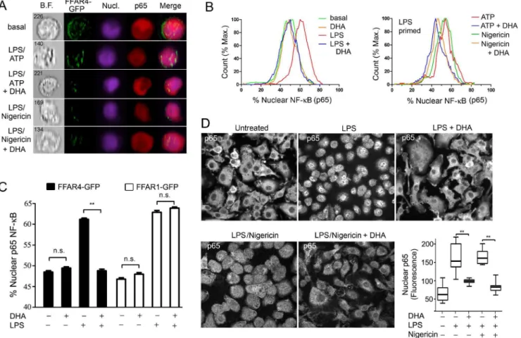
The reduced level of NLRP3 in LPS primed mouse BMDMs treated with DHA observed above; the reported inhibition of LPS induced phosphorylation of IKKb in DHA treated RAW 264.7 cells [2]; and the known requirement for NF-kB translocation for successful LPS priming [4] prompted us to examine whether DHA affected the nuclear translocation of NF-kB. As non-differentiated THP-1 cells express low levels of Ffar4 mRNA, we transfected them with thev3 FFA receptor FFAR4 (GPR120) fused to GFP. We used undifferentiated cells because of their lower basal expression of nuclear p65 NF-kB. We LPS primed the transfected cells in the presence or absence of DHA and performed a flow based imaging assay to determine the amount of nuclear p65 NF-kB in the FFAR4-GFP positive cells. We did a similar experiment, but also included an inflammasome activator. These results showed that DHA potently reduced the nuclear translocation of p65 NF-kB following LPS priming or LPS priming plus nigericin (Figure 2A & B). Next, we compared the translocation of p65 NF-kB in FFAR4-GFP or FFAR1-GFP expressing undifferentiated THP-1 cells. Focusing only on the FFAR4-GFP or FFAR1-GFP positive cells, we found that FFAR4 mediated the inhibitory effect of DHA, while FFAR1 did not (Figure 2C). Next, we checked whether DHA suppressed p65 NF-kB nuclear translocation following LPS priming and inflammasome activation in mouse BMDMs. In the absence of LPS priming the majority of p65 NF-kB resided in the cytosol, while exposure to LPS or LPS plus nigericin shifted a portion of p65 NF-kB to the nucleus, but the addition of DHA largely prevented this shift (Figure 2D). Thus, DHA signaling can dampen inflammasome activation by limiting the initial priming step likely by engaging FFAR4, not FFAR1.
Reducing FFAR4 expression limits the DHA-mediated suppression of IL-1b secretion
little effect (Figure 3C). Together these results indicate that DHA predominately uses FFAR4 to suppress NLRP3 inflammasome activity in mouse BMDMs and in a differentiated human monocyte cell line.
DHA triggers an increase in intracellular calcium and the recruitment of b-arrestins to FFAR4, which helps suppress IL-1b production
FFAR4 has been reported to signal via the heterotrimeric G-protein Gq [2]. Engagement of Gq-linked GPRs usually leads to an increase intracellular calcium levels by the activation of phospholipase Cb. To determine if DHA elicited a rise in intracellular calcium in mouse BMDMs, we challenged the cells, which had been pre-treated with pertussis toxin or not, with DHA. Pertussis toxin is a known inhibitor of Gi-linked receptors, which will not impact signaling through a Gq-linked receptor. Treatment of BMDMs with DHA resulted in modest, but prolonged increase in intracellular calcium, which was largely insensitive to pertussis toxin (Figure 4A).
GPRs signal not only by the activation of G-proteins, but also by the recruitment ofb-arrestins, which serve as a signaling platform for the activation of other signal transducers [9]. The suppression of Toll receptor induced IL-6 and TNF-a production by engagement of FFAR4 has been shown to depend onb -arrestin-2 [-arrestin-2]. Following engagement of FFAR4 b-arrestin2 recruited TAB1, which interacted with TAK1. This inhibited TLR4 induced activation of both NF-kB and Jun kinase. We tested
whether FFAR1 and FFAR4 recruited b-arrestin1 and/or b -arrestin2 following DHA treatment using bioluminescence reso-nance energy transfer (BRET) assays (Figure 4B). Following DHA treatment FFAR4 could recruit bothb-arrestin1 andb-arrestin2, although a stronger change in the BRET signal occurred withb -arrestin2. In contrast, substituting FFAR1 for FFAR4 resulted in little or no DHA induced change in the BRET signal with either b-arrestin. Next, we co-transfected HeLa cells with FFAR4-GFP andb-arrestin2-mCherry, stimulated the cells with DHA or not, and imaged the cells by confocal microscopy. FFRA4 localized predominately at the cell membrane although some of the protein was likely retained in intracellular compartments. In contrast,b -arrestin2 largely resided in the cytoplasm. DHA treatment resulted in a strong shift of b-arrestin-2 from the cytoplasm to the cell membrane and a partial internalization of FFAR4, which co-localized withb-arrestin2 in the cytoplasm (Figure 4C). We found similar results when we substituted THP-1 cells for the HeLa cells (data not shown). Together these results argue that FFAR4 rather than FFAR1 is the relevantv3 FFA receptor involved in limiting inflammation and likely inflammasome activation in mouse and human macrophages.
result argues that in BMDMs, b-arrestin2 preferentially targets FFAR4. To assess b-arrestin involvement in the DHA-mediated inhibition of inflammasome activity, we LPS primed BMDMs from
Arrb12/2and Arrb22/2 mice and treated them with ATP in the presence or absence of DHA. The results from these experiments demonstrated that loss ofb-arrestin2 partially reversed the DHA Figure 2. DHA inhibits the translocation of NF-kB to the nucleus.FFAR4-GFP expressing LPS primed non-differentiated THP-1 cells were stimulated with ATP or nigericin. DHA (100mM) was added or not at the priming step. PFA fixed cells were permeablized and then stained with p65
antibody and analysis was performed using an ImageStream instrument. Shown are the (A) Imaging results and (B) Flow cytometry results. (C) ImageStream analysis of nuclear p65 NF-kB in non-differentiated THP-1 cells expressing FFAR1-GFP or FFAR4-GFP and exposed to LPS, or not, in the presence of DHA (50mM), or not. (D) Confocal microscopy of BMDMs LPS primed and treated with nigericin in the presence, or absence of DHA, to
examine the status of NF-kB translocation by p65 immunostaining. Shown are representative individual images. Scale bar is 20mM. Whisker plot
shows the amount of nuclear p65 immunofluorescence in the nuclei of BMDMs treated as indicated. Data are representative of three independent experiments. **P,0.001, n.s-not significant.
doi:10.1371/journal.pone.0097957.g002
Figure 3. DHA-mediated suppression of inflammasome activity depends upon FFAR4.(A) Quantitative RT-PCR to detectFfar1,Gpr84, and
Ffar4mRNA expression in BMDMs. Cells were LPS primed and ATP treated. DHA (100mM) added during priming step, or not. Data is relative toGapdh
expression x 105. Experiment performed 2 times with similar results. (B) Quantitative RT-PCR to detectGPR84orFFAR4mRNA in LPS primed THP-1
cells expressing siRNAs targetingFFAR4orGPR84, respectively. The results were normalized tob-actin expression and shown as relative to control. (C) IL-1bELISA to assess inflammasome activity in FFAR1, GPR84, or FFAR4 knock-down cells. LPS primed THP-1 cells expressing siRNAs targetingFFAR1,
GPR84, orFFAR4were stimulated with ATP. DHA added during the priming step. Similar results from 3 experiments. *P,0.05, **P,0.001, ****P,
0.0001.
triggered suppression, while loss of b-arrestin1 had no effect (Figure 4E). These data indicate that b-arrestin2 signaling contributes to the DHA mediated reduction in inflammasome activation, but that other signaling pathways also contribute.
DHA increases autophagosome formation in macrophages
A previous report from our lab demonstrated that autophagy can limit inflammasome activity by delivering inflammasomes to autophagosomes for subsequent lysosome mediated destruction [10]. Suggesting that DHA might reduce inflammasome activity by inducing autophagy several reports have shown that exposure of a variety of cell lines tov3 FFA triggers autophagy [14,15]. To determine whether DHA induced autophagosomes in mouse BMDMs we isolated BMDMs from GFP-LC3 mice [8] and treated them with DHA. However, DHA treatment did not increase the number of cells with GFP-LC3 puncta (data not shown). When we LPS primed and treated the cells with nigericin the number of GFP-LC3 puncta in the BMDMs did increase. Furthermore, the addition of DHA significantly enhanced the number of cells with more than 5 GFP-LC3 puncta per cell (Figure 5A & B). Similar results were found when cells were stimulated with LPS+ ATP in the presence or absence of DHA (data not shown). To test whether autophagosome formation contributed to the suppressive effect of DHA on inflammasome
activity we compared mice incapable of forming autophagosomes to most stimuli to wild type mice. BMDMs fromAtg7flox/floxand Atg7flox/floxVav-1Cremice were stimulated with LPS+ATP in the presence of absence of DHA. The lack of ATG7 inhibited the suppressive effects of DHA on IL-1b secretion (Figure 5C). Western blot analysis of cells lysates confirmed the ELISA results (Figure 5D). The partial reduction in inflammasome activation in the ATG7 deficient macrophages is likely secondary to the effect of DHA on LPS priming while the inability of DHA to enhance autophagy may account for the sub-optimal inhibition. These results support a role for autophagy in DHA dependent suppression of inflammasome activation.
The mechanism by which DHA increases autophagosome formation is unknown although a recent study indicated that mTOR controlled autophagy required intracellular calcium signaling [16]. As both ATP and DHA are capable of eliciting an intracellular calcium flux we checked how the simultaneous addition of DHA and ATP affected the level of intracellular calcium in BMDMs. We exposed the cells to different concentra-tions of ATP, DHA, and to both signals together. We found that as previously described DHA triggered an increase in intracellular calcium. The addition of ATP alone elicited a higher increase in the peak intracellular calcium and a much sharper rate of increase that did DHA. The combination of ATP and DHA triggered a greater rise in intracellular calcium and a more prolonged increase Figure 4. DHA elicits a calcium flux and its suppression of inflammasome activity partially depends upon b-arrestin2 notb -arrestin1.(A) DHA elicited changes in intracellular Ca2+
in BMDMs treated with pertussis toxin (200 ng/ml), or not, for 2 hours prior to the assay. (B) BRET analysis using HeLa cells expressing a BRET donor (RLuc-b-arrestin1 orb-arrestin2-RLuc) and increasing amounts of a BRET acceptor (FFAR4-GFP or FFAR1-GFP) stimulated with LPS, or not, prior to addition of DHA. The BRET ratios were calculated and the change in BRET determined. (C) Confocal microscopy of HeLa cells expressing FFAR4-GFP andb-arrestin-2 mCherry following exposure to DHA. Red arrow indicatesb-arrestin-2 recruitment and the white arrows intracellular co-localization ofb-arrestin-2 and FFAR4. Scale bar is 10mM. (D) Intracellular calcium response to DHA of BMDMs
prepared from wild type andb-arrestin knock-out mice. (E) ELISA to measure IL-1bin cell supernatants conditioned by LPS primed and ATP treated BMDMs from WT,arrb12/2, andarrb22/2mice. DHA (100 M) was added at the priming step. Results are from 3 independent experiments. * P,0.05,
****P,0.0001.
than did either ATP or DHA alone. We are currently investigating whether the elevated intracellular calcium noted in these primary mouse macrophages is sufficient to trigger autophagosome formation. High concentrations of ATP have been shown to induced autophagy in human macrophages and macrophage cell lines[17].
In the course of our studies, Yan et al. published a report demonstrating thatv3 FFA suppressed macrophage NLRP3 and NLRP1b inflammasomes, but not AIM2 and NAIP5/NLRC4 inflammasomes [18]. They found roles for FFAR4, FFAR1, andb -arrestin-2 inv3 FFA signaling. They also demonstrated a ligand induced interaction between NLRP3 andb-arrestin-2. Our results differ slightly from those of Yan et al [18]. We found a strong suppression of all the tested inflammasome activators, perhaps because we used a higher concentration of DHA and included the DHA in the priming step, thereby reducing NF-kB activation and
limiting expression of some of the inflammasome components. We did not find a role for FFAR1 as we found very low levels of FFAR1 in mouse BMDMs. We did detect FFAR1 in differentiated THP-1 cells (Y. Williams-Bey, unpublished observation), but failed to find a recruitment ofb-arrestin-2 to FFAR1 following exposure of cells to DHA. Our data would support a much more important role for FFAR4 than FFAR1 in inflammasome suppression. Finally we provide an additional mechanism by whichv3 FFA limit inflammasome activity and that is by enhancing autophagy
In summary, we have shown thatv3 FFA can suppress NLRP3, AIM2, and NAIP/NLCR4 inflammasome activation. We focused on NLRP3 inflammasome activation and demonstrated a requirement for FFAR4. Mouse BMDMs expressFfar4and LPS stimulation increased Ffar4 mRNA levels. DHA stimulation recruited b-arrestin-1 and -2 to FFAR4 and caused receptor internalization, but onlyb-arrestin-2 helps mediated the suppres-Figure 5. DHA regulates inflammasome activity by promoting autophagy.(A) Confocal microscopy of BMDMs from GFP-LC3 mice primed with LPS and treated with nigericin. DHA (50mM) was added at the priming step. Scale bar is 5mM. (B) Number of cells with.5 GFP-LC3 puncta per cell were determined for each treatment group. Data are from 3 independent experiments. (C) ELISA to measure IL-1bin supernatants conditioned by BMDMs prepared fromAtg7flox/floxorAtg7flox/floxVav1-Cremice. The cells were primed with LPS and treated with ATP. DHA was added at the priming
step. Results are from 3 independent experiments. (D) Western blot analysis of supernatants and cell lysates from BMDMs prepared fromAtg7flox/flox
orAtg7flox/floxVav1-Cremice. Cells were LPS primed and treated with ATP. DHA was added at the time of priming. The numbers indicate the relative
expression of IL-1bin the cell supernatants. **P,0.001, ***P,0.0002, ****P,0.0001. (E) DHA and ATP triggered changes in intracellular Ca+2in mouse
BMDMs. The cells were exposed to the indicated concentrations of DHA and/or ATP. Results from one experiment performed in triplicate. Representative of three experiments performed.
sive effects of DHA. We identified two mechanisms by which DHA suppressed macrophage inflammasome activity, first, it impaired priming by inhibiting NF-kB activation likely via ab -arrestin-2 dependent mechanism and, second, it enhanced autophagy, thereby reducing inflammasome complex formation or presenting inflammasome components for destruction. Our studies support the further study and use of v3 FFA in those clinical situations characterized by excessive macrophage inflam-masome activity.
Acknowledgments
The authors would like to thank Dr. A. S. Fauci for his continued support.
Author Contributions
Conceived and designed the experiments: YWB CB CSS JHK. Performed the experiments: YWB CB AV NNH IYH. Analyzed the data: YWB CB AV NNH IYH CSS JHK. Wrote the paper: YWB CB JHK.
References
1. Kiecolt-Glaser JK, Belury MA, Andridge R, Malarkey WB, Hwang BS, et al. (2012) Omega-3 supplementation lowers inflammation in healthy middle-aged and older adults: a randomized controlled trial. Brain Behav Immun 26: 988– 995.
2. Oh DY, Talukdar S, Bae EJ, Imamura T, Morinaga H, et al. (2010) GPR120 is an omega-3 fatty acid receptor mediating potent anti-inflammatory and insulin-sensitizing effects. Cell 142: 687–698.
3. Nasti TH, Timares L (2012) Inflammasome activation of IL-1 family mediators in response to cutaneous photodamage. Photochem Photobiol 88: 1111–1125. 4. Schroder K, Tschopp J (2010) The inflammasomes. Cell 140: 821–832. 5. Wen H, Gris D, Lei Y, Jha S, Zhang L, et al. (2011) Fatty acid-induced
NLRP3-ASC inflammasome activation interferes with insulin signaling. Nat Immunol 12: 408–415.
6. Csak T, Ganz M, Pespisa J, Kodys K, Dolganiuc A, et al. (2011) Fatty acid and endotoxin activate inflammasomes in mouse hepatocytes that release danger signals to stimulate immune cells. Hepatology 54: 133–144.
7. Mortensen M, Ferguson DJ, Edelmann M, Kessler B, Morten KJ, et al. (2010) Loss of autophagy in erythroid cells leads to defective removal of mitochondria and severe anemia in vivo. Proc Natl Acad Sci U S A 107: 832–837. 8. Mizushima N (2009) Methods for monitoring autophagy using GFP-LC3
transgenic mice. Methods Enzymol 452: 13–23.
9. Luttrell LM, Lefkowitz RJ (2002) The role of beta-arrestins in the termination and transduction of G-protein-coupled receptor signals. J Cell Sci 115: 455–465. 10. Shi CS, Shenderov K, Huang NN, Kabat J, Abu-Asab M, et al. (2012) Activation of autophagy by inflammatory signals limits IL-1beta production by targeting ubiquitinated inflammasomes for destruction. Nat Immunol 13: 255– 263.
11. Bauernfeind FG, Horvath G, Stutz A, Alnemri ES, MacDonald K, et al. (2009) Cutting edge: NF-kappaB activating pattern recognition and cytokine receptors license NLRP3 inflammasome activation by regulating NLRP3 expression. J Immunol 183: 787–791.
12. Hara T, Hirasawa A, Ichimura A, Kimura I, Tsujimoto G (2011) Free fatty acid receptors FFAR1 and GPR120 as novel therapeutic targets for metabolic disorders. J Pharm Sci 100: 3594–3601.
13. Yonezawa T, Kurata R, Yoshida K, Murayama MA, Cui X, et al. (2013) Free fatty acids-sensing G protein-coupled receptors in drug targeting and therapeutics. Curr Med Chem 20: 3855–3871.
14. Shin S, Jing K, Jeong S, Kim N, Song KS, et al. (2013) The omega-3 polyunsaturated fatty acid DHA induces simultaneous apoptosis and autophagy via mitochondrial ROS-mediated Akt-mTOR signaling in prostate cancer cells expressing mutant p53. Biomed Res Int 2013: 568671.
15. Jing K, Song KS, Shin S, Kim N, Jeong S, et al. (2011) Docosahexaenoic acid induces autophagy through p53/AMPK/mTOR signaling and promotes apoptosis in human cancer cells harboring wild-type p53. Autophagy 7: 1348– 1358.
16. Decuypere JP, Kindt D, Luyten T, Welkenhuyzen K, Missiaen L, et al. (2013) mTOR-Controlled Autophagy Requires Intracellular Ca(2+) Signaling. PLoS One 8: e61020.
17. Biswas D, Qureshi OS, Lee WY, Croudace JE, Mura M, et al. (2008) ATP-induced autophagy is associated with rapid killing of intracellular mycobacteria within human monocytes/macrophages. BMC Immunol 9: 35.